National Diagnostic Protocol for...National Diagnostic Protocol for Plenodomus tracheiphilus the...
26
National Diagnostic Protocol for Plenodomus tracheiphilus the cause of mal secco PEST STATUS Not present in Australia PROTOCOL NUMBER NDP 26 VERSION NUMBER V1.1 PROTOCOL STATUS Endorsed ISSUE DATE April 2014 REVIEW DATE 2019 ISSUED BY SPHDS
Transcript of National Diagnostic Protocol for...National Diagnostic Protocol for Plenodomus tracheiphilus the...

National Diagnostic Protocol for Plenodomus tracheiphilus
the cause of mal secco
PEST STATUS Not present in Australia PROTOCOL NUMBER NDP 26 VERSION NUMBER V1.1 PROTOCOL STATUS Endorsed ISSUE DATE April 2014 REVIEW DATE 2019 ISSUED BY SPHDS
Prepared for the Subcommittee on Plant Health Diagnostic Standards (SPHDS)
This version of the National Diagnostic Protocol (NDP) for Plenodomus tracheiphilus is current as
at the date contained in the version control box on the front of this document.
NDPs are updated every 5 years or before this time if required (i.e. when new techniques become available).
The most current version of this document is available from the SPHDS website: http://plantbiosecuritydiagnostics.net.au/resource-hub/priority-pest-diagnostic-resources/
Where an IPPC diagnostic protocol exists it should be used in preference to the NDP. NDPs
may contain additional information to aid diagnosis. IPPC protocols are available on the IPPC website:
https://www.ippc.int/core-activities/standards-setting/ispms
2

TABLE OF CONTENTS
1 INTRODUCTION .......................................................................................... 4
1.1 Host range............................................................................................................................. 4
2.1 Effect on hosts ...................................................................................................................... 4
3.1 Stages of development .......................................................................................................... 4
4.1 Transmission ......................................................................................................................... 4
2 TAXONOMIC INFORMATION .......................................................................... 5
3 DETECTION ............................................................................................... 6
3.1 Symptom description ............................................................................................................. 6
3.1.1 Symptoms that are confused with mal secco ................................................................ 11
4 IDENTIFICATION ....................................................................................... 13
4.1 Isolation/culture techniques, morphological methods ........................................................... 13
4.1.1 Technique for initial fungal isolation .............................................................................. 13 4.1.2 Alternative technique for fungal isolation ....................................................................... 13 4.1.3 Technique for subculturing ............................................................................................ 14 4.1.4 Identification - morphology ............................................................................................ 15 4.1.5 Media recipes for fungal isolation and culture ............................................................... 18
4.2 Biochemical methods .......................................................................................................... 18
4.3 Molecular methods .............................................................................................................. 19
4.3.1 DNA extraction .............................................................................................................. 19 4.3.2 DNA amplification from pure fungi ................................................................................. 20 4.3.3 Validation results .......................................................................................................... 22 4.3.4 Real-time PCR .............................................................................................................. 23
5 CONFIRMATORY TESTING .......................................................................... 23
6 CONTACT FOR FURTHER INFORMATION ........................................................ 24
7 ACKNOWLEDGEMENTS .............................................................................. 24
8 REFERENCES .......................................................................................... 25
9 ADDITIONAL INFORMATION: ....................................................................... 26
3

1 INTRODUCTION
Plenodomus tracheiphilus (Petri) Gruyter, Aveskamp & Verkley (previously Phoma tracheiphila, Gruyter et al. 2013) is a mitosporic fungus that causes a destructive vascular disease of citrus named 'mal secco'. Mal secco (Italian for dry disease) was first reported in 1894 but the causal organism was not determined until 1929. It is difficult to control and even more so to eradicate as the disease is often well established by the time disease symptoms become obvious in the orchard.
1.1 Host range
The host range for P. tracheiphilus includes members of the Rutaceae family including members of the Citrus genus, Poncitrus trifoliata, Fortunella spp. and Severinia buxifolia (Solel and Oren 1975, Palm 1987).
Almost all citrus plants are susceptible to artificial inoculation by P. tracheiphilus (Perrotta and Graniti 1988). In the field, the disease is most severe on lemons (C. x limon) but citron (C. medica), bergamot (C. x bergamia) and lime (C. x aurantiifolia) are also very susceptible to natural infection.
2.1 Effect on hosts
Mal secco attacks trees of any age although the disease is more significant on young citrus trees. The disease reduces both the quantity and quality of production due to the cost of and difficulties with managing the disease and the replacement of susceptible cultivars with those that are more tolerant to the pathogen but have reduced production and fruit quality (Migheli et al. 2009; Nigro et al. 2011). Mal secco symptoms range from leaf and shoot chlorosis, wilting and dieback to complete canopy collapse.
3.1 Stages of development
Fungal spores (conidia) are water borne and enter the plant through wounds to cause infection. Wound entry points can be created by climatic events like wind, hail or frost damage and cultural practices like pruning and cultivation. Young tissue is particularly vulnerable to infection (Perrotta and Graniti 1988).
Conidial spores are produced by pycnidia in infected tissue and from phialides borne on free hyphae on exposed woody surfaces or debris. Once in the host the fungus reaches the lumen of xylem and then spreads systemically, mostly upward. Conidia also move in the xylem sap. Warm moist conditions are conducive to infection and disease development, with pycnidiospores needing 40 hours of moisture and temperatures in the range of 14-28°C (Perrotta and Graniti 1988). The optimum temperature for disease development is 20-25°C hence disease progress is most rapid in the spring and autumn. Temperatures above 30°C inhibit mycelial growth but do not kill the pathogen within infected tissues (Reichert and Chorin 1956, Perrotta and Graniti 1988). Infection periods vary in the Mediterranean region due to local climatic and seasonal conditions.
4.1 Transmission
Short range spread of mal secco is via fungal spores that are dispersed by water (rain splash, wind blown rain, overhead irrigation). Infected twigs and branches can remain infectious for several weeks and fungal propagules can survive within infected twigs in the soil for longer than 4 months (De Cicco et al. 1987).
The disease is spread over long distances via the movement of infected propagation material and plants. Symptomless infections can occur in stems, shoots, roots and fruit. P. tracheiphilus has been detected in lemon seeds as mycelium (Stepanov and Shaluishkina 1952; Ippolito et al. 1987, 1992) and reported in some other Citrus sp. (Ippolito et al. 1987; 1992). The pathogen colonises
4
the seed coat but not embryos. Therefore developing seedlings do not appear to be infected (Ippolito et al. 1987). Fruit infection occurs in lemons when the pathogen moves from infected branches into fruit. Infected fruit generally falls from the tree.
2 TAXONOMIC INFORMATION
Phylum: Ascomycota
Class: Dothideomycetes
Order: Pleosporales
Genus: Plenodomus
Species: tracheiphilus
Causal agent: Plenodomus tracheiphilus (Petri) Gruyter, Aveskamp & Verkley
Synonyms: Deuterophoma tracheiphila Petri, Bakerophoma tracheiphila (Petri) Ciferri, Phoma tracheiphila (Petri) Kantschaveli et Gikashvili
Detailed taxonomic descriptions can be found in Punithalingham and Holliday (1973), EPPO/OEPP (2007), Migheli et al. (2009) and Nigro et al. (2011).
5
3 DETECTION
Best practice in diagnostics requires that results be obtained by thorough investigation using more than one method.
The diagnostic methods that can be used to detect Plenodomus tracheiphilus include:
1. observation of symptoms consistent with mal secco
2. isolation of fungal isolate from infected tissue with morphological and molecular characteristics consistent with P. tracheiphilus
3. humid chamber incubation of withered twigs and morphology of resulting pycnidia and phialides consistent with P. tracheiphilus
4. positive reaction by PCR of infected tissue (bark or stem) or cultures isolated from infected tissue
Ideally, the same result should be obtained by two different laboratories using the same protocol, for each sample.
3.1 Symptom description
Mal secco attacks trees of any age although the disease is more significant on young citrus trees. Disease symptoms are most severe in spring and autumn. High temperatures inhibit the ability of the fungus to colonise xylem vessels, so the pathogen may not be detected in the summer flush of infected plants (De Patrizio et al. 2009).
Symptom expression and development depends upon infection mode. When infection starts in the canopy of adult plants, symptoms often appear in the uppermost shoots, with a slight discoloration of the primary and secondary veins. The disease develops showing leaf and shoot chlorosis, wilting of leaves and soft shoots (Figure 1), leaf fall and twig and branch dieback (Figures 2-5) (Reichert and Chorin 1956; Nigro et al. 2011). The petioles often remain for a period when leaves drop; petioles retain a green base but often are chlorotic or discoloured at the end (Figure 6). The wood of newly infected shoots, branches and trunks can be a salmon-pink or reddish-brown colour. In association with the wood discolouration is gum production within the xylem vessels. On occasion fallen leaves can show red discoloration of the midrib and secondary veins (Solel and Salerno 2000). A common response to infection is the sprouting of shoots from the base of affected branches and production of rootstock suckers (Perrotta and Graniti 1988).
When infection starts in the outer canopy, the pathogen moves slowly downwards from the shoots to the limbs, trunk and roots, eventually leading to tree death. When infection starts at the base of a shoot or branch the disease progresses more rapidly, whole shoots or branches quickly wilt followed by leaf and fruit drop (Nigro et al. 2011). Strains of P. tracheiphilus that differ in virulence have been identified in the field (Magnano di San Lio et al. 1992). However trials involving artificial inoculation using a large number of isolates from around the Mediterranean basin found no specialisation or significant variation in pathogenicity (Nigro et al. 2011).
P. tracheiphilus can invade fruits and seeds of infected lemon trees (Stepanov and Shaluishkina 1952; Ippolito et al. 1987). Unripe lemon fruits show partial or total yellowing of the peel when infection occurs whereas ripe fruits turn dark yellow to reddish when infected (Ippolito et al. 1987). Diseased fruits may fall to the ground or when branches desiccate quickly fruit can remain attached, show necrosis of the pericarp around the calyx and then mummify on the tree. Infected fruits are typically smaller and tougher than unaffected fruits and have discoloured seeds and vascular bundles. In addition to lemon, P. tracheiphilus can be isolated from fruits and seeds of other susceptible citrus species (Ippolito et al. 1987; 1992).
6

Figure 1: Branch wilting on lemon tree, Nizzanim Israel - first sign of mal secco (© NSW DPI)
Figure 2: Branch dieback in lemon tree, Nizzanim Israel (© NSW DPI)
Figure 3: Branch dieback in lemon tree, Eytan Israel (© NSW DPI)
7
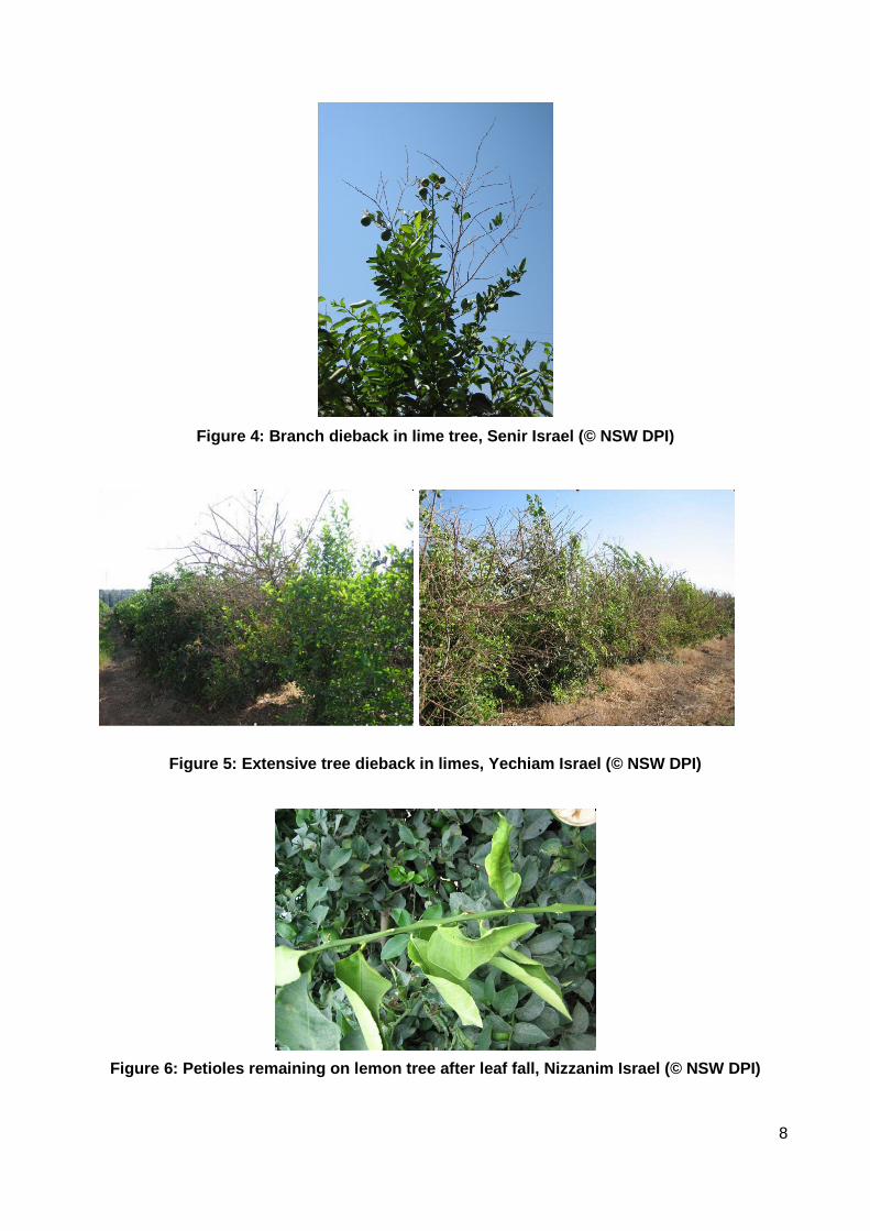
Figure 4: Branch dieback in lime tree, Senir Israel (© NSW DPI)
Figure 5: Extensive tree dieback in limes, Yechiam Israel (© NSW DPI)
Figure 6: Petioles remaining on lemon tree after leaf fall, Nizzanim Israel (© NSW DPI)
8
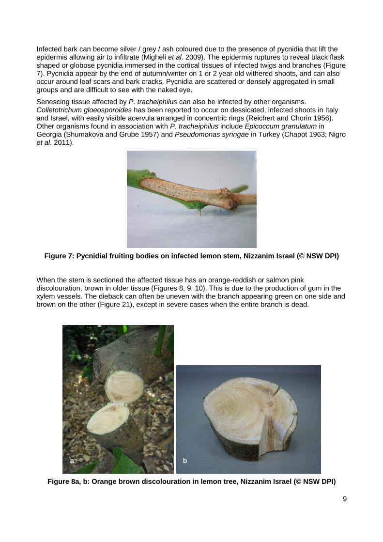
Infected bark can become silver / grey / ash coloured due to the presence of pycnidia that lift the epidermis allowing air to infiltrate (Migheli et al. 2009). The epidermis ruptures to reveal black flask shaped or globose pycnidia immersed in the cortical tissues of infected twigs and branches (Figure 7). Pycnidia appear by the end of autumn/winter on 1 or 2 year old withered shoots, and can also occur around leaf scars and bark cracks. Pycnidia are scattered or densely aggregated in small groups and are difficult to see with the naked eye.
Senescing tissue affected by P. tracheiphilus can also be infected by other organisms. Colletotrichum gloeosporoides has been reported to occur on dessicated, infected shoots in Italy and Israel, with easily visible acervula arranged in concentric rings (Reichert and Chorin 1956). Other organisms found in association with P. tracheiphilus include Epicoccum granulatum in Georgia (Shumakova and Grube 1957) and Pseudomonas syringae in Turkey (Chapot 1963; Nigro et al. 2011).
Figure 7: Pycnidial fruiting bodies on infected lemon stem, Nizzanim Israel (© NSW DPI)
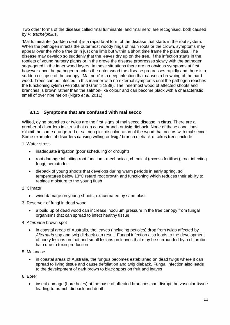
When the stem is sectioned the affected tissue has an orange-reddish or salmon pink discolouration, brown in older tissue (Figures 8, 9, 10). This is due to the production of gum in the xylem vessels. The dieback can often be uneven with the branch appearing green on one side and brown on the other (Figure 21), except in severe cases when the entire branch is dead.
Figure 8a, b: Orange brown discolouration in lemon tree, Nizzanim Israel (© NSW DPI)
a b
9
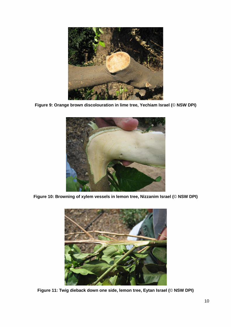
Figure 9: Orange brown discolouration in lime tree, Yechiam Israel (© NSW DPI)
Figure 10: Browning of xylem vessels in lemon tree, Nizzanim Israel (© NSW DPI)
Figure 11: Twig dieback down one side, lemon tree, Eytan Israel (© NSW DPI)
10
Two other forms of the disease called ‘mal fulminante’ and ‘mal nero’ are recognised, both caused by P. tracheiphilus.
‘Mal fulminante' (sudden death) is a rapid fatal form of the disease that starts in the root system. When the pathogen infects the outermost woody rings of main roots or the crown, symptoms may appear over the whole tree or in just one limb but within a short time frame the plant dies. The disease may develop so suddenly that the leaves dry up on the tree. If the infection starts in the rootlets of young nursery plants or in the grove the disease progresses slowly with the pathogen segregated in the inner wood layers. In these situations there are no obvious symptoms at first however once the pathogen reaches the outer wood the disease progresses rapidly and there is a sudden collapse of the canopy. ‘Mal nero' is a deep infection that causes a browning of the hard wood. Trees can be infected in this manner with no external symptoms until the pathogen reaches the functioning xylem (Perrotta and Graniti 1988). The innermost wood of affected shoots and branches is brown rather than the salmon-like colour and can become black with a characteristic smell of over ripe melon (Nigro et al. 2011).
3.1.1 Symptoms that are confused with mal secco
Wilted, dying branches or twigs are the first signs of mal secco disease in citrus. There are a number of disorders in citrus that can cause branch or twig dieback. None of these conditions exhibit the same orange-red or salmon pink discolouration of the wood that occurs with mal secco. Some examples of disorders causing wilting or twig / branch dieback of citrus trees include:
1. Water stress
• inadequate irrigation (poor scheduling or drought)
• root damage inhibiting root function - mechanical, chemical (excess fertiliser), root infecting fungi, nematodes
• dieback of young shoots that develops during warm periods in early spring, soil temperatures below 13°C retard root growth and functioning which reduces their ability to replace moisture to the young flush
2. Climate
• wind damage on young shoots, exacerbated by sand blast
3. Reservoir of fungi in dead wood
• a build up of dead wood can increase inoculum pressure in the tree canopy from fungal organisms that can spread to infect healthy tissue
4. Alternaria brown spot
• in coastal areas of Australia, the leaves (including petioles) drop from twigs affected by Alternaria spp and twig dieback can result. Fungal infection also leads to the development of corky lesions on fruit and small lesions on leaves that may be surrounded by a chlorotic halo due to toxin production
5. Melanose
• in coastal areas of Australia, the fungus becomes established on dead twigs where it can spread to living tissue and cause defoliation and twig dieback. Fungal infection also leads to the development of dark brown to black spots on fruit and leaves
6. Borer
• insect damage (bore holes) at the base of affected branches can disrupt the vascular tissue leading to branch dieback and death
11

7. Citrus blast
• infection by the bacterium Pseudomonas syringae pv. syringae induces water soaked lesions on twigs and leaf petioles of young succulent shoots, leaves wither, brown and often remain attached, petioles remain if leaves fall
8. Citrus blight
• general tree decline, canopy thinning, off season flush and root suckering, causal agent unknown
9. Sudden death
• sudden wilting of entire tree associated with excessive moisture and poor aeration for intermittent periods leading to a deterioration of root health
12

4 IDENTIFICATION
4.1 Isolation/culture techniques, morphological methods
P. tracheiphilus is readily isolated from infected branches that remain alive if processed within 3 days of collection. Tissue should be plated from the edge of a discoloured region.
If pycnidia are present, they can be mounted in distilled water or lactophenol blue and observed under the microscope. Pycnidial structures are described in Section 4.1.4 and Figure 15. Withered twigs may be incubated in a humid chamber for 12-24 hours. Incubation encourages spore tendrils (cirrhi) to protrude from the pycnidium which can be seen under a dissecting microscope.
Maximum fungal growth and pycnidia production on artificial media occurs between 20 and 25ºC (Salerno 1964).
4.1.1 Technique for initial fungal isolation
1. select infected stem 2. cut to a manageable size (20-100 mm) using secateurs 3. dip infected stem into 100% ethanol 4. light infected stem, let it burn for a few seconds 5. once the flame has extinguished, cut several thin segments of the stem into a sterile dish
using secateurs 6. plate stem pieces onto potato dextrose agar (PDA) plates (Figure 2), or carrot potato agar
(CPA). 7. incubate plates in the dark at 23+2°C for 6-12 days 8. check growth on plates daily to monitor growth of contaminants
4.1.2 Alternative technique for fungal isolation
1. immerse stem section in sterile distilled water for 30-60 min, dry on sterile filter paper, flame and plate on agar medium OR
2. surface sterilise stem section or leaves with 0.5-1% sodium hypochlorite or 50% ethanol for 40s to 2-5 min depending upon stem thickness or tissue type, rinse in sterile distilled water for 5 minutes and plate on agar medium.
3. wash fruit under running water then surface sterilise by spraying with 1% sodium hypochlorite solution and rinse with sterile distilled water, plate small tissue pieces from the top, inner part of the fruit onto agar medium.
Figure 12: Initial isolation by plating stems onto PDA (© NSW DPI)
13
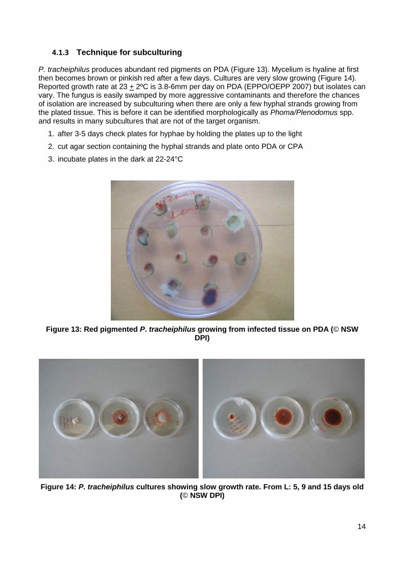
4.1.3 Technique for subculturing
P. tracheiphilus produces abundant red pigments on PDA (Figure 13). Mycelium is hyaline at first then becomes brown or pinkish red after a few days. Cultures are very slow growing (Figure 14). Reported growth rate at 23 + 2ºC is 3.8-6mm per day on PDA (EPPO/OEPP 2007) but isolates can vary. The fungus is easily swamped by more aggressive contaminants and therefore the chances of isolation are increased by subculturing when there are only a few hyphal strands growing from the plated tissue. This is before it can be identified morphologically as Phoma/Plenodomus spp. and results in many subcultures that are not of the target organism.
1. after 3-5 days check plates for hyphae by holding the plates up to the light
2. cut agar section containing the hyphal strands and plate onto PDA or CPA
3. incubate plates in the dark at 22-24°C
Figure 13: Red pigmented P. tracheiphilus growing from infected tissue on PDA (© NSW
DPI)
Figure 14: P. tracheiphilus cultures showing slow growth rate. From L: 5, 9 and 15 days old
(© NSW DPI)
14

4.1.4 Identification - morphology
Morphological description from Punithalingham and Holliday (1973), EPPO/OEPP (2007), Migheli et al. (2009), Nigro et al. (2011).
Mycelium: monoporic, made up of mono- and plurinucleate cells, young hyphae and apical cells are typically plurinucleate. Fungal growth on agar medium is initially white and fluffy. Culture pigmentation develops over time, mostly red but can range from light grey to very dark brown (Figure 16). Chromogenic and nonchromogenic variants have been identified (Graniti 1969).
Non chromogenic (or achromatic) strains: lack the red pigmentation, have brown aerial hyphae, lose the ability to form pycnidia.
Chromogenic (or chromatic) strains: high phenotypic instability in culture, hyaline hyphae, vary in their capacity to produce red pigments and in culture irreversibly lose their capacity to produce pycnidia, brown hyphae and sometimes hyphal conidia.
The high variability of phenotypic characters of fungal isolates is not reflected by genetic variability (Balmas et al. 2005; Ezra et al. 2007; Kalai et al. 2010).
Phialoconidia produced by phialides on free hyphae growing on exposed wood surfaces, wounded plant tissues and within the lumen of xylem vessels: conidia 1.5-3 x 3-8 μm, mucosal, hyaline, unicellular, uninucleate sometimes bi or trinucleate, straight or slightly curved with rounded apices (3-8 x 1.5-3 μm), phialides 12-30 x 3-6 μm.
Phialoconidia produced in culture on mono- or polyphilaides after 8-10 days incubation at 21+2ºC. Production of conidia is greater on CPA than PDA and the hyphal strands are more irregular. Conidia are generally observed directly on the culture plate. It is difficult to observe conidia on a microscope slide as they readily break from their stems. Repeated culturing does not appear to inhibit production of phialides or phialoconidia in most isolates.
Pycnidia: black, globose to lenticular, 60-165 x 45-140 μm diameter, necked when mature. Pycnidial wall is of uniform thickness and made up of randomly arranged polygonal scleroplectenchymatous cells. Repeated subculturing (1-2 times) reduces the ability of isolates to produce pycnidia. Pycnidia produced in culture may remain incomplete, be thin walled or open irregularly at maturity.
Pycnidial neck: diameter 45-70 μm, length up to 250 μm, cylindrical or tendentially obconical, often flared at the top and surrounded by a dense, dark hyphal mat that gathers under the epidermis, this cements the fruiting bodies to the epidermis and together in groups. Necks can be easily removed with the epidermis, this leaves behind widely and irregularly opened pycnidial bodies.
Pycnidial phialides: the surface of the pycnidial cavity is uniformly covered by small phialides 3-4.5 x 3-5.5 μm, irregularly saccula, widely conical or pyriform, tapering apically in a very short neck no more than 1 μm tall.
Pycnoconidia produced within pycnidial cavity by conidiogenous cells (phialides): minute, 0.5-1.5 x 2-4 μm, hyaline, unicellular, mononucleate and sometimes binucleate, shortly ellipsoid, irregularly pyriform and some slightly curved. Pycnoconidia can be extruded through ostioles in whitish cirrhi.
Blastoconidia: 15-17 x 7-9 μm, ovoid, subpyriform, sometimes bicellular, produced apically or intercalary on the hyphae inside host xylem vessels and in culture on liquid media.
15

Figure 15: A. Pycnidium, vertical section. B. Ostiolar region of pycnidium, surface view. C. Part of pycnidial wall and conidiogenous cells producing pycnidia, vertical section. D.
Conidia. E. Conidia being formed from hyphae. (Punithalingam and Holliday 1973).
16
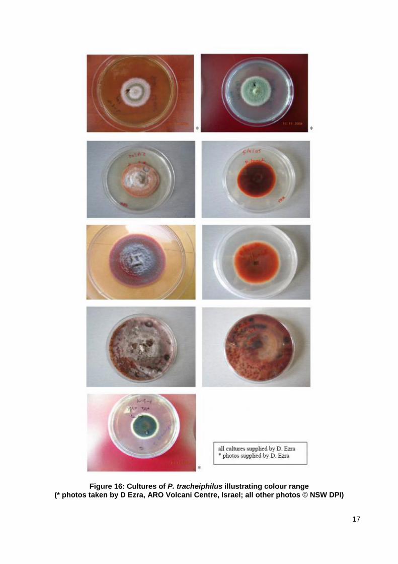
Figure 16: Cultures of P. tracheiphilus illustrating colour range
(* photos taken by D Ezra, ARO Volcani Centre, Israel; all other photos © NSW DPI)
17

4.1.5 Media recipes for fungal isolation and culture
4.1.5.1 Potato dextrose agar (PDA) 1. weigh 20 g of potatoes
2. wash, peel and cut into quarters
3. boil until soft
4. filter through a double layer of cheesecloth
5. add 20 g dextrose
6. add 15 g agar
7. make up to 1 L with water
N.B. PDA may also be prepared using a commercially available dehydrated powder
4.1.5.2 Carrot potato agar (CPA) 1. wash, peel and grate or blend carrots and potatoes
2. weigh 20 g of each
3. boil vegetables until soft
4. filter through a double layer of cheesecloth
5. add 15 g agar
6. make up to 1 L with water.
N.B. The carrot loses its colour in media over time if kept at room temperature.
4.1.5.3 Recipe for Potato Dextrose Broth (PDB) 1. add 4 g potato starch
2. add 20 g of dextrose
3. make up to 1 L with water
4. after autoclaving, adjust pH to 3.5 using tartaric acid
N.B. PDB may also be prepared using a commercially available dehydrated powder
4.2 Biochemical methods
An enzyme linked immunoassay (ELISA) has been developed to detect P. tracheiphilus in citrus tissue (Nachmias et al. 1979). The technique has potential but high background levels means the published method is not suitable for inclusion in a diagnostic protocol.
P. tracheiphila isolates that differ in their ability to produce pigments or do not produce phialoconidia or pycnidia can be identified by polyacrylamide gel electrophoresis (PAGE) analysis of mycelial proteins and isozymes (EPPO/OEPP 2007). This method is time consuming and has not been validated for use in Australia.
18
4.3 Molecular methods
The previous name of Phoma tracheiphila has continued to be used in this section as that is what has been used in the original testing.
4.3.1 DNA extraction
Molecular detection can be performed on DNA extracted from infected plant material and pure fungal cultures.
With regard to plant material, woody material (bark/stems), leaves or soft shoots may be used. However more tissue is needed if extractions are performed from leaf and shoot material.
Ezra et al. (2007) used the Genomic DNA Extraction Kit: Plant Samples (Cartagen®; Washington, USA) and the Rapid Homogenization: Plant leaf DNA amplification kit (Cartagen®; Washington, USA) for DNA extraction from infected plant material and pure fungal cultures respectively. Squares of cultured mycelia (0.5 cm2) were cut from one week old cultures with as much of the agar removed as possible by scraping. The culture pieces were then incubated at -80ºC for 10 mins in 1.5 mL eppendorf vials before extraction as per the kit instructions. Any piece of plant or fruit material used for fungal isolation can also be used for DNA extraction.
Infected leaf tissue was not available during the development of this protocol so no extraction methods for infected plant tissue have been evaluated but it is anticipated that commercially available extraction kits would work effectively in Australia.
Two methods are equally suitable for the extraction of DNA from pure fungal cultures of Phoma. tracheiphila with regard to throughput and quality of amplifiable template produced; the phenol/chloroform method and the high salt method. The EMAI laboratories have routinely used Qiagen DNeasy Plant kits (www.qiagen.com) to extract DNA from both plant material and pure fungal cultures for molecular detection of a range of organisms.
4.3.1.1 High salt method (Aljanabi and Martinez 1997) 1. dislodge at least 1cm2 of mycelia using sterile blade into 1.5 ml microcentrifuge tube 2. grind to a fine powder in liquid nitrogen using a micropestle 3. transfer powder to a sterile tube, to which has been added 400 μl of extraction buffer that
has been warmed to 35ºC in a water bath (400 mM NaCl, 10 mM Tris-Cl pH 8.0, 2 mM EDTA, 20 g/L SDS) and mix well
4. add 8 μl of 20 mg/ml Proteinase K (400 μg/ml final concentration), flick to mix 5. incubate at 65°C for a minimum of 2 hours (can be overnight) 6. add 300 μl saturated salt solution (6M NaCl) 7. vortex 30 seconds on high 8. microfuge 30 mins at 13,000 rpm. 9. transfer supernatant to a fresh tube and add equal volume isopropanol 10. mix by inversion 11. precipitate at -20°C for 1 hour 12. pellet the DNA by microfuging for 15 mins 13. aspirate the ethanol, wash pellet in 100 μl 70% ethanol, 14. spin at 13,000 rpm for 5 mins then decant 15. air dry pellet with lid open for about 10 mins
19
16. add 100 μl sterile TE buffer (10 mM Tris-Cl, pH 8.0, 1 mM EDTA), resuspend DNA by incubating overnight at 4°C
17. store at 4°C, use at 1:10 dilution in PCR
4.3.2 DNA amplification from pure fungi
There are 2 primer sets that can be used for the identification of Phoma tracheiphila (Table 1); Ezra et al. 2007 and Balmas et al. 2005. The two sets hybridise to the same region of the ITS in P. tracheiphila but are not identical, being separated by 4 nucleotides in the case of the forward primer and seven in the case of the reverse primer. Specificity and sensitivity of the two primer sets have been assessed and found to be equivalent (Donovan et al. 2007). The protocol was further refined by the authors following verification of the molecular method by separate laboratories.
Table 1: Sequence of oligonucleotide primers used during this study
Primer Name
Sequence (5'-3') Target Reference Amplicon Size (bp)
P.trach ITS F
CAGGGGATGGGCGCCAGCC Internal Transcribed Spacer (ITS) region
Ezra et al. (2007)
389
P.trach ITS R
CCGTCCTGCACAAGGGCAGTGG ITS region Ezra et al. (2007)
Pt-FOR GGATGGGCGCCAGCCTTC ITS region Balmas et al. (2005)
378
Pt-REV2 GCACAAGGGCAGTGGACAAA ITS region Balmas et al. (2005)
ß-tubulin DLH91
CAGCTCGAGCGCATGAACGTCTA ß-tubulin Hailstones et al. (2003)
1070
ß-tubulin DLH92
TGTACCAATGCAAGAAAGCCTT ß-tubulin Hailstones et al. (2003)
A pair of "universal" primers to the fungal ß-tubulin gene was used as an internal amplification control (IAC) (Table 1) in the Polymerase Chain Reaction (PCR) using either pairs of primers.
The presence of the IAC primers in the PCR will amplify a fragment in all fungal extracts and will thus indicate the performance efficiency of the amplification (Fig. 17). Extracts that are positive for P. tracheiphila will amplify both the ITS and ß-tubulin fragments (Fig. 17). On occasion extracts that are positive for P. tracheiphila will only produce the ITS fragment. This may not be ideal but the diagnosis can still be made. Extracts that are negative for P. tracheiphila will still amplify the ß-tubulin fragment, derived from other fungi present in the sample. Extracts that produce neither the ITS nor the ß-tubulin fragment may be recalcitrant to amplification. In this case a diagnosis cannot reliably be made and the extract should be retested.
Recommended reaction conditions for the mulitiplex of the ITS primers from Ezra et al. (2007) or Balmas et al. (2005) with the ß-tubulin primers are summarised in Table 2. The amount of template DNA used for molecular detection in a 15 µl reaction for either sets of diagnostic primers can range from 0.005 to 50 ng/µl (Fig. 17).
20

Table 2: Reaction conditions for multiplex of ITS primers from Ezra et al. (2007) or Balmas et al. (2005) with universal ß-tubulin primers
Reagents (initial concentration) Volume in each PCR tube (μl)
Final concentration
sdH2O 6.95
10x buffer 1.5 1x
DNTPs (2mM) 1.5 0.2 mM
MgCl2 (50 mM) 0.6 2.0 mM
Forward primer (10 μM )[ P.trach ITS F or Pt-FOR]
0.15 0.1 μM
Reverse primer (10 μM) [P.trach ITS R or Pt-REV2]
0.15 0.1 μM
Forward primer ß-tubulin (10μM) 1.5 1.0 μM
Reverse primer ß-tubulin (10 μM) 1.5 1.0 μM
Taq polymerase (5U/μl) 0.15 1 U
DNA template (5 ng) 1.0
Total reaction volume 15.0 Note: Taq polymerase used in development and validation included Platinum Taq (Life Technologies) and MyFi Taq (Bioline) and Bio 21071 (Biotaq).
Thermal cycling conditions for the two multiplex reactions are the same. The thermal cycling parameters included an initial denaturation cycle of 94 0C for 3 min; 7 cycles of 94 0C for 30 s (denaturation), 71 0C for 60 s with the annealing temperature decreased by 1 0C/cycle to 65 0C, 72 0C for 60 s (extension); 30 cycles of 94 0C for 30 s (denaturation), 66 0C for 60 s (annealing), 72 0C for 60 s (extension); and a final extension step of 72 0C for 5 mins. Thermocyclers used included Mastercycler (Eppendorf), Mastercycler TMA510572 and CT1000 (Bio-Rad).
Reaction products are separated by electrophoresis in 1% agarose gels in 1xTBE buffer (90mM Tris borate pH 8, 2mM EDTA) and stained with conventional ethidium bromide (Fig. 17) or preferably GelRed nucleic acid stain (http://www.biotium.com) according to manufacturer’s instructions. GelRed has been shown by manufacturer to be nonmutagenic and noncytotoxic and thus safer than EtBr. The gel is viewed under UV light with similar optical setting for both EtBr and GelRed. Band sizes are determined using a molecular weight marker.
21

Figure 17: Multiplex PCR of extracts from different Phoma species with ß-tubulin universal
primers (1070 bp) and Phoma tracheiphila specific primers from (A) Balmas et al. 2005 (378 bp) and (B) Ezra et al. 2007 (389 bp). (Copyright NSW DPI)
1- Marker (Bioline Hyperladder I) 2- Negative control (water) 3- Phoma glomerata (5 ng) 4- Phoma pomorum (5 ng) 5- Phoma prunicola (5 ng) 6- Phoma tracheiphila (50 ng) 7- Phoma tracheiphila (5 ng) 8- Phoma tracheiphila (0.5 ng ) 9- Phoma tracheiphila (0.05 ng) 10- Phoma tracheiphila (0.005 ng) 11- Phoma tracheiphila (3) (Italy, positive control, 50ng) 12- Phoma tracheiphila (4) (Italy, positive control, 50ng) 13- Negative control (water) 14- Marker (Bioline Hyperladder I)
4.3.3 Validation results
Two state laboratories have participated in the validation of the molecular protocol. Both pairs of ITS primers (Ezra and Balmas) were able to detect P. tracheiphila DNA to 0.005 ng/µL. The β-tubulin amplification however was not consistent across amplification, often appearing faint or negative at low template concentrations (below 0.05 ng). This is due to the fact that the β-tubulin gene is present in lower copy numbers than the ITS region in a fungal genome.
22
In cases of no amplification for β-tubulin, and a positive result for P. tracheiphila, confirmation of the molecular result may be achieved by using a higher concentration of template DNA in the PCR so as to enable amplification of both the ITS and beta tubulin products.
4.3.4 Real-time PCR
Protocols have been published for detection of Phoma tracheiphila by real-time PCR (Licciardello et al. 2006; De Montis et al. 2008; Russo et al. 2011). These techniques have been found to be more sensitive and less time consuming than conventional PCR and have the ability to detect the pathogen in symptomless tissue from infected hosts (Licciardello et al. 2006). Real-time PCR methods have not been validated for use in Australia.
5 CONFIRMATORY TESTING
Dr David Ezra (Plant Pathologist, Agricultural Research Organisation, Volcani Centre, Israel) is willing to act as an international expert to confirm results in the event of a suspected or actual incursion in Australia. Dr Ezra may be sent digital photos of suspect samples / trees, a DNA extract from suspect tissue or a killed fungal isolate. The preferred method for obtaining the killed fungal isolate would be:
1. grow fungal culture in liquid Potato Dextrose Broth (PDB) (section 4.1.5) 2. transfer mycelia to a 1.5mL eppendorf tube 3. centrifuge for 1 minute at 14000 rpm 4. pour supernatant from pellet 5. wash pellet with 1 ml sterile water 6. vortex then re-centrifuge for 1 minute at 14000 rpm 7. pour supernatant from pellet 8. resuspend in 1 ml sterile water, boil for 10 mins 9. close the tube tightly and air mail to Israel for examination
Dr Ezra may receive DNA extracts or dead organisms without a permit.
Department of Plant Pathology and Weed Research Agricultural Research Organisation The Volcani Centre PO Box 6, Bet Dagan 50250, Israel Email: [email protected] Tel: +972 3 9683555 Fax: +972 3 9683565
23
6 CONTACT FOR FURTHER INFORMATION
Nerida Donovan, Mui-Keng Tan, Aida Ghalayini.
NSW Department of Primary Industries Elizabeth Macarthur Agricultural Institute Woodbridge Rd, Menangle NSW 2568 Private Bag 4008, Narellan NSW 2567 Phone: +61 2 4640 6333 Fax: +61 2 4640 6300 Email: [email protected]; [email protected]; [email protected]
7 ACKNOWLEDGEMENTS
This protocol was developed by Nerida Donovan, Dr Mui-Keng Tan and Aida Ghalayini from the NSW Department of Primary Industries.
Assisting in the development of the Diagnostic Protocol: Dr David Ezra, Plant Pathologist with the Department of Plant Pathology and Weed Research, Agricultural Research Organisation, the Volcani Centre, Bet Dagan in Israel, provided cultures, protocols, images and advice and hosted Nerida Donovan during her study trip to Israel; Dr Vittoria Catara, Plant Pathologist with the Dipartimento Di Scienze E Tecnologie Fitosanitarie, University of Catania in Italy provided cultures and protocols.
Dr Michael Priest and Ms Norma Cother supplied Phoma isolates from the NSW Plant Pathology Herbarium located at Orange Agricultural Institute.
The protocol was reviewed and verified by Dr V. Lanoiselet (WA Dept of Agriculture), and the methods also verified by Dr L Tran-Nguyen (Department of Primary Industry and Fisheries, NT).
24
8 REFERENCES
Aljanabi SM, Martinez I (1997) Universal and rapid salt-extraction of high-quality genomic DNA for PCR-based techniques. Nucleic Acids Research 25: 4692-4693.
Balmas V, Scherm B, Ghignone S, Salem AOM, Cacciola SO, Migheli Q (2005) Characterisation of Phoma tracheiphila by RAPD-PCR, microsatellite-primed PCR and ITS rDNA sequencing and development of specific primers for in planta PCR detection. European Journal of Plant Pathology 111: 235-247
Chapot H (1963) Le ‘mal secco’. Al Awamia 9: 89-125
De Cicco V, Ippolito A, Salerno M (1987) Duration of the infective capacity of soil containing mal secco infected lemon twigs. Proceedings of the 7th Congress of the Mediterranean Phytopathological Union, Granada Spain 20-26 September 1987. pp 175-176
De Montis MA, Cacciola SO, Orru M, Balmas V, Chessa V, Maserti BE, Mascia L, Raudino F, Magnano di San Lio G, Migheli Q (2008) Development of real-time PCR systems based on SYBR® Green I and TaqMan® technologies for specific quantitative detection of Phoma tracheiphila in infected citrus. European Journal of Plant Pathology 120: 339-351
De Patrizio A, Raudino F, Pane A, Migheli Q, Magnano di San Lio G, Cacciola SO (2009) Evaluation of susceptibility of lemon cultivars to mal secco diease by a molecular method. Journal of Plant Pathology 91(4 Supplement): S4.59
Donovan NJ, Hailstones DL, Ghalayini A (2007) Development of a diagnostic protocol for Phoma tracheiphila. Final report to Department of Agriculture, Fishery and Forestry, 15 pages
EPPO/OEPP (2007) Phoma tracheiphila. Bulletin OEPP/EPPO 37, 521–527
Ezra D, Kroitor T, Sadowsky A (2007) Molecular characterization of Phoma tracheiphila, causal agent of Mal secco disease of citrus, in Israel. European Journal of Plant Pathology 118:183–191
Gillings M, Fahy P (1993) Genetic diversity of Pseudomonas solanacearum biovars 2 and N2 assessed using restriction endonuclease analysis of total genomic DNA. Plant Pathology 42: 744-753
Graniti A (1969) Host-parasite relations in citrus diseases as exemplified by Phytophthora gummosis and Deuterophoma 'mal secco'. In: Proceedings of the 1st International Citrus Symposium Volume 3, Riverside CA, University of California. Eds: Chapman HD. pp 1187- 1200
Gruyter J de, Woudenberg JHC, Aveskamp MM, Verkley GJM, Groenewald JZ, Crous PW (2013) Redisposition of phomo-like anamorphs in Pleosporales. Studies in Mycology 75:1-36, doi:10.3114/sim004
Hailstones D, Donovan N, Priest M (2003) Characterisation of fungi pathogenic to citrus to improve market access. Final report to Horticulture Australia Ltd, 91 pages
Ippolito A, De Cicco V, Cutuli G, Salerno M (1987) The role of infected citrus fruits and seeds in the spread of mal secco disease. Proceedings of the 7th Congress of the Mediterranean Phytopathology Union, Granada Spain. pp 166-7
Ippolito A, Maurantonio V, D’Anna R (1992) Role of infected seeds of citrus rootstocks in the spread of mal secco disease. Proceedings of the International Society of Citriculture 2: 877-878
25
Kalai L, Mnari-Hattab M, Hajlaoui MR (2010) Molecular diagnostics to assess the progression of Phoma tracheiphila in Citrus aurantium seedlings and analysis of genetic diversity of isolates recovered from different citrus species in Tunisia. Journal of Plant Pathology 92(3): 629-636
Licciardello G, Grasso FM, Bella P, Cirvilleri G, Grimaldi V, Catara V (2006) Identification and detection of Phoma tracheiphila, causal agent of citrus mal secco disease, by real-time polymerase reaction. Plant Disease 90: 1523-1530
Magnano di San Lio G, Cacciola SO, Pane A, Grasso S (1992) Relationship between xylem colonization and symptom expression in mal secco infected sour orange seedlings. Proceedings of the International Society of Citriculture 2: 873-876
Migheli Q, Cacciola SO, Balmas V, Pane A, Exra D, Magnano di San Lio G (2009) Mal secco disease caused by Phoma tracheiphila: a potential threat to lemon production worldwide. Plant Disease 93(9): 852-867
Nachmias A, Bar-Joseph M, Solel Z, Barash I (1979) Diagnosis of mal secco disease in lemon by enzyme-linked immunosorbent assay. Phytopathology 69(6): 559-561
Nigro F, Ippolito A, Salerno MG (2011) Mal secco disease of citrus: a journey through a century of research. Journal of Plant Pathology 93(3): 523-560
Palm ME (1987) Pests not known to occur in the United States or of limited distribution No. 91 Phoma tracheiphila. USDA APHIS-PPQ 81-50
Perrotta G, Graniti A (1988) Phoma tracheiphila (Petri) Kanchaveli & Gikashvili. In: European Handbook of Plant Diseases. Eds: IM Smith, J Dunez, RA Lelliott, DH Phillips, SA Archer. pp 396-398. Blackwell Scientific Publications, Oxford UK
Punithalingham E, Holliday P (1973) Deuterophoma tracheiphila. CMI Descriptions of Pathogenic Fungi and Bacteria No. 399
Reichert I, Chorin M (1956) Mal secco of citrus in Israel and neighbouring countries. Bulletin of the Research Council of Israel 5D(2-3): 176-180
Russo M, Grasso FM, Bella P, Licciardello G, Catara A, Catara V (2011) Molecular diagnostic tools for the detection and characterisation of Phoma tracheiphila. Acta Horticulturae 892: 207-214
Solel Z, Oren Y (1975) Outbreak of mal secco disease in Israel on normally tolerant citrus cultivars. Plant Disease Reporter 59(12): 945-946
Solel Z, Salerno M (2000) Mal secco. In: Compendium of Citrus Diseases 2nd Edition. Eds: LW Timmer, SM Garnsey, JH Graham. APS Press USA
Shipunov A, Newcombe G, Raghavendra KH, Anderson CL (2008) Hidden diversity of endophytic fungi in an invasive plant. American Journal of Botany 95(9): 1096-1108
Shumakova AA, Grube MM (1957) The role of Epicoccum granulatum Penzig in the disease Citrus withering. Report of Lenin Academy of Agricultural Science 22: 33-92
Stepanov KM, Shaluishkina VI (1952) Lemon fruit and seeds - sources of initial infectious desiccation. Microbiology Moscow 28: 48-51
9 ADDITIONAL INFORMATION:
EPPO have published a data sheet and a diagnostic protocol which are both available on line through the EPPO web site (http://www.eppo.org/) including the EPPO Bulletin (http://www.eppo.org/PUBLICATIONS/bulletin/bulletin.htm).
Information and photos of the disease can also be found on PaDIL (http://www.padil.gov.au).
26