National Criteria for Access to Community Radiology · iv National Criteria for Access to Community...
28
National Criteria for Access to Community Radiology 2015
Transcript of National Criteria for Access to Community Radiology · iv National Criteria for Access to Community...
National Criteria
for Access to
Community
Radiology
2015

Citation: Ministry of Health. 2015. National Criteria for Access to Community Radiology.
Wellington: Ministry of Health.
Published in March 2015
by the Ministry of Health
PO Box 5013, Wellington 6145, New Zealand
ISBN: 978-0-478-44481-0 (online)
HP 6116
This document is available at www.health.govt.nz
This work is licensed under the Creative Commons Attribution 4.0 International licence. In essence, you
are free to: share ie, copy and redistribute the material in any medium or format; adapt ie, remix, transform and build
upon the material. You must give appropriate credit, provide a link to the licence and indicate if changes were made.
National Criteria for Access to Community Radiology iii
Contents
Acknowledgements iv
Background 1
Purpose of these criteria 2
Primary and secondary care integration
Implementing these criteria 3
Scope of these criteria 3
Prioritisation and wait times 3
Managing demand 4
Criteria for access to radiology 7
X-ray 7
Ultrasound 12
CT scans 18
Paediatric imaging 20
Abbreviations 22
Endnotes 23

iv National Criteria for Access to Community Radiology
Acknowledgements
The Ministry of Health wishes to acknowledge and thank the following members of the National
Radiology Referral Criteria Review Panel for their participation and contribution in developing
the National Criteria for Access to Community Radiology:
Dr Kate Aitken (clinical leader and chair), radiology general practitioner (GP) liaison
(Waitemata DHB), clinical leader of the Northern Region Radiology Network and clinical
chair of the National Radiology Advisory Group
Margaret Colligan, nurse practitioner, Auckland DHB
Dr Vivienne Coppell, GP
Dr Dianne Davis, GP liaison, Northland DHB
Dr Kieran Holland, Canterbury DHB Community Referred Radiology Manager, Canterbury
Initiative
Dr Jim Kriechbaum, GP liaison, Auckland DHB
Dr Kim McAnulty, radiologist, Waikato DHB, national radiology clinical lead
Gerard Walker, Director Workwise Christchurch, Accident Compensation Corporation.
National Criteria for Access to Community Radiology 1
Background
Radiological investigation is a basic component of primary health care. Improving primary
health care practitioners’ ability to diagnose and manage conditions and to make more
appropriate referrals to secondary health care should lead to better patient outcomes.
The Ministry of Health originally developed the National Radiology Referral Guidelines in
2001. As a result of feedback from the sector, the Ministry has replaced the National Radiology
Referral Guidelines with this set of criteria. The move from guidelines to criteria is carefully
considered. Guidelines by definition identify the best practice management of a given condition,
but do not take into consideration resource limitations and (in the case of radiology) the need to
manage demand for diagnostic imaging or the access of primary care providers to specific types
of imaging.
These criteria were developed by a panel of clinicians comprising primary care, radiology,
nursing and occupational health representatives.
The process to develop these criteria included:
a stocktake of current access criteria across all DHBs
a review of DHBs’ existing access criteria
expert input and advice from specialists, particularly across primary care and radiology
services
a review of international literature on best practice.
These criteria will be updated, to consider new technology and changing clinical practice.
Primary and secondary care integration These criteria support the Ministry of Health’s strategic intent to provide better integrated care
between primary and secondary care. An integrated health system supports greater clinical
integration and the use of clinical networks.
Clinical pathways assist clinicians to choose the most appropriate diagnostic examinations in
the correct sequence, and are preferable to standalone access criteria. District health boards
need to develop and implement appropriate locally agreed clinical pathways for common
conditions presenting to primary and secondary care. The Ministry expects DHBs to develop
pathways according to broad clinical consensus and through primary and secondary care
partnerships.
The Ministry has developed these criteria in the absence of a full set of clinical pathways, which
include imaging steps. Locally agreed clinical pathways supersede these criteria.
2 National Criteria for Access to Community Radiology
Purpose of these criteria
The National Criteria for Access to Community Radiology has been developed to:
assist primary care practitioners to manage radiology patients effectively in the community
by ensuring they get appropriate access to diagnostics
provide district health boards (DHBs) with a minimum benchmark of service provision.
The criteria provide:
a nationally recommended minimum level of radiology access to help primary care
practitioners to manage patients in the community
a practical guide on radiology referral for primary care practitioners (including nurse
practitioners)
a basis for DHBs to develop local access criteria to prioritise resources to those with the
greatest clinical need and most potential to benefit.
These criteria are not mandatory. Some DHBs have already developed, or are in the process of
developing, their own criteria for access to radiology. In this case, DHBs can use the criteria to
check and update their own criteria. Other DHBs may find these criteria useful to help develop
their own criteria.
National Criteria for Access to Community Radiology 3
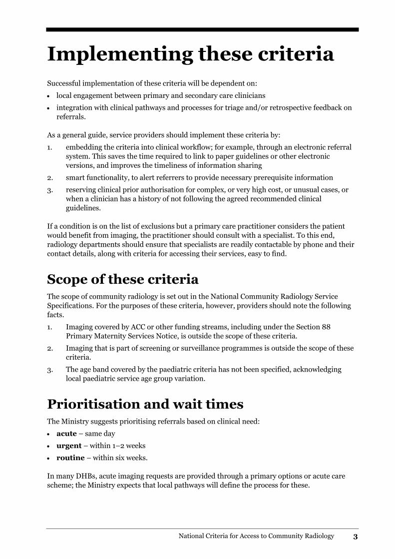
Implementing these criteria
Successful implementation of these criteria will be dependent on:
local engagement between primary and secondary care clinicians
integration with clinical pathways and processes for triage and/or retrospective feedback on
referrals.
As a general guide, service providers should implement these criteria by:
1. embedding the criteria into clinical workflow; for example, through an electronic referral
system. This saves the time required to link to paper guidelines or other electronic
versions, and improves the timeliness of information sharing
2. smart functionality, to alert referrers to provide necessary prerequisite information
3. reserving clinical prior authorisation for complex, or very high cost, or unusual cases, or
when a clinician has a history of not following the agreed recommended clinical
guidelines.
If a condition is on the list of exclusions but a primary care practitioner considers the patient
would benefit from imaging, the practitioner should consult with a specialist. To this end,
radiology departments should ensure that specialists are readily contactable by phone and their
contact details, along with criteria for accessing their services, easy to find.
Scope of these criteria The scope of community radiology is set out in the National Community Radiology Service
Specifications. For the purposes of these criteria, however, providers should note the following
facts.
1. Imaging covered by ACC or other funding streams, including under the Section 88
Primary Maternity Services Notice, is outside the scope of these criteria.
2. Imaging that is part of screening or surveillance programmes is outside the scope of these
criteria.
3. The age band covered by the paediatric criteria has not been specified, acknowledging
local paediatric service age group variation.
Prioritisation and wait times The Ministry suggests prioritising referrals based on clinical need:
acute – same day
urgent – within 1–2 weeks
routine – within six weeks.
In many DHBs, acute imaging requests are provided through a primary options or acute care
scheme; the Ministry expects that local pathways will define the process for these.
4 National Criteria for Access to Community Radiology
The Ministry encourages referrers to communicate expected wait times to their patients and
communicate with radiology services where they feel a referral is other than routine.
Provision of all routine imaging within six weeks is a ‘working towards’ benchmark in DHB
radiology departments.
The Ministry expects that reporting of all procedures will be completed within 24 to 48 hours,
and strongly recommends electronic distribution of reports. Radiology departments should
telephone significant findings to referrers on the day of imaging. All referrers should include
telephone numbers on the request form, to ensure ready contact.
Managing demand Managing the demand for diagnostic imaging is essential to:
ensure services are safe, efficient, effective and sustainable
manage radiology volumes and budgets, and reduce the wait time for patients in the
community.
Some factors that can impact on demand include:
lack of access to previous imaging reports or other clinical information
pressure from patients
factors affecting the clinician, such as inexperience.
Managing demand focuses on ensuring referrals are appropriate. The term ‘appropriate’ here
refers to a way of working based on agreed guidance: typically access criteria or clinical
pathways.
Best practice for referrals
Referrals may be inappropriate because a health practitioner refers a patient:
for a particular investigation when an alternative would have been preferable as it had
greater benefit and less risk
for an investigation at the wrong time
for an investigation when none was needed (either there was no relevant question to be
answered, there was no change in diagnosis or no management change would result).
It is also inappropriate not to refer a patient for an investigation when they need one.
Indications for diagnostic imaging may not always be clear-cut; primary health practitioners
should discuss with radiologists or refer for clinical review relevant specialists where
appropriate.
A useful investigation is one in which the result – positive or negative – may alter management
and improve the outcome for the patient. A significant number of radiological investigations do
not fulfil these aims, and may add unnecessarily to patient irradiation.
Health practitioners should take particular care in considering whether to order tests that
involve ionising radiation, especially in younger people.
National Criteria for Access to Community Radiology 5
A chest X-ray delivers approximately 0.04 mSv – the equivalent of eight days of natural
background radiation, while a CT of the abdomen and pelvis is approximately 14 mSv, or eight
years of natural background radiation. The Ministry expects all radiology providers to ensure
their equipment and imaging protocols are kept up to date, to deliver radiation doses that are as
low as practicably achievable.
The Ministry has developed the following principles to assist DHBs to establish effective
demand management processes.
Local governance
District health boards should establish formal local governance processes so that accountability
for managing the demand for community radiology referrals is clear and so that services can
maintain capacity and capability within budgets to the highest possible quality. The governance
process should allow for feedback on performance against the established guidelines and ‘fair’
usage expectations.
Managing budgets
All decision-makers (funders, providers and referrers) should regularly assess budgets and
volumes of referrals. In managing community radiology budgets, DHBs should make use of
alliancing arrangements, and make sure professions formally share information on clinical
management and budget decisions.
Prior authorisation
Prior authorisation from a DHB radiologist or relevant clinical specialist should only be required
for complex, or very high cost, or unusual cases, or when a referrer has a history of not following
the agreed recommended clinical pathways.
District health boards should make nominated consultants available to provide primary care
practitioners with advice on case management.
Clinical practice and ongoing education
District health boards should undertake regular clinical audit, to facilitate a shared
understanding of ‘reasonable practice’ between all decision-makers. They should offer clinical
education on the outcome of audits.
Legislative requirements of DHBs The Ministry of Health requires DHBs’ annual plans to ensure primary care services have direct
access to a complete suite of X-rays and ultrasound services (that is, abdomen, pelvis, renal,
small parts, deep venous thrombosis and musculoskeletal).
The Ministry also expects DHBs to provide mammography and fluoroscopy services; however,
these criteria do not apply to those services as service models and resource availability for them
vary across the country. Service provision of local nuclear medicine, double energy X-ray
absorption and magnetic resonance imaging currently varies. This document does not specify
minimum access criteria for these modalities; however, subsequent versions may do so.

6 National Criteria for Access to Community Radiology
These criteria fulfil the requirements of the National Community Radiology Service
Specifications, which require DHBs to define access criteria and expected waiting times for
diagnostic imaging. (These service specifications are due to be updated, but this requirement is
expected to remain.)
National Criteria for Access to Community Radiology 7
Criteria for access to
radiology
The following pages outline community radiology access criteria. The criteria indicate when
imaging is indicated and when it is not indicated, and provide guidance for referrers, under the
following headings:
X-ray
ultrasound
CT scans
paediatric imaging.
X-ray
Abdomen
Standard indications for X-ray referral:
diagnosis of constipation where patient history is unobtainable (eg, patient with autism or
special needs)
follow-up of radio-opaque (ie, evident on CT scout view) renal tract stones with a kidney,
ureter, bladder (KUB) X-ray
suspected renal tract stone according to local renal colic pathway criteria, where CT KUB is
unavailable.
Referral for community X-ray not typically indicated:
acute abdomen: discuss with acute surgical services or emergency services
vague central abdominal pain
suspected colorectal neoplasm (refer to colorectal cancer guidelines)
suspected constipation (other than in specific patient groups as above).
Ankle
Standard indications for X-ray referral:
undiagnosed pain present more than four weeks where the X-ray is expected to change
management
ankle pain with red flags
known osteoarthritis with symptoms meeting local criteria for surgical consideration (not
required if previously X-rayed within six months)
pain in previous arthroplasty
swelling, deformity or mass near the joint.

8 National Criteria for Access to Community Radiology
Red flags include:
persistent deep pain unrelated to activity
night pain in the absence of an obvious cause.
Referral for community X-ray not typically indicated:
suspected septic joint: refer for acute review at emergency department or orthopaedic
department
acute gout.
Chest
Standard indications for X-ray referral:
X-ray result will change patient management.
Referral for community X-ray not typically indicated:
screening for lung cancer in asymptomatic patient
pneumonia doesn’t require routine chest X-ray (CXR) follow-up unless there are risk factors
or red flags, including age > 50 years, significant smoking history, suspicious radiologic
findings on initial CXR or incomplete clinical resolution at six weeks (this is a guideline only
– there may be local pathways that apply) 1
routine assessment of hypertension
routine monitoring of known pulmonary sarcoidosis
routine X-ray for asbestos exposure surveillance
follow-up of nodules detected on chest X-ray or CT other than where recommended by
reporting or reviewing specialist (consider referral for respiratory specialist review)
initial investigation of heart murmur, unless signs of complications such as heart failure
routine follow-up of asymptomatic patients on amiodarone.
Elbow
Standard indications for X-ray referral:
undiagnosed pain present more than four weeks where the X-ray is expected to change
management
elbow pain with red flags
known osteoarthritis with symptoms meeting local criteria for surgical consideration (not
required if previously X-rayed within six months)
pain in previous arthroplasty
swelling, deformity or mass near the joint.
Red flags include:
persistent deep pain unrelated to activity
night pain in the absence of an obvious cause.
Referral for community X-ray not typically indicated:
suspected septic joint: refer for acute review
acute gout.
National Criteria for Access to Community Radiology 9
Hand/wrist
Standard indications for X-ray referral:
undiagnosed hand/wrist pain present more than four weeks where the X-ray is expected to
change management
hand/wrist pain with red flags
known osteoarthritis with symptoms meeting local criteria for surgical consideration (not
required if previously X-rayed within six months)
pain in previous arthroplasty
swelling, deformity or mass near the joint.
Red flags include:
persistent deep pain unrelated to activity
night pain in the absence of an obvious cause.
Referral for community X-ray not typically indicated:
suspected septic joint: refer for acute review
acute gout.
Guidance
Dedicated wrist views do not typically provide additional information to that obtained via
single postero-anterior (PA) hand view. Where inflammatory arthritis is suspected,
consider requesting an antero-posterior (AP) feet X-ray as well.
Hip
Standard indications for imaging referral:
undiagnosed hip pain present for more than four weeks where the X-ray is expected to
change management
hip pain with red flags
known osteoarthritis where symptoms meet local criteria for surgical consideration (not
required if previously X-rayed within six months)
pain in previous arthroplasty
swelling, deformity or mass near the joint.
Red flags include:
persistent deep pain unrelated to activity
night pain in the absence of an obvious cause.
Referral for community X-ray not typically indicated:
suspected septic arthritis: refer for acute review at emergency department or orthopaedic
department
mild symptoms and normal examination findings
follow-up of known or suspected osteoarthritis unless red flags develop or patient meets local
criteria for surgery.
10 National Criteria for Access to Community Radiology
Knee
Standard indications for X-ray referral:
undiagnosed knee pain present more than four weeks where the X-ray is expected to change
management
knee pain with red flags
known osteoarthritis with symptoms meeting local criteria for surgical consideration (not
required if previously X-rayed within six months)
pain in previous arthroplasty
swelling, deformity or mass near the joint.
Red flags include:
persistent deep pain unrelated to activity
night pain in the absence of an obvious cause.
Referral for community X-ray not typically indicated:
suspected septic arthritis: refer for acute review at emergency department or orthopaedic
department
mild symptoms and normal examination finding
follow-up of suspected or known osteoarthritis unless red flags develop or patient now meets
local clinical criteria for surgery
suspected meniscal and ligament injury.
Guidance
Routinely request standing knee X-rays. Such views demonstrate the magnitude of any
cartilage loss, which reflects the severity of any osteoarthritis.
Shoulder
Standard indications for X-ray referral:
undiagnosed shoulder pain present more than four weeks where the X-ray is expected to
change management
shoulder pain with red flags
known osteoarthritis with symptoms meeting local criteria for surgical consideration (not
required if previously X-rayed within six months)
pain in previous arthroplasty
swelling, deformity or mass near the joint.
Red flags include:
persistent deep pain unrelated to activity
night pain in the absence of an obvious cause.
National Criteria for Access to Community Radiology 11
Referral for community X-ray not typically indicated:
recent onset pain in the absence of red flags
frozen shoulder (unless the condition does not follow its expected natural history)
prerequisite for a trial of steroid injection (when a reasonable clinical diagnosis has been
made and red flags are excluded)
suspected septic arthritis: refer for acute review at emergency department or orthopaedic
department.
Sinuses
Guidance
Plain films are no longer recommended.
Skull
Standard indications for X-ray referral:
presence of a palpable vault abnormality that feels bony.
Referral for community X-ray imaging not typically indicated:
trauma: discuss with emergency department consultant. CT head may be appropriate
headache
epilepsy
cognitive impairment
middle or inner ear problems
suspected intracranial space occupying lesion.
Guidance
Refer suspected pituitary problems to a local relevant specialist.
Spine
Standard indications for X-ray referral:
spine pain more than eight weeks
spine pain with red flags
spine pain and osteoporosis or prolonged use of corticosteroids
focal neurological deficit (where recommended by local relevant specialist)
significant spinal deformity.
12 National Criteria for Access to Community Radiology
Red flags include:2
persistent deep pain unrelated to activity
night pain in the absence of an obvious cause
a history of cancer.
Referral for community X-ray not typically indicated:
acute uncomplicated spine pain without red flags (benign self-limiting condition).
Guidance
Where there is high clinical suspicion of infection or cancer, consult a local relevant
specialist.
Ultrasound
Abdomen
Standard indications for ultrasound referral:
abdominal mass or other palpable abdominal abnormality
painless jaundice without obvious cause
suspected gallstones: persistent/recurrent right upper quadrant pain
suspected pancreatic disease (limited resolution in obesity)
clinically suspected or radiologically suspected aortic aneurysm (AAA)
follow-up of AAA as per local guideline
abnormal liver function tests (LFTs); both gamma glutamyl transferase (GGT) and alanine
aminotransferase (ALT) elevated to greater than 1.5 times the upper limit of normal for more
than three months with no other clinical cause
abnormal LFTs suggestive of biliary tract obstruction or malignancy (persistently raised
alkaline phosphatase (ALP)/GGT± bilirubin).3
Referral for community ultrasound not typically indicated:
infective hepatitis
acute abdomen or suspected bowel obstruction (discuss with local relevant service)
dyspepsia
suspected colorectal neoplasm (refer to colorectal cancer guidelines)
clinically evident hernia in adults
screening for AAA.
Guidance
Discuss suspected pancreatic disease with a relevant local specialist. A CT scan may be
more appropriate.
National Criteria for Access to Community Radiology 13
Breast
Standard indications for ultrasound referral in the absence of local breast pathway:
women under 40 years of age with clinically benign or uncertain lump, or localised change in
texture
men with unexplained or suspicious unilateral breast enlargement
axillary lymph node enlargement or suspected lymph node enlargement in the absence of
obvious infectious cause.
Referral for community ultrasound not typically indicated:
breast pain alone
bilateral male breast enlargement.
Guidance
Referral to a local breast service for advice/assessment and multidisciplinary work-up,
is preferable, and where such a service is available locally (this supersedes these
recommendations).
Mammography (± ultrasound) is the appropriate investigation modality for women
over 40 years. If there is no breast clinic service available, refer these women directly
for mammography and ultrasound if required.
Carotid Doppler
Standard indications for imaging referral:
history of transient ischaemic attack or stroke with minor deficit where presentation meets
local pathway criteria
where no local pathway is in place and a relevant specialist has recommended a carotid
Doppler ultrasound.
Referral for community/outpatient imaging not typically indicated:
asymptomatic carotid bruits.
Groin
Standard indications for ultrasound referral:
non-reducible groin mass present for longer than three weeks. (If mass is suspicious of
cancer, discuss with local specialist.)
Referral for community ultrasound not typically indicated:
lymph nodes < 1.5 cm diameter and present less than three weeks
groin pain with no palpable mass.
Guidance
Most hernias can be diagnosed by clinical examination; ultrasound is rarely needed.
14 National Criteria for Access to Community Radiology
Hip
Referral for ultrasound not typically indicated:
suspected trochanteric bursitis. The underlying pathology in greater trochanteric pain
syndrome is most commonly gluteus tendinopathy, and ultrasound is not routinely required.
Referral for hip X-ray is recommended to identify bone or joint pathology.4
Neck
Standard indications for ultrasound referral:
salivary gland mass persisting for more than three weeks
suspected lymph node or undifferentiated neck mass – where swelling has persisted more
than three weeks, is > 1.5 cm size and there is no obvious infectious or other medical cause.5
Guidance
If a neck mass is suspicious for malignancy, discuss with a relevant local specialist.
If a patient has a prior history of a salivary gland tumour or cutaneous squamous cell
carcinoma SCC of head or face or has onset of facial nerve symptoms, discuss with
relevant surgical specialist; referral to a clinic may be more appropriate.
Pelvis
Standard indications for ultrasound referral:
intrauterine contraceptive device (IUCD) strings not visible on examination
post-menopausal bleeding after one year of amenorrhoea
pelvic mass on examination. Request a Ca125 and an urgent scan if there is a high index of
suspicion for ovarian malignancy
suspected ovarian cyst (unilateral pelvic pain for more than four weeks and/or pelvic mass or
unilateral tenderness)
pelvic pain more than six weeks unrelated to menstrual cycle, with pelvic inflammatory
disease excluded. Pre-referral expectation is that cervix has been visualised and swabs and
smear taken
abnormal pre-menopausal bleeding > 45 years old. Pre-referral expectation is that if IUCD
was present it has been removed for 3+ months.6
abnormal bleeding < 45 years old and one or more of the following risk factors for
endometrial hyperplasia:6
– weight >90 kg
– history of unopposed oestrogen or tamoxifen use
– nulliparity
– chronic anovulation ± infertility.

National Criteria for Access to Community Radiology 15
Referral for ultrasound not typically indicated:
routine follow-up of known fibroids7
follow-up of simple ovarian cyst < 5 cm diameter in asymptomatic premenopausal/low-risk
woman8
primary dysmenorrhoea
suspected endometriosis in the absence of a palpable mass
polycystic ovary syndrome where the required two out of three diagnostic criteria are fulfilled
by clinical and biochemical features (eg, oligomenorrhoea and clinical or biochemical
hyperandrogenism).9
Guidance
Refer women with acute non-pregnant pelvic pain in the absence of a palpable mass to
the appropriate specialty service.
For prolonged and/or heavy vagina bleeding after termination of pregnancy (TOP) or
post-partum, refer under Section 88 Primary Maternity notice up to two weeks post
miscarriage/TOP and six weeks post-partum).
Renal
Standard indications for ultrasound referral:
estimated glomerular filtration rate eGFR is consistently reduced for age after repeat testing
with the patient well hydrated:10
– < 70 years : eGFR is reduced to < 45 mls/min
– > 70 years: eGFR is reduced to < 30 mls/min
painless haematuria:
– persistent microscopic haematuria on two or more uncontaminated (epithelial cell count
< 15 x 106/L) mid-stream urinalyses (not dipstix), or
– macroscopic haematuria
polycystic kidneys: ultrasound screening when > 20 years age and a positive family history
with one or more first-degree relatives affected
recurrent urinary tract infections (UTI) in females with one or more of these risk factors for
an identifiable underlying cause:11
– repeated (more than two episodes) pyelonephritis (fever, chills, vomiting, costo-vertebral
angle tenderness)
– persistence of infection on urinalysis after completion of a prolonged three-week course of
appropriate antibiotics (ie, laboratory confirmed sensitivity)
– gross haematuria or persistent microscopic haematuria (> 15 x 106) on two separate
specimens) after resolution of infection
– recurrence of infection after three months of completed antibiotic prophylaxis
– urea-splitting organisms (eg, proteus, klebsiella, pseudomonas)
– history of abdomino-pelvic malignancy or immunocompromise
– history of urinary tract surgery or calculi
– obstructive symptoms with straining and weak stream
16 National Criteria for Access to Community Radiology
recurrent or persistent UTI in males
suspected renal colic in pregnancy. For all other patients, consider referral for CT KUB
suspected urinary retention with palpable/suspected enlarged bladder.
Referral for community ultrasound not typically indicated:
recurrent uncomplicated UTIs in adult females (underlying abnormalities are uncommon)
investigation of hypertension
elevated prostate-specific antigen
lower urinary tract symptoms
investigation of isolated proteinuria (discuss with local relevant specialist)
serial ultrasounds for polycystic kidneys, unless there are clinical symptoms.
Scrotum
Standard indications for imaging referral:
scrotal masses with concerning features (eg, testicular mass, painless, non-transilluminating,
rapidly growing (urgent urology referral recommended))
scrotal masses where either the clinical diagnosis is in doubt or it is unclear if the swelling is
testicular or extra-testicular
new hydrocoele in adults (may be secondary to testicular cancer).
Referral for community imaging not typically indicated:
non-solid (transilluminating) scrotal masses
hydrocoele in children
long-standing hydrocoele in adults
acute inflammatory conditions – only refer for ultrasound if symptoms and/or swelling fail to
resolve with antibiotics
chronic testicular pain in the absence of abnormality on examination.
Guidance
Refer urgently to surgical service for surgery if the following conditions are suspected:
testicular torsion
testicular cancer
strangulated inguinal hernia.
Scrotal masses can often be diagnosed clinically. If unsure, seek a second opinion from a
general practitioner colleague or specialist.
National Criteria for Access to Community Radiology 17
Shoulder
Standard indications for ultrasound referral:
pain and restricted movement that persists after eight weeks of conservative treatment
including physiotherapy and/or cortisone injection
when a full thickness tear is suspected and immediate surgical repair is being considered.
Guidance
Radiology is not a prerequisite for a trial of steroid injection when a reasonable clinical
diagnosis has been made and red flags have been excluded.
Soft tissue
Standard indications for community imaging referral:
soft tissue mass with red flags; however, specialist assessment is preferred, so only request
imaging if there is likely to be a delay before the patient is seen
suspicion of a foreign body where not covered by ACC.
Red flags include a soft tissue mass with any of the following characteristics):12
growing
> 5 cm in size
deep to deep fascia (limited mobility, less mobile with muscle flexion)
painful (most malignant lumps are painless; pain suggests nerve or bone involvement)
recurring after a previous excision.
Guidance
Apply caution in the use of ultrasound, as its ability to characterise solid mass lesions is
limited and incorrect diagnosis can lead to significant treatment delays.
Consider requesting a plain X-ray as well.
If a sarcoma is suspected, reserve biopsy for an orthopaedic or sarcoma specialist.
Thyroid
Standard indications for ultrasound referral:
palpable nodules
euthyroid goitre.
Referral for community ultrasound not typically indicated:
thryotoxicosis (with or without goitre)13
goitre with hypothyroidism.
18 National Criteria for Access to Community Radiology
Guidance
Red flags for thyroid malignancy, consider discussing with a local relevant specialist
service where a patient presents with:
< 20 years or > 60 years
history of head or neck malignancy
family history of thyroid cancer
rapid growth of a nodule
hard, ill-defined or fixed nodule
hoarseness, dysphagia or dysphonia
cervical lymphadenopathy.
Vascular
Standard indications for ultrasound referral:
pulsatile mass for investigation
suspected DVT (refer to local pathway if available)
proximal superficial thrombophlebitis in thigh.
Referral for community ultrasound not typically indicated:
suspected venous and arterial insufficiency – unless directed by local pathway.
Guidance
For patients with progressive uni- or bilateral lower limb oedema, consider referral for
abdomino-pelvic ultrasound, to exclude proximal lymphatic obstruction.
CT scans
CT head
Standard indications for CT referral:
undiagnosed cognitive impairment with one or more high-risk feature:14
– age < 60 years
– rapid (ie, one or two months) unexplained decline in cognition or function
– recent and significant head trauma
– unexplained neurological symptoms (eg, new onset of severe headache or seizures)
– history of cancer (especially in sites and types that metastasize to the brain)
– use of anticoagulants or history of bleeding disorder
– history of urinary incontinence and gait disorder early in the course of dementia (as may
be found in normal pressure hydrocephalus)
– any new localizing sign (eg, hemiparesis or a Babinski reflex)
– unusual or atypical cognitive symptoms or presentation (eg, progressive aphasia)
– gait disturbance
National Criteria for Access to Community Radiology 19
headaches where at least one of the following apply:
– new onset > 50 years
– change in pattern of headaches with increase in frequency or severity
– aggravated by exertion or Valsalva
– associated with nausea and vomiting
– background systemic illness with cerebral complications or involvement; especially
malignancy (breast, lung, melanoma).
Guidance
While CT may be appropriate as part of the work-up, initially discuss with local relevant
specialist for patients who have:
focal neurological signs
acute cognitive decline or change in personality.
CT abdomen
CT KUB
Standard indications for CT KUB referral:
non-pregnant patients with renal colic according to local pathway.
CT colonography15,16, 17, 18
Standard indications for CT colonography (CTC) referral in patients where colorectal cancer is
suspected:
symptomatic patients over 80 years
patients with co-morbidities when colonoscopy presents a higher risk (eg, patients on
warfarin therapy, respiratory risk from sedation)
patients presenting with abdominal mass
following failed or incomplete colonoscopy
patients with symptoms which are average to low risk for malignancy (patients who
previously would have been referred for barium enema).
Referral for CTC not typically indicated (ie, refer for colonoscopy):
diarrhoea as the predominant presenting symptom
known polyp syndromes (including familial) where biopsy/removal is likely to be required
suspected inflammatory bowel disease where mucosal visualisation and biopsy are required
for diagnosis
young patients (< 40 years).
Guidance
Referral is not typically indicated for either CTC or colonoscopy where there is:
abdominal pain alone
constipation as a single symptom
irritable bowel syndrome (consider specialist referral first)
20 National Criteria for Access to Community Radiology
uncomplicated CT-proven diverticulitis without suspicious radiological features.
The local DHB is likely to triage referrals for investigation of bowel symptoms to either
CTC or colonoscopy, depending on clinical presentation and resource availability.
CTC requires bowel preparation similar to colonoscopy, including fasting. The procedure
involves rectal air insufflation and changing position on the scanner table.
Where patient fitness level would preclude active treatment if a cancer is diagnosed, a
minimal preparation CT colon (MPCT ) should be considered. Discussion with local
radiologist recommended.
The ‘miss’ rate of lesions > 1 cm with both well-performed colonoscopy and CTC is
approximately 6 percent.
CTC is not intended for the detection of diminutive polyps <5mm.
CT sinus
Guidance
Sinus CT is not generally indicated without failed medical management. The main role of
sinus CT is for pre-surgical planning, rather than determining the need for surgery.
Paediatric imaging
X-ray – chest
Standard indications for X-ray referral:
lower respiratory tract disease (including asthma/bronchiolitis/pneumonia) unresponsive to
treatment
inhalation or suspected inhalation of foreign body.
Referral for X-ray not typically indicated:
incidental finding of a murmur
uncomplicated (afebrile) presentation of asthma/bronchiolitis.
X-ray – lower limb
Referral for X-ray not typically indicated:
Osgood-Schlatters, Sever’s and other apophysitides
X-ray – pelvis/hips
Standard indications for X-ray referral:
pain
limp
risk factors/soft signs or suspected developmental dysplasia of the hip (DDH).
National Criteria for Access to Community Radiology 21
Guidance
Capital femoral epiphyses ossify on average at 5–6 months of age; DDH can usually be
reliably excluded from this age onwards on X-ray.19
Slipped upper femoral epiphysis requires urgent orthopaedic referral.
Ultrasound – hips
Standard indication for ultrasound referral:
unstable or dislocated hip in child less than 3–4 months age; also refer to orthopaedic
specialist.
Referral for ultrasound not typically indicated:
soft signs (asymmetric buttock creases, leg length discrepancy, clicky hips) or risk factors
(breech presentation, family history): refer for plain X-ray at 5–6 months.
Ultrasound – neonatal spine
Standard indications for ultrasound referral:
sacral dimple or pit: non-simple (ie, with at least one of the following criteria):18
– outside the natal cleft (> 2.5 cm from anal verge in neonate)
– associated with cutaneous stigmata of spinal dysraphism – hairy tuft, haemangioma
– > 5 mm diameter
– deep (bottom of dimple not visible).
Referral for ultrasound not typically indicated:
simple isolated dimples within the gluteal cleft
child more than eight weeks age: ultrasound spine not technically feasible with ossification of
the posterior elements. Suggest discussion or review by local specialist.
Ultrasound – renal
Standard indications for ultrasound referral:
child < 12 months with first-time documented UTI
child of any age with recurrent UTI (no previous imaging)
child of any age with complicated UTI (eg, pyelonephritis, atypical UTI)
follow-up of antenatal hydronephrosis or other renal abnormality as recommended by
reporting radiologist.
Referral for ultrasound not typically indicated:
asymptomatic bacteriuria.
22 National Criteria for Access to Community Radiology
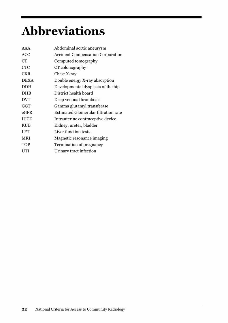
Abbreviations
AAA Abdominal aortic aneurysm
ACC Accident Compensation Corporation
CT Computed tomography
CTC CT colonography
CXR Chest X-ray
DEXA Double energy X-ray absorption
DDH Developmental dysplasia of the hip
DHB District health board
DVT Deep venous thrombosis
GGT Gamma glutamyl transferase
eGFR Estimated Glomerular filtration rate
IUCD Intrauterine contraceptive device
KUB Kidney, ureter, bladder
LFT Liver function tests
MRI Magnetic resonance imaging
TOP Termination of pregnancy
UTI Urinary tract infection
National Criteria for Access to Community Radiology 23
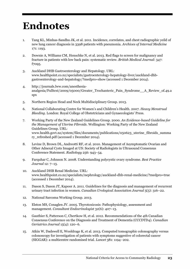
Endnotes
1. Tang KL, Minhas-Sandhu JK, et al. 2011. Incidence, correlates, and chest radiographic yeild of
new lung cancer diagnosis in 3398 patients with pneumonia. Archives of Internal Medicine
171: 1193.
2. Downie A, Williams CM, Henschke N, et al. 2013. Red flags to screen for malignancy and
fracture in patients with low back pain: systematic review. British Medical Journal: 347:
f7095.
3. Auckland DHB Gastroenterology and Hepatology. URL:
www.healthpoint.co.nz/specialists/gastroenterology-hepatology-liver/auckland-dhb-
gastroenterology-and-hepatology/?medpro=show (accessed 1 December 2014).
4. http://journals.lww.com/anesthesia-
analgesia/Fulltext/2009/05000/Greater_Trochanteric_Pain_Syndrome__A_Review_of.49.a
spx
5. Northern Region Head and Neck Multidisciplinary Group. 2013.
6. National Collaborating Centre for Women’s and Children’s Health. 2007. Heavy Menstrual
Bleeding. London: Royal College of Obstetricians and Gynaecologists’ Press.
7. Working Party of the New Zealand Guidelines Group. 2000. An Evidence-based Guideline for
the Management of Uterine Fibroids. Wellington: Working Party of the New Zealand
Guidelines Group. URL:
www.health.govt.nz/system/files/documents/publications/050623_uterine_fibroids_summa
ry_refreshed.pdf (accessed 1 December 2014).
8. Levine D, Brown DL, Andreotti RF, et al. 2010. Management of Asymptomatic Ovarian and
Other Adnexal Cysts Imaged at US: Society of Radiologists in Ultrasound Consensus
Conference Statement. Radiology 256: 943–54.
9. Farquhar C, Johnson N. 2008. Understanding polycystic ovary syndrome. Best Practice
Journal 12: 7–13.
10. Auckland DHB Renal Medicine. URL:
www.healthpoint.co.nz/specialists/nephrology/auckland-dhb-renal-medicine/?medpro=true
(accessed 1 December 2014).
11. Dason S, Dason JT, Kapoor A. 2011. Guidelines for the diagnosis and management of recurrent
urinary tract infection in women. Canadian Urological Association Journal 5(5): 316–22.
12. National Sarcoma Working Group. 2013.
13. Elston MS, Conaglen JV. 2005. Thyrotoxicosis: Pathophysiology, assessment and
management. Consultant Endocrinologist 32(6): 407–13.
14. Gauthier S, Patterson C, Chertkow H, et al. 2012. Recommendations of the 4th Canadian
Consensus Conference on the Diagnosis and Treatment of Dementia (CCCDTD4). Canadian
Geriatrics Journal 15(4): 120–6.
15. Atkin W, Dadswell E, Wooldrage K, et al. 2013. Computed tomographic colonography versus
colonoscopy for investigation of patients with symptoms suggestive of colorectal cancer
(SIGGAR): a multicentre randomised trial. Lancet 381: 1194–202.

24 National Criteria for Access to Community Radiology
16. Sanders A, Stevenson C, Pearson JF, et al. 2013. A novel pathway for investigation of colorectal
symptomswith colonoscopy or computed tomography colonography. New Zealand Medical
Journal 126(1382): 45.
17. Banerjee S, Van Dam J. 2006. CT colonography for colon cancer screening. Gastrointestinal
Endoscopy 63: 121–33.
18. Starship Clinical Guidelines. URL: www.starship.org.nz/for-health-professionals/starship-
clinical-guidelines (last accessed 2 December 2014).
19. Zywicke HA, Rozzelle CJ. 2011. Sacral Dimples. Pediatrics in Review 32(3): 109–14.