![9 9 1 1 0 0 2 2 , , 0 0 3 3 y y a MM a d d e e e e j j a M ... · 30.05.2019 · Legislative Updates from the Office of Rep. Nasif Majeed Representative Nasif Majeed [majeedla@ncleg.net]](https://static.fdocuments.net/doc/165x107/5fa4c96e56934812f045a434/9-9-1-1-0-0-2-2-0-0-3-3-y-y-a-mm-a-d-d-e-e-e-e-j-j-a-m-30052019-legislative.jpg)
nasif radiology
65
X-RAY Eng: Mohamed Gad
-
Upload
mohamed-gad -
Category
Technology
-
view
23 -
download
0
Transcript of nasif radiology

X-RAY
Eng: Mohamed Gad

CHAPTER 1: INTRODUCTION
NGOJO 3
X-rays were discovered in 1895 when Wilhelm Conrad Roentgen observed that a screen coated with a barium salt fluoresced when placed near a cathode ray tube. Roentgen concluded that a form of penetrating radiation was being emitted by the cathode ray tube and called the unknown rays, X-rays.

NGOJO 4
First X-ray Image
What are X-rays?• X rays are the ionizing electromagnetic radiation emitted from a highly evacuated high-voltage tube. Inner orbital electrons in the target anode are stimulated to emit x-radiation via bombardment by a stream of electrons from a heated cathode.
• X-rays, like gamma rays, are penetrating and carry enough energy to ionize atoms in their path. Nearly identical to gamma rays, x-rays require shielding to reduce their intensity and minimize the danger of tissue damage to personnel. Mishaps with x-rays can cause severe radiation burns and deep tissue damage and can lead to various cancers.
NGOJO 5

Radiation is energy in the form of waves or particles.
Radiation which is high enough in energy to cause ionization is
called ionizing radiation. It includes particles and rays given
off by radioactive material and high-voltage equipment.
Ionizing radiation includes x-rays, gamma-rays, beta particles,
alpha particles, and neutrons.
Without the use of monitoring equipment, humans are not able
to "find" ionizing radiation. In contrast to heat, light, odors and
noise, humans are not able to see, feel, taste, smell, or hear
ionizing radiation.
NGOJO 6
What is Radiation?

Units of Radiation Exposure and Dose
• Exposure (Roentgens)
• Dose Equivalence (Sievert)Relative biological effectiveness of different types of ionising radiation
• The Effective Dose Rate (Sievert)
• Absorbed dose (Gray)
NGOJO 7

DoseInternational Commission on Radiological Protection (ICRP) Prescribed Limits per annum• Members of public
• Radiation workers
20 mSv per annum above background150 mSv to eye500 mSv to hands
1 mSv per annum above background5 mSv to eye20 mSv to hands
•Pregnant women must receive no more than 2mSv per annum
NGOJO 8
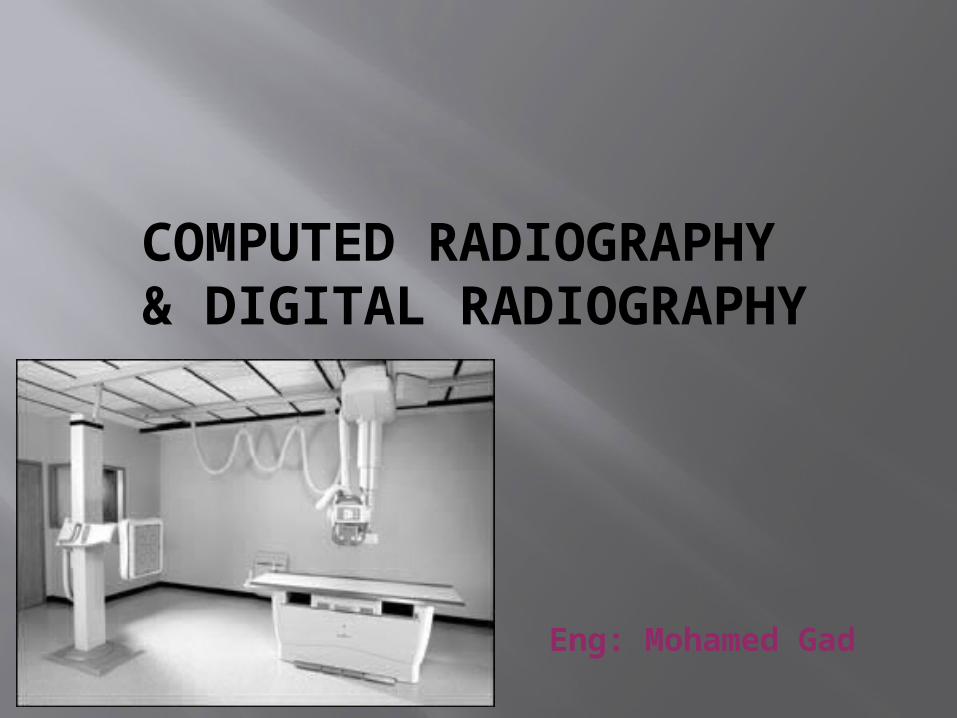
COMPUTED RADIOGRAPHY & DIGITAL RADIOGRAPHY
Eng: Mohamed Gad
Digital X-ray Technology
There are two ways to obtain Digital X-rays:
• Computed Radiography
• Digital Radiography

Computed Radiography (CR)
• A Digital way of doing general radiography
with Conventional X-ray machines
What is CR?
• Computed Radiography (CR) is a process of capturing radiographic data from a conventional X-ray machine and processing the data digitally to produce crisp and high quality radiographic images

What is CR?…
• For exposure, an Imaging Plate (IP) is placed in a cassette instead of a piece of film. The IP captures and "stores" the X-rays
• The image is "developed" in a CR reader instead of a film processor. The CR reader extracts the information stored in the plate and produces a digital image
What is CR?…
• Computed Radiography is a digital image acquisition process that produces images that have much better contrast than a Conventional X-ray film-screen system
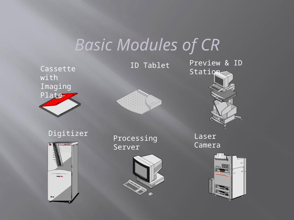
Basic Modules of CR
M A TRIX LR 3300
Digitizer
Preview & ID Station
Processing Server
ID Tablet
Laser Camera
Cassette withImaging Plate
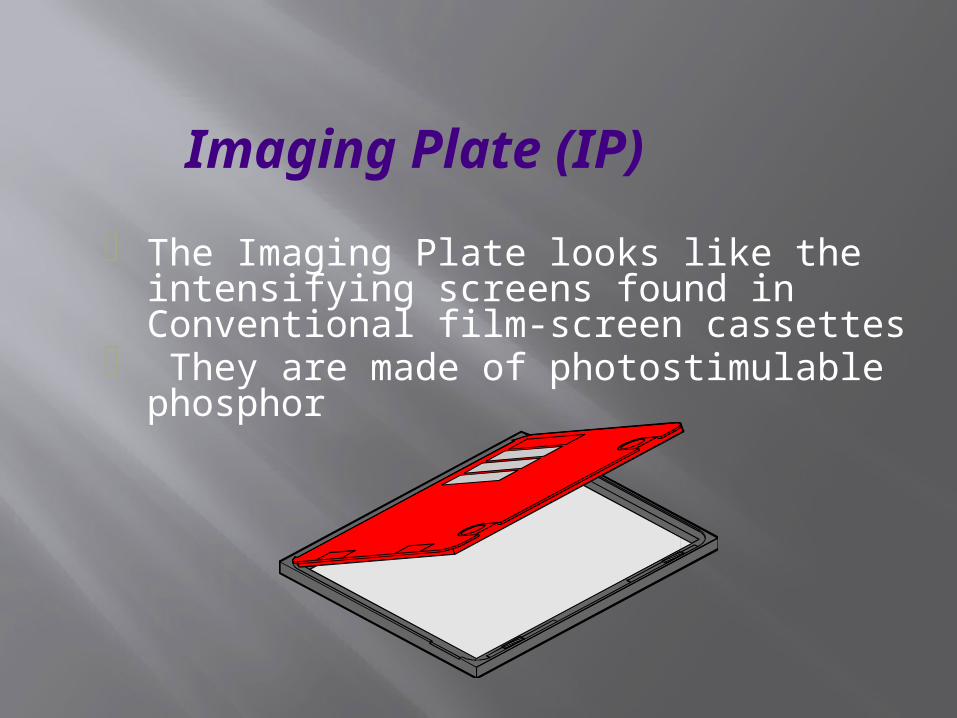
Imaging Plate (IP)
• The Imaging Plate looks like the intensifying screens found in Conventional film-screen cassettes
• They are made of photostimulable phosphor
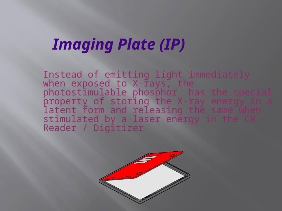
Imaging Plate (IP)
• Instead of emitting light immediately when exposed to X-rays, the photostimulable phosphor has the special property of storing the X-ray energy in a latent form and releasing the same when stimulated by a laser energy in the CR Reader / Digitizer
Imaging Plate (IP)…
• Storage phosphors are unique because they respond to a very wide range of X-ray exposures
• This latitude gives the flexibility in selecting X-ray technique and takes care of under or over exposure
• Regardless of the exposure, the image can be displayed correctly
• As a consequence, retakes due to inappropriate exposures are drastically reduced
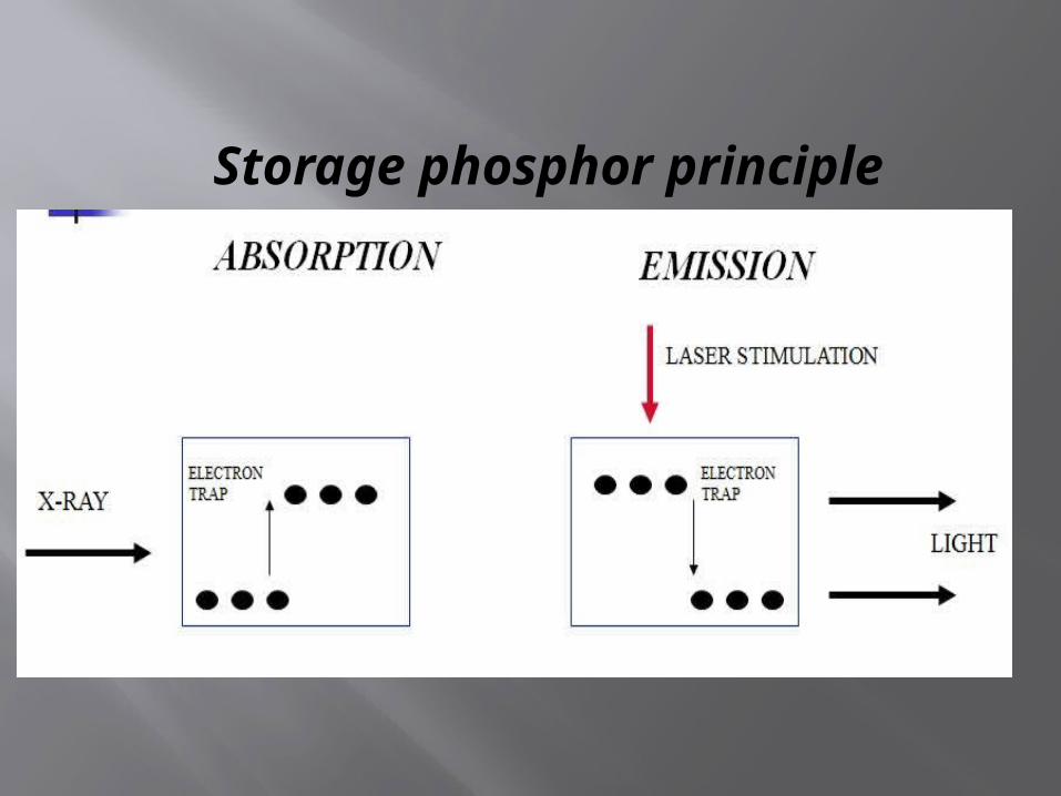
Storage phosphor principle
laser stimulation
EmissionEmissionAbsorptionAbsorption
x-rays
electrontrap
electrontrap
Storage phosphor principle…
• The imaging plate is coated with photostimulable phosphor, also called storage phosphor
• The phosphor material is generally a kind of Bariumfluorohalide
• The Imaging Plate contains not only the phosphor layer, but also a protective coat, a conductive layer, support and laminate layers

Storage phosphor principle…
• Incident X-rays excite electrons into a higher energy level (electron traps)
• A latent image is created in the form of “stored energy”
• Stimulation with a scanning laser beam releases electrons
• Typical wavelength of the stimulating laser is 633 nm• Falling back, electrons emit luminescent light • Typical wavelength of the emitted light is 390 nm

CR Readout
Storage phosphor principle…
• The emitted light intensity is proportional to the original incident X-ray intensity
• The emitted light is captured with an optical array and a photomultiplier and is digitized
• The residual image is erased from the plate by an intense light source, which returns all electrons to their original state. This makes the plate ready to be reused for new exposures
• The storage phosphor plate fits inside a standard size cassette and is exposed to X-rays exactly like film
• The X-ray energy is stored on the plate in the form of latent energy
How is a Storage Phosphor plate exposed?

Patient ID Station • Before exposing the cassette, the patient
demographic and exam data is stored on the microchip attached on cassette
• This is done by inserting the cassette in a slot of ID station and entering the data with the help of keyboard
• When cassette is inserted in digitizer after X-ray exposure, the digitizer reads both patient data as well as X-ray exposure data
• The two data are combined to display images along with patient data
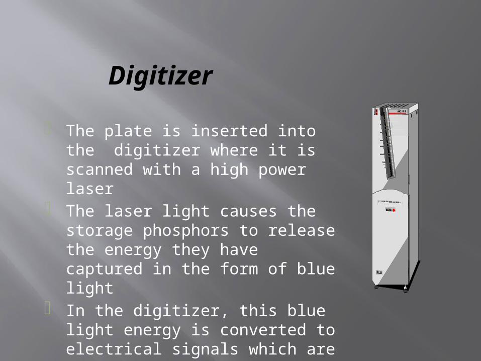
Digitizer
• The plate is inserted into the digitizer where it is scanned with a high power laser
• The laser light causes the storage phosphors to release the energy they have captured in the form of blue light
• In the digitizer, this blue light energy is converted to electrical signals which are then digitized to produce digital images
What happens to a Storage Phosphor Plate after it is scanned?
• After exposure and scanning, the phosphor plate is "erased" by exposing to a bright light exposure within the digitizer
• The previous image stored in the phosphors is removed and the plate is ready to be exposed again
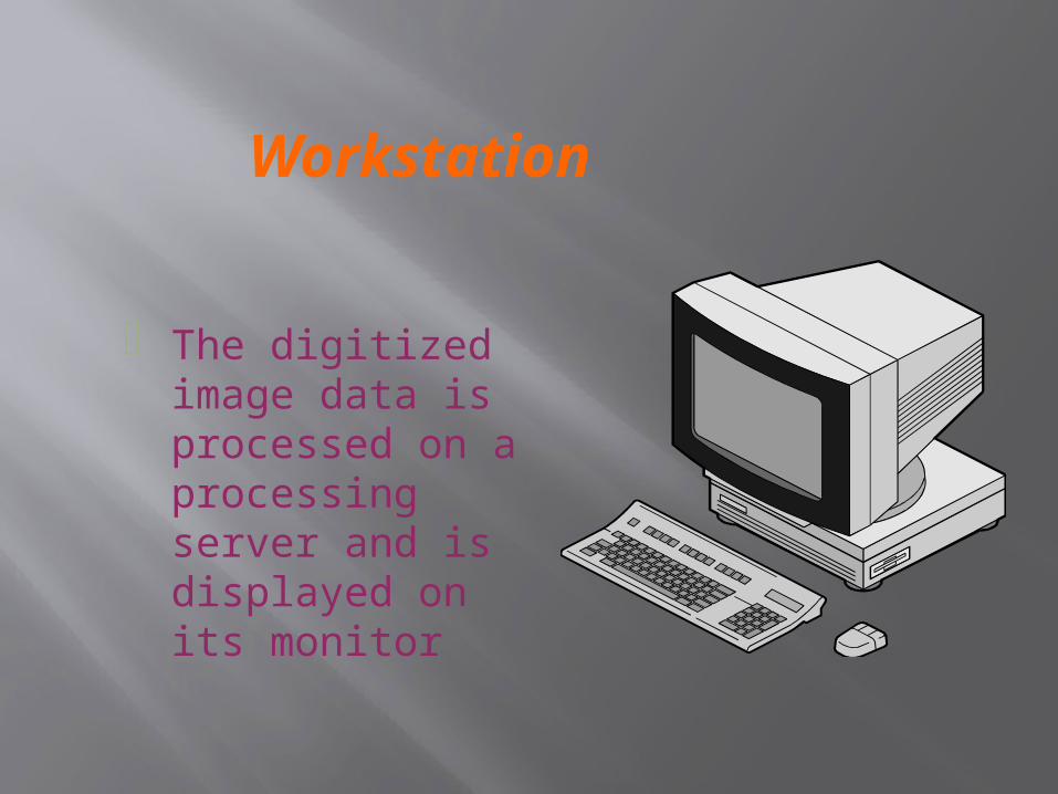
Workstation
• The digitized image data is processed on a processing server and is displayed on its monitor
How many times can we use a Storage Phosphor Plate?
• The life of a phosphor plate depends on how carefully it is handled. Physical damage to the plate will limit its useful life
• If properly cared for, a plate will produce thousands of images
• Imaging Plates are known to last more than 50000 Exposure Cycles
Does CR require X-ray machine replacement?
• No, CR uses the existing X-ray equipment• One CR system can support multiple x-ray
rooms
How is the workflow different with CR ?
• Instead of taking the film cassette to a dark room for processing, the technologist takes the cassette with imaging plate to the CR reader for digital processing of the image
• Instead of manually taking the films to the reporting radiologists , the softcopy images reach the workstation almost immediately

How is the workflow different with CR ?…
• The time required to acquire a Digital image is much less compared to conventional darkroom process
• The film is the first product in Conventional where as the film is the last product in CR
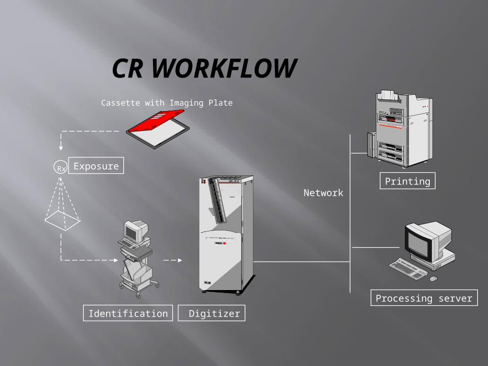
CR WORKFLOW
Rx
Network
Digitizer
Printing
Identification
Processing server
Exposure
Cassette with Imaging Plate

Rx
Network
Digitizer
Printing
Identification
Processing server
Exposure
Cassette with Imaging Plate
The cassettes fit into X-ray table. After the exposure, the cassette is identified in the ID-station. Here patient and exam related information is stored. Next the digitizer reads the identification data, handles the plates, reads the image and sends out a raw dataset in DICOM-format. The automatic processing server processes the image according to the type of exam. For each type of exam, an optimized image processing parameter set-up is used. The processing server then pushes the processed image to the preview station for previewing. After approval the image is routed to other destinations such as a printer, a review station and an archive server.

Digital Radiography
This technique is performed by digital X-ray machines with flat panel detectors
Digital Radiography
Digital Radiography uses two types of detectors:• Direct• Indirect
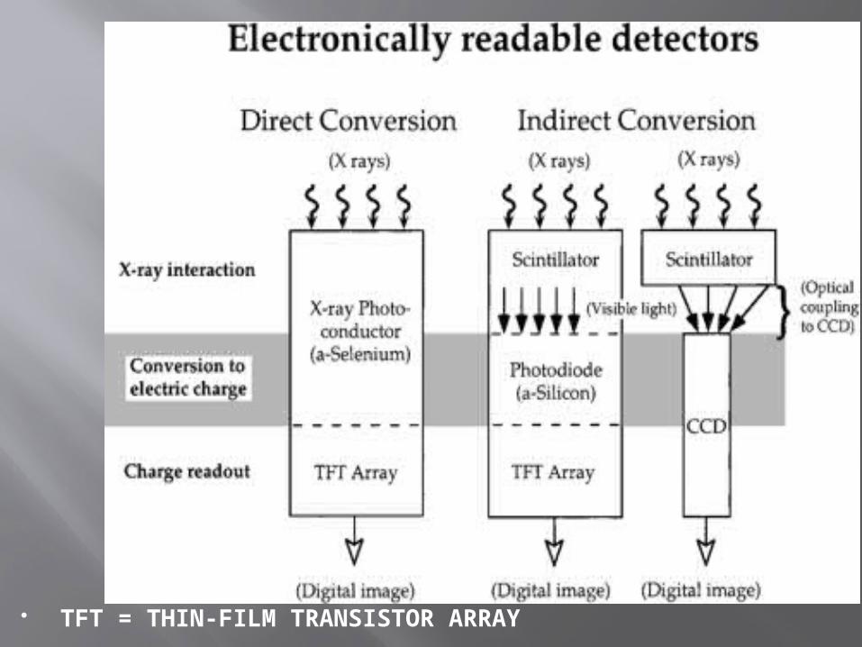
TFT = THIN-FILM TRANSISTOR ARRAY
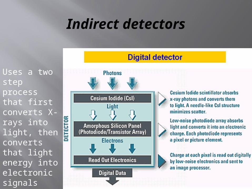
Indirect detectors
Uses a two step process that first converts X-rays into light, then converts that light energy into electronic signals

Direct detectors
• Direct detectors automatically convert X-rays into electronic signals.
X-rays interact with semiconductor material Amorphous selenium
X-rays converted directly into electrical charge No intermediate steps
Direct detectors
• The flat panel detector consists of an amorphous selenium semiconductor X-ray absorber coating over a thin-film transistor array of amorphous silicon
• In this system,X-ray photons are immediately converted into electronic signal
• This immediate conversion eliminates the need for additional steps to capture and convert incident X-ray energy
• Corrective image processing which can result in increased image noise is reduced with the highly efficient X-ray energy conversion of direct DR

Limitations of Conventional X-ray process (X-ray film / Screen/Darkroom)
• Film has a limited exposure latitude i.e less detail contrast
• Time consuming & cumbersome• Intolerant to exposure errors• Repeat X-rays ( More radiation exposure )• Film wastage
Limitations of Conventional X-ray process (X-ray film / Screen/Darkroom)…
• Cannot be duplicated without loss of quality• Film storage is a problem• Scatter radiation reduces contrast and increases
patient dose• Quality control is an issue
What are the advantages of Digital X-rays?
• Post processing (soft tissue and bony details can be viewed at same time )
• Reduction in hazardous X-ray dose to patients • More info on one image • Constant image quality• Possibility of viewing X-ray images wherever
needed
What are the advantages of Digital X-rays?…
• Digital images are of extremely high quality • Digital images have a future scope of better
image management • Facility of giving multiple images of
investigative studies on a single high definition laser film

How is Digital X-ray similar to Conventional basic radiography process ?…
• Radiography consists of following functions:
-Image data acquisition
-Image processing
-Reproduction of image
-Storage
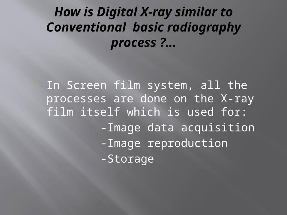
How is Digital X-ray similar to Conventional basic radiography process ?…
• In Screen film system, all the processes are done on the X-ray film itself which is used for:
-Image data acquisition
-Image reproduction
-Storage
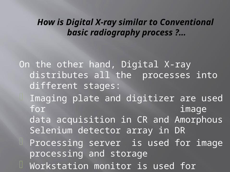
How is Digital X-ray similar to Conventional basic radiography process ?…
On the other hand, Digital X-ray distributes all the processes into different stages:
• Imaging plate and digitizer are used for image data acquisition in CR and Amorphous Selenium detector array in DR
• Processing server is used for image processing and storage
• Workstation monitor is used for image reproduction

What is the role of Digital X-ray in PACS Environment?
• Digital X-ray is the only film less way to link the existing general radiography set up into the digital environment of PACS
Features of Digital X-ray
• Image enhancement• Printing• Annotation• Black border• Panoramic dental package • Full leg / Full Spine

Under/over exposure
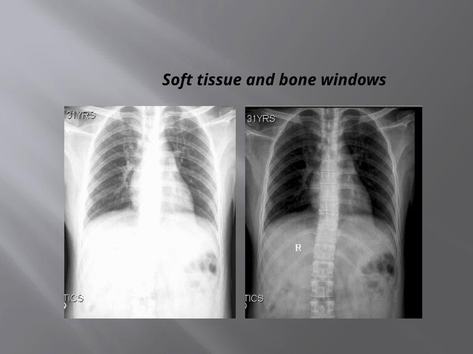
Soft tissue and bone windows

Digital image manipulation
• Image pre-processing • Scale the data to appropriate range• Contrast enhancement – Anatomy specific
grayscale manipulation• Spatial frequency enhancement
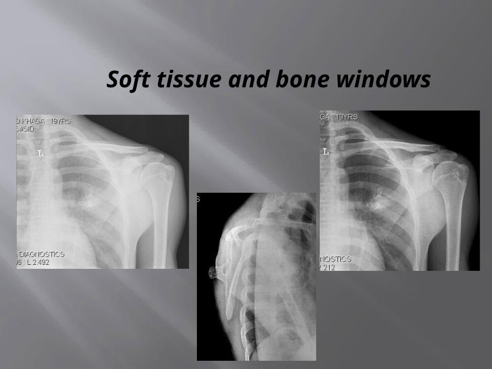
Soft tissue and bone windows
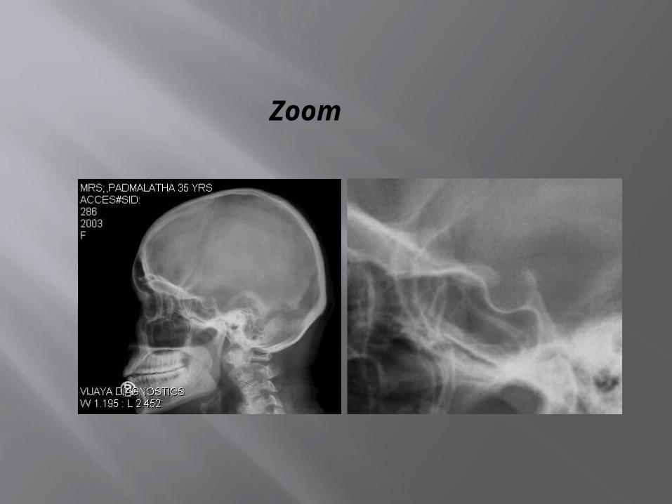
Zoom
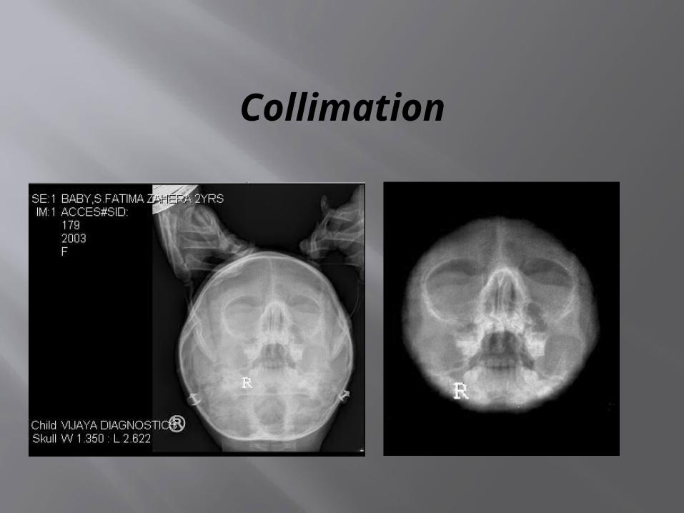
Collimation

Collimation
MISS.PADMINI 20YRS,F 14/08/2004 20:15:01SKULL, MASTIODS 23
MISS.PADMINI 20YRS,F 14/08/2004 20:17:01SKULL, MASTIODS 23
MISS.PADMINI 20YRS,F 14/08/2004 20:15:01SKULL, MASTIODS 23
MISS.PADMINI 20YRS,F 14/08/2004 20:17:01SKULL, MASTIODS 23

Magnifying glass
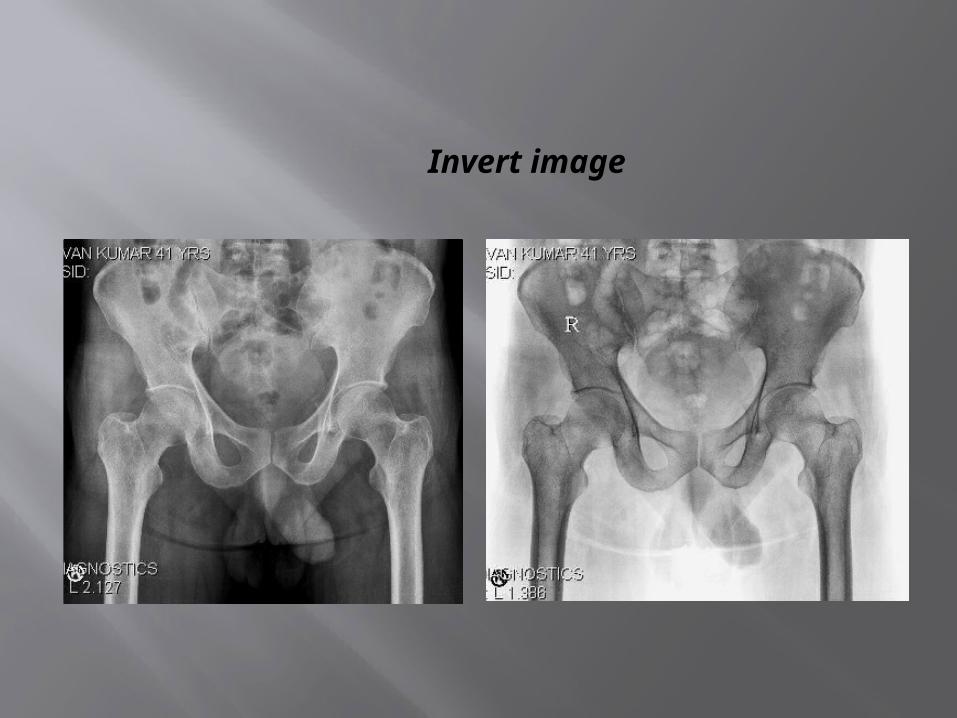
Invert image

Invert image
DIGITIZER, EMERGENCY 1O/07/2004 22:3O:28 DIGITIZER, EMERGENCY 1O/07/2004 22:3O:28
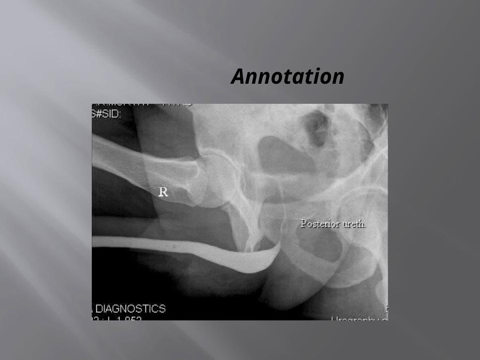
Annotation
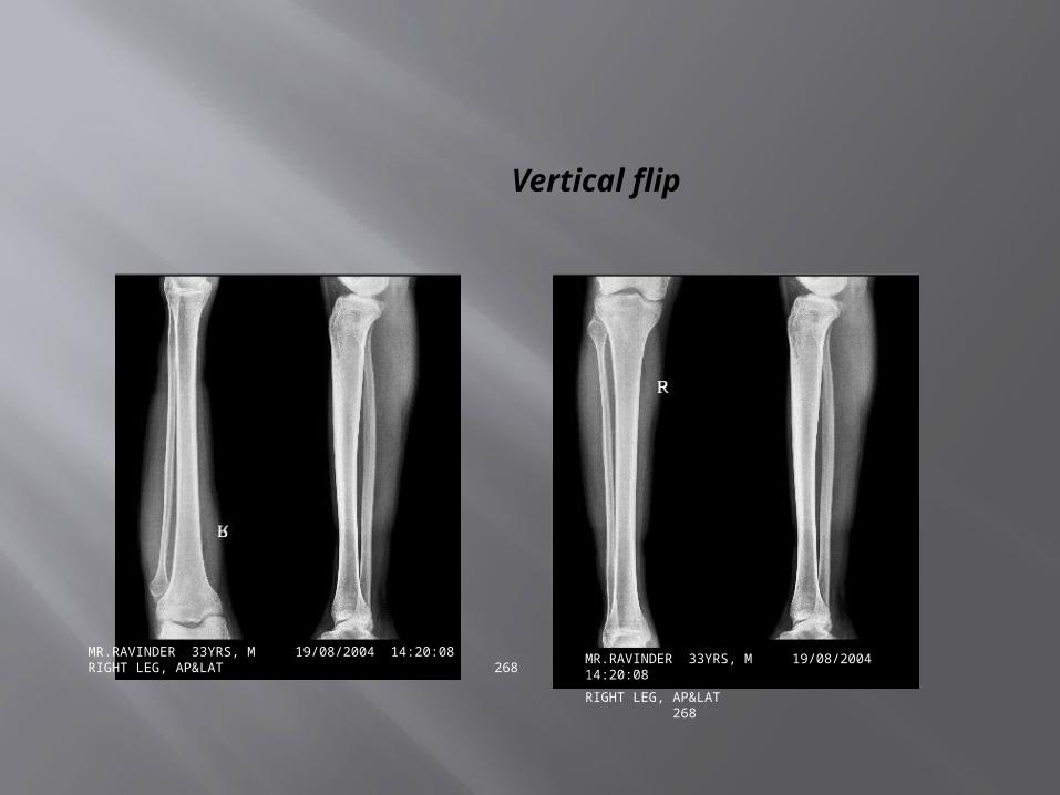
Vertical flip
MR.RAVINDER 33YRS, M 19/08/2004 14:20:08RIGHT LEG, AP&LAT 268
MR.RAVINDER 33YRS, M 19/08/2004 14:20:08
RIGHT LEG, AP&LAT 268
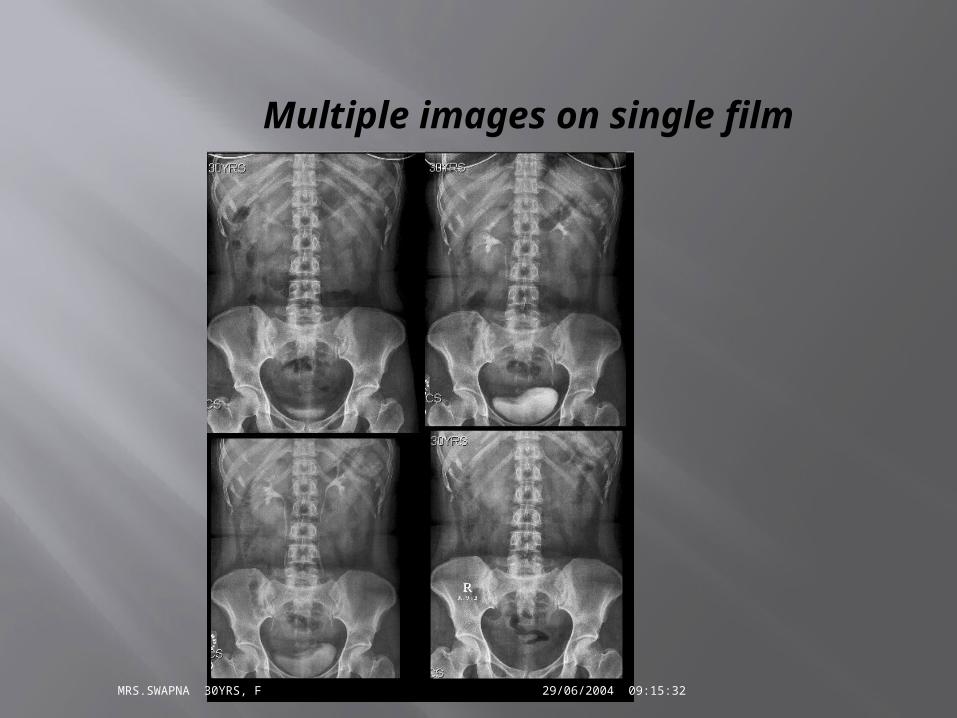
Multiple images on single film
MRS.SWAPNA 30YRS, F 29/06/2004 09:15:32

Multiple images on single film
MRS.FARIDA 30YRS ,F 12/8/2004 09:15:23BARIUM MEAL FOLLOW THROUGH 286
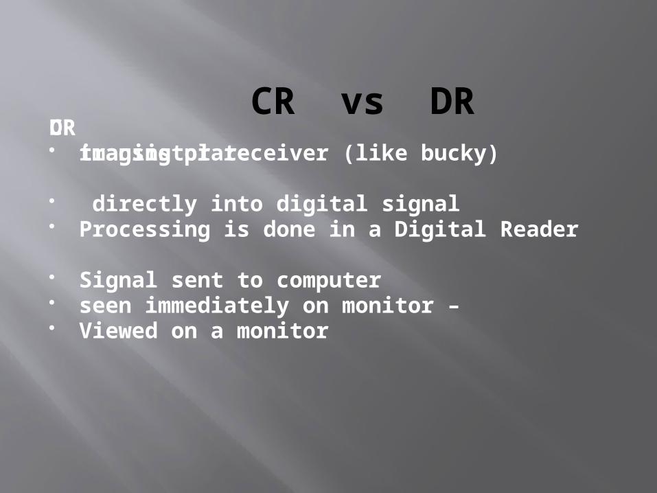
CR vs DRCR imaging plate
Processing is done in a Digital Reader
Signal sent to computer
Viewed on a monitor
DR transistor receiver (like bucky)
directly into digital signal
seen immediately on monitor –