Nasal Septum Resection
13
Surgery of the nasal septum and turbinates Abstract The following article presents nasal septum and turbinate surgery. First an overview with special consideration of the anatomical and physiolo- Christoph Matthias 1 gical background is given followed by indications for surgical procedures. 1 ENT Department, Grosshadern Medical Center, Key steps of the gold standard procedure first described by Cottle and common variations are presented. Furthermore, some techniques Ludwig-Maximilians- University, Munich, Germany dealing with special problems of the septumplasty are discussed fol- lowed by an overview on complications and long term results. However, it should be mentioned that studies on surgical procedures of the nasal septum are still not sufficient as higher evidence levels are very rare. Within a separated chapter techniques for closure of septum perfora- tions are presented and indications particularly in the background of the standard procedure of bridge flaps forwarded by Schultz-Coulon are discussed. The second part focusses on turbinate surgery. Accordingly, anatomical and physiological basics are presented followed by indica- tions for surgical procedures and the surgical steps of different proced- ures as well as postsurgical treatment and long term results. Keywords: reduced nasal breathing, septumplasty, septumperforation, turbinate surgery 1 Introduction The nose represents the entrance of the respiratory tract and has several functions: 1. the passageway for the air stream, 2. it harbours chemical sensory functions, 3. it warms and moistions the air and 4. it plays a role in the defence against foreign bodies of the surrounding envir- onment [1], [2], [3], [4], [5], [6], [7]. The nose is comprised of a separated organ that harbours two passageways. However, as every nasal cavity has its own blood supply and innervation it can be seen as two organs that usually work together but in several situations can be regulated separately [2]. The most frequent symptom is obstruced nasal breathing. Problems with the other functions are less frequent. Since the beginning of modern rhinosurgery at the end of the 19 th century several methods have been developed to objectively measure the nasal airstream. The first method was the cold mirror to measure the condensing dust during expir- ation, forwarded by Zwaarademaker. For a long time reli- able surgical procedures to correct impaired nasal breathing were lacking. With the presentation of the submucous septum resection by Killian a reproducible procedure was described which could be evaluated in studies by different surgeons. Most of them were done during the 70s and 80s and are presented in the article by Mlynski for the congress 2005 [8]. During the 70s and 80s the septumplasty described by Cottle progressively took over and this procedures can be seen with its several variations as a gold standard nowadays. The correction of a deviated nasal septum combined with reduction of the nasal turbinates is often seen as a begin- ners operation. Taking all the different techniques into consideration that are necessary to solve all septum pathologies with reliable long term results it can be one of the most difficult procedures in rhinosurgery an be very demanding even for the experienced surgeon. It is nearly impossible to present all surgical techniques in this art- icle. Accordingly, it is the authors intention to give an overview which techniques should be used in the common pathologies and to present some possible solutions for typical problems. 2 Septum surgery 2.1. Anatomical and physiological background The nasal septum is separated into a bony and cartilagin- ous part. The bony part is comprised of the lamina per- pendicularis as a part of the ethmoid of the vomer as well as bony processes of the maxilla and palate. The car- tilaginous part is comprised of the lamina quadrangularis and its extensions to the lateral and alar cartilages. The lamina perpendicularis of the ethmoid forms the upper and anterior part of the septum is continuously connected to the lamina cribrosa. The vomer extends from below the sphenoid sinus anteriorly along the nasal floor. The cartilage of the lamina quadrangularis combines lamina perpendicularis, ethmoid, nasal dorsum and the vomer in the anterior part of the septum [9]. Frequently, a small sphenoid process is located between the ethmoidal part of the bony septum and the vomer which can particularly be seen in children and is a common cause for a conse- cutive development of a septum deviation. The upper 1/13 GMS Current Topics in Otorhinolaryngology - Head and Neck Surgery 2007, Vol. 6, ISSN 1865-1011 Review Article OPEN ACCESS
-
Upload
albert-gheorghe -
Category
Documents
-
view
62 -
download
0
description
Medical
Transcript of Nasal Septum Resection

Surgery of the nasal septum and turbinates
AbstractThe following article presents nasal septum and turbinate surgery. Firstan overview with special consideration of the anatomical and physiolo-
Christoph Matthias1
gical background is given followed by indications for surgical procedures.1 ENT Department,GrosshadernMedical Center,
Key steps of the gold standard procedure first described by Cottle andcommon variations are presented. Furthermore, some techniques Ludwig-Maximilians-
University, Munich, Germanydealing with special problems of the septumplasty are discussed fol-lowed by an overview on complications and long term results. However,it should bementioned that studies on surgical procedures of the nasalseptum are still not sufficient as higher evidence levels are very rare.Within a separated chapter techniques for closure of septum perfora-tions are presented and indications particularly in the background ofthe standard procedure of bridge flaps forwarded by Schultz-Coulon arediscussed. The second part focusses on turbinate surgery. Accordingly,anatomical and physiological basics are presented followed by indica-tions for surgical procedures and the surgical steps of different proced-ures as well as postsurgical treatment and long term results.
Keywords: reduced nasal breathing, septumplasty, septumperforation,turbinate surgery
1 IntroductionThe nose represents the entrance of the respiratory tractand has several functions: 1. the passageway for the airstream, 2. it harbours chemical sensory functions, 3. itwarms and moistions the air and 4. it plays a role in thedefence against foreign bodies of the surrounding envir-onment [1], [2], [3], [4], [5], [6], [7].The nose is comprised of a separated organ that harbourstwo passageways. However, as every nasal cavity has itsown blood supply and innervation it can be seen as twoorgans that usually work together but in several situationscan be regulated separately [2]. The most frequentsymptom is obstruced nasal breathing. Problems withthe other functions are less frequent. Since the beginningof modern rhinosurgery at the end of the 19th centuryseveral methods have been developed to objectivelymeasure the nasal airstream. The first method was thecold mirror to measure the condensing dust during expir-ation, forwarded by Zwaarademaker. For a long time reli-able surgical procedures to correct impaired nasalbreathing were lacking. With the presentation of thesubmucous septum resection by Killian a reproducibleprocedure was described which could be evaluated instudies by different surgeons. Most of them were doneduring the 70s and 80s and are presented in the articleby Mlynski for the congress 2005 [8]. During the 70s and80s the septumplasty described by Cottle progressivelytook over and this procedures can be seen with its severalvariations as a gold standard nowadays.The correction of a deviated nasal septum combined withreduction of the nasal turbinates is often seen as a begin-ners operation. Taking all the different techniques into
consideration that are necessary to solve all septumpathologies with reliable long term results it can be oneof themost difficult procedures in rhinosurgery an be verydemanding even for the experienced surgeon. It is nearlyimpossible to present all surgical techniques in this art-icle. Accordingly, it is the authors intention to give anoverviewwhich techniques should be used in the commonpathologies and to present some possible solutions fortypical problems.
2 Septum surgery
2.1. Anatomical and physiologicalbackground
The nasal septum is separated into a bony and cartilagin-ous part. The bony part is comprised of the lamina per-pendicularis as a part of the ethmoid of the vomer as wellas bony processes of the maxilla and palate. The car-tilaginous part is comprised of the lamina quadrangularisand its extensions to the lateral and alar cartilages. Thelamina perpendicularis of the ethmoid forms the upperand anterior part of the septum is continuously connectedto the lamina cribrosa. The vomer extends from belowthe sphenoid sinus anteriorly along the nasal floor. Thecartilage of the lamina quadrangularis combines laminaperpendicularis, ethmoid, nasal dorsum and the vomerin the anterior part of the septum [9]. Frequently, a smallsphenoid process is located between the ethmoidal partof the bony septum and the vomer which can particularlybe seen in children and is a common cause for a conse-cutive development of a septum deviation. The upper
1/13GMS Current Topics in Otorhinolaryngology - Head and Neck Surgery 2007, Vol. 6, ISSN 1865-1011
Review ArticleOPEN ACCESS
margin of the septum cartilage is closely connected tothe upper lateral cartilages and usually forms an angleof 10 to 15°. Anteriorly to the upper lateral cartilages arethe alar cartilages that lie close to the septum cartilagein the nasal tip and columellar area. The area betweenthe upper lateral cartilages and the alar cartilagesrepresents the anterior septum angle which can be seenas a supratip depression. The inferior (or posterior)septum angle is the structure where the septum is fixedto the anterior nasal spine. Anterior to that the medialcrura of the alar cartilages are located which are separ-ated from the cartilaginous septum by a small part ofmembranous septum, frequently named weak internaltriangle. The embryological development of the cartilagin-ous septum and the cephalic two thirds of the upper lat-eral cartilages is one entity [9], [10], [11], [12].The connection between septum cartilage and premaxillaor vomer has several pecularities. While some fibers ofthe perichondrium and periost run parallel with thecartilage and bone many of them cross around thepremaxilla to the opposite site. The fibers of the septumperichondrium have a similar crossing direction whichresults in a kind of pseudo-joint of the cartilaginousseptum and the bony premaxilla allowing a slight lateralmovement of the nasal septum. Accordingly, while it re-duces the risk of fractioning under lateral compressionit supports lateralisation of the lower septum duringgrowth [12], [13], [14].The submucosa of the inner nasal lining comprises of anintense venous plexus particularly in the area of the in-ferior turbinates and the posterior parts of the nasalseptum. In the region of the anterior septum a vascularconfluence of branches of the arteria ethmoidalis anterior,a. sphenopalatina and a. labialis superior is located. Thisarea, also named Kiesselbachs plexus particularly playsa role in anterior nasal bleeding.The nasal airsteam can be regulated by filling the venousplexus [15]. 1932 Schaeffer described these venous si-nus as corpora cavernosa, that are particularly locatedin the area of the nasal turbinates and the correspondingparts of the nasal septum. Meanwhile, the role of thesevenous sinus in the control of the nasal airstream is wellinvestigated and its capability to intensively swell andcompletely obstruct the nasal cavity is well documented[15], [16]. The localisation of these venous swellingbodies in the anterior part of the nasal cavity is very im-portant fort he control of the nasal airstream as this isthe narrowest part of the nasal cavity, therefore namednasal valve. Thus, it represents about 80% of the totalnasal resistance [2], [9], [17]. The nasal resistance isregulated by three different components: the nasal en-trance, the nasal valve and the passageway along theturbinates. The nasal airstream is regulated by the sym-pathic nerve system. Further details are described in theliterature [2], [3], [6], [7], [8], [9], [17], [18], [19].
2.2. Indications for septum surgery
Since the beginning of rhinosurgery at the end of the 19th
century it is well known that the nasal septum very rarelylies absolutely straight within the skull but usually showsa more or less pronounced deviation that can be foundin up to 90% of cases investigated. As this deviation cannot be judged to be pathological several authors separatea physiological deviation from a pathological septum de-viation. A physiological septum deviation is defined as adeviation without subjective or objective reduction of thenasal breathing. Accordingly, a pathological septum devi-ation has to be defined as a septum deviation with sub-jective reduction of nasal breathing. Thus, the problemof precisely defining the septum deviation is evident [17],[20]. To precisely evaluate the resistance of the nasalairway today several diagnostic procedures are available.First the rhinomanometry has to be mentioned, whichallows the identification of a nasal obstruction but is notable to exactly identify the underlying anatomical struc-tures. If this procedure is performed in advance and aftersympathomimetic treatment it can reliably be used toseparate between mucosal and bony or cartilaginousreasons for the reduced nasal airflow. Accordingly, thisprocedure is recommended in the guidelines by our soci-ety [21]. More intensive procedures like rhinoresistometry,acoustic rhinometry or long-term-rhinoflowmetry in theircombination allow the precise anatomical identificationof the obstruction but are rarely performed in clinicalroutine [2], [8], [16], [22]. Finally, the diagnosis of theunderlying reasons for reduced nasal breathing is verymuch dependent on the clinical experience of the investi-gator. The abovementioned procedures are able to assistin identifying the anatomical structures that should befocussed on.The identification of a deviated septum in a patient whocomplains reduced nasal breathing frequently results inthe indication of a septumplasty that consecutively isperformed using the procedure described by Cottle or inone of the variations [23]. It is assumed that this is oneof the main reasons for the poor long term results ofseptumplasty as very commonly, the problems of the noseare far more complex. The septum deviation itself givesrise for compensatory changes in the nose [16], [20],[24]. Thus, usually the investigator can find an atrophyof the nasal turbinate on the convex side while the con-cave side presents enlarged. It is assumed that the tur-binates regulate the nasal air stream in a way that furtherfunctions like warming up and moistioning are fulfilledeven in the deviated nose [2], [17], [22], [24].To better compare and standardise the surgical proced-ures it often has been tried to establish a classificationfor septum deviation. Particularly, the classifications for-warded by Guyuron and coworkers 1999 and Sciuto andcoworkers 1999 should be mentioned [25], [26]. Bothare based on the differentiation between horizontal andvertical and combined deviations. The classification for-warded by Sciuto and coworkers is presented in Figure1. If it is possible to clearly classify the septum deviation
2/13GMS Current Topics in Otorhinolaryngology - Head and Neck Surgery 2007, Vol. 6, ISSN 1865-1011
Matthias: Surgery of the nasal septum and turbinates

it is recommended to adapt the surgical procedure. Inexample, in the presence of an isolated horizontal devi-ation a surgical procedure is recommended that only re-duces the tension of the septum. In vertical-caudal devi-ations an excision of the deviation and consecutive reim-plantation should be performed while high vertical orcombined deviations frequentlymake a septumexchangenecessary. The success rates for this diagnostic andtherapeutic procedure given by the authors seem to beintensively improved, however, it is nearly impossible tofind out whether this improvement is attributable to theclassification system itself or due to the greater surgicalexperience of the investigators. As it is nearly impossibleto investigate only one variable in the end the selectionof the surgical procedure very much depends on theclinical experience of the surgeon. The complaints of thepatient, all visible pathologies of the nose and, if possible,the results of further diagnostic procedures should betaken into consideration to plan the surgical procedure.Classification systems like the presented are helpful inthis process.
Figure 1: Classification of septum deviation (from Sciuto andcoworkers): A: Horizontally, parallel to the premaxilla runningdeviation of the lamina quadrangularis. B: Vertically runningdeviation of the lamina quadrangularis. B1: Caudal-verticallyrunning deviation inferiorly to the insertion of the upper lateralcartilages. B2: Mid-vertically running deviation with the axis inthe area of the insertion of the upper lateral cartilages. B3:High-vertically running deviation with the axis in the area ofthe bony nasal dorsum. C: Combined deviation of A and B.
2.3. Surgical procedures
Several surgical procedures have been described to cor-rect a deviated nasal septum [23], [27], [28], [29], [30],[31], [32], [33], [34], [35], [36], [37], [38], [39], [40],[41]. The conservative, standard procedure today initiallybegins with a hemitransfiction incision, followed by adissection of the caudal edge of the septal cartilage andthe elevation of the mucosa of the opposite site of thehemitransfiction incision. If possible, it is recommendedto leave the mucosa attached on one side of the septumto better support the mucosal bloodflow. Additionally, in-ferior tunnels should be prepared to reach the premaxilla
and the vomer region. Resection of cartilage and boneshould be performed very conservatively in this technique,particularly in circumscribed deviations and in children.The complete caudal and inferior septum can be reachedvia the hemitransfiction incision after removing the mu-coperichondriumandmucoperiost on both sides. Further-more, the bony ridge of the premaxilla and vomer can beexposed and all bony and cartilaginous deviations canbe corrected. In all resections it is important to leave an“L”-form to support the nasal dorsum at the anteriornasal spine. Depending on the stability of the cartilagethe width of this columellar strut should be around 1 cm.When fractures of the anterior septum are present it ismendatory to reconstruct this “L”-frame using columellarstruts or by performing a septum exchange or an extracor-poral septumplasty [29], [30]. In this procedure thecomplete cartilaginous septum is freed from the upperlateral cartilages and is straightened outside the nose.Inmost cases the surgeonwill find sufficient cartilaginousstructures in upper or posterior parts of the septum thatallow functional reconstruction of the “L”-frame by rotatingthe remaining septum. Meticulous suturing of the reim-planted cartilage is particularly important in key areaslike anterior nasal spine, columellar and K-region (car-tilaginous-bony connection of the nasal dorsum).
Surgical steps
Hemitransfiction incision: After injection of 1 ml supra-renin containing local anaestetics into the columellaranterior septum the hemitransfiction incision is placedto find the entrance between cartilage and perichondrium.The exact localisation of the hemitransfiction incision ismuch more important than the side of incision. Whilemost surgeons prefer the right side for the incision nearlyall pathologies can also be corrected from a left side in-cision. Only in cases with severe deviations of the anteriorseptum, i.e. in cleft nose deformities, the incision shouldbe placed on the concave side of the deviation. Further-more, it is important to place the incision into the vestibu-lar skin and to avoidmucosal incisions as they predisposefurther injuries of themucosal lining and heavier bleeding.The incision should be performed from cranial to caudalto avoid accidental injury of the alar cartilages and domeregion. The next step includes themeticulous preparationof the subperichondreal lining. The entrance under theperichondrium should be located at the ventro-caudalseptum as this area is much more stable than more cra-nial parts and accidental injuries of the caudal septumshould be avoided. In cases of severe deviations it maybe necessary to free the concave side first as the convexside might be accessible only after mobilisation of thecorresponding cartilaginous area (Figure 2). Incisions onthe cartilaginous surface should be avoided as theythemselves can give rise to new deviations. The prepara-tion of the anterior nasal spine is the next step and de-pending on the pathology themobilisation of the cartilagin-ous septum from the anterior nasal spine. In cases ofdeviation of the bony septum particularly of the vomer
3/13GMS Current Topics in Otorhinolaryngology - Head and Neck Surgery 2007, Vol. 6, ISSN 1865-1011
Matthias: Surgery of the nasal septum and turbinates
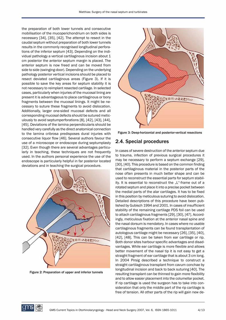
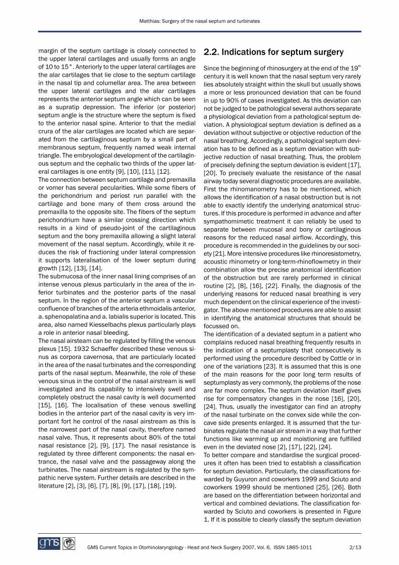
the preparation of both lower tunnels and consecutivemobilisation of the mucoperichondrium on both sides isnecessary [34], [35], [42]. The attempt to resect in thecaudal septumwithout preparation of both lower tunnelsresults in the commonly recognised longitudinal perfora-tions of the inferior septum [43]. Depending on the indi-vidual pathology a vertical cartilaginous incision about 1cm posterior the anterior septum margin is placed. Theanterior septum is now freed and can be moved fromside to side (swinging-door). Depending on the underlyingpathology posterior vertical incisions should be placed toresect deviated cartilaginous areas (Figure 3). If it ispossible to save the key areas for septum stability it isnot necessary to reimplant resected cartilage. In selectedcases, particularly when injuries of themucosal lining arepresent it is advantageous to place cartilaginous or bonyfragments between the mucosal linings. It might be ne-cessary to suture these fragments to avoid dislocation.Additionally, larger one-sided mucosal defects and allcorrespondingmucosal defects should be suturedmetic-ulously to avoid septumperforations [8], [42], [43], [44],[45]. Deviations of the lamina perpendicularis should behandled very carefully as the direct anatomical connectionto the lamina cribrosa predisposes dural injuries withconsecutive liquor flow [46]. Several authors favour theuse of a microscope or endoscope during septumplasty[32]. Even though there are several advantages particu-larly in teaching, these techniques are not frequentlyused. In the authors personal experience the use of theendoscope is particularly helpful in far posterior locateddeviations and in teaching the surgical procedure.
Figure 2: Preparation of upper and inferior tunnels
Figure 3: Deep-horizontal and posterior-vertical resections
2.4. Special procedures
In cases of severe destruction of the anterior septum dueto trauma, infection of previous surgical procedures itmay be necessary to perform a septum exchange [29],[30], [40]. This procedure is based on the common findingthat cartilaginous material in the posterior parts of thenose often presents in much better shape and can beused to reconstruct the essential parts for septum stabil-ity. It is essential to reconstruct the „L“-frame out of arotated septumand place it into a precise pocket betweenthe medial parts of the alar cartilages. It has to be fixedin this position bymeticulous suturing to avoid dislocation.Detailed descriptions of this procedure have been pub-lished by Gubisch 1994 and 2001. In cases of insufficientstability of the remaining cartilage PDS foil can be usedto attach cartilaginous fragments [29], [30], [47]. Accord-ingly, meticulous fixation at the anterior nasal spine andthe nasal dorsum ismendatory. In cases where no usablecartilaginous fragments can be found transplantation ofautologous cartilage might be necessary [26], [35], [40],[42], [48]. This can be taken from ear cartilage or rip.Both donor sites harbour specific advantages and disad-vantages. While ear cartilage is more flexible and allowsbetter movement of the nasal tip it is not easy to get astraight fragment of ear cartilage that is about 3 cm long.In 2004 Pirsig described a technique to construct astraight cartilaginous transplant from cavum conchae bylongitudinal incision and back to back suturing [40]. Theresulting transplant can be thinned to gainmore flexibilityand to allow easier placement into the columellar pocket.If rip cartilage is used the surgeon has to take into con-sideration that only the middle part of the rip cartilage isfree of tension. All other parts of the rip will gain new de-
4/13GMS Current Topics in Otorhinolaryngology - Head and Neck Surgery 2007, Vol. 6, ISSN 1865-1011
Matthias: Surgery of the nasal septum and turbinates

viations due to their own tension. Even though gettingenough cartilaginous material hardly ever is a problemthe patient suffers from a painful additional surgicalprocedure and scar. Furthermore, while the higher stabil-ity of rip cartilage may be advantageous in several cases(i.e. saddle nose deformities) it results in a less flexiblenasal tip. Additionally, the use of bony transplants fromthe bony septum or skull has been described but the useof these transplants imply an even more reduced flexibil-ity. The use of foreign material inside the septum is usu-ally associated with high extrusion rates over time, how-ever, recent studies on porous polyethylene could showthat this alloplastic material can be sucessfully used inthe septum or nasal valve area in selected cases whenthe surgeon consequently follows several rules, i.e. tometiculously cover all the foreign material with healthytissue.
Septumplasty in the cartilaginous deviatednose
With the exception of the congenitally deviated nose, i.e.in cleft nose deformities, trauma or previous surgery arethe most common causes for the deviated nose. Due tothe close correlation of the bony and cartilaginousframework of the nose a bony fracture often has an im-pact on cartilaginous structures like septum, upper lateralcartilages or alar cartilages. To achieve good results inthe correction of deviated noses it is essential to analyseall anatomical components and correct them individually.Thus, during the development of modern rhinoplasty theconviction grew that the septumplasty plays a essentialrole in the management of the deviated nose [25], [26],[44], [49], [50], [51]. In many cases a high horizontaldeviation requires a septum exchange. If an open ap-proach is performed the total septum can easily bereached and fixed after correction. Several authors favourthe implantation of a one sided spreader graft betweenthe septumand the upper lateral cartilage on the concaveside to achieve a long lasting straightening of the nose[41], [49], [50]. Foda even mentioned the use of anoverlong bony spreader graft to achieve this effect [51].
The severe antero-caudal septumdeviation(subluxatio septi)
Deviations of the anterior septum tend to give rise to aprolapsing anterior septummargin into one of the nostrilswith a consecutive asymmetry of the nasal basis. The keypart in correcting this pathology is, as Foda describes,the complete mobilisation of the ventro-kaudal and cra-nial septum [51]. Most of the techniques described sofar are based on the concept of separating septum cartil-age from upper lateral cartilages and consecutive refixa-tion with spreader grafts. This may be difficult in severalcases of severe septumdeviations particularly when aclosed approach is used. Thus, Bocchieri and coworkers2002 mentioned the possibility to correct the deviatednose using a „septal crossbar graft“ [50]. In this technique
a one sided spreader graft is placed under the high hori-zontal septum deviation on the concave side betweenthree incisions of the cranial cartilaginous septum afterpreparation of the mucoperichondrium (Figure 4).
Figure 4: “Septal Crossbar Graft”
Visible changes of the nose induced bycorrections of the nasal septum
While changes of the cosmetic aspect of the nose shouldgenerally be avoided during septumplasty the experiencedsurgeon can use corrections at the cartilaginous septumto achieve changes in the cosmetic aspect of the nose.Accordingly, a careful resection of a cartilaginous strip atthe caudal septum can shorten the nose and reducecolumellar show (Figure 5). The excision of a triangle atthe caudal septum with its basis at the anterior septumangle results in a rotation of the nasal tip (Figure 6). Fur-thermore, the reduction of an overlong septum in thearea of the inferior septum angle reduces the fullness ofthe nasolabial angle (Figure 7) [41]. It has to be men-tioned at this point that corrections in these areas of theseptum should only be made by an experiencedrhinosurgeon who has a clear view about the extent ofthe corrections necessary to achieve the changes inten-ded.
5/13GMS Current Topics in Otorhinolaryngology - Head and Neck Surgery 2007, Vol. 6, ISSN 1865-1011
Matthias: Surgery of the nasal septum and turbinates

Figure 5: Resection of an overlong septum
Figure 6: Resection of a triangle based at the anterior septumangle
Figure 7: Resection at the posterior-inferior septum angle
2.5. Postsurgical treatment
The key role of meticulous fixation of all implantedmater-ial inside the nasal septum has already beenmentioned.Accordingly, the reconstructed nasal septum itself canbe fixed using mattress sutures. Some authors recom-mend to use fibrin glue, however, it has often been de-scribed that this fixation is not reliable. There is goodevidence that in most reconstructed septums the use ofsplints (Doyle, Reuther) for about four days is sufficient,while there is no sufficient study material on the benefitof nasal packing. In the authors experience nasal packingis only necessary in turbinate resections or heavy bleedingduring surgery. More details about various methods ofnasal packing are described in the congress article byMlynski [8]. Even though the postsurgical treatment afterseptumplasty usually is not very intensive the patientshould be seen for about 14 days on a regular basis.Particularly a short time after removing the splints aseptum hematoma is not uncommon. It can easily beseen by the typical ballooning of the septum. In mostcases suctioning through the hemitransfiction incision isnot sufficient, but reexploration of the complete septumand removing all blood clots is necessary. Immediate re-vision surgery in mandatory as infection and abscessformation with consecutive necrosis of cartilaginous ma-terial has to be avoided [42], [45], [52].
2.6. Complications after septumplasty
The critical role of the anterior margin of the septum withits possible influence on the cosmetic aspect of the nosehas been mentioned above. Accordingly, many of thecomplications after septumplasty described in the litera-ture are mainly due to mistakes in the indication ortechnical procedure itself. Unfortunately, good studieson the complications after septumplasty are lacking.
Infections
Infections after septumplasty or rhinoplasty are muchless frequent than it could be expected from a surgicalprocedure that usually is performed in an unsterile envir-onment. Even though potentially pathological bacterialike staphylococci or streptococci are present the infectionrate usually lies under 3% [44], [52], [53], [54]. It will risein revision surgery and more extensive procedures likeseptum exchange. In revision cases with the use of freetransplants in 75% of cases Pirsig and Schäfer foundminor infections in 25% and more severe infections in6% of cases [53]. Taking these studies into considerationan antibiotic treatment can not be recommended forroutine septumplasty. Even though there is no sufficientstudy based evidence most authors recommend the useof antibiotics in revision cases, in complications likehematomas and in cases were free transplants or allo-plastic material has been used.
6/13GMS Current Topics in Otorhinolaryngology - Head and Neck Surgery 2007, Vol. 6, ISSN 1865-1011
Matthias: Surgery of the nasal septum and turbinates

Hematomas
Postsurgical bleeding is the most frequent complicationafter septumplasty occuring in 2 to 7% of all cases [28],[43]. Superficial ulcer and bleeding from incisions particu-larly from branches of the palatinal artery or thepremaxilla are common causes. Even more frequent arebleedings fromwounds after turbinate surgery, a surgicalprocedure frequently performed together with sep-tumplasty. Thus, the use of nasal packing is recommen-ded in cases in which resections of the turbinates havebeen performed.
Abscess
Usually, a septum abscess develops from hematomaafter surgery or trauma. Commonly found bacteria arestaphylococci, streptococci and haemophilus influencae.Good studies on the frequency of abscess developmentafter septumplasty are lacking. There is a strong necessityto immediately reoperate as there is the risk of necrosisof cartilaginous tissue. Intravenous antibiotics are usuallyapplied additionally. If further cartilage is affected thetypical changes are saddle nose deformities and retrac-tion of the columella [35], [53].
Intranasal adhesions (synechia)
Intranasal adhesions are relatively common after sep-tumplasty in combination with turbinate surgery [54],[55], [56], [57]. In retrospective studies in up to 36% ofcases intranasal adhesions could be found, however notall of them were functionally relevant [57]. Investigationsby Pirsig on more than 2000 patients could show thatthe use of nasal splinting for 4 to 7 days could avoid in-tranasal adhesions in almost all cases [45]. Crusting andmucosal atrophy which was a common complication aftersubmucosal septum resection (Killian procedure) [39],[56] is very unusual after the above mentioned Cottleprocedure [37], [42], [45], [58].
Septum perforation
Extensive submucosal resections are the most frequentreason for septum perforations after septumplasty. Theexact frequency is very difficult to determine as long termstudies are lacking and a clear differentiation of theseverity or a classification of septum perforations arelacking. Described frequencies rage from3 und 25% [27],[38], [49], [54]. As a general rule all large one-sided per-forations and all corresponding perforations should me-ticulously be sutured and cartilage should be positionedbetween themucoperichondrial sheets to prevent septumperforations.
Changes of the outer form of the nose
Major changes of the outer form of the nose after sep-tumplasty are less common with the Cottle procedure
compared to Killian`s septum resection [52]. Typicalchanges are saddle nose deformities or minor forms ofthese changes like cranial rotation of the nasal tip,widening of the nasolabial angle, shortening of the tip orretraction of the columella. These changes are mainlydue to insufficient stability of the “L”-frame and thus in-sufficient protection of the nasal dorsum and nasal tipfrom the anterior nasal spine. The frequency of thesechanges varies in the literature from 5 und 60% [37],[45], [48], [59], reflecting the problem of clear definitionof these changes. In most of these cases a rhinoplastyis necessary to correct all these changes [46], [47].
Smelling disorders
Swelling and scar formation in the area of the olfactorymucosa can result in a complete or incomplete loss ofolfaction [60], [61], [62]. As patients are often unable todifferentiate taste and smelling it is recommended to doa routine olfactometry prior to septum surgery. Addition-ally, olfactory dysfunction usually is not realised by thepatient immediately after surgery with additional diffi-culties to attribute the dysfunction to the surgical proced-ure. Preliminary dysfunction of smelling is regularly foundimmediately after septum surgery and mainly due tomucosal swelling and crust formation. The risk of perman-ent hyposmia or anosmia should be under 1% [61], [62].
Rare complications
The possibility of dural injury at the anterior skull basewith consecutive liquor leakage has already been men-tioned. Other rare complications are impaired visualfunction up to blindness due to retrograde flow of localanaestetics via the ethmoidal artery into the ophthalmicvessels. However, these are very rare complications andfrequencies are not given in the literature.
2.7. Long term results
Septumplasty is one of the most frequent performedsurgical procedures in otolaryngology. Accordingly, onewould expect to have sufficient studies with high evidencelevels to prove the benefit and long term results of thisoperation. By contrast, the study level is quite poor. Mostfollow up studies evaluate after 3 to 6 month. Severalscores have been developed, however non achievedwidespread acceptance [63], [64]. The few long termstudies available give evidence that the benefit afterseptumplasty is not as long lasting as often supposed.Thus, Konstantinidis found out, that 2 to 3 years aftersurgery only 50% of the patients feel to benefit from sur-gery [59]. These patients predominantly had deviationsof the anterior septum.
Septumperforation
In cases of typical symptoms like crusting, bleeding andreduced breathing small nasoseptal defects can effect-
7/13GMS Current Topics in Otorhinolaryngology - Head and Neck Surgery 2007, Vol. 6, ISSN 1865-1011
Matthias: Surgery of the nasal septum and turbinates

ively be treated with the established surgical procedures.In contrast, the closure of large septum defects repre-sents one of the most challenging rhinosurgical proced-ures [64], [65], [66]. Within the last decades more than40 different techniques have been described. Surgicalprocedures are derived from six different strategies: freetissue transfer [67], [68], septal mucosa flaps [68], [69],inferior turbinate flaps [70], oral vestibular flaps [67],endonasal mucosa advancement [65], [66], [69], andthe use of a fronto-temporal or paramedian forehead flap[71]. Most procedures, like free tissue transfer andseptum mucosa flaps are only suitable in small defects.Other techniques carry specific risks like necrosis in oralvestibular flaps. Thus, the closure rate of septam perfor-ation varies between 40% and 90% depending on thedefect size and the technique used. Schultz-Coulon de-scribed a success rate of 92.5% in 403 patients sufferingseptum perforations using bipedicled nasal mucosa flaps[66]. The author assesses the limitation of this techniquein defects exceeding 50-60% of the vertical septum high.In this large defects a separation and mobilisation of thelower lateral cartilages is necessary in the technique for-warded, a procedure known to be functionally and aes-thetically risky. Consequently, the author sees no surgicalalternative for larger vertical septum perforations [66].Established alternative procedures like obturator implant-ations give poor results [72] or necessitate external in-cisions with major aesthetic disturbance like alar baseincisions or lateral rhinotomy [71], [73]. Another proced-ure uses the midfacial degloving advancement which isa time consuming procedure and resulted in the authors’series in nasal valve stenosis in 20% of the cases [74].The use of a subcutaneous pericranial flap to closeseptum defects was first discribed by Paloma andcoworkers but the authors used a bicoronal approach incombination with an open rhinoplasty [75].We describe a new technique for the reconstruction oflarge septaum defects using a galeo-periostal foreheadflap. This technique has been developed by Berghausand the author within the last three years. The techniquerequires a 4 cm hairline incision combined with standardclosed rhinoplasty procedures. The subcutanous galeo-periostal forehead flap consists of the cranial periost andloose connective tissue adjacent to the periost, the sub-galeal fascia. The flap is strongly vascularized by the deepbranches of the supraorbital and supratrochlear vessels.The forehead flap is harvested in the subdermal planeand is pedicled at the supratrochlear and supraorbitalvessels. It is rotated through an open nasal roof into theseptum. The flap is fashioned through a 4 cmparamedianhairline incision (Figure 8). Only skin, subdermal tissueand galea are incised to facilitate further preparationwhile the flap is still spanned in the donor region. Thedissection runs in the subdermal layer down to the super-ior rim of the orbit. A small vertical skin incision can facili-tate safe identification of the supratrochlear vesselsduring preparation of a small pedicle.
Figure 8: The subcutanous galea-periost flap
The distal part of the flap should be adapted to the hori-zontal extension of the perforation while the axial lengthof the flap easily reaches 6-7 cm sufficient enough toreach the nasal floor. This allows the reconstruction ofseptum perforations without vertical limitations in size.After complete preparation of the subdermal layer theflap is cut down to the bone.The further preparation involves standard procedures ofclosed rhinoplasty. A hemitransfiction incision is used toadvance the mucosal margins of the perforation whiledécollement of the nasal dorsum is performed throughintercartilaginous incisions. After division of the upperlateral cartilages from their attachment to the dorsalseptum paramedian osteotomies are performed to widenthe nasal roof. Lateral osteotomies usually are necessaryparticularly if a dorsal hump is removed in the sameprocedure. After complete tunnelling between foreheadand nasal dorsum the flap will be rotated 180° throughthe open nasal roof into the separated mucosal marginsof the perforation (Figure 9 a)The flap will meticulously be sutured to the defectmarginsusing rapid Vicryl 4-0 (Figure 9 b). Nasal splinting for 5days and additional cleaning of the nasal cavity over 3weeks seems sufficient. Surgery resulted in completeclosure of large septum defects in 8 out of 9 cases (Figure9 c).The galeo-periostal forehead flap represents a safe pro-cedure for the reconstruction of anterior skull base de-fects [76], [77]. Due to its good vascularisation the flapis widely used to cover free transplants like bone, carti-lage or implants in poorly vascularized parts of the mid-face [77]. The donor region of the flap is easy to reachand the blood supply is very reliable provided by branchesof the supratrochlear and supraorbital vessels. On thebasis of this previous experience with the galeo-periostal
8/13GMS Current Topics in Otorhinolaryngology - Head and Neck Surgery 2007, Vol. 6, ISSN 1865-1011
Matthias: Surgery of the nasal septum and turbinates

Figure 9: Closure of a septum perforation using the subcutanous galea-periost flap. a: Preoperative perforation. b: During surgerywith sutures positioned. c: 6 month after surgery.
forehead flap we have chosen it for septum reconstruc-tion.Due to lacking long term follow-up and large numbers ofpatients treated with the procedure described successrates can not be presented up to now. Clamping of thepedicle in the area of the open roof is a possible risk.Additionally, a second step may be necessary to closethe open roof. In the cases treated by the authors aesthet-ic results made a second operation unnecessary. Thetechnique presented here seems to be a very promisingprocedure to close large septum perforations particularlywith large vertical extensions. The vertical high of theperforation is not important in determining the difficultyof the procedure and success rate of the closure as theflap is positioned into the defect from the nasal dorsum.This is a great advantage of the technique presented asthe vertical extension of the perforation is the limitingfactor in nearly all other procedures available.
3 Surgery of the nasal turbinates
3.1. Anatomical and physiologicalbackground
The inferior nasal turbinates are comprised of the turbin-ate bone with the mucoperiost above, a submucouscavernous plexus and the respiratory mucosa. The bonehas its own ossification centre, that arises at the 5th
month during development [9], [14]. The bony surface isvery irregular, perforated and interspersed with numerousvessels. Therefore, the mucoperiost is very strongly at-tached to the bone. The bone has contact to the ethmoid,the palate and the lacrimal sac. The cavernous plexus isparticularly well developed in the anterior part of the in-ferior turbinate and is regulated by autonome endocrineinnervation. Thus, the nasal turbinate is able to regulateits expansion. The number of goblet cells is much higherin the mucosa of the inferior nasal turbinate comparedto the middle turbinate [6], [9], [18]. The bone of themiddle turbinate is part of the ethmoid sinus [9], [78]. In30% of all cases the turbinate is pneumatised (conchabullosa). Its cavernous plexus is much less developed
compared to the inferior turbinate [6], [9], [18]. Similarto septum deviation enlarged turbinates are a very com-mon finding in patients suffering from reduced nasalbreathing. Underlying reasons are: allergic, vasomotoricor medication induced rhinitis chronical sinusitis, hor-mone induced rhinitis, and compensatory hypertrophyon the concave side of a septumdeviation [34].
3.2. Indication for nasal turbinatesurgery
According to the indication for septum surgery, it has tobe taken into consideration that the nose has to be seenas one system and the anatomical reasons for obstructionhavemeticulously to be evaluated. Therefore, it is import-ant to keep in mind that numerous reasons for turbinatehypertrophy are systematic or reflect a reaction of theentire respiratory tract. Besides the clinical finding therhinomanometry before and after a-mimetic reduction isvery helpful [21]. It could be shown that the long termresult after turbinate surgery is very poor if the underlyingreason for hypertrophy is not meticulously evaluated [5].Including techniques that are very rarely used today (i.e.neurectomy, kryosurgery, injection of steroids) more than20 different techniques including the use of several lasersystems have been described for turbinate surgery [79],[80], [81], [82], [83], [84], [85], [86], [87], [88]. Thesesurgical procedures are based on three different prin-ciples: the lateralisation that only changes the positionof the turbinate, resection and coagulation procedures.The indication for surgical resection is generally agreedin cases where septumplasty is performed under generalanaesthesia, in cases with a hyperplastic turbinal boneor in recurrent hypertrophy after lasersurgery.
3.3. Surgical procedures
The most aggressive procedure, total turbinectomy, isable to markedly increase nasal breathing, but this pro-cedure should be avoided due to common severe com-plications like crusting and postoperative bleeding [8].By contrast, several authors still recommend this proced-ure still as the most effective procedure to improve nasal
9/13GMS Current Topics in Otorhinolaryngology - Head and Neck Surgery 2007, Vol. 6, ISSN 1865-1011
Matthias: Surgery of the nasal septum and turbinates

Figure 10: Laserturbinotomy (diode-laser). a: Beginning of the procedure. b: End of the procedure. c: Before and 1 year aftertreatment.
breathing even in areas of dry and hot climate [89]. Un-fortunately, good data and high evidence level studiesare not available. Partial turbinectomy, in which the headof the inferior turbinate is resected, is also very effectiveand associated with less complication rates. Due to scarformation the nasal cycle might severely be irritated asregulation from thermical stimuli is mainly induced by theanterior nasal turbinate [27]. Submucous resection, inwhich only parts of the turbinal bone and soft tissue willbe resected is a very frequently performed procedure.Accordingly, a very low complication rate has been attrib-uted to this procedure [8]. Turbinoplasty, in which resec-tion of the lateral mucosa and parts of the turbinal boneis performed is of similar effectivity and low complicationrate. Radiofrequency, a very mild procedure that caneasily be performed as an outpatient procedure, repres-ents a further effective alternative as progressively studiescould prove long lasting effectivity [80], [85], [90].Increasingly, the laser is used for turbinate surgery [81],[82], [83], [86], [87], [88], [90], [91], [92], [93]. It hassome conceptional advantages but also implies somedisadvantages. Thus, laserturbinotomy can be performedunder general anaesthetics, postoperative bleeding isvery rare and even if it occurs it usually is less severe sothat nasal packing is generally not necessary [83], [92].Conceptional disadvantage is the partial destruction ofthe mucosal surface that prolongs the healing process.Reduced energy can reduce the destruction of themucos-al surface but the volume effect is also reduced. However,submucous laser application has meanwhile been de-scribed to minimise this problem [92]. To support the in-
dication for laserturbinotomy, rhinomanometry in advanceand after application of local a-mimeticmedication shouldbe performed to simulate the benefit of turbinate reduc-tion. Laserturbinotomy should be avoided in cases withadditional paranasal sinus infection, nasal polyposis,deviated septum or other anatomical variations likeseptum deviation or stenosis of the nasal valve [81], [82],[83], [86], [87], [88], [90], [91], [92], [93]. Particularlythe severity of a septum deviation that still justifieslaserturbinotomy is very much dependent on the invest-igators experience. In example, laserturbinotomy on theconvex side of a septumdeviation could be correlatedwith an increased risk for septumperforation [92], [93].Numerous different laser systems have been used forlaserturbninotomy. The most frequently available CO2-laser is not ideal as it lacks the possibility for fiber trans-mission and applies only minor coagulation which in-ceases the risk of postoperative bleeding and adhesions.The KTP-laser is fiber transmissible and has a high ad-sorption for bloodmaking this system very attractive [89],[90]. The Holmium-YAG-laser is also usable for laser-turbinoplasty. It is used in pulsed mode but the depth ofthe effect is only about 0.4 mm. With the application ofseveral stripes a good volume effect can be achieved.Nd-YAG-laser is most frequently used in contact modeand multiple spots are applied. The effect can invademuch deeper making this system better for reduction ofturbinate hyperplasia [79]. In our experience the diodelaser is the best system available as it applies the effectup to about 3 mm depth. We use this system in continu-
10/13GMS Current Topics in Otorhinolaryngology - Head and Neck Surgery 2007, Vol. 6, ISSN 1865-1011
Matthias: Surgery of the nasal septum and turbinates

ous-wave-mode and apply longitudinal stripes on theturbinate surface (Figure 10) [81].
3.4. Postsurgical treatment
As mentioned above an intensive postsurgical treatmentafter laserturbinotomy in mandatory as crust formationoccurs with nearly all laser systems used. Creams likeJellin-Neomycin or Bepanthen and additional ointmentwith NaCl are good choices. As the volume effect usuallydevelops within 2 weeks local α-mimetics may be used.
4 ConclusionsSurgery of the nasal septum and turbinates are amongthemost frequently performed procedures in otolaryngo-logy. They are performed in varying techniques for dec-ades. By contrast, the studymaterial for evaluation of thedifferent techniques is still unsatisfactory. This is partiallydue to the fact that the indication for surgery is verysubjective and mainly depends on the experience of thesurgeon. Even though a variety of procedures to object-ively measure nasal function are available today the effortto exactly evaluate the nasal pathology is very high andthus not applied in routine clinical work. Everyotolaryngologist should have detailed anatomical andpathophysiological knowledge of the inner nose. Thesurgeon has to be experienced in the different techniquesavailable today to be able to perform these proceduressavely and effectively. The surgeon must be aware thatthe basic techniques are not sufficient to solve all septumproblems. It is the author`s intention to point out that itshould be avoided that relatively inexperienced surgeonsuse aggressive surgical procedures and thus producemore harm than benefit. The procedure is particularlydemanding if it is integrated into rhinoplasty as it itselfcan influence the outer aspect of the nose. In rhinoplasty,a good cosmetic result can be achieved by experiencedcolleagues from other specialisations, however, a goodresult that considers all cosmetic and functional aspectsof the nose can only be achieved by the experiencedotolaryngologist.
5 AcknowledgementI particularly want to thank Alexander Berghaus, professorand chairman of our department for his very helpful criti-cism and supplement and for leaving some figures (Figure2 and Figure 3). Additionally, I want to thank AndreasLeunig, consultant otolaryngologist of our department forhis support and picture material (Figure 8). Dr. MiriamHavel assisted in literature search and Mr. Martin Müllerin writing the manuscript.
References1. Cole P. The Respiratory Role of the Upper Airways. A Selective
Clinical and Pathological Review. Saint Louis, Mittelohr: MosbyYear Book 1993, 164-165.
2. Eccles R. Nasal Airflow in Health and Disease. Acta Otolaryngol.2000; 120: 580-595.
3. Hanf G, Schierhorn K, Brunnée T, Matthias C, Kunkel G.Neuromodulation vonMastzellen in humaner Nasenschleimhaut.Histaminfreisetzung durch Neuropeptide in vitro. Allergologie1997; 20: 121-127.
4. Kamani T, Yilmaz T, Surucus Turan E, Brent KA. Scanning ElectronMicroscopy of Ciliae and Saccharine Test for Ciliary Function inSeptal Deviations. Laryngoscope 2006; 116: 586-590.
5. Mabry RL. Chronic Nasal Obstruction. In: Gates G, editor. CurrentTherapy in Otorhinolarnygology, Head and Neck Surgery. Toronto:BC Decker; 1987. p. 274.
6. Matthias C, de Suuza P, Merker HJ. Morphological Investigationson the Epithelium and Subepithelial Connective Tissue of theHuman Paranasal SinusMucosa. J Rhinol. 1997; 4 (2): 129-138.
7. Wiesmiller K, Keck T, Rettinger G, Leiacker R, Dzida R, LindemannI. Nasal Air Conditioning in Patients before and after Septoplastywith Bilateral Turbinoplasty. Laryngoscope 2006; 116 (6): 890-894.
8. Mlynski G. Wiederherstellende Verfahren bei gestörter Funktionder oberen Atemwege. Nasale Atmung. Laryno-Rhino-Otologie2005; 84 Suppl.1: 101-117.
9. Maran AGD, Lund VJ. Nasal Anatomy. In: Maran AGD, Lund VJ,editors. Clinical Rhinology. Stuttgart: Thieme; 1990. p. 5.
10. McKinney P, Johnson P, Walloch J. Anatomy of the Nasal Hump.Plast Reconstr Surg. 1986; 77: 404-407.
11. Straatsma BR, Straatsma CR. The Anatomical Relationship ofthe Lateral Nasal Cartilage to the Nasal Bone and theCartilaginous Nasal Septum. Plast Reconstr Surg. 1951; 8: 443-445.
12. Converse JM. The Cartilaginous Structures of the Nose. Ann OtolRhinol Laryngol. 1955; 64: 220.
13. O'Neal RM, Beil RJ, Schlesinger J. Surgical Anatomy of the Nose.Clin Plast Surg. 1996; 23 (2): 195-222.
14. Dingman RO, Natvig P. Surgical Anatomy in Aesthetic andCorrective Rhinoplasty. Plast Surg Clin. 1977; 4: 111-114.
15. Cauna N. Blood and Nerve Supply of the Nasal Lining. In: ProctorDF, Andersen I, editors. The Nose, Upper Airways Physiology andthe Athmospheric Environment. Amsterdam: Elsevier; 1982. p.45-69.
16. Haight JSJ, Cole P. Site and Function of the Nasal Valve.Laryngoscope 1983; 93: 49-55.
17. Bridger GP. Physiology of the Nasal Valve. Arch Otolaryngol. 1970;92: 543-553.
18. Matthias C, de Suuza P, Merker HJ. Electron Microscopic andImmunomorphological Investigations of theMucosa of theHumanParanasal Sinuses. Eur Arch Otorhinolaryngol. 1997; 254: 230-235.
19. Schierhorn K, Zang M, Matthias C, Kunkel G. Influence of Ozoneand Nitrogen Dioxide upon Histamine and Interleukin Formationin a Human Nasal Mucosa System. Am J Respir Cell Mol Biol.1999; 20 (5): 1013-1019.
20. Yigit O, Akgul G, Alkan S, Uslu B. Dadas B. Changes Occurring inthe Nasal Mucociliary Transport in Patients with One-SidedSeptum Deviation. Rhinology 2005; 43: 257-260.
11/13GMS Current Topics in Otorhinolaryngology - Head and Neck Surgery 2007, Vol. 6, ISSN 1865-1011
Matthias: Surgery of the nasal septum and turbinates

21. Ganzer U, Arnold W. Leitlinien: Septumplastik.http://www.hno.org/publikationen/leitlinien.html.
22. van Dishoek HAE, Leiden MD. The Part of the Valve and theTurbinates in Total Nasal Resistance. Int Rhinol. 1965; 3: 19-26.
23. Cottle JM. The Maxilla-Premaxillary Approach to Extensive NasalSeptum Sirgery. Arch Otolaryngol Head Neck Surg. 1958; 68:301-306.
24. Berger G, Gass S, Ophir B. The Histopathology of the HypertrophicInferior Turbinate. Arch Otolarnygol Head Neck Surg. 2006; 132(6): 588-594.
25. Guyuron B, Uzzo CD, Scull H. A practical classification ofseptonasal deviation and an effective guide to septal surgery.Plast Reconstruct Surg 1999; 104 (7): 2202-2212 .
26. Sciuto S, Bernadesci D. Exzision and Replacement of NasalSeptum in Aesthetic and Functional Nose Surgery: Setting Criteriaand Establishing Indications. Rhinology 1999; 37: 74-79.
27. Carroll T, Ladner K, Meyers AD. Alternative Surgical DissectionTechniques. Otolaryngol Clin North Am 2005; 38 (2): 397-411.
28. Fjermedal O, Saunte C, Pedersen S. Septoplasty and / orSubmucous Resection? 5 Years Nasal Septum Operation. JLaryngol Otol. 1988; 102: 796-798.
29. Gubisch W. The Extracorporeal Septumplasty: Technique toCorrect Difficult Nasal Deformities. Plast Reconstr Surg. 1995;95 (4): 672-681.
30. Gubisch W. 20 Jahre mit der extrakorporalen Septumkorrektur.Laryngo-Rhino-Otol. 2002; 81: 22-30.
31. Hellmich S. Septumplastik. Laryngol-Rhinol-Otol. 1997; 76: 663-666.
32. Hwang PH, McLaughlin RB, Lanza DC, Kennedy D. EndoscopicSeptoplasty: Indications, Technique, and Results. OtolaryngolHead Neck Surg. 1999; 120 (5): 678-682.
33. Kastenbauer ER. Eingriffe an der Nasenscheidewand. Laryngol-Rhinol-Otol. 1997; 76: A 93 - A 103.
34. King HC, Mabry RL. Surgical Approaches to Correcting NasalObstruction. In: King HC, Mabry RL. A Practical Guide to theManagement of Nasal and Sinus Disorders. Stuttgart: Thieme;1993. p. 94.
35. Marschall HAH, Johnston MN, Jones NS. Principles of SeptalCorrection. J Laryngol Otol. 2004; 118: 129-134.
36. Masing H, Hellmich S. Erfahrungen mit konserviertem Knorpelbei Wiederaufbau der Nase. Z Laryngol Rhinol. 1968; 47: 904-914.
37. Mayer B, Henkes H. Mini-Septumplastik - für Funktion und Form.Larnygo-Rhino-Otol. 1990; 69: 303-307.
38. NewmanMH. Surgery of the Nasal Septum. Clin Plast Surg. 1996;23 (2): 271-279.
39. PeacockMR.Mucous Resection of the Nasal Septum. J LaryngolOtol. 1981; 95: 341-356.
40. Pirsig W, Kern EB, Ferser T. Reconstruction of Anterior NasalSeptum: Back-to-Back Autogenous Ear Cartilage Graft.Laryngoscope 2004; 114: 627-638.
41. Toriumi DM, Becker DG. Septoplasty. In: Rhinoplasty: DissectionManual. Philadelphia: Lippincott; 1999. p. 31.
42. Schultz-Coulon HJ. Anmerkungen zur Septumplastik. HNO 2006;54: 59-70.
43. Stockstead VP, Vase P. Perforations of the Nasal SeptumFollowing Operative Procedures. Rhinology 1978; 16: 123-138.
44. Lawson W, Kessler S, Biller JF. Unusual and Fatal Complicationsof Rhinoplasty. Arch Otolaryngol. 1983; 109: 164-169.
45. Miller T. Immediate Postoperative Complications of Septoplastiesand Septorhinoplasties. Trans Pac Coast Ophthalmol Soc. 1976;57: 201-205.
46. Schwab JA, Pirsig W. Complications of Septal Surgery. FacialPlast Surg. 1997; 13 (1): 3-14.
47. Boenisch M, Tamas H, Nolst-Trenité GJ. Influence ofPolydioxanone Foil on growing Septal Cartilage after Surgery inan Animal Model: New Aspects of Cartilage Healing andRegeneration. Arch Facial Plast Surg 2003; 5(4): 316-319.
48. Rettinger G. Autogene und allogene Knorpeltransplantate in derKopf- und Halschirurgie. Eur Arch Oto-Rhino-Laryngol. Suppl.1992; 1: 127-162.
49. Holt GR, Garner ET, McLarey D. Postoperative Sequelae andComplications of Rhinoplasty. Otolaryngol Clin North Am. 1987;20: 853-876.
50. Boccieri A, Pascali M. Septal Crossbar Graft for the Correctionof the Crooked Nose. Plast Reconstr Surg. 2003; 111 (2): 629-638.
51. Foda HMT. The Role of Septal Surgery in Management of theDeviated Nose. Plast Reconstr Surg. 2005; 406-415.
52. Tzadik A, Gilbert SE, Sade J. Complications of SubmucousResections of the Nasal Septum. Arch Otorhinlaryngol. 1988;245: 74-76.
53. Pirsig W, Schäfer J. The Importance of Antibiotic Treatment inFunctional and Aesthetic Rhinosurgery. Rhinology Suppl. 1998;4: 3-11.
54. Weimert TA, Yoder MG. Antibiotics and Nasal Surgery.Laryngoscope 1980; 90: 667-672.
55. Eschelmann LT, Schleunig AJ, Brummett RE. ProphylacticAntibiotics and Otolaryngologic Surgery. A Double Blind Study.Trans Am Acad Ophthalmol Otolaryngol. 1971; 75: 387-394.
56. Huizing EH. The Management of Septal Abscesses. Facial PlastSurg. 1986; 3 (4): 243-252.
57. Bewarder S, Pirsig W. Long-Term Results of Submucous SeptalResection. Laryngol Rhinol. 1978; 57: 922-931.
58. White A, Murray JA. Intransal Adhesion Formation FollowingSurgery for Chronic Nasal Obstruction. Clin Otolaryngol. 1988;13: 139-143.
59. Konstantinidis I, Triaridis S, Triaridis A, Karagianidis K, KontzoglouG. Long-Term Results Following Nasal Septum Surgery: Focuson Patients' Satisfaction. Auris Nasus Larynx 2005; 32: 369-374.
60. Pfaar, O, Hüttenbrink KB, Hummel T. Assessment of OlfactoryFunction after Septoplasty: A Longitudinal Study. Rhinology 2004;43: 195-199.
61. Stevens CN, Stevens MG. Quantitative Effects of Nasal Surgeryon Olfaction. Am J Otolaryngol. 1985; 6: 264-267.
62. Kimmelmann CP. The Risk of Olfaction from Nasal Surgery.Laryngoscope 1994; 104: 981-988.
63. Stewart MG, Smith TL, Weaver EM,Witsell, DL, Yueah B, HannleyMT, Johnson JT. Outcomes after Nasal Septoplasty: Results fromthe Nasal Obstruction Septoplasty Effectiveness (NOSE) Study.Otolaryngol Head Neck Surg. 2004; 130: 283-290.
64. Rowe-Jones, J. Nasal Surgery: Evidence of Efficacy. Septal andTurbinate Surgery. Rhinology 2004; 42 (4): 248-250.
65. Schultz-Coulon HJ. Experiences with the bridge-flap techniquefor the repair of large nasal septal perforations. Rhinology1994;32: 25-33.
66. Schultz-Coulon HJ. Three layer repair of nasoseptal defects.Otolaryngol Head Neck Surg 2005;132(2): 213-217.
67. Kridel RWH. Septal perforation repair. Otolaryngol Clin North Am1999;32(4): 695-724.
12/13GMS Current Topics in Otorhinolaryngology - Head and Neck Surgery 2007, Vol. 6, ISSN 1865-1011
Matthias: Surgery of the nasal septum and turbinates

68. Woolford TJ, Jones NS. Repair of nasal septal perforations usinglocal mucosal flaps and a composite cartilage graft. J LaryngolOtol 2001;115: 22-25.
69. Newton JR, White PS, Lee MSW. Nasal septal perforation repairusing open septoplasty and unilateral bipedicled flaps. J LaryngolOtol 2003;117: 52-55.
70. Stoor P, Grenman R. Bioactive glass and turbinate flaps in therepair of nasal septal perforations. Ann Otol Rhinol Laryngol2004;113: 655-661.
71. Meyer R, Berghaus A. Closure of perforations of the septumincluding a single session method for large defects. Head NeckSurg 1983;8: 390-400.
72. Osma Ü, Cüreoglu S, Akbulut N. The results of septal bottoninsertion in the management of nasal septal perforation. JLaryngol Otol 1999;113: 823-824.
73. Kastenbauer ER, Masing H. Chirurgie der inneren Nase,Versorgung von Nasenverletzungen. In: Naumann HH, editor.Kopf- und Halschirurgie, Band 1, Teil 1. 1985. p. 403-408.
74. Romo 3rd T, Foster CA, Korovin GS. Repair of nasal septalperforation utilizing the midface degloving technique. ArchOtolaryngol 1988;114: 739-742.
75. Paloma V, Samper A, Cervera-Pas F J. Surgical technique forreconstruction of the nasal septum: the pericranial flap. Headand Neck. 2000: 90-94.
76. Fukuta K, Avery C, Jachson YT. Long term complications of thegalea-frontalis myofascial flap in craniofacial surgery. Eur J PlastSurg 1993; 16: 174-176.
77. Argenta LC, Friedman RJ, Dingman RO, Duus EC. The versatilityof pericranial flaps. Plast Reconstr Surg 1985; 76(5): 695-702.
78. Cook PR, Begegni A, Cullen Bryant W, Davis WE. Effect of PartialMiddle Turbinectomy on Nasal Airflow and Resistance.Otolaryngol Head Neck Surg. 1995; 113 (4): 413-419.
79. Chang CW, RiesWR. Surgical Treatment of the Inferior Turbinate:New Techniques. Current Opin Otolaryngol Head Neck Surg.2004; 12 (1): 53-57.
80. CavaliereM,Mottola G, ImmaM. Comparison of the Effectivenessand Safety of Radiofrequency Turbinoplasty and TraditionalSurgical Technique in Treatment of Inferior TurbinateHypertrophy. Otolaryngol Head Neck Surg. 2005; 133 (6): 972-978.
81. Hoffmann P, Rudert H. CO2- und Nd-YAG-Laser: Vergleich zweierVerfahren zur Nasenmuschelreduktion. Arch Otorhinolaryngol.Suppl. 1992; 2: 116-117.
82. Hopf JUG, Hopf M, Koffroth-Becker C. Minimal invasive Chirurgieobstruktiver Erkrankungen der Nasemit demDiodenlaser. LaserMed. 1999; 14 (4): 106-115.
83. Janda P, Sroka R, Baumgartner R, Grevers G, Leunig A. LaserTreatment of Hyperplastic Inferior Nasal Turbinates: A Review.Lasers Surg Med. 2001; 28 (5): 404-413.
84. Jovanovic S, Dokic D. Nd:YAG-Laserchirurgie in der Behandlungder allergischen Rhinitis. Laryngol Rhinol Otol. 1995; 74: 419-422.
85. Kezirian J, Powell NB, Riley RW, Hester IE. Incidence ofComplications of Radiofrequency Treatment of the Upper Airway.Laryngoscope 2005; 115 (7): 1298-1304.
86. Krespi YP, Slatkine M. Nd:YAG-Fiber Delivery System forSubmucosal Interstitial Coagulation of Nasal Turbinates. LaserSurg Med. 1994; 15: 217-248.
87. Lippert BM,Werner JA. Behandlung der hyperplastischen unterenNasenmuscheln. HNO 2000; 48 (4): 267-274.
88. Lippert BM, Werner JA. Long-Term Results after LaserTurbinektomy. Laser Surg Med 1998; 22: 126-134.
89. Talmon Y, Samet A, Gilbey P. Total inferior Turbinectomy:Operative Results and Technique. Ann Otol Rhinol Laryngol 2000;109:1117-1119.
90. Porter MW, Hales NW, Nease CI, Krempel GA. Long-Term Resultsof Inferior Turbinate Hypertrophy with Radiofrequency Treatment.A New Standard of Care? Laryngoscope 2006; 116 (4): 554-557.
91. De la Chaux R, Dreher A, Clemens C, Rasp G, Leunig A.Respiratory Sleep Disorders: Benefit from Laser-Surgery. MMW2004; 146 (47): 49-50.
92. Oswal V, Hopf JUG, Hopf M, Scherer H. Endonasal LaserApplications. In: Oswal V, Remacle M, editors. Principles andPractice of Lasers in Otorhinolaryngology and Head and NeckSurgery. The Hague, Holland; 2002. p. 163.
93. Oswal V, Krespi YJ, Kacker A. Nasal Turbinate Surgery. In: OswalV, Remacle M, editors. Principles and Practice of Lasers inOtorhinolaryngology and Head and Neck Surgery. The Hague,Holland; 2002. p. 221.
94. Raynor EM. Powered endoscopic septoplasty for septal deviationsand isolated spurs. Arch Facial Plast Surg. 2005; 7(6): 410-412.
95. Campbell JB, Watson MG, Shenoi PM. The Role of IntranasalSplints in the Prevention of Postoperative Adhesions. J LaryngolOtol. 1987; 101: 1140-1143.
96. Shone GR, Clegg RT. Nasal Adhesions. J Laryngol Otol. 198; 101:555-557.
97. Fairbanks DN. Closure of nasal septal perforations. ArchOtolaryngol 1980;106(8): 509-513.
98. Federspil PA, Schneider M. Der individuell angepassteNasenscheidewandobturator. Laryngo-Rhino-Otol. 2006; 85:323-325.
99. Berghaus A, Jovanovic S. Technique and indications of extendedsublabial rhinotomy ("midfacial degloving"). Rhinology 1991; 29(2):105-110.
Corresponding author:Prof. Dr. med. Christoph MatthiasENT-Dept., Grosshadern Medical Center, Marchioninistr.15, 81377 Munich, Phone: 089/7095-2990, Fax:089/7095-88 [email protected]
Please cite asMatthias C. Surgery of the nasal septum and turbinates. GMS Curr TopOtorhinolaryngol Head Neck Surg. 2007;6:Doc10.
This article is freely available fromhttp://www.egms.de/en/journals/cto/2008-6/cto000044.shtml
Copyright©2008 Matthias. This is an Open Access article distributed under theterms of the Creative Commons Attribution License(http://creativecommons.org/licenses/by-nc-nd/3.0/deed.en). Youare free: to Share — to copy, distribute and transmit the work, providedthe original author and source are credited.
13/13GMS Current Topics in Otorhinolaryngology - Head and Neck Surgery 2007, Vol. 6, ISSN 1865-1011
Matthias: Surgery of the nasal septum and turbinates