![1.Set up 110 µl mix for each primer/DNA combo on ice! 1.1.1 µl 100x F primer (1 pMol/µl = 1µM final []) 2.1.1 µl 100x R primer 3.11 µl 10x PCR buffer 4.2.2.](https://static.fdocuments.net/doc/165x107/56649ce05503460f949aa81d/1set-up-110-l-mix-for-each-primerdna-combo-on-ice-111-l-100x-f-primer.jpg)
Nanosep® Centrifugal Devices - Protocols for Use · Nanosep® Centrifugal Devices - Protocols for...
28
Nanosep® Centrifugal Devices - Protocols for Use Ultrafiltration Fundamentals Purification and Handling of DNA Fragments PCR: Before and After Protein Purification and Handling Miscellaneous Protocols Appendices References Ordering Information Ultrafiltration Fundamentals Background Ultrafiltration (UF) is a membrane separation technique used to separate extremely small particles and dissolved molecules in fluids. The primary basis for separation is molecular size, although other factors such as molecule shape and charge can also play a role. Molecules larger than the membrane pores will be retained at the surface of the membrane (not in the polymer matrix as they are retained in microporous membranes) and concentrated during the ultrafiltration process. Compared to non-membrane processes (chromatography, dialysis, solvent extraction, or centrifugation), ultrafiltration: Is far gentler to the molecules being processed. Does not require an organic extraction which may denature labile proteins. Maintains the ionic and pH milieu. Is fast and relatively inexpensive. Can be performed at low temperatures (for example, in the cold room). Is very efficient and can simultaneously concentrate and purify molecules. The retention properties of ultrafiltration membranes are expressed as Molecular Weight Cutoff (MWCO). This value refers to the approximate molecular weight (MW) of a dilute globular solute (i.e., a typical protein) which is 90% retained by the membrane. However, a molecule’s shape can have a direct effect on its retention by a membrane. For example, linear molecules like DNA may find their way through pores that will retain a globular species of the same molecular weight. There are three generic applications for ultrafiltration: 1. Concentration. Ultrafiltration is a very convenient method for the concentration of dilute protein or DNA/RNA samples. It is gentle (does not shear DNA as large as 100 Kb or cause loss of enzymatic activity in proteins) and is very efficient (usually over 90% recovery). 2. Desalting and Buffer Exchange (Diafiltration). Ultrafiltration provides a very convenient and efficient way to remove or exchange salts, remove detergents, separate free from bound molecules, remove low molecular weight materials, or rapidly change the ionic or pH environment. 3. Fractionation. Ultrafiltration will not accomplish a sharp separation of two molecules with similar molecular weights. The molecules to be separated should differ by at least one order of magnitude (10X) in size for effective separation. Fractionation using ultrafiltration is effective in applications such as the preparation of protein-free filtrates, separation of unbound or unincorporated label from DNA and protein samples, and the purification of PCR products from synthesis reactions. Choosing the Correct Device Devices are available in a range of sizes that accommodate the following sample volumes (Table 1): Table 1 Device Selection Based on Volume Filtered Contact Us: www.pall.com/contact Nanosep® Centrifugal Devices - Protocols for Use
-
Upload
truongtruc -
Category
Documents
-
view
216 -
download
0
Transcript of Nanosep® Centrifugal Devices - Protocols for Use · Nanosep® Centrifugal Devices - Protocols for...

Nanosep® Centrifugal Devices - Protocols for Use
Ultrafiltration Fundamentals
Purification and Handling of DNA Fragments
PCR: Before and After
Protein Purification and Handling
Miscellaneous Protocols
Appendices
References
Ordering Information
Ultrafiltration Fundamentals
BackgroundUltrafiltration (UF) is a membrane separation technique used to separate extremely small particles anddissolved molecules in fluids. The primary basis for separation is molecular size, although other factorssuch as molecule shape and charge can also play a role. Molecules larger than the membrane pores willbe retained at the surface of the membrane (not in the polymer matrix as they are retained inmicroporous membranes) and concentrated during the ultrafiltration process.
Compared to non-membrane processes (chromatography, dialysis, solvent extraction, or centrifugation),ultrafiltration:
Is far gentler to the molecules being processed.
Does not require an organic extraction which may denature labile proteins.
Maintains the ionic and pH milieu.
Is fast and relatively inexpensive.
Can be performed at low temperatures (for example, in the cold room).
Is very efficient and can simultaneously concentrate and purify molecules.
The retention properties of ultrafiltration membranes are expressed as Molecular Weight Cutoff (MWCO).This value refers to the approximate molecular weight (MW) of a dilute globular solute (i.e., a typicalprotein) which is 90% retained by the membrane. However, a molecule’s shape can have a direct effect onits retention by a membrane. For example, linear molecules like DNA may find their way through poresthat will retain a globular species of the same molecular weight.
There are three generic applications for ultrafiltration:
1. Concentration. Ultrafiltration is a very convenient method for the concentration of dilute protein orDNA/RNA samples. It is gentle (does not shear DNA as large as 100 Kb or cause loss of enzymaticactivity in proteins) and is very efficient (usually over 90% recovery).
2. Desalting and Buffer Exchange (Diafiltration). Ultrafiltration provides a very convenient and efficientway to remove or exchange salts, remove detergents, separate free from bound molecules, removelow molecular weight materials, or rapidly change the ionic or pH environment.
3. Fractionation. Ultrafiltration will not accomplish a sharp separation of two molecules with similarmolecular weights. The molecules to be separated should differ by at least one order of magnitude(10X) in size for effective separation. Fractionation using ultrafiltration is effective in applicationssuch as the preparation of protein-free filtrates, separation of unbound or unincorporated labelfrom DNA and protein samples, and the purification of PCR products from synthesis reactions.
Choosing the Correct DeviceDevices are available in a range of sizes that accommodate the following sample volumes (Table 1):
Table 1Device Selection Based on Volume Filtered
Contact Us: www.pall.com/contact
Nanosep® Centrifugal Devices - Protocols for Use

Sample Volume Use
50 µL to 500 µL Nanosep Device
500 µL to 3.5 mL Microsep™ Device
3 mL to 15 mL Macrosep® Device
15 mL to 60 mL Jumbosep™ Device
Choosing the Correct MWCOOnce sample volume is determined, the next step is to select the appropriate MWCO (for ultrafiltration)or pore size (for microfiltration). MWCOs are nominal ratings based on the ability to retain > 90% of asolute of a known molecular weight (in Kilodaltons). Tables 2 and 3 provide retention characteristics ofdifferent MWCO membranes for some solutes. For proteins, it is recommended that a MWCO be selectedthat is 3 to 6 times smaller than the molecular weight of the solute being retained. If flow rate is aconsideration, choose a membrane with a MWCO at the lower end of this range (3X); if the main concernis retention, choose a tighter membrane (6X).
It is important to recognize that retention of a molecule by a UF membrane is determined by a variety offactors, among which its molecular weight serves only as a general indicator. Therefore, choosing theappropriate MWCO for a specific application requires the consideration of a number of factors includingmolecular shape, electrical charge, sample concentration, sample composition, and operating conditions.Because different manufacturers use different molecules to define the MWCO of their membranes, it isimportant to perform pilot experiments to verify membrane performance in a particular application.
Common variables that increase molecule passage:
Sample concentration less than 1 mg/mL (Figure 1).
Linear versus globular molecules.
High transmembrane pressure created by g-force in centrifugal concentrators. (This is especiallyimportant in the case of linear molecules, for example DNA fragments. Decreasing the g-force canincrease retention of molecules by a membrane.)
Buffer composition that favors breakup of molecules.
pH and ionic conditions that change the molecule (for example, cause conformational changes oraggregation).
Common variables that decrease molecule passage:
Sample concentration higher than 1 mg/mL.
Buffer conditions that permit molecules to aggregate.
Presence of other molecules that increase sample concentration.
Lower transmembrane pressure. (In the case of centrifugal concentrators, lower g-force.)
Adsorption to the membrane or device.
Low temperature (4 °C versus 24 °C).
Table 2MWCO Selection for Nucleic Acid Applications
MWCO Base Pairs (DS) Bases (SS)
1K 5-16 Bp 9-32 Bs
3K 16-32 Bp 32-65 Bs
5K 25-50 Bp 50-95 Bs
10K 50-145 Bp 95-285 Bs
30K 145-285 Bp 285-570 Bs
50K 240-475 Bp 475-950 Bs
100K 475-1,450 Bp 950-2,900 Bs
300K 1,450-2,900 Bp 2,900-5,700 Bs

1,000K 4,800-9,500 Bp > 9,500 Bs
Table 3MWCO Selection for Protein Applications
MWCO
MembraneNominalPore Size
Biomolecule Size
BiomoleculeMolecularWeight
1K 3K-10K
3K 10K-20K
5K 15K-30K
10K 30K-90K
30K 90K-180K
50K 5 nm 15-30 nm 150K-300K
100K 10 nm 30-90 nm 300K-900K
300K 35 nm 90-200 nm 900K-1,800K
1,000K 100 nm 300-600 nm > 3,000K
Figure 1Retention and Passage Dilute Protein Samples
Ten 400 µL samples of a dilute solution of BSA (100 ng/mL) were centrifuged in the indicated MWCONanosep devices. Retentate was recovered in 40 µL of phosphate buffered saline (10X concentrated relativeto the original sample). 20 µL aliquots of the retenate and filtrate from each device were denatured andelectrophoresed on a 4 to 20% polyacrylamide gel and stained with coomassie blue. At higher initialconcentrations, some BSA would normally be retained by the 100K device (data not shown) while at aninitial concentration of 100 ng/mL, no BSA was detected in the retentate.
Figure 2aRetention and Passage of Dilute Protein Samples
A 500 µL sample of a 100 µg/mL DNA solution containing 50 bp and 500 bp double-stranded DNAfragments was centrifuged at 5,000 x g in Nanosep devices to a final volume of 50 µL. Recovered sampleswere quantitated using absorbance at 260 nm. We found that the 100K device is able to separate thesefragments; the 50 bp fragment was recovered in the filtrate, and the 500 bp fragment was recovered in theretentate.
Figure 2bRecovery of Dilute DNA Samples

Nanosep 30K devices were used to filter dilute radioactive DNA fragments. In order to accuratelyquantitate DNA recovery from dilute samples, PCR products (100 and 400 bp) were dual labeled to low-specific activity with 32P-dCTP and 32P-dATP and prepared for filtration. After synthesis, unincorporatednucleotides as well as termination products were removed by ultrafiltration using a 30K Nanosep device.The resulting retentate was checked for size and quantitated using gel electrophoresis. Labeled DNA inquantities ranging from 100 ng all the way down to 2 ng per device was diluted to 500 µL using TE. Thesamples (in triplicate) were centrifuged at 5,000 x g for 10 minutes (spun to dryness) and recovered in twowashes of 20 µL water. The resulting retentate was added to a counting vial containing scintillationsolution and counted. Nanosep devices reliably recover small, dilute DNA samples.
Top
Purification and Handling of DNA Fragments
BackgroundFrom a biochemical standpoint, DNA is a rather simple molecule and, unlike proteins, chemicalextraction methods cannot separate one similarly-sized DNA molecule from another. Gel electrophoresisseparates DNA molecules at a very high resolution based on size. However, the majority of scientists needto do much more than simply analyze restriction digest DNA fragments on agarose gels. For theseapplications, DNA molecules are cut, ligated, sequenced, transformed, electrophoresed, and labeled. Inaddition, desalting, buffer exchange, or elution from agarose may be required during any of a number ofprocedures. The handling of these sensitive and often very small and dilute biomolecules requires specificproactive laboratory procedures to prevent the loss or contamination of valuable samples.
Purification and recovery of DNA molecules from either agarose gels or other complex reaction solutionscan be accomplished in several ways.
1. Electroelution, using an electric charge with either dialysis bags or a specialized electrophoresisdevice to capture DNA migrating out of a gel fragment. Often this method requires vigilance, andthe recovered DNA sample requires a concentration/desalting step.
2. Affinity Chromatography, using either glass beads, resins, or glass fiber membranes to bind DNAand elute off after washing. This method is labor intensive, can cause shearing, and leaves glassparticles, ethanol, and salts in the DNA sample.
3. Precipitation, using differential solubility to selectively separate DNA from other alcohol-solublecomponents. This method results in poor recoveries from dilute DNA solutions. It is also timeconsuming, and gel and buffer contaminants can be co-precipitated. In addition, over-dried pelletsmay not redissolve properly.
4. Column Chromatography relies on size separation in a gel bed. This technique often requires theaddition of carrier tRNA to achieve high yields. Column chromatography requires significanthandling and dilutes the original sample.
5. Microfiltration/Ultrafiltration allows the rapid purification and concentration of the DNA usingNanosep devices. With this method there is very little handling, yields are high, DNA binding is low(Figure 3), DNA is undamaged, and the concentrated DNA is free of contaminants and ready forfurther reactions.
In general, DNA molecular weights do not correspond well with the MWCO because DNA is a long linearmolecule (Table 2).
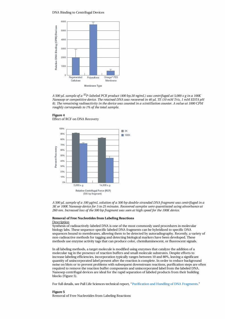
Effective retention of DNA by an ultrafiltration membrane requires a reduction in g-force, otherwise DNAcan be forced through many MWCO membranes regardless of size (Figure 4).
Figure 3
DNA Binding to Centrifugal Devices
A 500 µL sample of a 32P-labeled PCR product (400 bp,50 ng/mL) was centrifuged at 5,000 x g in a 100KNanosep or competitive device. The retained DNA was recovered in 40 µL TE (10 mM Tris, 1 mM EDTA pH8). The remaining radioactivity in the device was counted in a scintillation counter. A value at 1000 CPMroughly corresponds to 1% of the total sample.
Figure 4Effect of RCF on DNA Recovery
A 500 µL sample of a 100 µg/mL solution of a 500 bp double-stranded DNA fragment was centrifuged in a3K or 100K Nanosep device for 5 to 25 minutes. Recovered samples were quantitated using absorbance at260 nm. Increased loss of the 500 bp fragment was seen at high speed for the 100K device.
Removal of Free Nucleotides from Labeling ReactionsDescriptionSynthesis of radioactively-labeled DNA is one of the most commonly used procedures in molecularbiology labs. These sequence-specific labeled DNA fragments can be hybridized to specific DNAsequences bound to membranes, allowing them to be detected by autoradiography. Recently, a variety ofnon-radioactive methods for tagging and detecting biological markers have been developed. Thesemethods use enzyme activity tags that can produce color, chemiluminescent, or fluorescent signals.
In all labeling methods, a target molecule is modified using enzymes that catalyze the addition of amolecular tag in the presence of reaction buffers and small molecule substrates. Despite efforts toincrease labeling efficiencies, incorporation typically ranges between 10 and 80%, leaving a significantquantity of unincorporated label present after the reaction is complete. In order to reduce backgroundnoise on blots or to prevent problems with subsequent downstream reactions, purification steps are oftenrequired to remove the reaction buffer components and unincorporated label from the labeled DNA.Nanosep centrifugal devices are ideal for the rapid separation of labeled products from their buildingblocks (Figure 5).
For full details, see Pall Life Sciences technical report, “Purification and Handling of DNA Fragments.”
Figure 5Removal of Free Nucleotides from Labeling Reactions

A PCR labeling reaction was set up using 100 ng pUC18, 20 nmole primers, and PCR Supermix (LifeTechnologies) supplemented with 10 uCi 32P-dCTP resulting in 400 bp low-specific-activity PCR productswith about 5% of the available label incorporated and 95% free nucleotides for each reaction. PCRreactions were centrifuged in a 30K Nanosep device at 5,000 x g for 15 minutes and recovered using two 20µL TE (10 mM Tris, 1 mM EDTA, pH 8) rinses and equivalent loads were electrophoresed using a 10% TrisBorate polyacrylamide gel. RET= Retained DNA, FT= Flow-Through. The 30K Nanosep device is clearlycapable of removing the large number of free nucleotides completely without losing the valuable 400 bpPCR product in the process.
Step-by-Step Procedure
1. Dilute the labeling reaction to 500 µL with TE (10 mM Tris/1 mM EDTA, pH 8).
2. Transfer the diluted sample into a separate, appropriate Nanosep device (Table 2). Note: Randomhexamer synthesized labeling reactions generally produce smaller and more variably-sized DNAfragments and work best with a 30K device.
3. Centrifuge at 5,000 x g for 10 to 15 minutes; do not overspin.
4. Optional: To ensure small molecule removal, rinse retentate with 100 to 400 µL TE and repeatcentrifugation.
5. Recover retained DNA from the membrane surface with water or TE by rinsing the membranesurface with a pipette tip. Highest yields result from two rinses of 20 µL each.
6. The labeled molecule’s specific activity can be approximated by counting the radioactivity of theretentate and comparing this to the counts in the filtrate* or starting material.
*While developing your protocol, you should save the filtrate in the event that a different MWCO isneeded.
Single-tube DNA Purification and Cloning Using Ultrafiltration DevicesDescriptionTraditional methods for subcloning DNA fragments or shuttling sequences between vectors involve theligation of restriction digest fragments from chromosomes, plasmids, cosmids, or PCR products intospecific sites on a selected cloning vector. In order to desalt, remove primers or adapters, or concentrateDNA fragments, researchers generally use gel electrophoresis and precipitation steps that are laborintensive and time consuming. Using Nanosep centrifugal ultrafiltration devices for subcloning PCR-generated fragments results in a rapid process that is more streamlined than traditional methods.
After optimizing the parameters for effective target DNA purification, PCR products were synthesizedwith terminal adapter sequences containing restriction sites. These adapters were digested, generating theappropriate cohesive ends for ligation (2). The resulting DNA products were then concentrated anddesalted while simultaneously allowing the primers and adapter fragments to pass into the discardedfiltrate during a five-minute centrifugation step. The resulting ligation-ready DNA fragments were ligatedto digested vector directly within the ultrafiltration device, and then transformed into competent cells (1).Using Pall Nanosep ultrafiltration devices achieves significant savings in time, labor, and starting materials,while the resulting yield of transformed recombinants roughly equals that of the traditional techniques.
For full details, see Pall Life Sciences technical report, “Single-tube DNA Purification and Cloning UsingUltrafiltration Devices.”
Figure 6Effect of Terminal Restriction Digestion on Subsequent Ultrafiltration Retention

A radioactive, dilute (100 ng) PCR product amplified from a cloned fragment of Ceratopteris phytochromeDNA template was repeatedly purified using a Nanosep 100K device. The restriction digest products areshown for the first (HinDIII) and second (BamHI) restriction digests. (-) = sample prior to Nanosep deviceconcentration, (+) = recovered concentrated sample after filtration. Equivalent band intensitiesapproximate 100% recovery.
Figure 7PCR Products Resulting from the Amplification of DNA Templates from Individual Recombinant Colonies
The 620 bp band visualized on an agarose gel indicates successful recombination of the amplified genefragment into the vector. The absence of a 620-bp product indicates a spurious recombination event. Thelengthy standard cloning procedure was used as a control.
Step-by-Step ProcedurePCR
1. Prepare PCR reaction (25 µL) using an appropriate PCR Master Mix supplemented MgCl2, 0.2 mMprimers, and 1 µL genomic template DNA.
2. Incubate PCR reactions in a preheated thermocycler at 90 °C for 1 minute, then subject sample to 35cycles of 30 seconds at 94 °C, 30 seconds at 45 °C, and 60 seconds at 72 °C, followed by a postcycleextension for 3 minutes at 72 °C (or use your favorite conditions).
3. Remove a 5 µL aliquot of each reaction; electrophorese to confirm the size and purity of theproducts and to quantitate the fragments for subsequent cloning.
Nanosep Device Sample Preparation
1. Digest 0.1 to 1 µg of PCR product (either together or in separate tubes) and 0.2 µg of vector with theappropriate restriction enzymes directly in a Nanosep 100K device for 2 hours at 37 °C.
2. Heat inactivate the enzyme at 95 °C for 5 minutes.
3. Centrifuge through the Nanosep device at 1,500 x g for 5 minutes, and wash the retentate twice byaddition of 50 µL sdH2O.
4. If sequential digests are required due to buffer incompatibilities, repeat steps 1 and 2 with eachenzyme.
5. Resuspend in 8.5 µL sdH2O, 1 µL 10x ligation buffer with ATP, and 0.5 µL ligase. Ligation wasallowed to proceed directly in the Nanosep device at 17 °C overnight.
6. If digested in separate tubes, resuspend the vector in 8.5 µL sdH20 and transfer to the insert tube forligation.
Transformation and Recombinant AnalysisAdd 3 µL aliquot of ligation product to transform competent cells. Follow standard transformationprocedures.
Plate onto selective culture media to select for transformants.
DNA Purification/Buffer Exchange for Restriction DigestionsDescriptionTo be effectively digested or modified, DNA should be free of contaminants such as phenol, chloroform,alcohol, EDTA, detergents, drugs (like actinomycin and distamycin frequently used in DNA studies), orexcessive salts. All of these compounds can interfere with restriction endonuclease activity causing eitherpartial digestion or altered specificity (star activity).

Commonly-used protocols recommend dialysis or ethanol precipitation, followed by a wash-and-dryregimen prior to restriction digestion of the DNA sample. Very often, cleaving a DNA substrate with twoor more enzymes requires the use of different buffers to satisfy the buffer requirements of each enzyme(Figure 8). Discontinuous diafiltration (a series of concentration and dilution steps) can be used for theeasy and efficient removal of small molecular weight contaminants and salts from the DNA sample. Whilethis protocol is written for serial restriction digests, this basic desalting/buffer exchange protocol can bemodified to fit a variety of applications.
Figure 8DNA Desalting and Enzyme Activity
The Nru I restriction enzyme requires a unique restriction buffer for the optimal activity. Lane M = sizemarkers-Lambda DNA EcoR1 & Hind III digest, Lane 1 = (negative control) Lambda DNA digested withNru 1 in NEBuffer 1, Lane 2 = (positive control) Lambda DNA in Nru I buffer incubated with Nru I, Lane 3= Lambda DNA in NEBuffer 1 incubated with Nru I, Lane 4 = the same DNA sample from Lane 3,following buffer exchange and digestion with Nru I. Use of a 100K Nanosep device ensures Nru I activityfollowing desalting.
Step-by-Step Procedure
1. Select the Nanosep device with the proper MWCO (Table 2). Note: Increased solutes in the solutionwill decrease the passage of DNA and may require the use of a higher MWCO device.
2. Dilute the digested DNA sample to 500 µL with the single strength (1X) buffer to be used for thesecond digestion.
3. Centrifuge at 5,000 x g for 5 (100K) to 30 (3K) minutes.
4. Optional: To perform discontinuous diafiltration: Add a sufficient amount of buffer to bring theretentate volume to 500 µL and centrifuge again. Usually two cycles of dilution and concentrationwill remove over 99% of salts and over 90% of small molecular weight contaminants. If a higher levelof purity is desired, repeat the dilution and concentration steps for a third time. Multiple diafiltrationsteps will decrease overall yields; therefore, quality versus yield considerations must be made.
5. Resuspend in appropriate 1X restriction digestion buffer, add the second enzyme, digest accordingto manufacturer’s instructions (repeat step 3), and recover double-cut DNA in at least 20 µL wateror buffer.

Purification of DNA Fragments from Agarose GelsDescriptionNumerous methods have been developed to purify electrophoretically separated DNA fragments awayfrom the agarose gel matrix. These methods allow specific, electrophoretically separated DNA fragmentsto be used in further analysis and cloning procedures.
The isolation and purification of DNA fragments from a gel slice is done in two simple steps. First, theDNA is eluted from the agarose slurry. The agarose gel matrix is retained by a Nanosep MF devicecontaining a 0.2 µm or 0.45 µm membrane, and the gel buffer (containing DNA) freely flows through.Second, DNA is concentrated and washed using a Nanosep device containing a MWCO membraneadequate to retain the DNA (see Table 2) and wash away contaminants. This method is more rapid andreliable than the standard precipitation or dialysis methods (Figure 9).
Our data show that the DNA recovery by Nanosep 100K devices was greater than 90% based on bandintensity. The four different molecular weight bands represent independent experiments. PrecipitatedDNA bands also gave high yields but would contain many gel buffer contaminants not found inultrafiltered DNA samples. DNA recoveries from the competitive devices were generally less reliable,ranging from 0 to 80%. One popular competitor regularly suffered from device seal failures in about 1 in10 devices tested.
For full details, see Pall Life Sciences technical report, “Purification and Handling of DNA Fragments.”
Figure 9Recovery of DNA Fragments from Agarose Gels
A 0.8% agarose TAE (Tris-acetate, EDTA) preparative gel (not shown) was electrophoresed with 4 µg totalDNA from a commercially available 1Kb ladder (Life Technologies). Gel slices for 4 different molecularweight bands (MW) were excised, frozen, macerated and centrifuged using a 0.2 µm device. The DNAcontaining filtrate was diluted and aliquoted into four fractions. One 400 µL fraction was precipitated

containing filtrate was diluted and aliquoted into four fractions. One 400 µL fraction was precipitatedusing 1/10th volume sodium acetate and 2 volumes ethanol, incubated 2 hours at -20 °C, centrifuged athigh speed for 30 minutes, dried and resuspended in 20 µL TE. The other 400 µL fractions were centrifugedin either a Nanosep 100K (10 minutes) or competitive 100K (20 minutes) device and recovered in 20 µL TE(two 10 µL rinses). The samples were electrophoresed in a 0.8% agarose TAE gel along with marker lanescontaining 1 µg of a 1Kb ladder. Equal band intensities approximate 100% recovery.
Step-by-Step Procedure
1. Wear gloves, lab coat, and UV-resistant face protection to prevent UV burns from thetransilluminator.
2. Minimize the UV exposure of the gel on the transilluminator to the sample to prevent DNA damage.
3. Cut out the desired DNA band and trim away excess agarose. Freeze at -20 °C (optional, for highestyields) and macerate the agar chunk with a pipette tip.
4. Add up to 400 µL of the agar slurry to a Nanosep MF device (0.45 µm or 0.2 µm); centrifuge at 5,000x g for 5 minutes.
5. Collect the filtrate from the tube bottom and heat to 65 °C for 5 minutes to inactivate any DNasesintroduced during electrophoresis or gel handling.
6. Transfer the above filtrate to the appropriate MWCO device and centrifuge at 5,000 x g for 10 to 15minutes (Table 2). Higher spin speeds will reduce DNA recovery.
7. Optional: To ensure small molecule removal, rinse retentate with 100 to 400 µL 10 mM Tris/1 mMEDTA (TE) pH 8 and repeat centrifugation.
8. Recover the retained sample with a pipette tip. To maximize recovery, rinse the retentate cup twicewith 10 to 20 µL TE or water. Use a larger bore pipette tip to prevent shearing of fragments largerthan 5 Kb.
Purification of DNA Fragments from Enzyme-digested Agarose GelsDescriptionRecently, enzymes that are able to digest agarose have been used to free DNA from gel slices. While theDNA is now free in solution, a number of soluble contaminants remain that can inhibit downstreamhandling and modification procedures. Because agarase-digested agarose is in a liquid state, it is now ableto pass through a UF membrane as a small molecule. This allows DNA from the original sample to bepurified and concentrated in one spin (Figure 10).
Figure 10Recovery of DNA Fragments from Agarase Digested Gels
A 400 bp PCR product pool was electrophoresed in 8 aliquots in a 1% Low-Melting-Point (LMP) agaroseTAE gel. The eight individual bands were excised and treated with agarase according to manufacturer’sinstructions (Life Technologies). After agarase digestion, two samples were precipitated with sodiumacetate and ethanol according to the agarase manufacturer’s protocol. The other six samples werecentrifuged in Nanosep or competitive 30K UF centrifugal devices for 15 minutes (Nanosep device) to 30minutes (competitor). The retentate was recovered in 30 µL TE. PPT = Ethanol Precipitation, Comp-RC =Competitor Regenerated Cellulose, Comp-PS = Competitor Polysulfone, Nanosep Device = high-fluxpolyethersulfone. The Nanosep devices gave the most reproducible and highest yields of all devices andtechniques.

Step-by-Step Procedure
1. Wear gloves, lab coat, and UV-resistant face protection to prevent UV burns from thetransilluminator.
2. Minimize the UV exposure of the gel on the transilluminator to the sample to prevent DNA damage.
3. Electrophorese the DNA sample(s) in Low-Melting-Point agarose.
4. Cut out the desired DNA band and trim away excess agarose. Take care handling the LMP gel as ittends to be softer than normal agarose.
5. Follow the manufacturer’s instructions for agarase digestion. After digestion is complete, dilute thesample to 500 µL with TE (10 mM Tris/1 mM EDTA, pH 8) or water.
6. Centrifuge the digested-diluted sample for 10 to 15 minutes at 5,000 x g in a 30K or 100K NanosepUF device (Table 2).
7. Recover the retained DNA with a pipette tip. To maximize recovery, rinse the retentate cup twicewith 10 to 20 µL TE or water. Take care not to pipette any remaining undissolved gel materialadhering to the membrane.
8. Optional: To ensure complete small molecule removal, rinse retentate with 100 to 400 µL TE andrepeat centrifugation and recovery.
Purification of Partial-digest DNA Fragments from a Sucrose GradientDescriptionIsolating a population of large DNA fragments for use in the construction of gene libraries is still bestaccomplished by separating the DNA fragment pools using a sucrose gradient. Once the appropriatefraction has been identified, the next step is to purify the DNA from the sucrose and buffer mixture.Diafiltration may be needed to completely remove the sucrose prior to ligation. Partial digestionfragments are generally larger than 1 Kb, and a 100K Nanosep device is recommended (Figure 11).
Figure 11Recovery of DNA Fragments from a Sucrose Gradient Fraction
A partial Sau3A digest of yeast genomic DNA was centrifuged on a sucrose gradient for 18 hours at 56,000rpm. A series of 2 mL fractions was collected and 40 µL was electrophoresed to check quality and fractionsize range. Fraction #4 (see arrow) was diafiltered twice using a 100K device. This fraction was ligated intoa cosmid vector and resulted in a 10,000 clone subgenomic library used successfully to clone a gene bychromosome walking.
Step-by-Step Procedure
1. Dilute the appropriate fractions with at least one volume of TE (10 mM Tris/1 mM EDTA, pH 8) orwater.
2. Aliquot each diluted fraction into a separate Nanosep 100K device (500 µL each) or use a larger spindevice for larger volumes (a Microsep™ or Macrosep® device).

3. Centrifuge at 5,000 x g for 10 minutes; discard filtrate.
4. Optional: To perform discontinuous diafiltration, add a sufficient amount of buffer to bring thesample volume to 500 µL and centrifuge again. Usually two cycles of dilution and concentration willremove over 99% of salts and over 90% of small molecular weight contaminants. If a higher level ofpurity is desired, repeat the dilution and concentration steps for a third time. Multiple diafiltrationsteps will decrease overall yields; therefore, quality versus yield considerations must be made.
5. Recover the retained sample with a pipette tip. To maximize recovery, rinse the retentate cup twicewith 10 to 20 µL TE or water. Use a large-bore pipette tip to prevent shearing large DNA fragments.
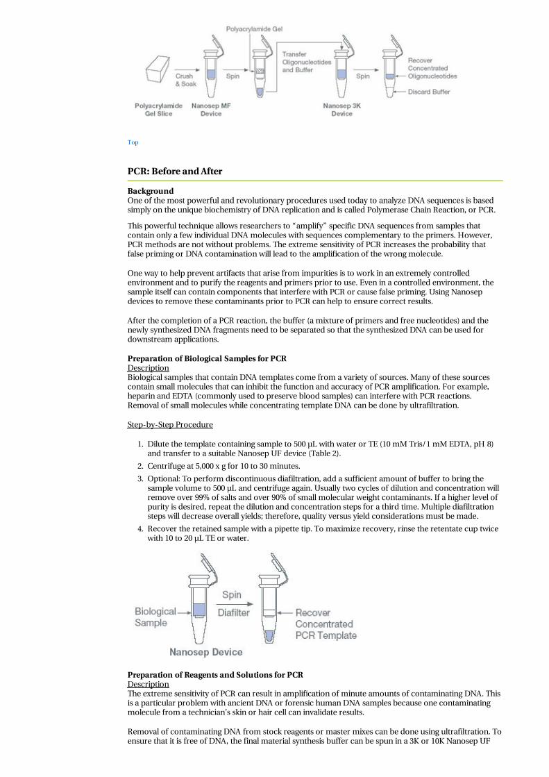
Purification of Oligonucleotides from Polyacrylamide GelsDescriptionOligonucleotides often require gel purification to separate shorter synthesis-failure sequences from full-length synthetic products. Once the desired band has been excised from the gel, it is necessary to recoverthe oligonucleotide from the gel matrix. The “crush and soak” technique is the method of choice forelution from polyacrylamide gels because it is inexpensive, easy, and can be accomplished withoutmonitoring. Nanosep MF devices can be used to remove gel debris, followed by ultrafiltration using aNanosep 3K device to concentrate and desalt the oligonucleotides.
Step-by-Step Procedure
1. Electrophorese oligonucleotides in a polyacrylamide gel. Use 20% acrylamide/8 M urea foroligonucleotides < 25 bases; 15% acrylamide/8 M urea for oligonucleotides 25 to 60 bases, and 12%acrylamide/8 M urea for 60 to 100 bases.
2. Visualize oligonucleotide bands by autoradiography (if radiolabeled). If unlabeled, detect using UVshadowing by placing the gel on a fluorescent TLC plate and using short-wavelength UV lamp toobserve fluorescence quenching.
3. Excise each oligonucleotide band with a new razor blade.
4. Place the gel slice into a microcentrifuge tube and grind it into a fine powder with a microcentrifugepestle. Add 0.5 mL of extraction buffer and vortex. Incubate the slurry at 37 °C for 2 hours orovernight at room temperature.Extraction buffers:
Deionized water
0.5 M NaCl, 2M triethylammonium acetate
50 mM triethylammonium acetate
0.5 M NaCl, 0.1 M Tris-HCl (pH 7.0), 1 mM EDTA
5. To remove acrylamide fragments, transfer the gel slurry to a Nanosep MF device and spin at 14,000x g for 10 minutes. Rinse the upper chamber with 50 µL TE (10 mM Tris/1 mM EDTA, pH 8) and spinagain.
6. To simultaneously concentrate the oligonucleotides and remove dissolved urea and salts, transferthe filtrate to a Nanosep 3K device and centrifuge at 5,000 x g. Oligonulcleotides larger than 30 bpmay be centrifuged at 14,000 x g.
7. Optional: To perform discontinuous diafiltration, add a sufficient amount of buffer to bring thesample volume to 500 µL and centrifuge again. Usually two cycles of dilution and concentration willremove over 99% of salts and over 90% of small molecular weight contaminants. If a higher level ofpurity is desired, repeat the dilution and concentration steps for a third time. Multiple diafiltrationsteps will decrease overall yields; therefore, quality versus yield considerations must be made.
8. Recover the retained sample with a pipette tip. To maximize recovery, rinse the retentate cup twicewith 10 to 20 µL TE or water.
Top
PCR: Before and After
BackgroundOne of the most powerful and revolutionary procedures used today to analyze DNA sequences is basedsimply on the unique biochemistry of DNA replication and is called Polymerase Chain Reaction, or PCR.
This powerful technique allows researchers to “amplify” specific DNA sequences from samples thatcontain only a few individual DNA molecules with sequences complementary to the primers. However,PCR methods are not without problems. The extreme sensitivity of PCR increases the probability thatfalse priming or DNA contamination will lead to the amplification of the wrong molecule.
One way to help prevent artifacts that arise from impurities is to work in an extremely controlledenvironment and to purify the reagents and primers prior to use. Even in a controlled environment, thesample itself can contain components that interfere with PCR or cause false priming. Using Nanosepdevices to remove these contaminants prior to PCR can help to ensure correct results.
After the completion of a PCR reaction, the buffer (a mixture of primers and free nucleotides) and thenewly synthesized DNA fragments need to be separated so that the synthesized DNA can be used fordownstream applications.
Preparation of Biological Samples for PCRDescriptionBiological samples that contain DNA templates come from a variety of sources. Many of these sourcescontain small molecules that can inhibit the function and accuracy of PCR amplification. For example,heparin and EDTA (commonly used to preserve blood samples) can interfere with PCR reactions.Removal of small molecules while concentrating template DNA can be done by ultrafiltration.
Step-by-Step Procedure
1. Dilute the template containing sample to 500 µL with water or TE (10 mM Tris/1 mM EDTA, pH 8)and transfer to a suitable Nanosep UF device (Table 2).
2. Centrifuge at 5,000 x g for 10 to 30 minutes.
3. Optional: To perform discontinuous diafiltration, add a sufficient amount of buffer to bring thesample volume to 500 µL and centrifuge again. Usually two cycles of dilution and concentration willremove over 99% of salts and over 90% of small molecular weight contaminants. If a higher level ofpurity is desired, repeat the dilution and concentration steps for a third time. Multiple diafiltrationsteps will decrease overall yields; therefore, quality versus yield considerations must be made.
4. Recover the retained sample with a pipette tip. To maximize recovery, rinse the retentate cup twicewith 10 to 20 µL TE or water.
Preparation of Reagents and Solutions for PCRDescriptionThe extreme sensitivity of PCR can result in amplification of minute amounts of contaminating DNA. Thisis a particular problem with ancient DNA or forensic human DNA samples because one contaminatingmolecule from a technician’s skin or hair cell can invalidate results.
Removal of contaminating DNA from stock reagents or master mixes can be done using ultrafiltration. Toensure that it is free of DNA, the final material synthesis buffer can be spun in a 3K or 10K Nanosep UF

device prior to aliquoting into PCR tubes (Figure 12). Note: Never bring amplified samples into the samework area that is used to prepare reagents or set up future PCR reactions. Always use aerosol-resistanttips for pipetting.
For full details, see Pall Life Sciences technical report, “Nanosep Centrifugal Ultrafiltration Devices andPCR: Before and After.”
Figure 12Removal of DNA Template from PCR Stock Solutions
DNA template sample of 500 ng plasmid pUC18 in TE was divided into seven 200 µL fractions, onefraction was kept as a control (START, 33 ng/µL) and the other fractions were centrifuged in Nanosepultrafiltration devices. Following centrifugation, 3 µL was added to each PCR reaction containing 45 µLPCR mix (Life Technologies) and 2 µL of a 20 nmole pUC18-complementary primer mix. A PCR reaction of25 cycles under standard conditions was performed and 25 µL of each reaction was electrophoresed andstained with ethidium bromide. No PCR product was detected using filtrate samples from the 3K, 10K, and30K devices, demonstrating that they effectively removed the template spike. Filtrate from the 100K devicegave a weak PCR product band, suggesting that some of the template was able to pass through this device.DNA template passed freely through the 0.2 µm and 300K devices.
Step-by-Step Procedure
1. Always use new or “set-aside” chemicals to make stock PCR reagents. Use care to avoid adding anynew contaminants during preparation.
2. Prepare reagents away from the area used to process PCR products.
3. Centrifuge the stock reagents or master mixes (without primers, templates, or enzymes) in anappropriate 3K or 10K UF device (Table 2).
4. Transfer filtrate to sterile tubes. Sub-aliquotes are suggested to minimize future contamination ofentire stocks.
5. For extra insurance, ultrafiltration can be used to filter the master mix or commercial buffer miximmediately prior to the start of PCR.
Purification and Recovery of PCR PrimersDescriptionSynthetic oligonucleotides are a critical component of the PCR reaction as they act as primers for thesynthesis of DNA from DNA sequence-specific start points. Oligonucleotides are synthesized by chemicalmeans. After synthesis, purification steps are required to purify the full-length oligonucleotides from thesynthesis mixture. Prior to PCR, desalting is required to remove residual by-products from the synthesis,cleavage, and deprotection procedures. Ultrafiltration using centrifugal concentrators is an efficient, high-yield way to desalt and concentrate oligonucleotides (Figure 13).
For full details, see Pall Life Sciences technical report, “Nanosep® Centrifugal Ultrafiltration Devices andPCR: Before and After."

Figure 13Purification and Recovery of 25 bp Oligonucleotides
A 400 µL solution containing 50 ng end-labeled 25 bp oligonucleotides was filtered, in duplicate, usingdifferent molecular weight cutoff (MWCO) devices. These devices were centrifuged for 10 minutes (100K,30K, 10K) to 30 minutes (3K) at 5,000 x g. The retained material was resuspended in 40 µL TE, diluted to400 µL, and then recovered. Radiolabeled DNA was quantified using a scintillation counter. The Nanosep3K device retained close to 90% of the radiolabeled oligonucleotide. The choice of a higher MWCO deviceallowed for a shorter spin time but resulted in decreased yields. If the intention was to pass all of theprimers, then the 100K device would be the best choice.
Step-by-Step Procedure
1. For oligonucleotides up to 50 bases long, use a Nanosep 3K device. For longer oligonucleotides, theNanosep 10K device can be used.
2. Place the oligonucleotide solution in the sample reservoir. Dilute the sample to a volume of 500 µLwith TE (10 mM Tris/1 mM EDTA, pH 8) or water.
3. Optional: To perform discontinuous diafiltration, add a sufficient amount of buffer to bring thesample volume to 500 µL and centrifuge again. Usually two cycles of dilution and concentration willremove over 99% of salts and over 90% of small molecular weight contaminants. If a higher level ofpurity is desired, repeat the dilution and concentration steps for a third time. Multiple diafiltrationsteps will decrease overall yields; therefore, quality versus yield considerations must be made.
4. Recover the retained sample with a pipette tip. To maximize recovery, rinse the retentate cup twicewith 10 to 20 µL TE or water.
Cleanup and Recovery of PCR ProductsDescriptionThe final PCR reaction may contain up to a microgram of amplified DNA that can be used for a variety ofmolecular biology applications. These applications may be more or less sensitive to the residualcomponents of the PCR reaction mix. Certain restriction enzymes and particularly DNA ligase are verysensitive to the presence of contaminants in DNA samples. Because a PCR reaction mixture contains avariety of salts, free nucleotides, glycerol, proteins, and primers, most downstream applications willrequire some sort of cleanup of the PCR product.
Purification of the PCR product away from buffer components, free nucleotides, and primers can beperformed in a variety of ways:
1. Precipitation, using chemical solubility properties to selectively separate DNA. The primarydrawbacks for using this method to purify PCR products are the time involved and incompleteremoval of co-precipitating buffer components and contaminants.

2. Chromatography, using size exclusion resin or affinity to glass to purify DNA from the PCR mixturecomponents. This technique is costly, generally requires significant handling, and samples must beconcentrated after elution from the matrix.
3. Ultrafiltration involves the isolation and concentration of PCR products using size exclusionmembrane devices (Figure 14). It is rapid, there is very little handling, yields are high, DNA isundamaged, and the concentrated DNA is free of contaminants that may inhibit downstreamreactions (Figure 15).
For full details, see Pall Life Sciences technical report, “Nanosep® Centrifugal Ultrafiltration Devices andPCR: Before and After."
Step-by-Step Procedure
1. Avoid pipetting the oil or wax into the subsequent steps. Oil can be removed by freezing the sampleat -70 °C and drawing off the surface liquid, or the sample can be successfully removed by carefulpipetting with a narrow bore pipette tip from below the surface of the oil.
2. Dilute the reaction mixture to 500 µL with water or TE (10 mM Tris/1 mM EDTA, pH 8) and transferto a Nanosep 30K or 100K UF device (Table 2).
3. Centrifuge at 5,000 x g for 10 minutes (greater speeds increase PCR product loss).
4. Optional: To perform discontinuous diafiltration, add a sufficient amount of buffer to bring thesample volume to 500 µL and centrifuge again. Usually two cycles of dilution and concentration willremove over 99% of salts and over 90% of small molecular weight contaminants. If a higher level ofpurity is desired, repeat the dilution and concentration steps for a third time. Multiple diafiltrationsteps will decrease overall yields; therefore, quality versus yield considerations must be made.
5. Recover the retained sample with a pipette tip. To maximize recovery, rinse the retentate cup twicewith 10 to 20 µL TE or water.
Figure 14Purification and Recovery of PCR Products
A small amount of 32P-labeled dCTP was added to the PCR reaction mix (Life Technologies) containing32P-end labeled primers. The reaction was run for 30 cycles using standard conditions. Ten reactions werepooled and 100 µL aliquots were diluted to 500 µL and centrifuged in the indicated MWCO devices. Theretained material was recovered in 20 µL TE, electrophoresed using a 10% polyacrylamide Tris Borate gel(BioRad) and analyzed by autoradiography. The 100K Nanosep device demonstrated the best combinationof PCR product retention along with complete primer and nucleotide removal. If the desire is to ensure theremoval of the buffer and free nucleotides but not primers, then the 30K or 10K devices will retain the PCRproduct while removing buffer components. Cleanup using the 10K device may be used if the PCR productis smaller than 200 bp and the presence of primers does not inhibit downstream applications.
Figure 15Biological Activity of Purified PCR Products
A pooled mixture of PCR products was divided in half. One fraction was precipitated with 1/10th volumesodium acetate and two volumes ethanol, chilled at -20 °C for 2 hours, centrifuged at high speed for 30minutes, rinsed with 70% ethanol and air dried prior to resuspension in 40 µL water. The second fractionwas diluted to 500 µL and centrifuged for 15 minutes in a 30K Nanosep device. The retained material was

resuspended in 40 µL water and divided into 10 µL aliquots for analysis. One fraction was electrophoresedas the uncut (UC) control and the others were diluted into a 20 µL restriction digest reaction according tomanufacturer’s instructions (EcoRI = RI, BamHI = BH1, Xba1 = Xba). These samples were digested for 30minutes at 37 °C, electrophoresed in a 1.5% agarose gel, and DNA bands were visualized by staining withethidium bromide. Our data show clearly that the XbaI enzyme is unable to digest to completion the 400bp PCR fragment from a sample that has been precipitated. In contrast, the sample that was rapidlypurified with the 30K device was completely digested using XbaI, indicating that use of this device removedcontaminants that could interfere with digestion.
Top
Protein Purification and Handling
BackgroundUltrafiltration has been used successfully for years and is an excellent and gentle method to purify andconcentrate protein samples. Ultrafiltration can be used to replace precipitation, evaporation, dialysis,lyophylization, and gel filtration to concentrate and desalt protein without significant protein loss (Figure16).
Advantages of ultrafiltration:
The most gentle method for concentrating proteins.
Minimal denaturation of protein compared to precipitation.
Maintains ionic strength, hence does not result in hyperconcentration of salts.
Produces high yields.
Much faster than dialysis and less expensive than lyophilization.
Ultrafiltration, however, is a separation technique rather than a fractionation technique. With very fewexceptions, the use of ultrafiltration for protein fractionation remains impractical unless the two proteinsto be separated have at least a ten-fold difference in their molecular weights.
Figure 16Protein Binding and Recovery
Nanosep devices (Omega membrane) and competitive devices (Comp-PES, Comp-PS) were used to filterand recover radioactively-labeled BSA (125I-BSA). A 1 µg sample of labeled BSA was diluted into 500 µLphosphate buffered saline and centrifuged in a fixed angle rotor using instructions from the respectivedevice manufacturers for speed and spin duration. The resulting retentate was recovered in 40 µL PBS andplaced directly into counting vials containing scintillation solution. After recovering the retentate, theupper receivers were submerged in separate vials containing scintillation solution. The graph contains theresults of counting (PerkinElmer, Gaithersburg, MD, USA) of two independent experiments where eachdevice was analyzed in triplicate.
Protein Concentration from Dilute SolutionsDescriptionOne of the most popular and successful UF applications is concentration of dilute protein solutionscontaining antibodies, enzymes, growth factors, etc. Protein purification schemes include cell disruption,followed by initial fractionation, then a secondary fractionation, and finally a polishing step.
After these steps are completed, the dilute protein-containing solutions need to be concentrated prior touse in downstream applications. Ultrafiltration is an efficient method of protein concentration anddesalting under gentle conditions without significant loss of biological activity (Table 4).

Table 4Typical Protein Retentate Recovery/Passage
Solute Solute MW (Kd)MWCO 3K 10K 30K 100K 300K
Spin Time (min.) 15 10 8 5 3
Vitamin B12 1,335 % Recovery 7 - - - -
Aprotinin 6,200 % Recovery 99 51 11 - -
Cytochrome C 12,400 % Recovery 100 89 77 1.8 -
Chymotrypsinogen A 25,000 % Recovery - 97 94 2.1 -
Ovalbumin 45,000 % Recovery - 97 92 3 -
BSA 67,000 % Recovery - - 100 26 1.5
Phosphorylase B 97,400 % Recovery - - 95 91 1
IgG 156,000 % Recovery - - - 97 1.5
Thyroglobulin (1 mg/mL) 677,000 % Recovery - - - 100 91
Samples of 0.5 mL of a 1.0 mg/mL solution were centrifuged at 14,000 x g and were concentrated to avolume of 10 to 60 µL.
Step-by-Step Procedure
1. Choose the appropriate device depending on the sample volume (Table 1).
2. Place dilute protein samples into the sample reservoir of the device with the appropriate MWCOmembrane.
3. Note: Increased solute concentration in the sample can decrease the passage of a specific proteinand the use of a higher MWCO device may be required. If larger sample volumes are to beconcentrated, a pilot experiment is suggested using 100 µL of the sample in a Nanosep device todetermine which membrane is best suited for the specific large-scale application.
4. Centrifuge the device at the recommended g-force; longer spin times will be required for solute-laden samples or lower MWCO devices.
5. After concentration, the protein sample can be pipetted from the retentate chamber. Higher yieldscan be achieved if a small volume of buffer (20 µL for a Nanosep device) is used to rinse themembrane surface for remaining protein samples.
Improving the Recovery of Proteins from Dilute Solutions (Passivation)DescriptionFor very dilute protein solutions (< 10 µg/mL), concentrate recovery in UF devices is often notquantitative (Figure 1). Pall centrifugal devices have been specifically constructed of materials thatminimize nonspecific binding. However, certain proteins, particularly when dilute, can be problematic.The extent of nonspecific binding varies with the structure of the individual protein. Proteins containingcharged or hydrophobic domains tend to show a high affinity toward various surfaces that may lead toirreversible binding.
Strategies that reduce adsorptive loss of proteins on surfaces are either based on pretreatment of thesurface to fill the exposed binding sites or by changes in the composition of the solution, usually byaddition of protein (often albumin), detergents, or salts. In most cases, pretreating (passivating) the devicebefore concentration of dilute protein solutions can improve recovery (Figure 17).
Figure 17Use of Passivation to Increase Protein Recoveries

0.1 µg/mL and 1.0 µg/mL bovine serum albumin (BSA) solution were centrifuged in pretreated 30KNanosep devices. Pretreatment involved incubating the devices filled with either 1% BSA, 5% SodiumDodecyl Sulfate (SDS), or 5% Tween 20 for 1 hour. The device was then rinsed and used immediately.Passivation increased the recovery of protein, particularly for the most dilute samples.
Step-by-Step Procedure
1. Add 500 µL of the sterile passivation solution to the Nanosep device (see below). Close the cap andincubate the solution in the device for at least one hour at room temperature.Passivation solutions:
1% BSA in PBS
5% SDS in distilled water
5% Tween-20 in distilled water
5% Triton-X in distilled water
5% PEG compound in distilled water
1% IgG in PBS
2. Discard the passivation solution by either pouring or pipetting it out of the device, and rinse theNanosep device thoroughly with sterile distilled water.
3. To ensure that residual passivation solution is removed, add 500 µL of distilled water to the deviceand centrifuge at 14,000 x g for 5 to 10 minutes. Discard the filtrate.
4. The device can be used immediately or stored for later use. If the device is to be used later, add 100µL sterile distilled water to the sample reservoir and store at 4 °C to retard bacterial growth.Important: Do not allow the membrane to dry out once the device has been passivated.
Protein Desalting or Buffer Exchange (Diafiltration)DescriptionCentrifugal concentrators are ideal for the removal or exchange of salts. Desalting by dialysis is time-

consuming and works best when the concentration differential between the two solutions is large. Dialysisdoes not concentrate dilute samples and may result in even further dilution.
A single round of protein concentration using ultrafiltration results in a sample with essentially the samebuffer composition as the starting material. To remove salts or exchange buffers, the concentratedsample is diluted with the new buffer or water and centrifuged a second time (this process is calleddiscontinuous diafiltration). The dilution/concentration steps can be repeated until the required amountof salt is removed/replaced (Table 5).
Table 5Desalting Estimates
Spin No. Salt ConcentrationAfter Spin
Percent SaltRemoval
1 500 mM 95%
2 25 mM 99.75%
3 1.25 mM 99.99%
4 0.06 mM 100%
Estimates are made for concentrating 500 µL to 25 µL each spin.
Step-by-Step Procedure
1. Select the Nanosep device with a MWCO three times smaller than the MW of the protein to beretained (Table 3).
2. To concentrate the sample without changing salt concentration: pipette 500 µL of sample into thesample reservoir and centrifuge at the speed and time needed to concentrate the sample. Spin timeswill depend on sample concentration and MWCO.
3. To desalt or exchange buffer, first concentrate the sample (step 2) then add new buffer or water todilute the concentrated sample to 500 µL. Centrifuge again. This process can be repeated to achievea lower salt concentration. Usually two cycles of dilution and concentration will remove over 99% ofsalts and over 90% of small molecular weight contaminants. If a higher level of purity is desired,repeat the dilution and concentration steps for a third time. Multiple diafiltration steps will decreaseoverall yields; therefore, quality versus yield considerations must be made.
4. Recover the retained sample with a pipette tip. To maximize recovery, rinse the retentate cup twicewith 10 to 20 µL new buffer or water.
Gross Fractionation of Complex Protein SolutionsDescriptionAlthough ultrafiltration is primarily a separation technique, under some conditions it can be used for thegross fractionation of proteins that differ significantly in size (Figure 18). In order to fractionate or enrichfor a particular protein, the following must be considered:
The proteins must have at least 10-fold difference in MW.
The MW of retained protein should be at least 3 times the MWCO of membrane.
The passing protein should be at least 3 times smaller than the MWCO of membrane.
The sample concentration should be 5 mg/mL or less.
Centrifugation should be performed at lower than maximum recommended g-force (500 to 1,000 x g).
It is important to remember that separation is rarely absolute and is better described as an enrichment.
Figure 18Gross Fractionation of Proteins

A 500 µL sample of a 5.0 mg/mL protein solution containing IgG (156 kD) and Cytochrome C (12.4 kD) wascentrifuged at 1,000 x g for 30 minutes in a Nanosep 100K device. The retentate was recovered in 500 µL; 15µL samples of the retentate and filtrate were analyzed on 10% NuPAGE Bis-Tris polyacrylamide gel. Lane1 = NOVEX SeeBlue Pre-Stained Protein Standards. Lane 2 = Mixture of IgG and Cytochrome C. Lane 3 =Retentate. Lane 4 = Filtrate. After two spins (not shown), over 95% of Cytochrome C was found in filtratewhile more than 85% of the IgG was retained by the membrane.
Step-by-Step Procedure
1. Fill the Nanosep device (usually 100K or 300K) with 500 µL of the protein mixture.
2. Centrifuge at low speed (500 to 1,000 x g) for approximately 20 minutes.
3. Transfer the filtrate from the bottom receiver to a new tube for storage. The filtrate can beconcentrated in a lower MWCO device if needed.
4. To diafilter the retentate, fill the sample reservoir to 500 µL with appropriate buffer. Mix briefly,centrifuge, and collect filtrate as before.
5. Remove or resuspend the retentate in the appropriate buffer and transfer it to a fresh tube forstorage.
Concentration of Monoclonal Antibodies from Culture SupernatantsDescriptionThe concentration of monoclonal antibodies (mAbs) in culture supernatants is often too low to be usefulfor most in vitro and in vivo immunological techniques. Concentrated mAbs are obtained from ascitesfluid induced in hybridoma cells. However, development of ascites from hybridoma cells is a complicatedand time-consuming procedure, and antibodies still need to be purified prior to use.
Ultrafiltration can be used to rapidly purify and recover concentrated mAbs directly from culturesupernatants. Use of a 100K MWCO membrane allows for the concentration of antibodies while at thesame time removing serum albumin and other low molecular weight proteins present in the culturemedia.
Step-by-Step Procedure
1. Add 0.5 mL (Nanosep 100K device), 3 mL (Microsep 100K device), or up to 15 mL (Macrosep 100Kdevice) of the culture supernatant to the centrifugal device.
2. Centrifuge at 5,000 x g for 30 minutes.
3. Recover mAbs by pipetting the retentate from the sample reservoir.
4. Optional: To perform discontinuous diafiltration, add sufficient amount of buffer to bring thesample volume to 3 mL (Microsep device) or 12 mL (Macrosep device) and centrifuge again. Usuallytwo cycles of dilution and concentration will remove over 99% of salts and over 90% of smallmolecular weight contaminants. If a higher level of purity is desired, repeat the dilution andconcentration steps for a third time. Multiple diafiltration steps will decrease overall yields;therefore, quality versus yield considerations must be made.

Top
Miscellaneous Protocols
Preparation of Protein-free Samples for Analytical ApplicationsDescriptionPeptides and other low molecular weight bioactive materials can be analyzed and purified from complexbiological sources using a variety of techniques such as HPLC, GC, and GC/MS. Ultrafiltration is aneffective and fast way to remove proteins that may interfere with the analysis of other small moleculesprior to these procedures (Table 6).
Table 6Protein Removal for Analytical Applications
Cyto C (12.5 kD) BSA (67 kD)
Nanosep® Device MWCO 3K 10K 3K 10K
% of Protein in Filtrate <1% 6.5% 0 0
A 500 µL protein sample of 1.0 mg/mL in phosphate buffer saline was centrifuged at 14,000 x g. A sampleof filtrate (50 µL) was analyzed using a TOSOHAAS, TSK-GEL G3000WXL, 10 µm, 1000 x 6.0 mm HPLCcolumn and detected at 280 nm.
Step-by-Step Procedure
1. Select a Nanosep device with a 3K or 10K MWCO membrane, depending on the size of the protein tobe removed.
2. Dilute the sample if possible to 500 µL. This step helps minimize membrane fouling; however,smaller volumes can be used.
3. Place the dilute sample into the upper sample reservoir.
4. Centrifuge the Nanosep device at 5,000 to 14,000 x g for up to 30 minutes.
5. Collect the protein-free filtrates from the bottom filtrate receiver. The sample is now ready forfurther analysis.
6. If needed, the concentrated/retained proteins can be recovered from the sample reservoir.
Purification of RNA Transcripts from Polyacrylamide GelsDescriptionA number of applications require the purification of synthetic RNA transcripts from polyacrylamide gels(PAGE). Often this is done to remove short or incomplete transcripts which may interfere withdownstream applications. Once the synthetic mRNA band has been identified and excised from the gel, itis necessary to recover RNA from the gel matrix. The “crush and soak” method for mRNA recovery is

often the method of choice because it is inexpensive, easy, and can be accomplished without extensivemonitoring. Once the mRNA is released into solution, it can be desalted and concentrated in one easy stepusing ultrafiltration devices (Figure 19).
Figure 19Recovery of RNA from Polyacrylamide Gels
RNA Century Markers prepared by in vitro transcription of Century Markers Templates (Ambion) wereelectrophoresed in 6% TBE (Tris-borate, EDTA)-urea gel. Gel slices containing RNA fragments from 100 to500 bases long were excised, macerated with the Eppendorf fitting pestle and mixed with the extractionbuffer. After 2 hours incubation at 37 °C, each slurry was transferred to a Nanosep MF 0.2 µm device andcentrifuged at 14,000 x g. Each filtrate was transferred to a Nanosep 10K device and centrifuged at 5,000 x gfor 15 minutes and diafiltered with TE. The desalted retentate was recovered in 20 µL TE andelectrophoresed in a 6% TBE-urea gel alongside their respective RNA Markers. Equal band intensitiesapproximate 100% recovery. Our recoveries ranged from 50 to 90%; no smearing due to RNase activity wasdetected.
Step-by-Step Procedure
1. Electrophorese the synthetic RNA transcripts in a 5% polyacrylamide/8 M urea PAGE gel.
2. Visualize RNA by staining the gel with ethidium bromide, or in the case of radioactive RNA, by briefexposure of the gel to X-ray film. (Do not overexpose the autoradiogram; a thinner band widthallows a more accurate excision of the desired mRNA species.)
3. Place the excised band containing RNA in a standard 1.5 mL microcentrifuge tube.
4. Crush the excised band with microcentrifuge fitting pestle and add 500 µL gel extraction buffer (0.5M Ammonium Acetate, 2 mM EDTA, 0.1% SDS).
5. Vortex and incubate at 37 °C from 2 hours to overnight depending on the size of the transcript.Small transcripts (< 200 nucleotides) will elute in approximately 2 hours; larger transcripts requirelonger incubation.
6. Transfer the slurry into a Nanosep MF device and spin at 14,000 x g for 10 minutes to remove theacrylamide and collect the mRNA in the filtrate.
7. RNA from the filtrate can be concentrated and desalted in a Nanosep device by centrifuging thefiltrate at 5,000 x g in a device with the appropriate MWCO. For RNA molecules smaller than 50bases, use a Nanosep 3K device, 50 to 500 bases use a Nanosep 10K device, 500 to 1000 bases use aNanosep 30K device, and larger than 1000 bases use a Nanosep 100K device.
8. To remove salts (diafilter), add 500 µL of water or buffer to the upper sample receiver andcentrifuge again. If necessary, repeat the dilution and concentration step.
Fast and Efficient Elution of Proteins from Polyacrylamide GelsDescriptionPolyacrylamide Gel Electrophoresis (PAGE) is both an analytical and preparative tool widely used toseparate proteins in an electric field according to their size, shape and charge. Recent advances in 2D-PAGE have revolutionized proteomics and its applications in biomedicine, biotechnology orpharmaceuticals. Complex protein samples are first separated by 2D-PAGE, stained, and then scanned,allowing for detailed measurements of expression levels and protein mapping. Further proteincharacterization often requires the extraction of specific proteins from the polyacrylamide gel matrix forfurther analyses such as mass spectrometry, HPLC, amino acid sequencing or composition analyses, andpost transcriptional modification analyses (3,4).
Nanosep centrifugal devices provide a fast and inexpensive alternative to commonly used methods for theelution and concentration of proteins that have been separated on polyacrylamide gels. While Nanosepcentrifugal devices allow for rapid and efficient elution of proteins from polyacrylamide gels, the amountof starting polyacrylamide gel material is the limiting factor for high protein recoveries. Reducing theamount of polyacrylamide not only increases the yields of eluted protein but also reduces centrifugationtimes significantly, a factor of consideration when eluting biologically active proteins (4).
For an in-depth scientific discussion on this protocol, see Pall Life Sciences technical report, “Fast andEfficient Elution of Proteins from Polyacrylamide Gels Using Nanosep Centrifugal Units."
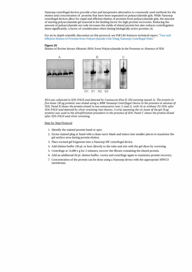
Figure 20Elution of Bovine Serum Albumin (BSA) from Polyacrylamide in the Presense or Absence of SDS
BSA was subjected to SDS-PAGE and detected by Coomassie Blue R-250 staining (panel A). The protein infive lanes (30 µg protein) was eluted using a 300K Nanosep Centrifugal Device in the presence or absence ofSDS. Panel B shows the protein eluted in two consecutive runs (1 and 2), with (S) or without (N) SDS, afterSDS-PAGE and detected by silver straining (not shown). A strip spanning the six lanes of the gel (8 µgprotein) was used in the ultrafiltration procedure in the presence of SDS. Panel C shows the protein elutedafter SDS-PAGE and silver straining.
Step-by-Step Protocol
1. Identify the stained protein band or spot.
2. Excise stained plug or band with a clean razor blade and mince into smaller pieces to maximize thegel surface area during protein elution.
3. Place excised gel fragments into a Nanosep MF centrifugal device.
4. Add elution buffer (50 µL or less) directly to the tube and mix with the gel slices by vortexing.
5. Centrifuge at 14,000 x g for 2 minutes; recover the filtrate containing the eluted protein.
6. Add an additional 20 µL elution buffer, vortex and centrifuge again to maximize protein recovery.
7. Concentration of the protein can be done using a Nanosep device with the appropriate MWCOmembrane.

Top
Appendices
Sanitizing Nanosep DevicesNanosep centrifugal devices are sold non-sterile but can easily be sanitized prior to use with 70% ethanolby performing the following:
1. Fill device with 70% molecular biology grade ethanol. Close the cap and centrifuge at maximum g-force until all liquid passes through the membrane (10 to 30 minutes, depending on MWCO).
2. Discard filtrate. To remove residual ethanol, fill device with sterile water or the desired buffer andcentrifuge again. Note: Use device within 20 minutes to prevent irreversible membrane damagecaused by dehydration.
Nanosep Device Centrifugation TimesFiltration rates in centrifugal devices are affected by several operating parameters including membraneMWCO and material of construction, sample composition and viscosity, the centrifugal force used, andthe operating temperature (Table 7).
Table 7Approximate Spin Times in Nanosep Devices

Nanosep DeviceMWCO Color Code RCF Spin Time at 25 °C
3K gray 14,000 x g 20 minutes
10K blue 14,000 x g 15 minutes
30K red 14,000 x g 8 minutes
100K clear 14,000 x g 5 minutes
300K orange 14,000 x g 3 minutes
A 1.0 mg/mL 500 µL sample containing five proteins of molecular weights ranging from 200 Kd to 6 Kdwas concentrated down to 20 to 25 µL. (B-amylase, 200 Kd; Alcohol dehydrogenase, 150 Kd; Bovine serumalbumin, 66 Kd; Trypsin inhibitor, 20 Kd; Aprotinin, 6 Kd).
Effect of UF Membrane Material on Filtration RatesPall Omega membrane (modified polyethersulfone) has a very fast flow rate compared to UF membranemade of regenerated cellulose. Figure 18 compares the time required to concentrate 500 µL of 0.1 mg/mLBSA sample in a Nanosep device containing the Omega 10K MWCO membrane with a device containing a10K regenerated cellulose membrane. Both devices were spun at 5,000 x g.
Figure 21Filtration Rates in UF Devices with Different Membranes
Effect of G-force on Filtration RatesCentrifuge speed has a direct effect on the selectivity of membrane as well as the duration of the spin. A500 µL sample (0.1 mg/mL IgG) was concentrated in a Nanosep 100K device spun at different centrifugalforces (Figure 22). Higher g-forces result in faster filtration rates and a corresponding increase inmolecule passage.
Figure 22Effect of g-force on Filtration Rates

Effect of Sample Concentration on Filtration RatesSample concentration cannot only effect spin times but also may effect the performance of thenominally-rated MWCO. As protein concentration increases, there is a corresponding decrease inmolecule passage (Figure 23).
Samples containing 500 µL IgG with initial concentrations of 0.01 mg/mL to 1 mg/mL were centrifuged at14,000 x g in Nanosep 100K devices. Samples with higher starting concentrations require longer spin timesto process.
Figure 23Effect of Sample Concentration on Filtration Rates
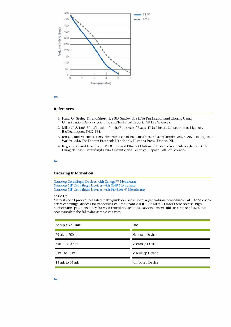
Effect of Temperature on Filtration RatesAs temperature decreases, there is a corresponding increase in centrifugation time (Figure 24). A 500 µLsample of 0.1 mg/mL IgG was centrifuged in a Nanosep 100K device at 21 °C or 4 °C.
Figure 24Effect of Temperature on Filtration Rates
Top
References
1. Fang, Q., Seeley, K., and Short, T. 2000. Single-tube DNA Purification and Cloning UsingUltrafiltration Devices. Scientific and Technical Report, Pall Life Sciences.
2. Miller, J. S. 1988. Ultrafiltration for the Removal of Excess DNA Linkers Subsequent to Ligation.BioTechniques. 5:632-634.
3. Jeno, P. and M. Horst. 1996. Electroelution of Proteins from Polyacrylamide Gels, p. 207-214. In J. M.Walker (ed.), The Protein Protocols Handbook. Humana Press, Totowa, NJ.
4. Reguera, G. and Leschine, S. 2000. Fast and Efficient Elution of Proteins from Polyacrylamide GelsUsing Nanosep Centrifugal Units. Scientific and Technical Report, Pall Life Sciences.
Top
Ordering Information
Nanosep Centrifugal Devices with Omega™ MembraneNanosep MF Centrifugal Devices with GHP MembraneNanosep MF Centrifugal Devices with Bio-Inert® Membrane
Scale UpMany if not all procedures listed in this guide can scale up to larger-volume procedures. Pall Life Sciencesoffers centrifugal devices for processing volumes from < 100 µL to 60 mL. Order these precise, highperformance products today for your critical applications. Devices are available in a range of sizes thataccommodate the following sample volumes:
Sample Volume Use
50 µL to 500 µL Nanosep Device
500 µL to 3.5 mL Microsep Device
3 mL to 15 mL Macrosep Device
15 mL to 60 mL Jumbosep Device
Top


![pET Express & Purify Kits User Manual - Takara Bio Manual/PT5018-1.pdf15 µl pET6xHN-C Vector (In-Fusion Ready) [100 ng/µl] 10 µl pET6xHN-GFPuv Vector [500 ng/µl] 15 µl 1.1 kb](https://static.fdocuments.net/doc/165x107/5e7b57982623d66a901d15a7/pet-express-purify-kits-user-manual-takara-bio-manualpt5018-1pdf-15-l.jpg)