NANOMATERIAL ENVIRONMENTAL, HEALTH, AND SAFETY PROJECT...
133
NANOMATERIAL ENVIRONMENTAL, HEALTH, AND SAFETY PROJECT COMMITTEE Mission The mission of the HESI Nanomaterial Environmental, Health, and Safety (EHS) Project Committee was to improve the science associated with developing toxicological and safety evaluations for engineered nanomaterials and to increase the fundamental understanding of the behavior of these materials in biological systems and the environment. 2009 Participants: BASF Corporation The Coca-Cola Company The Dow Chemical Company East Carolina State University L’Oréal Corporation North Carolina State University The Procter & Gamble Company University of Rochester US Centers for Disease Control and Prevention National Institute of Occupational Safety and Health US Consumer Product Safety Commission US Environmental Protection Agency US Food and Drug Administration US National Institutes of Health National Institute of Environmental Health Sciences Committee Publications Balshaw, D.M., M. Philbert, and W.A. Suk, Research strategies for safety evaluation of nanomaterials, Part III: nanoscale technologies for assessing risk and improving public health. Toxicol Sci, 2005. 88(2): p. 298-306. Borm, P., et al., Research strategies for safety evaluation of nanomaterials, part V: role of dissolution in biological fate and effects of nanoscale particles. Toxicol Sci, 2006. 90(1): p. 23-32. Holsapple, M.P., et al., Research strategies for safety evaluation of nanomaterials, part II: toxicological and safety evaluation of nanomaterials, current challenges and data needs. Toxicol Sci, 2005. 88(1): p. 12-7. Holsapple, M.P., et al., Research strategies for safety evaluation of nanomaterials, part II: toxicological and safety evaluation of nanomaterials, current challenges and data needs. Toxicol Sci, 2005. 88(1): p. 12-7. Holsapple, M.P. and L.D. Lehman-McKeeman, Forum series: research strategies for safety evaluation of nanomaterials. Toxicol Sci, 2005. 87(2): p. 315. Thomas, K., et al., Research strategies for safety evaluation of nanomaterials, part VIII: International efforts to develop risk-based safety evaluations for nanomaterials. Toxicol Sci, 2006. 92(1): p. 23-32.
Transcript of NANOMATERIAL ENVIRONMENTAL, HEALTH, AND SAFETY PROJECT...
NANOMATERIAL ENVIRONMENTAL, HEALTH, AND SAFETY PROJECT COMMITTEE Mission The mission of the HESI Nanomaterial Environmental, Health, and Safety (EHS) Project Committee was to improve the science associated with developing toxicological and safety evaluations for engineered nanomaterials and to increase the fundamental understanding of the behavior of these materials in biological systems and the environment. 2009 Participants: BASF Corporation The Coca-Cola Company The Dow Chemical Company East Carolina State University L’Oréal Corporation North Carolina State University The Procter & Gamble Company University of Rochester US Centers for Disease Control and Prevention National Institute of Occupational Safety and Health US Consumer Product Safety Commission US Environmental Protection Agency US Food and Drug Administration US National Institutes of Health National Institute of Environmental Health Sciences Committee Publications Balshaw, D.M., M. Philbert, and W.A. Suk, Research strategies for safety evaluation of nanomaterials, Part III: nanoscale technologies for assessing risk and improving public health. Toxicol Sci, 2005. 88(2): p. 298-306. Borm, P., et al., Research strategies for safety evaluation of nanomaterials, part V: role of dissolution in biological fate and effects of nanoscale particles. Toxicol Sci, 2006. 90(1): p. 23-32. Holsapple, M.P., et al., Research strategies for safety evaluation of nanomaterials, part II: toxicological and safety evaluation of nanomaterials, current challenges and data needs. Toxicol Sci, 2005. 88(1): p. 12-7. Holsapple, M.P., et al., Research strategies for safety evaluation of nanomaterials, part II: toxicological and safety evaluation of nanomaterials, current challenges and data needs. Toxicol Sci, 2005. 88(1): p. 12-7. Holsapple, M.P. and L.D. Lehman-McKeeman, Forum series: research strategies for safety evaluation of nanomaterials. Toxicol Sci, 2005. 87(2): p. 315. Thomas, K., et al., Research strategies for safety evaluation of nanomaterials, part VIII: International efforts to develop risk-based safety evaluations for nanomaterials. Toxicol Sci, 2006. 92(1): p. 23-32.
Thomas, K. and P. Sayre, Research strategies for safety evaluation of nanomaterials, Part I: evaluating the human health implications of exposure to nanoscale materials. Toxicol Sci, 2005. 87(2): p. 316-21. Thomas, T., et al., Research strategies for safety evaluation of nanomaterials, part VII: evaluating consumer exposure to nanoscale materials. Toxicol Sci, 2006. 91(1): p. 14-9. Tsuji, J.S., et al., Research strategies for safety evaluation of nanomaterials, part IV: risk assessment of nanoparticles. Toxicol Sci, 2006. 89(1): p. 42-50. Committee Presentation and Data Resources January 19, 2009: HESI Nanomaterial Environmental, Health, and Safety Committee Presentation. "Nanomaterial Environmental, Health, and Safety." Presented at the 2009 HESI Annual Meeting, Tucson, Arizona. Presentation by Ms. Nancy Doerrer, HESI.
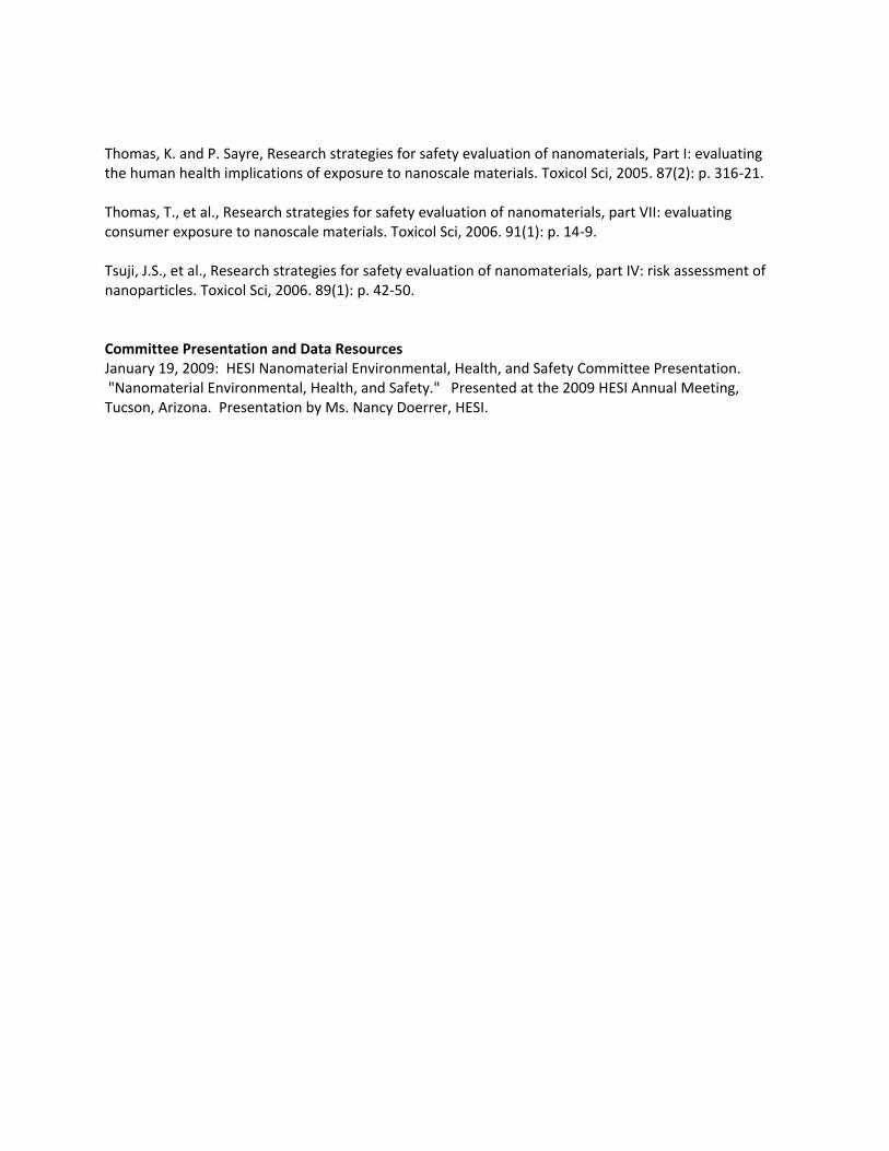
HESI Nanomaterial Environmental, Health and Safety Project Committee
Chair:Raymond M. David, PhD, DABT
(BASF Corporation)
Vice-Chair:Hon-Wing Leung, PhD, DABT, CIH
(Arkema Inc.)
January 19, 2009, Assembly of Members MeetingHESI Annual Meeting
Tucson, AZ

2008 Participation
INDUSTRY
ArkemaBASFCoca-Cola Dow ChemicalL’OrealProcter & Gamble
PUBLIC
CDC National Institute of Occupational Safety and Health (NIOSH)
East Carolina UniversityNIH National Institute of Environmental
Health Sciences (NIEHS)North Carolina State UniversityUniversity of RochesterUS Consumer Product Safety
Commission (CPSC)US Environmental Protection AgencyUS Food and Drug Administration
Project Goals
• Determine the current knowledge-base and research needs for toxicology and safety evaluations of engineered nanomaterials.
• Identify unresolved scientific issues, research needs, and/or data gaps that would facilitate the development of a comprehensive risk assessment for nanomaterials.
• Develop a better understanding of the fundamental behavior of nanomaterials.
Eight-Part Series inToxicological Sciences (2005-2006)
Research strategies for safety evaluation of nanomaterials:
1. Human health implications of exposure2. Toxicological and safety evaluation3. Nanoscale technologies4. Risk assessment of nanoparticles5. Role of dissolution in biological fate and effects
of nanoscale particles6. Characterization of nanoscale particles7. Consumer exposure8. International efforts to develop risk-based
safety evaluations
Research Objectives
• Explore human health effects associated with pulmonary exposure to the same well-characterized materials in in vivo and in vitro test systems.
• Evaluate the distribution and fate of nanomaterials in biological systems.
• Multi-sector consortium established to conduct research.
Pulmonary Toxicity Studies:In Vivo
MaterialsTitanium dioxide (TiO2)multi-walled carbon nanotubes (MWCNT)carbon black
Routes of Administration (rat)Nose-only inhalation chamber (BASF)Pharyngeal aspiration (NIOSH)Exposure levels were consistent from test system to test system.
ResultsSlight differences were observed in responses to different nanoparticles.

Fate Studies (Tissue Distribution) Following Systemic or Lung Exposure
(Procter & Gamble)
MaterialNanoscale polystyrene fluorescent beads
Routes of Administration (rat)pharyngeal aspirationiv injection (systemic exposure)
ResultsSmall particles (20 nm) distribute differently from larger ones (100-1000 nm).Smaller particles are not as persistent.Distribution is route-specific.
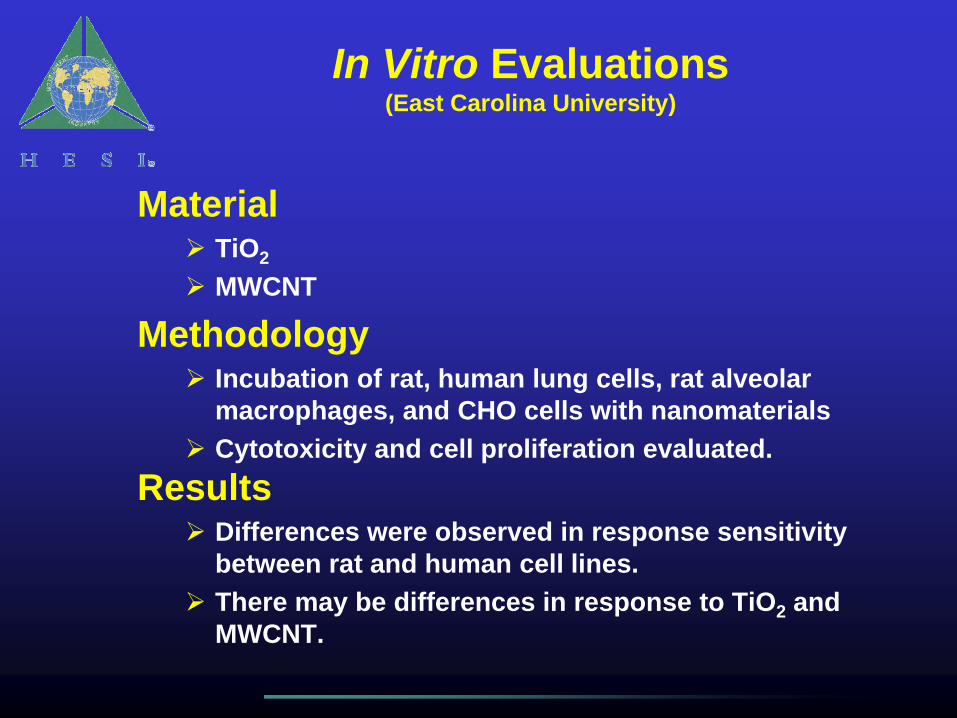
In Vitro Evaluations(East Carolina University)
MaterialTiO2
MWCNT
MethodologyIncubation of rat, human lung cells, rat alveolar macrophages, and CHO cells with nanomaterialsCytotoxicity and cell proliferation evaluated.
ResultsDifferences were observed in response sensitivity between rat and human cell lines. There may be differences in response to TiO2 and MWCNT.
2009 Project Committee Activity
Webinar
• February 2009• “Genotoxicity of Nanomaterials”• ~ 50 invited participants• Speakers from BASF and University of
Copenhagen
Project Committee Sunset
• Project Committee has elected to disband, effective March 31, 2009.
• Interested parties are encouraged to submit targeted proposals on nanomaterial safety and toxicity to HESI for consideration via the Emerging Issues process.

February 9, 2009: HESI Nanomaterial EHS Project Committee Webinar Session, Washington, DC
ILSI Health and Environmental Sciences Institute
HESI WebinarGENOTOXICITY OF NANOMATERIALS
February 9, 200910:30 am – 12:00 pm (US Eastern)
Moderator:Raymond M. David, PhD, DABT
(BASF Corporation)Chair, HESI Project Committee on Nanomaterial
Environmental, Health and Safety
Purpose
Provide a state-of-the-science review of the published literature on genotoxicity testing of nanomaterials
• Are existing and validated in vitro and in vivo assays appropriate?
• Which assays should be used?• What technical issues are associated with
delivery of nanomaterials in the test systems?
Background Materials(distributed to confirmed webinar participants in advance)
Gonzales et al. 2008. Genotoxicity of engineered nanomaterials: a critical review. Nanotoxicol2(4), 252-273.
Landsiedel et al. 2008. Genotoxicity investigations on nanomaterials: methods, preparation, characterization of test material, artifacts and limitations – many questions, some answers. Mutat Res doi:10.1016:j.mrrev.2008.10.002.
Webinar Format
• Presentations by 2 speakers (30 minutes each, including Q&A)
• Panel discussion (20 minutes)
• Outcome: Recommendations regarding genotoxicity testing of nanomaterials
Confirmed Participants(approximately 50 invited)
Industry (France, Germany, UK, US)• Pharmaceutical• Consumer products• Chemical
Government• US: FDA, NIEHS, CPSC, National Cancer Institute• Canada: Health Canada• Italy: European Commission, JRC• Japan: National Institute of Health Sciences• Germany: BfR Federal Institute for Risk Assessment• The Netherlands: TNO
Academia (Denmark, Russia, UK, US)• University of Copenhagen (Denmark)• University of Rochester (US)• University of Surrey (UK)• School of Medicine, Swansea University (UK)• Medical University, Kazan (Russian Federation)
Speakers
Dr. Robert Landsiedel (BASF SE)• Genotoxicity of Nanomaterials: Appropriate
Testing Methods and Preparation of the Test Material
Dr. Steffen Loft (University of Copenhagen)• In Vivo / In Vitro Associations of Oxidative
Stress-Induced Genotoxicity of Nanomaterials

Asking Questions(“Chat Box”)
As you listen to each presentation, you may have questions or comments.
How to ask questions:• Type your question/comment in the
“chat box” in the lower right column. • Questions will be taken in the order
submitted. • Time may not be available for all
questions.
Post-Webinar Availabilityof Presentations
PDF versions of the presentations (as approved by the two speakers) will be made available post-webinar to participants.
Webinar Summary
HESI anticipates writing a brief summarypaper for publication in a scientific peer-reviewed journal. When this publication becomes available, webinar participants will be notified.
Genotoxicity Testingof
Nanomaterials
Robert Landsiedel, MSc, PhD, DABT
BASF SE
Experimental Toxicology and Ecology, Ludwigshafen, Germany
HESI-ILSIWebinar
Landsiedel 02Dec08 2
Content
• General Thoughts on Nanomaterials in the Body
• Test and Testmethod Overview
• Role of the Particle Size, the Testsubstanceand the Testmethod
• Recomendations
• Conclusion
• Example:Inhalation study with ex vivo Comet assay in the lung
Landsiedel 02Dec08 3
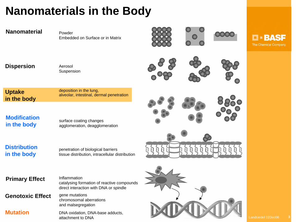
Nanomaterials in the Body
deposition in the lung,alveolar, intestinal, dermal penetration
Dispersion
Modificationin the body
Nanomaterial
penetration of biological barrierstissue distribution, intracellular distribution
Inflammationcatalysing formation of reactive compoundsdirect interaction with DNA or spindle
PowderEmbedded on Surface or in Matrix
Distributionin the body
Primary Effect
gene mutationschromosomal aberrationsand malsegregation
Mutation
Uptakein the body
DNA oxidation, DNA-base adducts,attachment to DNA
Genotoxic Effect
surface coating changesagglomeration, deagglomeration
AerosolSuspension
Landsiedel 02Dec08 4
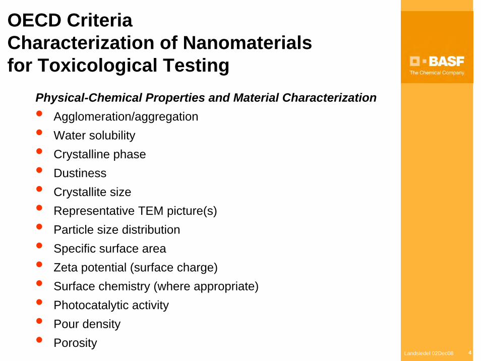
OECD Criteria Characterization of Nanomaterialsfor Toxicological Testing
Physical-Chemical Properties and Material Characterization • Agglomeration/aggregation • Water solubility • Crystalline phase • Dustiness • Crystallite size • Representative TEM picture(s) • Particle size distribution • Specific surface area • Zeta potential (surface charge) • Surface chemistry (where appropriate) • Photocatalytic activity • Pour density • Porosity
Landsiedel 02Dec08 5
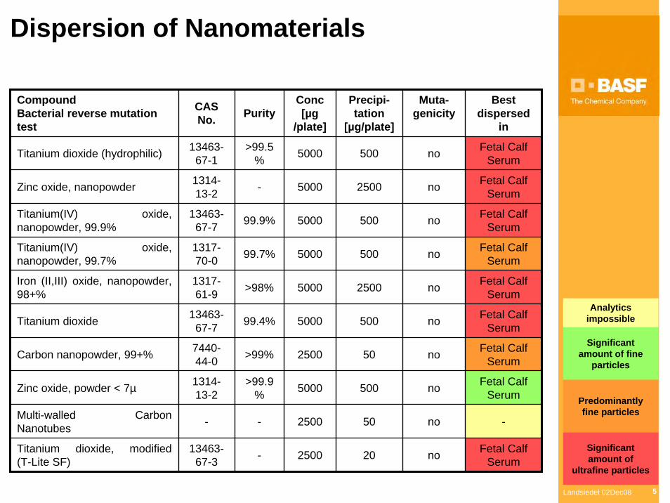
Dispersion of Nanomaterials
CompoundBacterial reverse mutation test
CAS No. Purity
Conc[µg
/plate]
Precipi-tation
[µg/plate]
Muta-genicity
500 no
no
no
no
no
no
no
no
Multi-walled Carbon Nanotubes - - 2500 50 no -
Titanium dioxide, modified(T-Lite SF)
13463-67-3 - 2500 20 no Fetal Calf
Serum
2500
500
500
2500
500
50
500
Best dispersed
in
Titanium dioxide (hydrophilic) 13463-67-1
>99.5% 5000 Fetal Calf
Serum
Zinc oxide, nanopowder 1314-13-2 - 5000 Fetal Calf
Serum
Titanium(IV) oxide, nanopowder, 99.9%
13463-67-7 99.9% 5000 Fetal Calf
Serum
Titanium(IV) oxide, nanopowder, 99.7%
1317-70-0 99.7% 5000 Fetal Calf
Serum
Iron (II,III) oxide, nanopowder, 98+%
1317-61-9 >98% 5000 Fetal Calf
Serum
Titanium dioxide 13463-67-7 99.4% 5000 Fetal Calf
Serum
Carbon nanopowder, 99+% 7440-44-0 >99% 2500 Fetal Calf
Serum
Zinc oxide, powder < 7µ 1314-13-2
>99.9% 5000 Fetal Calf
Serum
Analyticsimpossible
Significantamount of fine
particles
Predominantlyfine particles
Significantamount of
ultrafine particles
Landsiedel 02Dec08 6
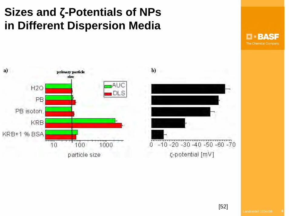
Sizes and ζ-Potentials of NPsin Different Dispersion Media
[52]
Landsiedel 02Dec08 7
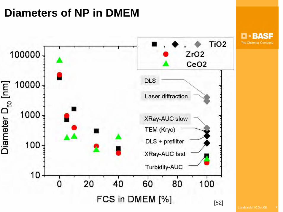
Diameters of NP in DMEM
[52]
Landsiedel 02Dec08 8
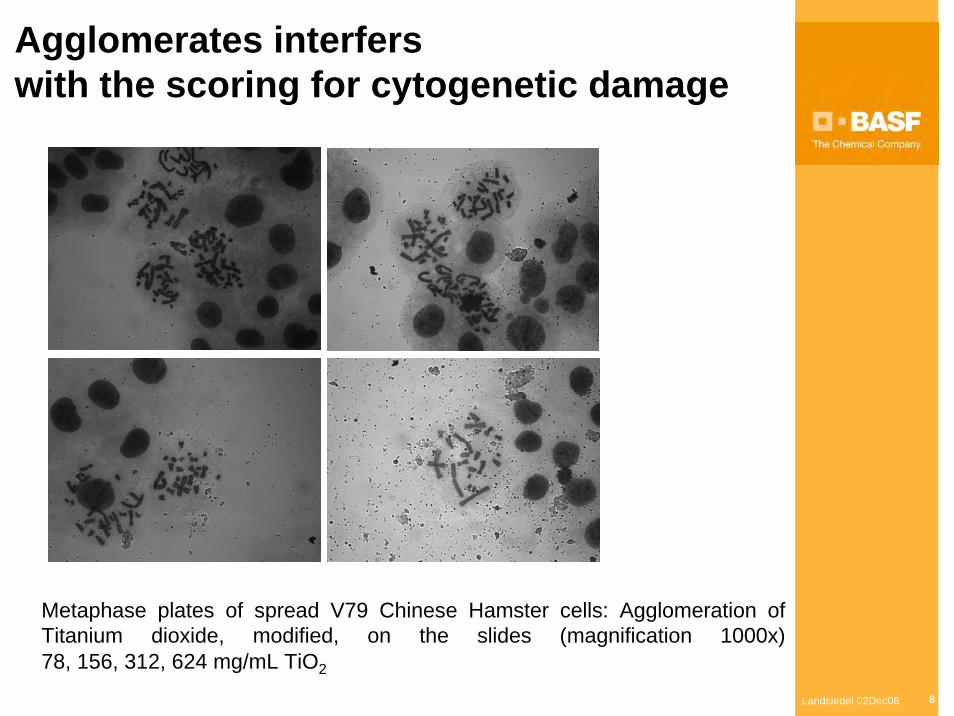
Agglomerates interferswith the scoring for cytogenetic damage
Metaphase plates of spread V79 Chinese Hamster cells: Agglomeration of Titanium dioxide, modified, on the slides (magnification 1000x)78, 156, 312, 624 mg/mL TiO2
Landsiedel 02Dec08 9
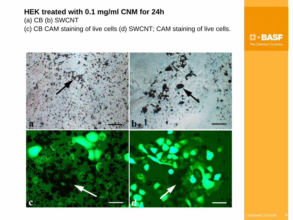
HEK treated with 0.1 mg/ml CNM for 24h(a) CB (b) SWCNT(c) CB CAM staining of live cells (d) SWCNT; CAM staining of live cells.
Landsiedel 02Dec08 10
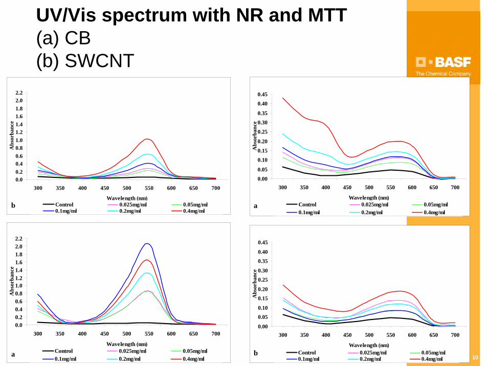
UV/Vis spectrum with NR and MTT(a) CB (b) SWCNT
0.00.20.40.60.81.01.21.41.61.82.02.2
300 350 400 450 500 550 600 650 700
Wavelength (nm)
Abs
orba
nce
Control 0.025mg/ml 0.05mg/ml0.1mg/ml 0.2mg/ml 0.4mg/ml
a
0.00.20.40.60.81.01.21.41.61.82.02.2
300 350 400 450 500 550 600 650 700
Wavelength (nm)
Abs
orba
nce
Control 0.025mg/ml 0.05mg/ml0.1mg/ml 0.2mg/ml 0.4mg/ml
b
0.000.05
0.100.150.20
0.250.300.35
0.400.45
300 350 400 450 500 550 600 650 700
Wavelength (nm)
Abs
orba
nce
Control 0.025mg/ml 0.05mg/ml0.1mg/ml 0.2mg/ml 0.4mg/ml
a
0.00
0.050.10
0.150.20
0.250.30
0.350.40
0.45
300 350 400 450 500 550 600 650 700
Wavelength (nm)
Abs
orba
nce
Control 0.025mg/ml 0.05mg/ml0.1mg/ml 0.2mg/ml 0.4mg/ml
b
Landsiedel 02Dec08 11
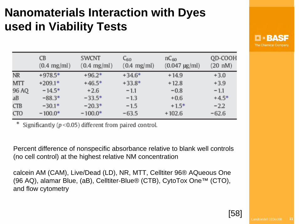
Nanomaterials Interaction with Dyesused in Viability Tests
Percent difference of nonspecific absorbance relative to blank well controls (no cell control) at the highest relative NM concentration
calcein AM (CAM), Live/Dead (LD), NR, MTT, Celltiter 96® AQueous One (96 AQ), alamar Blue, (aB), Celltiter-Blue® (CTB), CytoTox One™ (CTO), and flow cytometry
[58]
Landsiedel 02Dec08 12
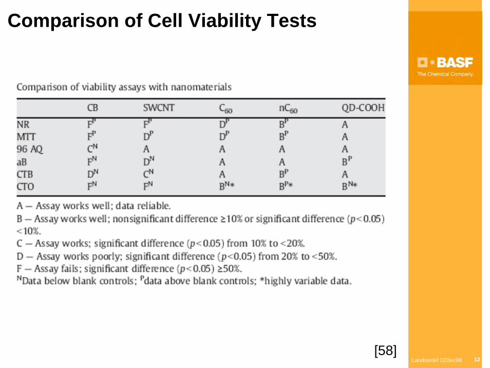
Comparison of Cell Viability Tests
[58]
Landsiedel 02Dec08 13
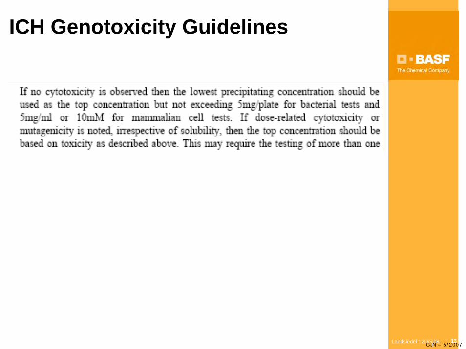
ICH Genotoxicity Guidelines
GJN – 5/2007
Landsiedel 02Dec08 14
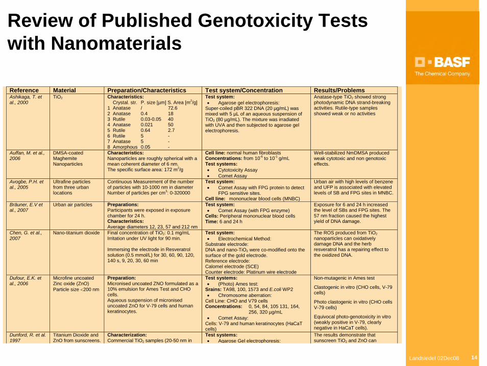
Review of Published Genotoxicity Tests with Nanomaterials
Reference Material Preparation/Characteristics Test system/Concentration Results/Problems Ashikaga, T. et al., 2000
TiO2 Characteristics: Crystal. str. P. size [µm] S. Area [m2/g] 1 Anatase / 72.6 2 Anatase 0.4 18 3 Rutile 0.03-0.05 40 4 Anatase 0.021 50 5 Rutile 0.64 2.7 6 Rutile 5 - 7 Anatase 5 - 8 Amorphous 0.05 -
Test system: • Agarose gel electrophoresis:
Super-coiled pBR 322 DNA (20 µg/mL) was mixed with 5 µL of an aqueous suspension of TiO2 (80 µg/mL). The mixture was irradiated with UVA and then subjected to agarose gel electrophoresis.
Anatase-type TiO2 showed strong photodynamic DNA strand-breaking activities. Rutile-type samples showed weak or no activities
Auffan, M. et al., 2006
DMSA-coated Maghemite Nanoparticles
Characteristics: Nanoparticles are roughly spherical with a mean coherent diameter of 6 nm. The specific surface area: 172 m2/g
Cell line: normal human fibroblasts Concentrations: from 10-6 to 10-1 g/mL Test systems: • Cytotoxicity Assay • Comet Assay
Well-stabilized NmDMSA produced weak cytotoxic and non genotoxic effects.
Avogbe, P.H. et al., 2005
Ultrafine particles from three urban locations
Continuous Measurement of the number of particles with 10-1000 nm in diameter Number of particles per cm3: 0-320000
Test system: • Comet Assay with FPG protein to detect
FPG sensitive sites. Cell line: mononuclear blood cells (MNBC)
Urban air with high levels of benzene and UFP is associated with elevated levels of SB and FPG sites in MNBC.
Bräuner, E.V et al., 2007
Urban air particles Preparations: Participants were exposed in exposure chamber for 24 h. Characteristics: Average diameters 12, 23, 57 and 212 nm
Test system: • Comet Assay (with FPG enzyme)
Cells: Peripheral mononuclear blood cells Time: 6 and 24 h
Exposure for 6 and 24 h increased the level of SBs and FPG sites. The 57 nm fraction caused the highest yield of DNA damage.
Chen, G. et al., 2007
Nano-titanium dioxide Final concentration of TiO2: 0.1 mg/mL Irritation under UV light for 90 min. Immersing the electrode in Resveratrol solution (0.5 mmol/L) for 30, 60, 90, 120, 140 s, 9, 20, 30, 60 min
Test system: • Electrochemical Method:
Substrate electrode: DNA and nano-TiO2 were co-modified onto the surface of the gold electrode. Reference electrode: Calomel electrode (SCE) Counter electrode: Platinum wire electrode
The ROS produced from TiO2 nanoparticles can oxidatively damage DNA and the herb resveratrol has a repairing effect to the oxidized DNA.
Dufour, E.K. et al., 2006
Microfine uncoated Zinc oxide (ZnO) Particle size <200 nm
Preparation: Micronised uncoated ZNO formulated as a 10% emulsion for Ames Test and CHO cells. Aqueous suspension of micronised uncoated ZnO for V-79 cells and human keratinocytes.
Test systems: • (Photo) Ames test:
Srains: TA98, 100, 1573 and E.coli WP2 • Chromosome aberration:
Cell Line: CHO and V79 cells Concentrations: 0, 54, 84, 105 131, 164, 256, 320 µg/mL • Comet Assay:
Cells: V-79 and human keratinocytes (HaCaT cells)
Non-mutagenic in Ames test
Clastogenic in vitro (CHO cells, V-79 cells)
Photo clastogenic in vitro (CHO cells V-79 cells)
Equivocal photo-genotoxicity in vitro (weakly positive in V-79, clearly negative in HaCaT cells).
Dunford, R. et al. 1997
Titanium Dioxide and ZnO from sunscreens.
Characterization: Commercial TiO2 samples (20-50 nm in
Test systems: • Agarose Gel electrophoresis:
The results demonstrate that sunscreen TiO2 and ZnO can
Landsiedel 02Dec08 15
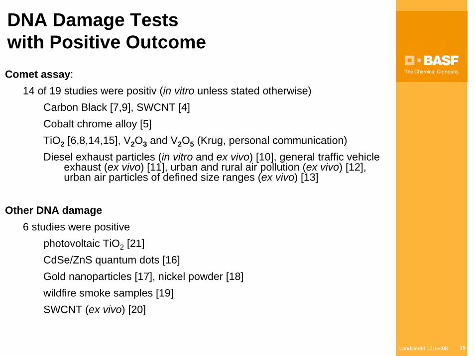
DNA Damage Tests with Positive OutcomeComet assay:
14 of 19 studies were positiv (in vitro unless stated otherwise)Carbon Black [7,9], SWCNT [4]Cobalt chrome alloy [5]TiO2 [6,8,14,15], V2O3 and V2O5 (Krug, personal communication)Diesel exhaust particles (in vitro and ex vivo) [10], general traffic vehicle
exhaust (ex vivo) [11], urban and rural air pollution (ex vivo) [12], urban air particles of defined size ranges (ex vivo) [13]
Other DNA damage6 studies were positive
photovoltaic TiO2 [21]CdSe/ZnS quantum dots [16]Gold nanoparticles [17], nickel powder [18]wildfire smoke samples [19]SWCNT (ex vivo) [20]
Landsiedel 02Dec08 16
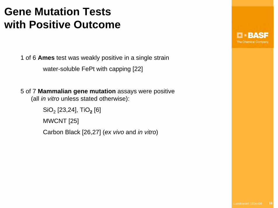
Gene Mutation Tests with Positive Outcome
1 of 6 Ames test was weakly positive in a single strain
water-soluble FePt with capping [22]
5 of 7 Mammalian gene mutation assays were positive(all in vitro unless stated otherwise):
SiO2 [23,24], TiO2 [6]
MWCNT [25]
Carbon Black [26,27] (ex vivo and in vitro)
Landsiedel 02Dec08 17
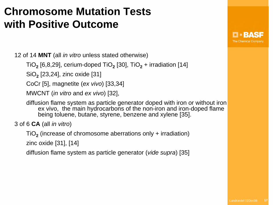
Chromosome Mutation Tests with Positive Outcome
12 of 14 MNT (all in vitro unless stated otherwise)TiO2 [6,8,29], cerium-doped TiO2 [30], TiO2 + irradiation [14]SiO2 [23,24], zinc oxide [31]CoCr [5], magnetite (ex vivo) [33,34]MWCNT (in vitro and ex vivo) [32],diffusion flame system as particle generator doped with iron or without iron
ex vivo, the main hydrocarbons of the non-iron and iron-doped flame being toluene, butane, styrene, benzene and xylene [35].
3 of 6 CA (all in vitro)TiO2 (increase of chromosome aberrations only + irradiation)zinc oxide [31], [14] diffusion flame system as particle generator (vide supra) [35]
Landsiedel 02Dec08 18
DNA-damage-dependent Signalling, Biomarkers and Special Methods
Carbon Black Printex 90 in A549 type II [7]
p53 phosphorylation
phosphorylated p53BP1
single-strand DNA breaks (Comet assay)
phosphorylated BRCA1.
Carbon black particles of larger size showed none of the responses
TiO2 (P25) dispersed with calf thymus DNA and irradiated [36]
DNA and RNA damage visualized by scanning micrographs
Landsiedel 02Dec08 19
DNA Damage Tests with Negative Outcome
5 of 19 Comet assays were negative (all in vitro unless stated otherwise)TiO2 [14]Carbon Black [38]SiO2 [23,24]Maghemite coated with DMSA [37]vehicle exhaust (ex vivo) (no increase in DNA strand breaks as
determined by Comet assay, but oxidative DNA damage in terms of FPG-sensitive sites) [11]
1 Test on DNA damage (8-oxoguanine) was negativeafter intratracheal instillation in rats (ex vivo)
TiO2 [39]
Landsiedel 02Dec08 20
Gene Mutation Tests with Negative Outcome
5 of 6 Ames test were negative
TiO2 [14,40]
zinc oxide [31]
SWCNT [4]
silica-coated magnetic nanoparticles labeled with rhodamine B isothiocyanate “MNPs@SiO2(RITC)” [41]
2 of 7 Mammalian gene mutation tests were negative
TiO2 in vitro [14]
diesel exhaust particles ex vivo (cII mutation frequency in lung tissue of transgenic MutaTMMice exposed by inhalation
Landsiedel 02Dec08 21
Chromosome Mutation Testswith Negative Outcome
3 of 6 CA were negative (all in vitro)
TiO2 [40,44]
“MNPs@SiO2(RITC)” [41]
2 of 14 MNT were negative (all in vitro)
TiO2 [42]
V2O3 and V2O5 [Krug, personal communication]
Landsiedel 02Dec08 22
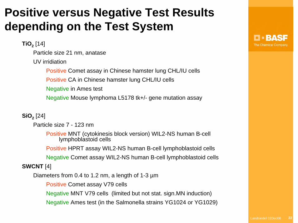
Positive versus Negative Test Results depending on the Test System
TiO2 [14] Particle size 21 nm, anataseUV irridiation
Positive Comet assay in Chinese hamster lung CHL/IU cellsPositive CA in Chinese hamster lung CHL/IU cellsNegative in Ames testNegative Mouse lymphoma L5178 tk+/- gene mutation assay
SiO2 [24] Particle size 7 - 123 nm
Positive MNT (cytokinesis block version) WIL2-NS human B-cell lymphoblastoid cells
Positive HPRT assay WIL2-NS human B-cell lymphoblastoid cells Negative Comet assay WIL2-NS human B-cell lymphoblastoid cells
SWCNT [4] Diameters from 0.4 to 1.2 nm, a length of 1-3 µm
Positive Comet assay V79 cells Negative MNT V79 cells (limited but not stat. sign.MN induction) Negative Ames test (in the Salmonella strains YG1024 or YG1029)
Landsiedel 02Dec08 23
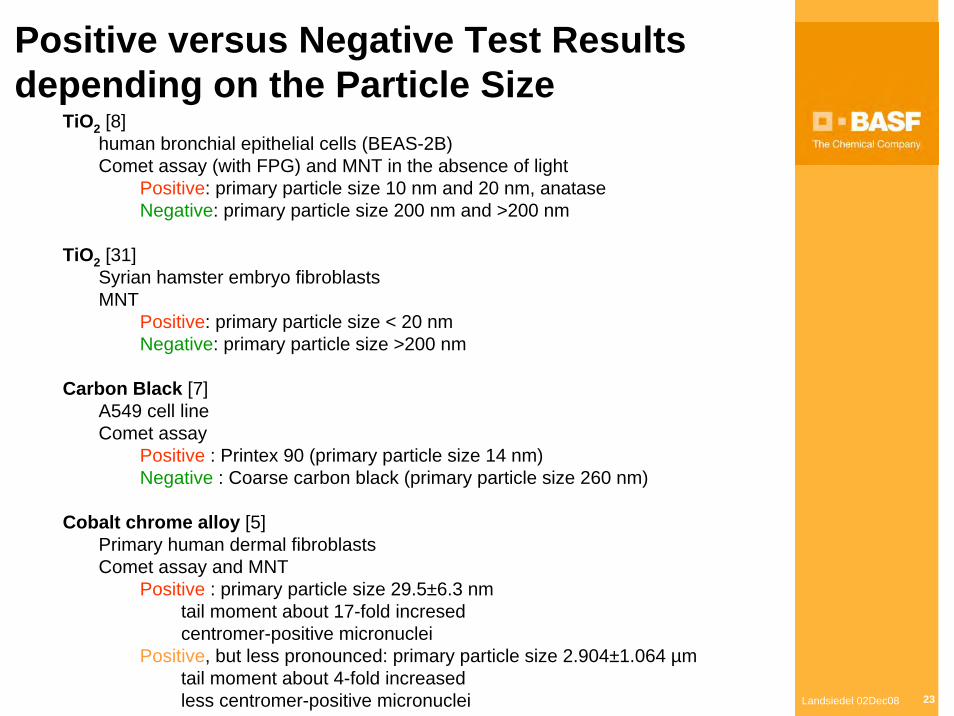
Positive versus Negative Test Resultsdepending on the Particle Size
TiO2 [8]human bronchial epithelial cells (BEAS-2B)Comet assay (with FPG) and MNT in the absence of light
Positive: primary particle size 10 nm and 20 nm, anataseNegative: primary particle size 200 nm and >200 nm
TiO2 [31]Syrian hamster embryo fibroblastsMNT
Positive: primary particle size < 20 nmNegative: primary particle size >200 nm
Carbon Black [7]A549 cell line Comet assay
Positive : Printex 90 (primary particle size 14 nm)Negative : Coarse carbon black (primary particle size 260 nm)
Cobalt chrome alloy [5]Primary human dermal fibroblasts Comet assay and MNT
Positive : primary particle size 29.5±6.3 nm tail moment about 17-fold incresedcentromer-positive micronuclei
Positive, but less pronounced: primary particle size 2.904±1.064 µmtail moment about 4-fold increasedless centromer-positive micronuclei
Landsiedel 02Dec08 24
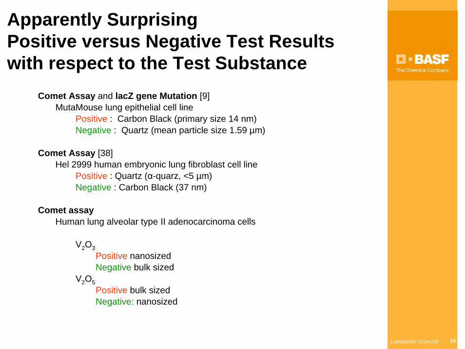
Apparently SurprisingPositive versus Negative Test Resultswith respect to the Test Substance
Comet Assay and lacZ gene Mutation [9]MutaMouse lung epithelial cell line
Positive : Carbon Black (primary size 14 nm)Negative : Quartz (mean particle size 1.59 µm)
Comet Assay [38] Hel 2999 human embryonic lung fibroblast cell line
Positive : Quartz (α-quarz, <5 µm) Negative : Carbon Black (37 nm)
Comet assayHuman lung alveolar type II adenocarcinoma cells
V2O3Positive nanosizedNegative bulk sized
V2O5Positive bulk sized Negative: nanosized
Landsiedel 02Dec08 25
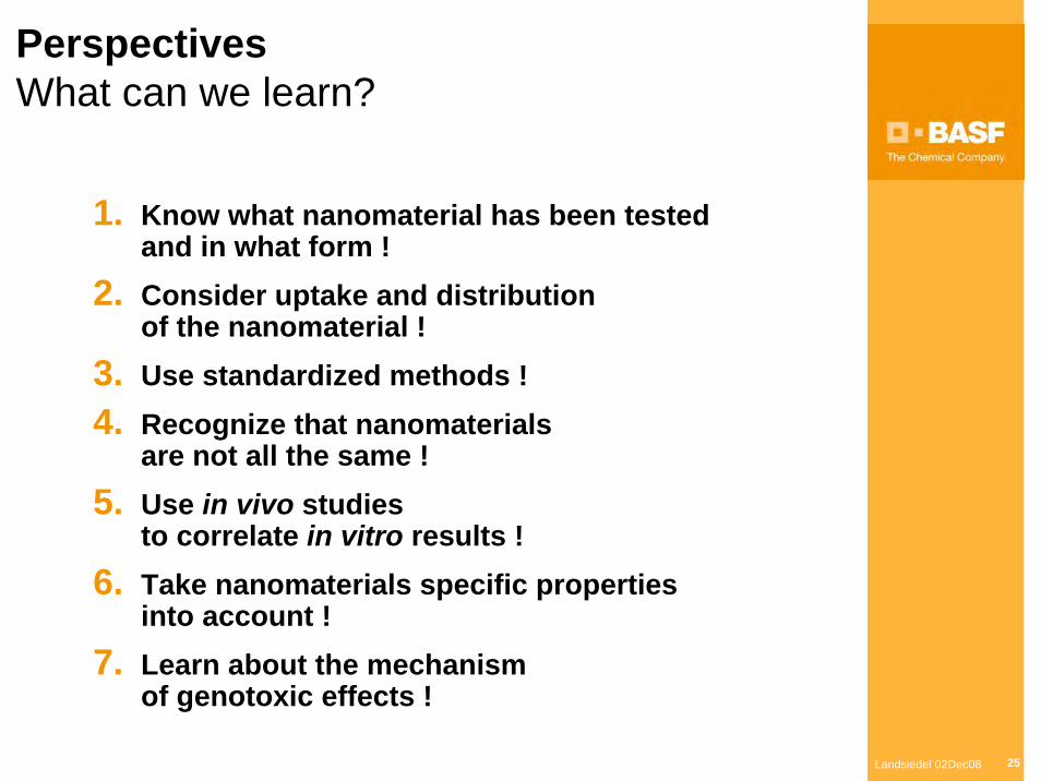
PerspectivesWhat can we learn?
1. Know what nanomaterial has been testedand in what form !
2. Consider uptake and distributionof the nanomaterial !
3. Use standardized methods !4. Recognize that nanomaterials
are not all the same !5. Use in vivo studies
to correlate in vitro results !6. Take nanomaterials specific properties
into account !7. Learn about the mechanism
of genotoxic effects !
Landsiedel 02Dec08 26
Conclusions
Experiences with other, non-nano, substances (molecules and larger particles) taught us, that mechanisms of genotoxic effects can be diverse and their elucidation can be demanding, while there often is an immediate need to assess the genotoxic hazard.
Thus a practical and pragmatic approach is the use of a battery of standard genotoxicity testing methods covering a wide range of mechanisms.
Application of these standard methods to nanomaterialsdemands, however, several adaptations and the interpretation of results from the genotoxicity tests may need additional considerations.
Landsiedel 02Dec08 27
EXAMPLE:
Inhalation study with ex vivo COMET Assay
Landsiedel 02Dec08 28
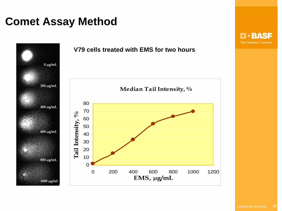
Comet Assay Method
0 µg/mL
200 µg/mL
400 µg/mL
600 µg/mL
800 µg/mL
1000 µg/ml
V79 cells treated with EMS for two hours
Median Tail Intensity, %
01020304050607080
0 200 400 600 800 1000 1200EMS, µg/mL
Tail
Inte
nsit
y, %
Landsiedel 02Dec08 29
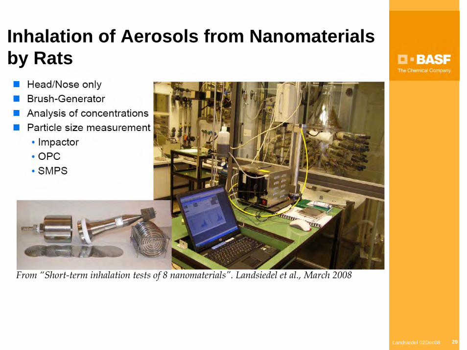
Inhalation of Aerosols from Nanomaterialsby Rats
From “Short-term inhalation tests of 8 nanomaterials”. Landsiedel et al., March 2008
Landsiedel 02Dec08 30
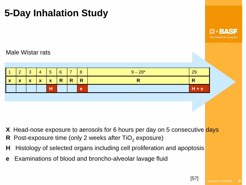
Male Wistar rats
X Head-nose exposure to aerosols for 6 hours per day on 5 consecutive daysR Post-exposure time (only 2 weeks after TiO2 exposure)H Histology of selected organs including cell proliferation and apoptosis
e Examinations of blood and broncho-alveolar lavage fluid
5-Day Inhalation Study
1 2 3 4 5 6 7 8 9 – 28* 29
x x x x x R R R R RH e H + e
[57]
Landsiedel 02Dec08 31
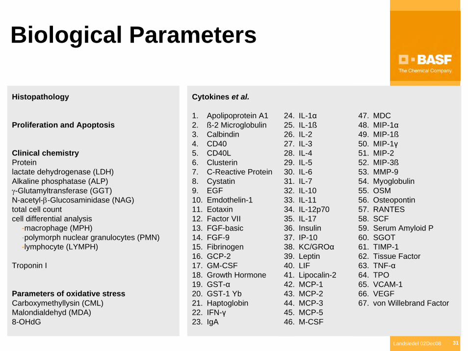
Biological Parameters
Cytokines et al.
1. Apolipoprotein A12. ß-2 Microglobulin3. Calbindin4. CD405. CD40L6. Clusterin7. C-Reactive Protein8. Cystatin9. EGF10. Emdothelin-111. Eotaxin12. Factor VII13. FGF-basic14. FGF-915. Fibrinogen16. GCP-217. GM-CSF18. Growth Hormone19. GST-α20. GST-1 Yb21. Haptoglobin22. IFN-γ23. IgA
24. IL-1α25. IL-1ß26. IL-227. IL-328. IL-429. IL-530. IL-631. IL-732. IL-1033. IL-1134. IL-12p7035. IL-1736. Insulin37. IP-1038. KC/GROα39. Leptin40. LIF41. Lipocalin-242. MCP-143. MCP-244. MCP-345. MCP-546. M-CSF
47. MDC48. MIP-1α49. MIP-1ß50. MIP-1γ51. MIP-252. MIP-3ß53. MMP-954. Myoglobulin55. OSM56. Osteopontin57. RANTES58. SCF59. Serum Amyloid P60. SGOT61. TIMP-162. Tissue Factor63. TNF-α64. TPO65. VCAM-166. VEGF67. von Willebrand Factor
Histopathology
Proliferation and Apoptosis
Clinical chemistry Proteinlactate dehydrogenase (LDH)Alkaline phosphatase (ALP)γ-Glutamyltransferase (GGT)N-acetyl-β-Glucosaminidase (NAG)total cell countcell differential analysis
-macrophage (MPH)-polymorph nuclear granulocytes (PMN)-lymphocyte (LYMPH)
Troponin I
Parameters of oxidative stressCarboxymethyllysin (CML)Malondialdehyd (MDA)8-OHdG
Landsiedel 02Dec08 32
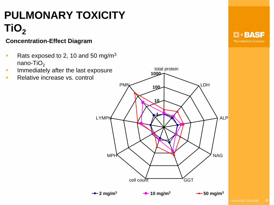
1
10
100
1000total protein
LDH
ALP
NAG
GGTcell count
MPH
LYMPH
PMN
2 mg/m3 10 mg/m3 50 mg/m3
Concentration-Effect Diagram
Rats exposed to 2, 10 and 50 mg/m3
nano-TiO2Immediately after the last exposureRelative increase vs. control
PULMONARY TOXICITY TiO2
Landsiedel 02Dec08 33
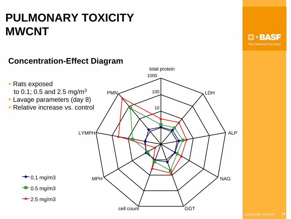
PULMONARY TOXICITYMWCNT
10
100
1000total protein
LDH
ALP
NAG
GGTcell count
MPH
LYMPH
PMN
0.1 mg/m3
0.5 mg/m3
2.5 mg/m3
Concentration-Effect Diagram
Rats exposedto 0.1; 0.5 and 2.5 mg/m3
Lavage parameters (day 8)Relative increase vs. control

Landsiedel 02Dec08 34
Preparation of Lung cells
• Perfusion • Lavage• Enzyme instillation • Enzymatic digestion
• Collagenase IV
• Trypsin
• DNAse I
• Cell isolation• 230 µm and 73.7 µm MESH
• Percoll 1.040 g/ml gradient
• Viability measurement by Trypan Blue dye exclusion method
Landsiedel 02Dec08 35
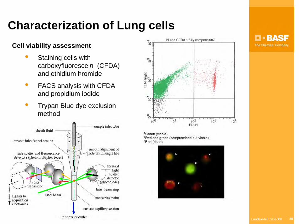
Characterization of Lung cellsCell viability assessment
• Staining cells with carboxyfluorescein (CFDA) and ethidium bromide
• FACS analysis with CFDA and propidium iodide
• Trypan Blue dye exclusion method
Landsiedel 02Dec08 36
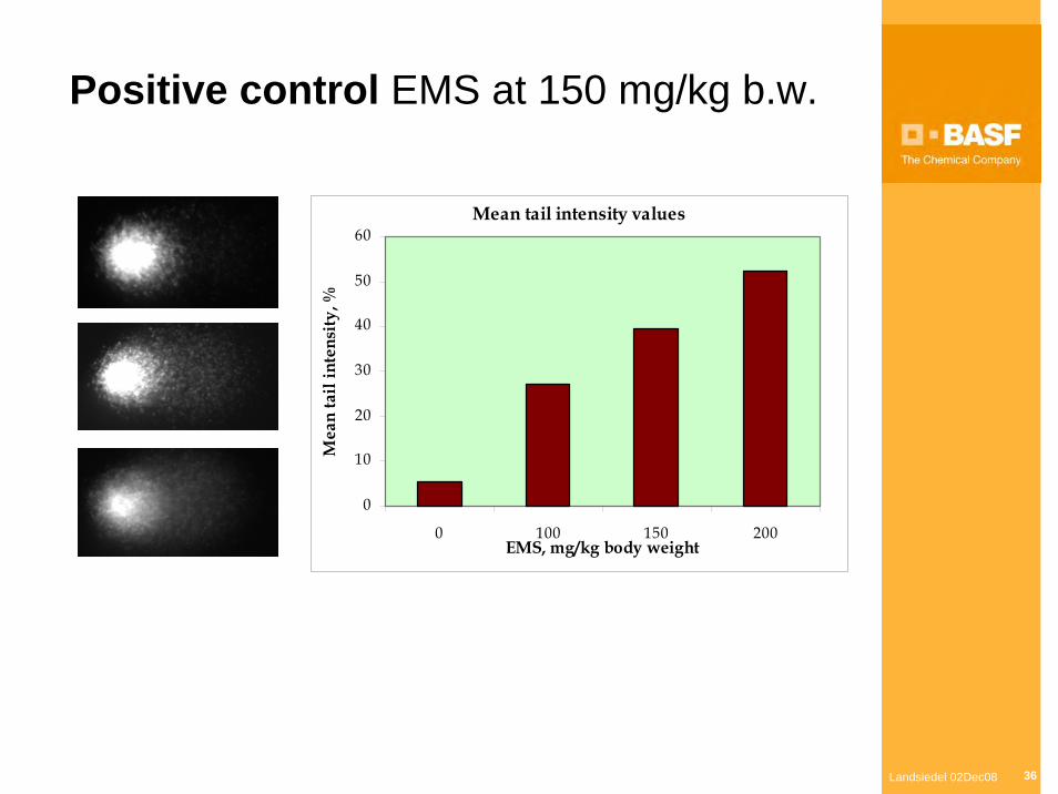
Mean tail intensity values
0
10
20
30
40
50
60
0 100 150 200EMS, mg/kg body weight
Mea
n ta
il in
tens
ity, %
Positive control EMS at 150 mg/kg b.w.
Landsiedel 02Dec08 37
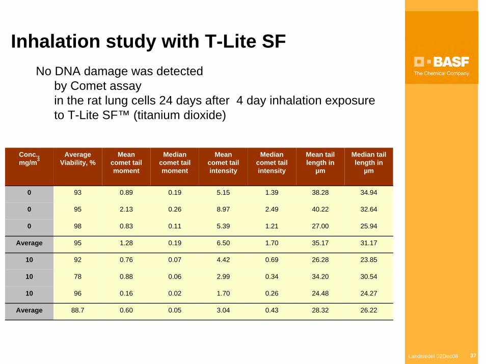
Inhalation study with T-Lite SF
Conc., mg/m3
Average Viability, %
Mean comet tail moment
Median comet tail moment
Mean comet tail intensity
Median comet tail intensity
Mean tail length in
µm
Median tail length in
µm
0 93 0.89 0.19 5.15 1.39 38.28 34.94
0 95 2.13 0.26 8.97 2.49 40.22 32.64
0 98 0.83 0.11 5.39 1.21 27.00 25.94
Average 95 1.28 0.19 6.50 1.70 35.17 31.17
10 92 0.76 0.07 4.42 0.69 26.28 23.85
10 78 0.88 0.06 2.99 0.34 34.20 30.54
10 96 0.16 0.02 1.70 0.26 24.48 24.27
Average 88.7 0.60 0.05 3.04 0.43 28.32 26.22
No DNA damage was detectedby Comet assayin the rat lung cells 24 days after 4 day inhalation exposureto T-Lite SF™ (titanium dioxide)
Landsiedel 02Dec08 38
Thank you !

Landsiedel 02Dec08 39
REFERENCES[1] D.B .Warheit, C.M. Sayes, K.L. Reed, K.A. Swain, Health effects related to nanoparticle exposures: Environmental, health and safety
considerations for assessing hazards and risks, Pharmacol. Ther. 120 (2008) 35-42.[2] V.L. Colvin, The potential environmental impact of engineered nanomaterials, Nature Biotechnol. 21 (2003) 1166-1170.[3] Landsiedel R, Schulz M, Kapp MD, Oesch F: "Genotoxicity Investigations on Nanomaterials: Methods, Preparation and Characterization of
Test Material, Potential Artifacts and Limitations - Many Questions, Some Answers", Mutation Research (in press)[4] E.R. Kisin, A.R. Murray, M. J. Keane, X.C. Shi, D. Schwegler-Berry, O. Gorelik, S. Arepalli, V. Castranova, W.E. Wallace, V.E. Kagan, A.A.
Shvedova, Single-walled carbon nanotubes: Geno- and cytotoxic effects in lung fibroblast V79 cells, J. Toxicol. Environ. Health, Part A, 70 (2007) 2071–2079.
[5] I. Papageorgiou, C. Brown, R. Schins, S. Singh, R. Newson, S. Davis, J. Fisher, E. Ingham, C.P. Case, The effect of nano- and micron-sized particles of cobalt–chromium alloy on human fibroblasts in vitro, Biomaterials 28 (2007) 2946-2958.
[6] J.J. Wang, B.J. Sanderson, H Wang, Cyto- and genotoxicity of ultrafine TiO2 particles in cultured human lymphoblastoid cells, Mutat. Res. 628 (2007) 99-106.
[7] R.M. Mroz, R.P. Schins, H. Li, L.A. Jimenez, E.M. Drost, A. Holownia, W. MacNee, K. Donaldson, Nanoparticle-driven DNA damage mimics irradiation-related carcinogenesis pathways, Eur. Respir. J. 31 (2008) 241-251.
[8] J.R. Gurr, A.S. Wang, C.H. Chen, K.Y. Jan KY, Ultrafine titanium dioxide particles in the absence of photoactivation can induce oxidative damage to human bronchial epithelial cells, Toxicology 213 (2005) 66-73.
[9] N.R. Jacobsen, A.T. Saber, P. White , P. Møller, G. Pojana, U. Vogel, S. Loft, J. Gingerich, L. Soper, G.R. Douglas, H. Wallin, Increased mutant frequency by carbon black, but not quartz, in the lacZ and cII transgenes of muta mouse lung epithelial cells, Environ. Mol. Mutagen. 48 (2007) 451-461.
[10] M. Dybdahl, L. Risom, J. Bornholdt, H. Autrup , S. Loft , H. Wallin, Inflammatory and genotoxic effects of diesel particles in vitro and in vivo, Mutat. Res. 562 (2004) 119-131.
[11] P.S. Vinzents, P. Møller, M Sørensen, L. E. Knudsen, O. Hertel, F. P. Jensen, B. Schibye, S. Loft, Personal exposure to ultrafine particles and oxidative DNA damage, Environ. Health Perspect. (2005) 1485-1490.
[12] P.H. Avogbe, L. Ayi-Fanou, H. Autrup, S. Loft, B. Fayomi , A. Sanni , P. Vinzents, P. Møller, Ultrafine particulate matter and high-level benzene urban air pollution in relation to oxidative DNA damage, Carcinogenesis 26(2005) 613-620.
[13] E.V. Bräuner, L. Forchhammer, P. Møller, J. Simonsen, M. Glasius, P. Wåhlin, O. Raaschou-Nielsen, S. Loft, Exposure to ultrafine particles from ambient air and oxidative stress–induced DNA damage, Environ. Health Perspect. 115 (2007) 1177-1182.
[14] Y. Nakagawa, S. Wakuri, K. Sakamoto, N. Tanaka, The photogenotoxicity of titanium dioxide particles, Mutat. Res. 394 (1997) 125-132. [15] R. Dunford, A. Salinaro, L. Cai, N. Serpone, S. Horikoshi, H. Hidaka, J. Knowland, Chemical oxidation and DNA damage catalysed by
inorganic sunscreen ingredients, FEBS Lett. 418 (1997) 87-90.[16] M. Green, E. Howman, Semiconductor quantum dots and free radical induced DNA nicking, Chem. Commun. 121 (2005) 121–123.[17] Y. Zheng, D.J. Hunting, P. Ayotte, L. Sanche, Radiosensitization of DNA by gold nanoparticles irradiated with high-energy electrons,
Radiat. Res. 169 (2008) 19-27.[18] Q. Zhang, Y. Kusaka, K. Sato, K. Nakakuki, N. Kohyama, K. Donaldson, Differences in the extent of inflammation caused by intratracheal
exposure to three ultrafine metals: role of free radicals, J. Toxicol. Environ. Health, Part A, 53 (1998) 423–438. [19] S.S. Leonard, V. Castranova, B.T. Chen, D. Schwegler-Berry, M. Hoover, C. Piacitelli, D.M. Gaughan, Particle size-dependent radical
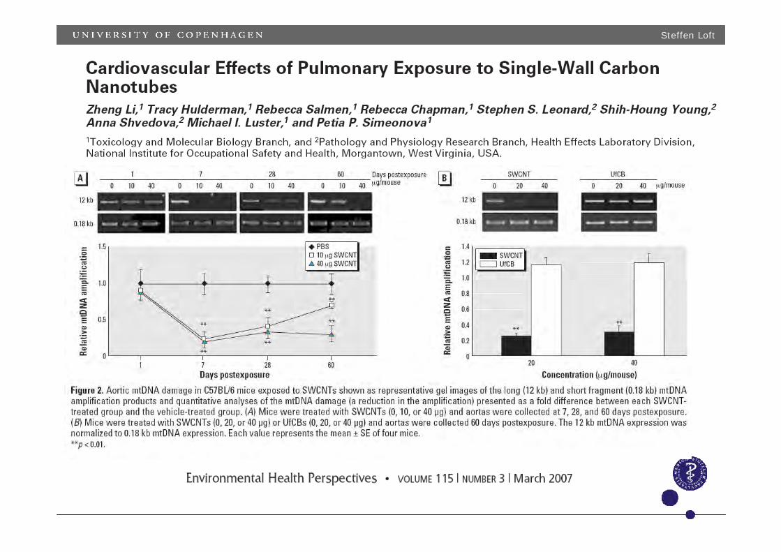
generation from wildland fire smoke, Toxicology 236 (2007) 103–113.[20] Z. Li, T. Hulderman, R. Salmen, R. Chapman, S.S. Leonard, S.H. Young, A. Shvedova, M.I. Luster, P.P. Simeonova, Cardiovascular
effects of pulmonary exposure to single-wall carbon nanotubes, Environ. Health Perspect. 115 (2007) 377-382.
Landsiedel 02Dec08 40
REFERENCES continued[21] G. Chen, J. Zhao, X. Liu, G. Gao, J. Huang , G. Li, Electrochemical sensing DNA damage with nano-titanium dioxide and repair with a
medicinal herb species resveratrol. J. Biotechnol. 127 (2007) 653-656.[22] S. Maenosono, T. Suzuki, S. Saita, Mutagenicity of water-soluble FePt nanoparticles in Ames test, J. Toxicol. Sci. 32 (2007) 575-579. [23] J.J.Wang, B.J.S. Sanderson, H. Wang, Cytotoxicity and genotoxicity of ultrafine crystalline SiO2 particulate in cultured human
lymphoblastoid cells, Environ. Mol. Mutagenesis 48 (2007) 151-157.[24] J.J. Wang, H. Wang, B.J.S. Sanderson, Ultrafine quartz-induced damage in human lymphoblastoid cells in vitro using three genetic
damage end-points, Toxicol. Mechanisms Methods 17 ( 2007) :223–232. [25] L. Zhu, D.W. Chang, L. Dai, Y. Hong, DNA damage induced by multiwalled carbon nanotubes in mouse embryonic stem cells, Nano Lett.
7 (2007) 3592-3597.[26] H.G. Claycamp, Phenol sensitization of DNA to subsequent oxidative damage in 8-hydroxyguanine assays. Carcinogenesis 13 (1992)
1289–1292.[27] E. Driscoll, L.C. Deyo, J.M. Carter, B.W. Howard, G. Hassenbein, T.A. Bertram, Effect of particle exposure and particle-elicited
inflammatory cells on mutation in rat alveolar epithelial cells, Carcinogenesis 18 (1997) 423-430.[28] G. Oberdörster, A. Maynard, K. Donaldson, V. Castranova, J. Fitzpatrick, K. Ausman, J. Carter, B. Karn, W. Kreyling, D. Lai, S. Olin, N.
Monteiro-Riviere, D. Warheit, H. Yang, Principles for characterizing the potential human health effects from exposure to nanomaterials: elements of a screening strategy, Particle Fibre Toxicol. 2 (2005) 8-43.
[29] Q. Rahman, M. Lohani, E. Dopp, H. Pemsel, L. Jonas, D.G. Weiss, D. Schiffmann, Evidence that ultrafine titanium dioxide induces micronuclei and apoptosis in syrian hamster embryo fibroblasts, Environ. Health Perspect. 110 (2002) 797-800.
[30] L. Wang, J. Mao, G.H. Zhang, M.J. Tu, Nano-cerium-element-doped titanium dioxide induces apoptosis of Bel 7402 human hepatomacells in the presence of visible light, World J. Gastroenterol. 13(2007) 4011-4014.
[31] E.K. Dufour, T. Kumaravel, G.J. Nohynek, D. Kirkland, H. Toutain, Clastogenicity, photo-clastogenicity or pseudo-photo-clastogenicity: Genotoxic effects of zinc oxide in the dark, in pre-irradiated or simultaneously irradiated Chinese hamster ovary cells, Mutat. Res. 607 (2006) 215-224.
[32] J. Muller, I. Decordier , P.H. Hoet, N. Lombaert, L. Thomassen, F. Huaux, D. Lison, M. Kirsch-Volders, Clastogenic and aneugenic effects of multi-wall carbon nanotubes in epithelial cells, Carcinogenesis 29 (2008) 427-433.
[33] M.L.L. Freitas, L.P. Silva, R.B. Azevedo, V.A.P. Garcia, L.M. Lacava, C.K. Grisolia, C.M. Lucci, P.C. Morais, M.F. Da Silva, N. Buske, R. Curi, Z.G.M. Lacava, A double-coated magnetite-based magnetic fluid evaluation by cytometry and genetic tests, J. Magnetism Magnetic Materials 252 (2002) 396–398.
[34] N. Sadeghiani, L.S. Barbosa, L.P. Silva, R.B. Azevedo, P.C. Morais, Z.G.M. Lacava, Genotoxicity and inflammatory investigation in mice treated with magnetite nanoparticles surface coated with polyaspartic acid, J. Magnetism Magnetic Materials 289 (2005) 466–468.
[35] J.H. Park, K.T. Han, K.J. Eu, J.S. Kim, K.H. Chung, B. Park, G.S.Yang, K.H. Lee, M.H. Cho, Diffusion flame-derived fine particulate matters doped with iron caused genotoxicity in B6C3F1 mice, Toxicol. Ind. Health 21(2005 57-65.
[36] H. Hidaka, S. Horikoshi, N. Serpone, J. Knowland, In vitro photochemical damage to DNA. RNA and their bases by an inorganic sunscreen agent on exposure to UVA and UVB radiation, J. Photochem. Photobiol. A: Chem. 111 (1997 ) 205-213.
[37] M. Auffan, L. Decome, J. Rose, T. Orsiere, M. Demeo, V. Briois, C. Chaneac, L. Olivi, J.L. Berge - Lefranc A. Botta, M.R . Wiesner, J.Y. Bottero, In Vitro Interactions between DMSA-coated maghemite nanoparticles and human fibroblasts: a physicochemical and cyto-genotoxical study, Environ. Sci. Technol. (2006) 40 4367-4373.
[38] B.Z. Zhong, W.Z. Whong, T.M. Ong, Detection of mineral-dust-induced DNA damage in two mammalian cell lines using the alkaline single cell gel/comet assay, Mutat Res. 393 (1997) 181-187.
[39] B. Rehn, F. Seiler, S. Rehn, J. Bruch, M. Maierd, Investigations on the inflammatory and genotoxic lung effects of two types of titanium dioxide: untreated and surface treated, Toxicol. Appl. Pharmacol. 189 (2003) 84–95.
[40] D.B. Warheit, R. A. Hoke, C. Finlay, E. M. Donner,K. L. Reed, C. M. Sayes, Development of a base set of toxicity tests using ultrafine TiO2 particles as a component of nanoparticle risk management, Toxicol. Lett. 171 (2007) 99–110.
Landsiedel 02Dec08 41
REFERENCES continued[41] J.S. Kim, T.J. Yoon, K.N. Yu, B.G. Kim, S.J. Park, H.W. Kim, K.H. Lee, S.B. Park, J.K. Lee, M.H. Cho, Toxicity and tissue distribution of
magnetic nanoparticles in mice, Toxicol. Sci. 89 (2005) 338–347. [42] K. Linnainmaa, P. Kivipensas, H. Vainio, Toxicity and cytogenetic studies of ultrafine titanium dioxide in cultured rat liver epithelial cells,
Toxicol. in Vitro 11 (1997) 329-335. [43] D.R. Haynes, S.D. Rogers, D.W. Howie, M.J. Pearcy, B. Vernon-Roberts, Drug inhibition of the macrophage response to metal wear
particles in vitro, Clin. Orthop. Relat. Res. 323 (1996) 316-326.[44] E. Theogaraj, S. Riley, L. Hughes, M. Maier, D. Kirkland, An investigation of the photo-clastogenic potential of ultrafine titanium dioxide
particles, Mutat. Res. 634 (2007) 205-19. [45] K. Donaldson, C.L. Tran CL, An introduction to the short-term toxicology of respirable industrial fibres, Mutat Res. 553 (2004) 5-9. [46] L.K. Duncan, J.R. Jinschek, P.J. Vikesland, C60 colloid formation in aqueous systems: effects of preparation method on size, structure,
and surface charge, Environ. Sci. Technol. 42 (2008) 173-178. [47] J.A. Brant, J. Labille, J.Y. Bottero, M.R. Wiesner, Characterizing the impact of preparation method on fullerene cluster structure and
chemistry, Langmuir. 22 (2006) 3878-3885. [48] Z. Markovic, B. Todorovic-Markovic, D. Kleut, N. Nikolic, S. Vranjes-Djuric, M. Misirkic, L. Vucicevic, K. Janjetovic, A. Isakovic, L. Harhaji,
B. Babic-Stojic, M. Dramicanin, V. Trajkovic, The mechanism of cell-damaging reactive oxygen generation by colloidal fullerenes, Biomaterials 28 (2007) 5437-5448.
[49] L.K. Limbach, Y. Li, R.N. Grass, T.J. Brunner, M.A. Hintermann, M. Muller, D. Gunther, W.J. Stark, Oxide nanoparticle uptake in human lung fibroblasts: effects of particle size, agglomeration, and diffusion at low concentrations, Environ. Sci. Technol. 39 (2005) 9370-9376.
[50] R.S. Kane, A.D. Stroock, Nanobiotechnology: protein-nanomaterial interactions, Biotechnol. Prog. 23 (2007) 316-319.[51] T. Cedervall, I. Lynch, S. Lindman, T. Berggard, E. Thulin, H. Nilsson, K. A. Dawson, S. Linse, Understanding the nanoparticle-protein
corona using methods to quantify exchange rates and affinities of proteins for nanoparticles, Proc. Natl. Acad. Sci. U S A, 104 (2007) 2050-2055.
[52] C. Schulze, A. Kroll, C.M. Lehr, U.F. Schäfer, K. Becker, J. Schnekenburger, C.Schulze Isfort, R. Landsiedel, W. Wohleben, Not ready to use - overcoming pitfalls when dispersing nanoparticles in physiological media, Nanotoxicology, 2 (2008) 51-61.
[53] L. Guo, A. Von Dem Bussche, M. Buechner, A. Yan, A.B. Kane, R.H. Hurt, Adsorption of essential micronutrients by carbon nanotubesand the implications for nanotoxicity testing, Small, 4 (2008) 721-727.
[54] A. Isakovic, Z. Markovic, B. Todorovic-Markovic, N. Nikolic, S. Vranjes-Djuric, M. Mirkovic, M. Dramicanin, L. Harhaji, N. Raicevic, Z. Nikolic, V. Trajkovic, Distinct cytotoxic mechanisms of pristine versus hydroxylated fullerene, Toxicol. Sci. 91 (2006) 173-183.
[55] R. Singh, D. Panatarotto, D. McCarthy, O. Chaloin, J. Hoebeke, C.D. Partidos, J.P. Briand, M. Prato, A. Bianco, K. Kostarelos, Binding and condenstation of plasmid DNA onto functionalized carbon nanotubes: toward the construction of nanotube-based gene delivery vectors, J. Am. Chem. Soc. 127 (2005) 4388-4396.
[56] Y. Pan, S. Neuss, A. Leifert, M. Fischler, F. Wen, U. Simon, G. Schmid, W. Brandau, W. Jahnen-Dechent, Size-dependent cytotoxicity of gold nanoparticles, Small 3 (2007) 1941-1949.
[57] Ma-Hock L, Burkhardt S, Strauss V, Gamer A, Wiench K, van Ravenzwaay B, Landsiedel R: Development of a short-term inhalation test
in rats using nano-titanium dioxide as a model substance, Inhalation Toxicology, 21:102–118, 2009
[58] N.A. Monteiro-Riviere �, A.O. Inman, L.W. Zhang: Limitations and relative utility of screening assays to assess engineered nanoparticle toxicity in a human cell line, Toxicology and Applied Pharmacology 234 (2009) 222–235
Steffen Loft
HESI-ILSI Webinar February 9, 2009
AIRPOLIFE
In vivo in vitro associations of oxidative stress-induced genotoxicity of nanomaterials
Steffen Loft, Dept.of Environmental Health, University of Copenhagen, Denmark
Steffen Loft
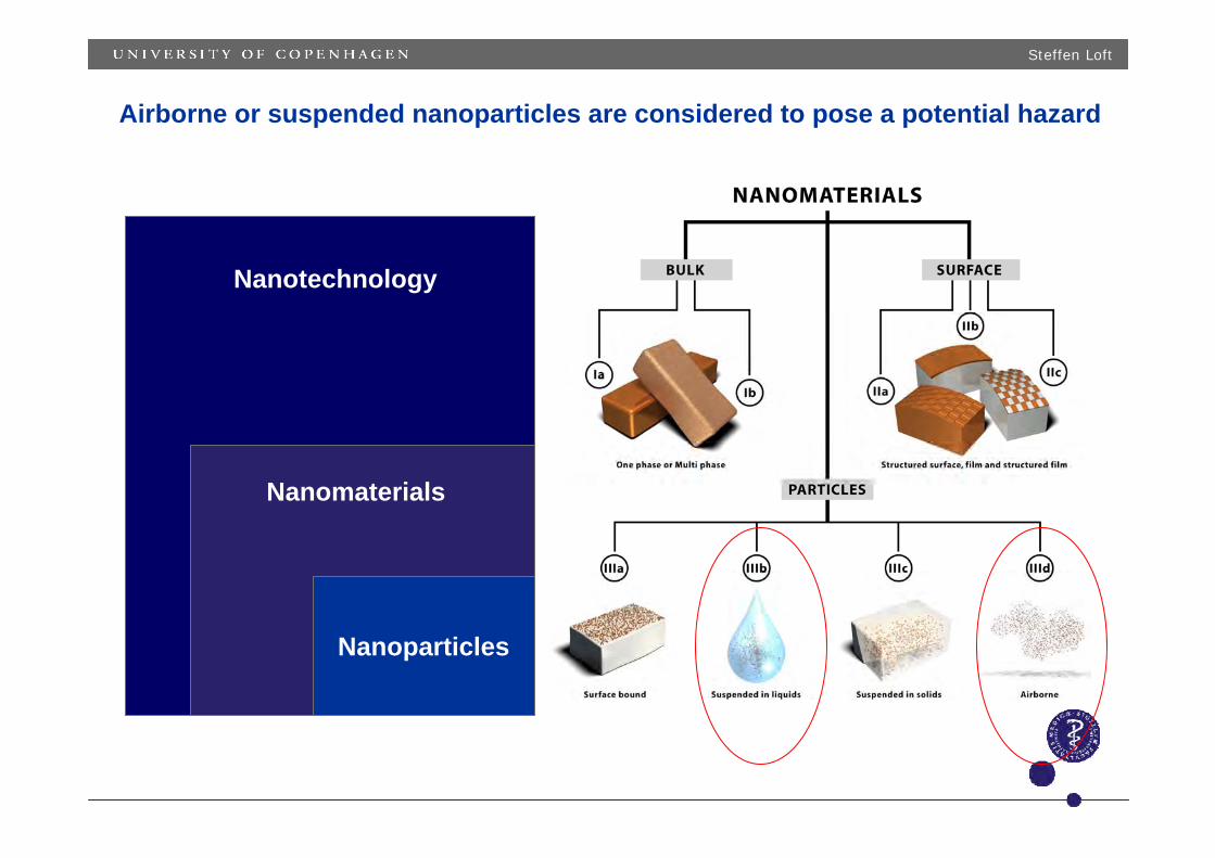
Airborne or suspended nanoparticles are considered to pose a potential hazard
Nanotechnology
Nanomaterials
Nanoparticles
Steffen Loft
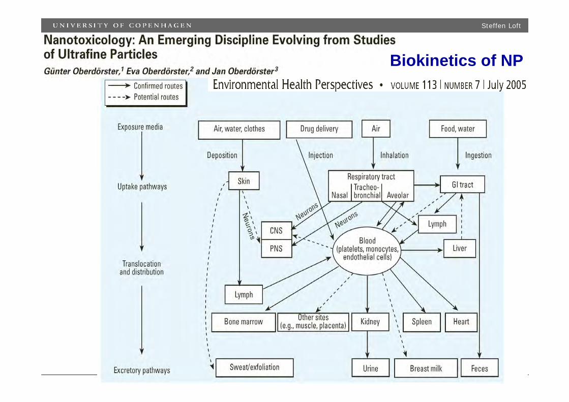
Biokinetics of NP
Steffen Loft
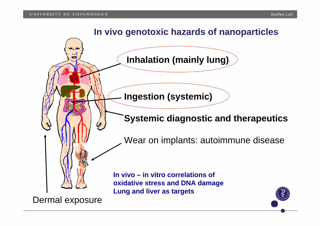
Inhalation (mainly lung)
Ingestion (systemic)
Systemic diagnostic and therapeutics
Wear on implants: autoimmune disease
Dermal exposure
In vivo genotoxic hazards of nanoparticles
In vivo – in vitro correlations of oxidative stress and DNA damageLung and liver as targets

Steffen Loft
Steffen Loft
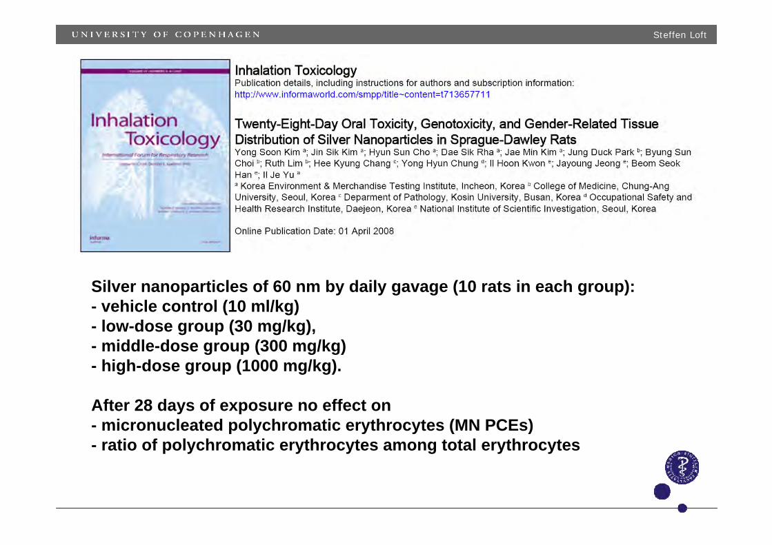
Silver nanoparticles of 60 nm by daily gavage (10 rats in each group): - vehicle control (10 ml/kg)- low-dose group (30 mg/kg), - middle-dose group (300 mg/kg)- high-dose group (1000 mg/kg).
After 28 days of exposure no effect on- micronucleated polychromatic erythrocytes (MN PCEs) - ratio of polychromatic erythrocytes among total erythrocytes
Steffen Loft
Steffen Loft
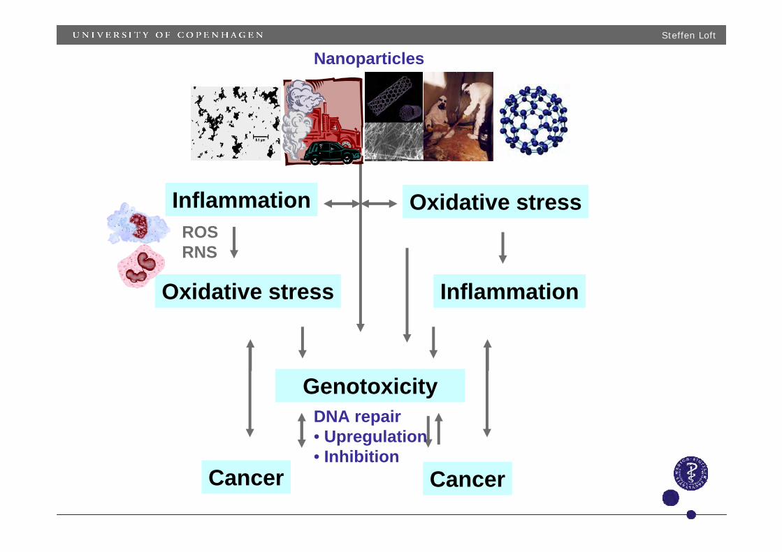
Inflammation
Genotoxicity
Cancer
Oxidative stress Inflammation
Cancer
Oxidative stress
Nanoparticles
ROSRNS
DNA repair• Upregulation• Inhibition
Steffen Loft
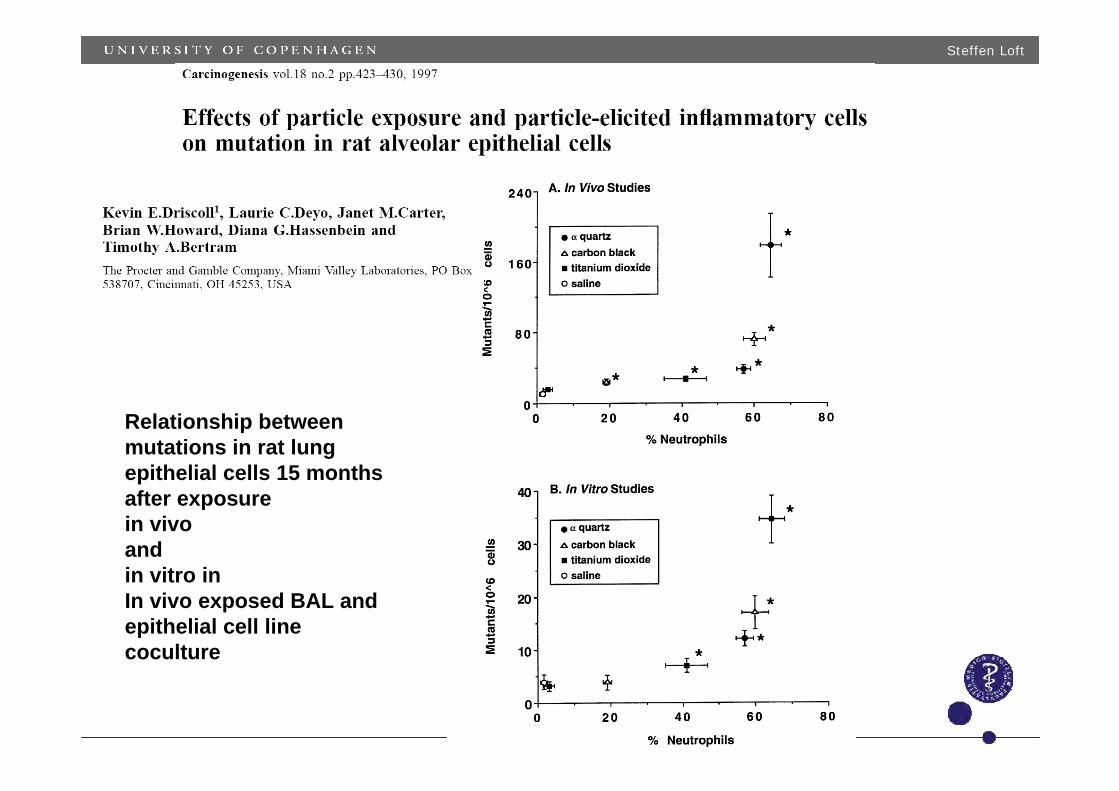
Relationship betweenmutations in rat lungepithelial cells 15 monthsafter exposurein vivoand in vitro in In vivo exposed BAL and epithelial cell line coculture
Steffen Loft
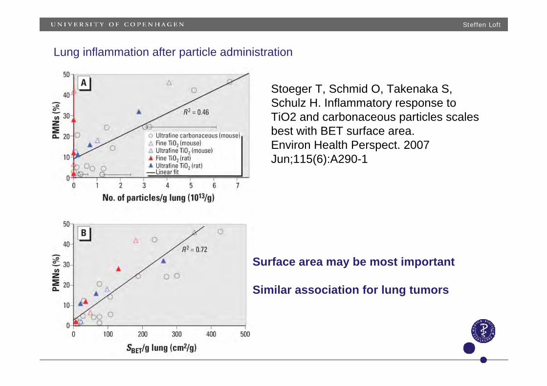
Lung inflammation after particle administration
Stoeger T, Schmid O, Takenaka S, Schulz H. Inflammatory response to TiO2 and carbonaceous particles scalesbest with BET surface area. Environ Health Perspect. 2007 Jun;115(6):A290-1
Surface area may be most important
Similar association for lung tumors
Steffen Loft
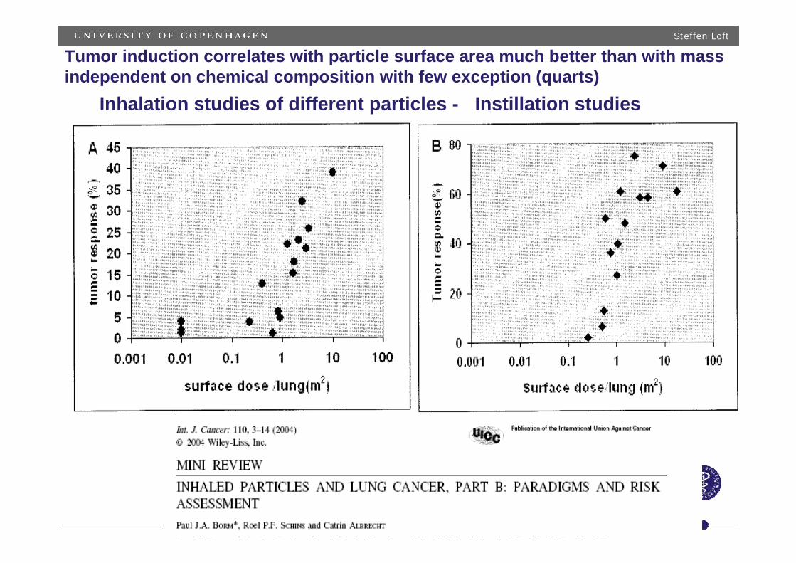
Tumor induction correlates with particle surface area much better than with massindependent on chemical composition with few exception (quarts)
Inhalation studies of different particles - Instillation studies
Steffen Loft
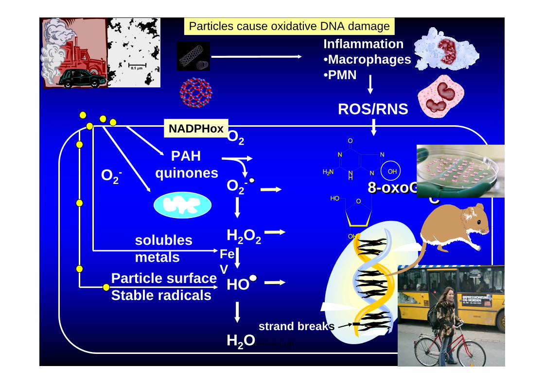
O2
O2-
H2O2
HO
H2O
NADPHox
FeV
PAHquinones
Inflammation•Macrophages•PMN
ROS/RNS
O2-
solublesmetals
Particle surfaceStable radicals
8-oxoG-8-oxoG- T-AT-AACAC
strand breaks
Particles cause oxidative DNA damage

Steffen Loft
DNA damage by particles in A549 lung epithelial cell and in isolated DNADieselparticles
Increased strand breaks, guanine oxidation and TNF, IL1, 6, 8 mRNAexpression (20-500 ug/ml) in cells by NIST 1650 or 2975 diesel or streetparticles
Similarly increased strand breaks in human lymphocytes (from 20 ug/mL)
Only effect of street particles and not of diesel particles on 8-oxodG in isolated DNA (HPLC-EC)
EM of UF
Dybdahl et al., Mutation Res 2004Danielsen et al. Particle Fibre Toxicol 2008
TSP from street filter (µg/ml)
8-ox
odG
per
10
6 dG
0
500
1000
1500
2000
2500
0 10 20 30 40 50
Street particles8-oxodG by HPLC-EC
FPG enzyme for guanine oxidation
Steffen Loft
0.1
1
10
100
0.1 1 10 100 1000
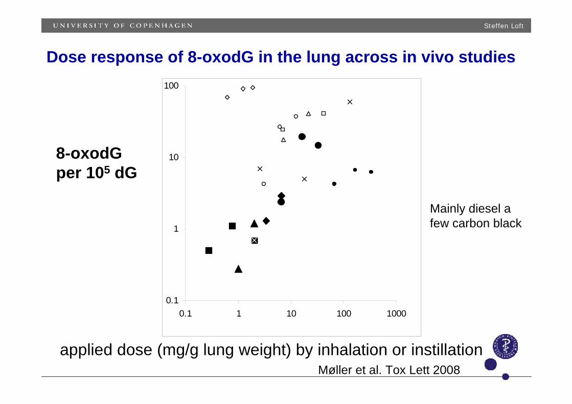
Dose response of 8-oxodG in the lung across in vivo studies
applied dose (mg/g lung weight) by inhalation or instillation
8-oxodG per 105 dG
Mainly diesel a few carbon black
Møller et al. Tox Lett 2008
Steffen Loft
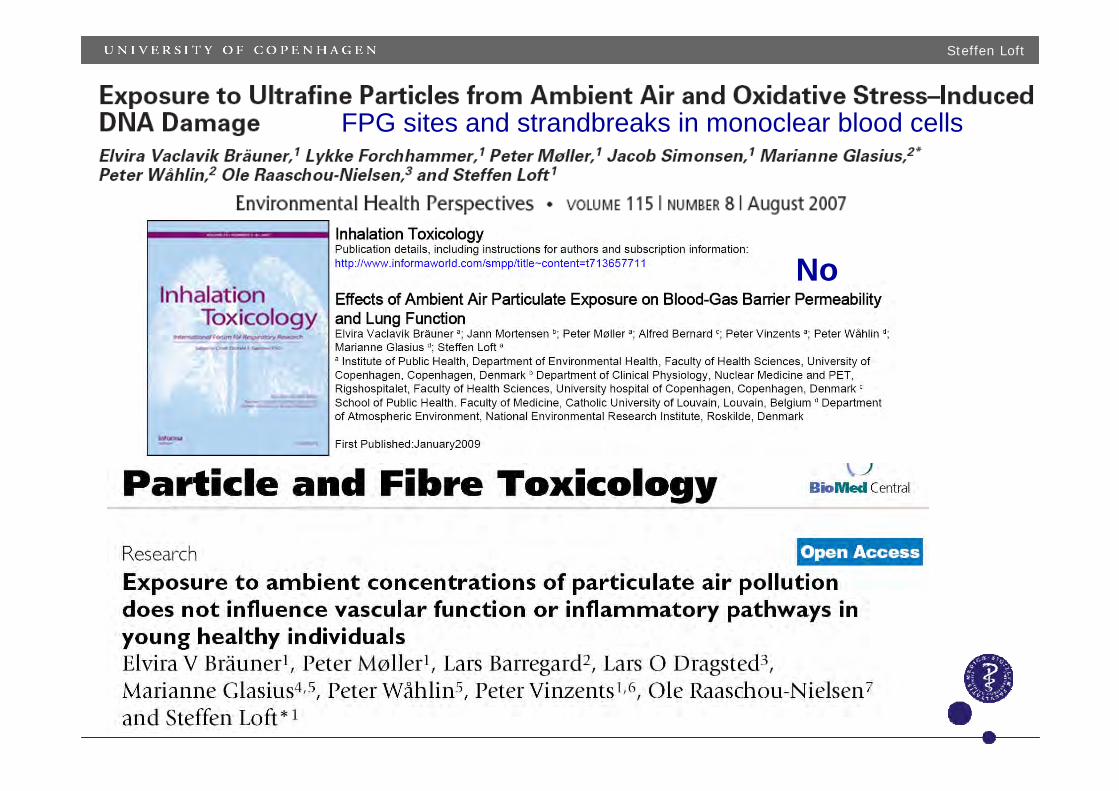
No
FPG sites and strandbreaks in monoclear blood cells
Steffen Loft
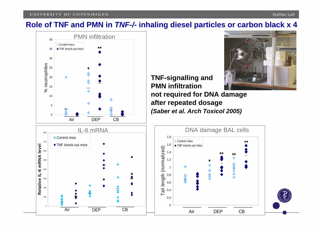
Role of TNF and PMN in TNF-/- inhaling diesel particles or carbon black x 4
DNA damage BAL cells
PMN infiltration
IL-6 mRNA
0
5
10
15
20
25
30
35
40
% n
eutro
phile
sControl miceTNF knock-out mice
Air DEP CB
0
100
200
300
400
500
600
700
800
Rel
ativ
e IL
-6 m
RN
A le
vel
Control mice
TNF knock-out mice
Air DEP CB0
0,2
0,4
0,6
0,8
1
1,2
1,4
1,6
1,8
Tail
leng
th (n
orm
aliz
ed)
Control miceTNF knock-out mice
Air DEP CB
TNF-signalling andPMN infiltrationnot required for DNA damageafter repeated dosage(Saber et al. Arch Toxicol 2005)

Steffen Loft
Compare wild type and ApoE-/- mice for susceptibility (carbon black)
Compare inhalation and instillation for effect (carbon black)
Use instillation to compare a nanoparticle battery• Carbon black 14 nm• C60 0.7 nm• SWCNT 0.9-1.7 x <1000 nm• Au 2 nm• Quantum dots 5 nm
Steffen Loft
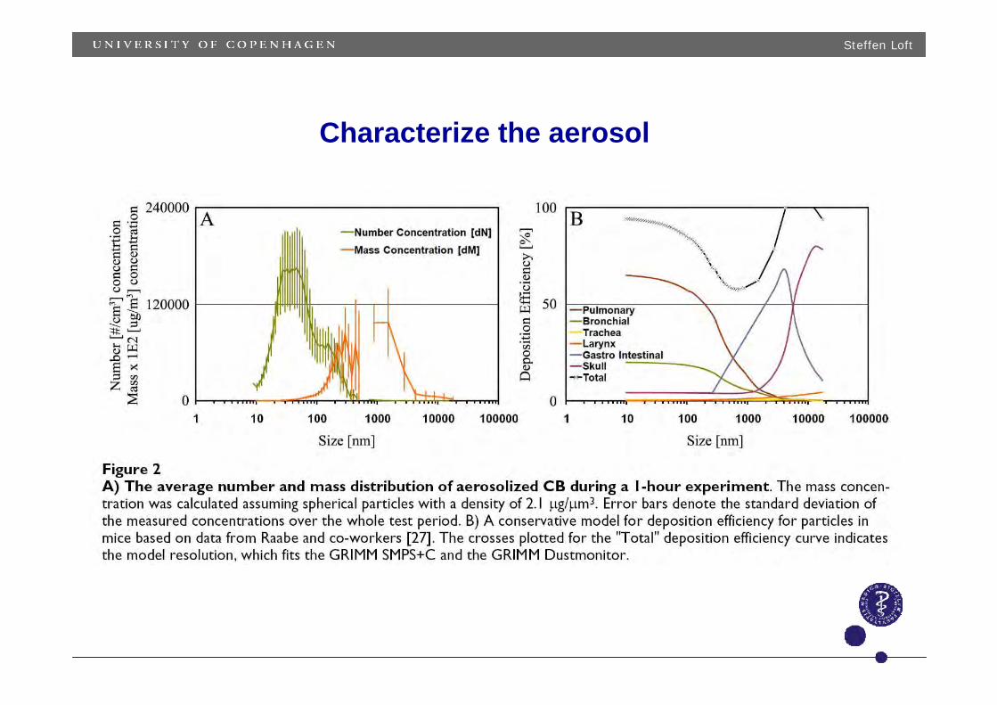
Characterize the aerosol
Steffen Loft
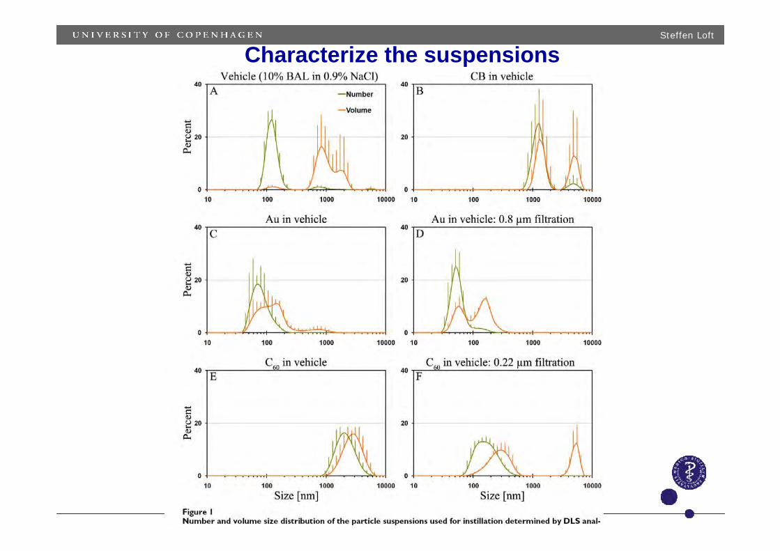
Characterize the suspensions
Steffen Loft
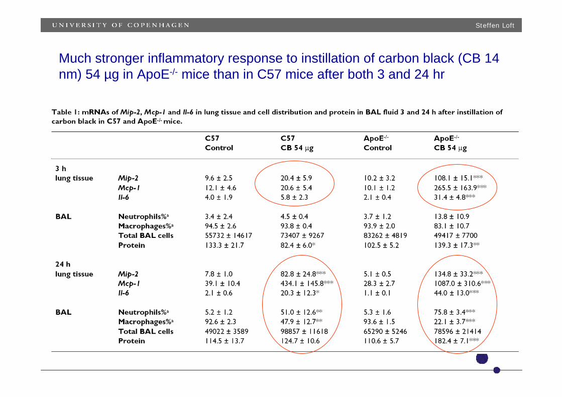
Much stronger inflammatory response to instillation of carbon black (CB 14 nm) 54 µg in ApoE-/- mice than in C57 mice after both 3 and 24 hr

Steffen Loft
Much stronger inflammatory response to instillation of carbon black (CB 14 nm) 18 or 54 µg than to inhalation of the same dose in ApoE-/- mice after 24 hr
Steffen Loft
control Au C60 SWCNT CB QD620 QD621
SB (%T)PMN
MCP-10
10
20
30
40
50
60 SB (%T)PMNMCP-1
Lung mRNA% in BALIn BAL
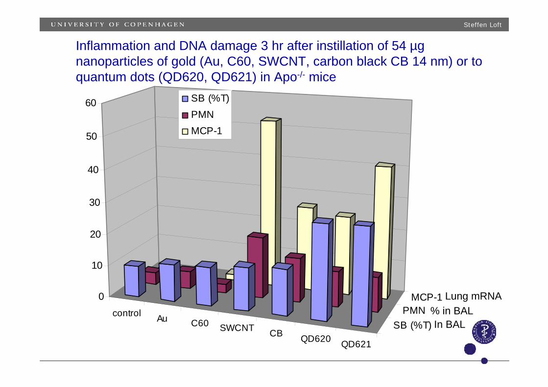
Inflammation and DNA damage 3 hr after instillation of 54 µg nanoparticles of gold (Au, C60, SWCNT, carbon black CB 14 nm) or to quantum dots (QD620, QD621) in Apo-/- mice
Steffen Loft
Animal experiments show increased oxidative stress, DNA damage and gene expression in colon, liver and lungs after low oral doses of diesel particles in feedor by gavage and without signs of inflammation or mutagenicity (after 21 days)
Steffen Loft
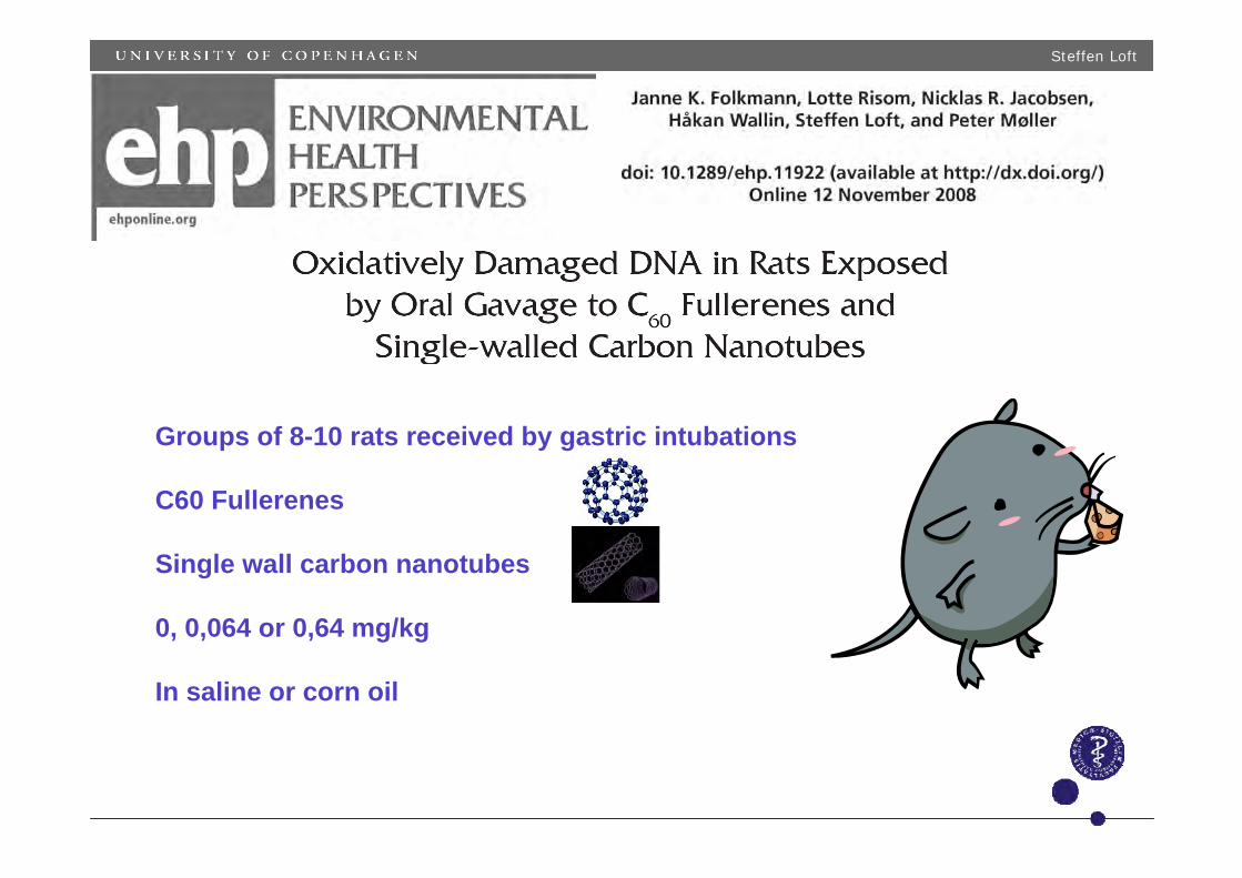
Groups of 8-10 rats received by gastric intubations
C60 Fullerenes
Single wall carbon nanotubes
0, 0,064 or 0,64 mg/kg
In saline or corn oil
Steffen Loft
0
10
20
30
40
50
C60 SWCNT
Control Low Low HighHigh
Dose
DN
A in
cisi
ons
(a.u
.)
0
1
2
3
4
5
6
7
C60 SWCNTControl Low Low HighHigh
* ***
Dose
8-ox
odG
/106
dG
0
1
2
3
4
5
6
7
Control Low LowHigh High
**
C60 SWCNT
*
Dose
8-ox
odG
/106
dG
05
1015202530
control low high low high
salinecorn oil
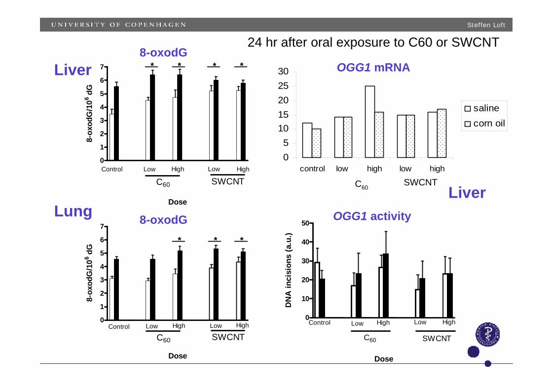
8-oxodGOGG1 mRNA
OGG1 activity
Liver
LungLiver
C60SWCNT
8-oxodG
24 hr after oral exposure to C60 or SWCNT
Steffen Loft
0
0.1
0.2
0.3
0.064 mg/kg 0.64 mg/kg
Dose of particle
Diffe
renc
e in
8-o
xodG
C60 SWCNT SRM2975
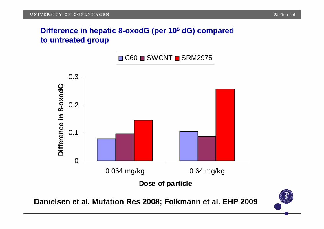
Difference in hepatic 8-oxodG (per 105 dG) comparedto untreated group
Danielsen et al. Mutation Res 2008; Folkmann et al. EHP 2009
Steffen Loft
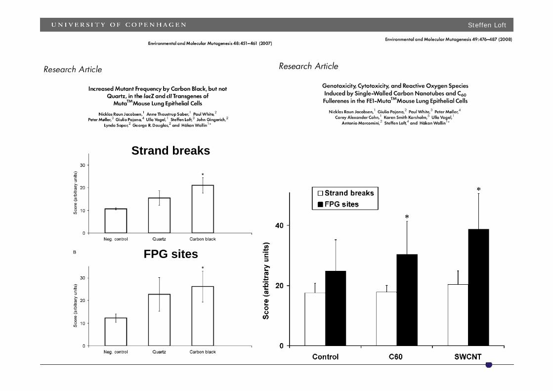
Strand breaks
FPG sites
Steffen Loft
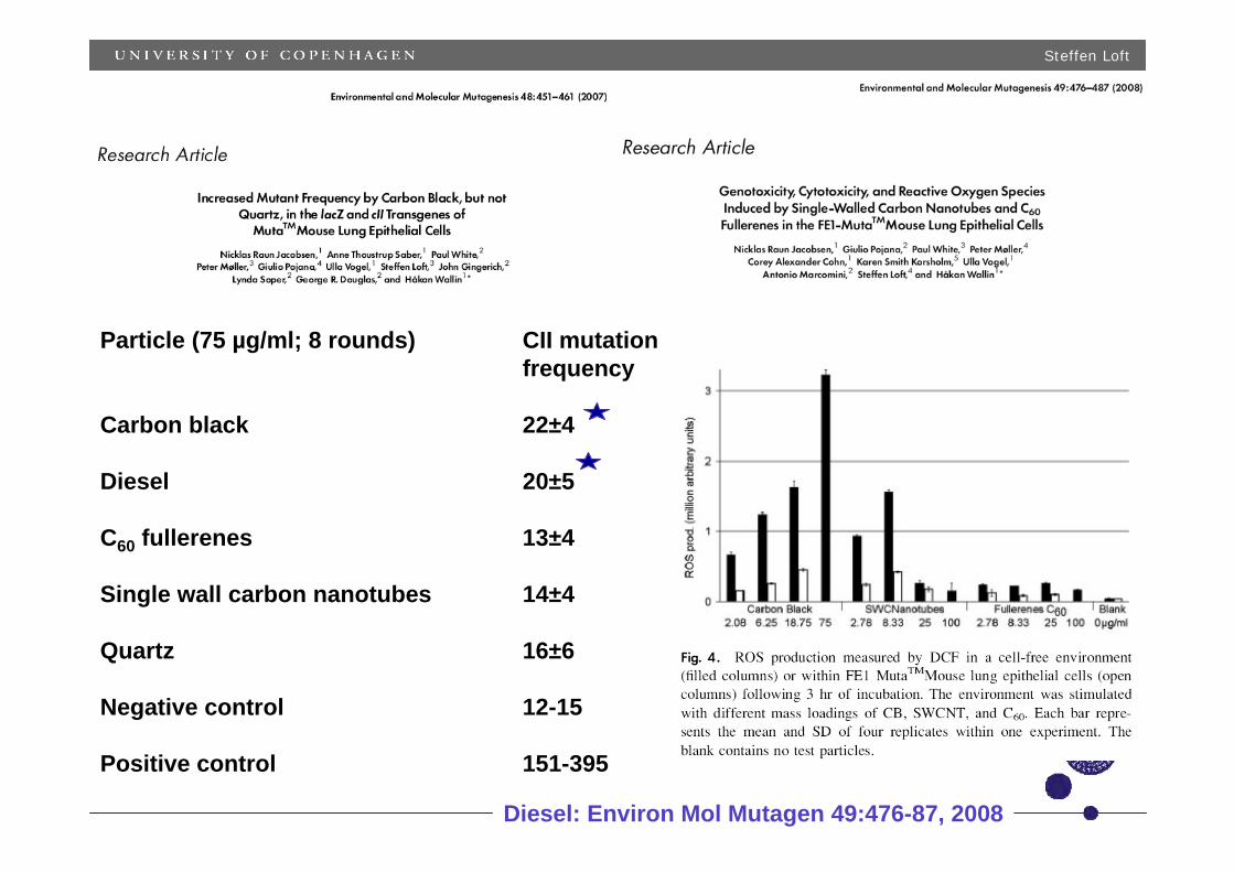
Particle (75 µg/ml; 8 rounds) CII mutationfrequency
Carbon black 22±4
Diesel 20±5
C60 fullerenes 13±4
Single wall carbon nanotubes 14±4
Quartz 16±6
Negative control 12-15
Positive control 151-395
Diesel: Environ Mol Mutagen 49:476-87, 2008
Steffen Loft
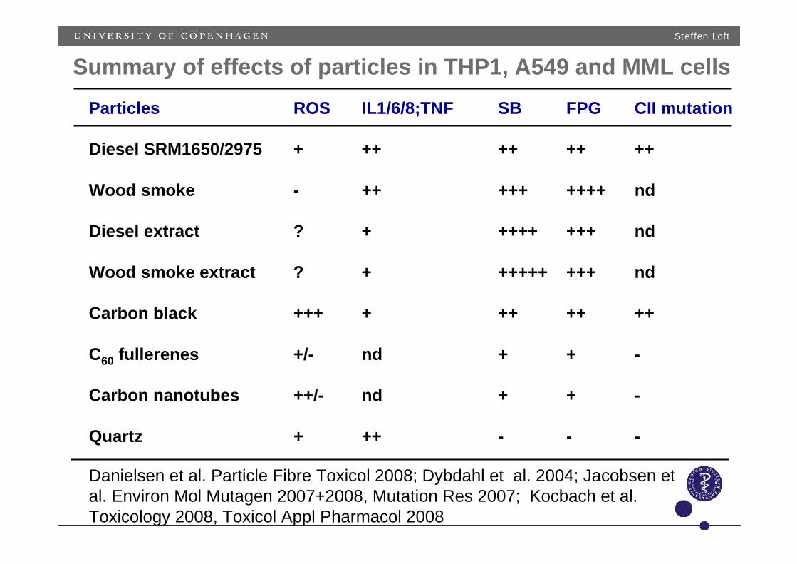
Particles ROS IL1/6/8;TNF SB FPG CII mutation
Diesel SRM1650/2975 + ++ ++ ++ ++
Wood smoke - ++ +++ ++++ nd
Diesel extract ? + ++++ +++ nd
Wood smoke extract ? + +++++ +++ nd
Carbon black +++ + ++ ++ ++
C60 fullerenes +/- nd + + -
Carbon nanotubes ++/- nd + + -
Quartz + ++ - - -
Summary of effects of particles in THP1, A549 and MML cells
Danielsen et al. Particle Fibre Toxicol 2008; Dybdahl et al. 2004; Jacobsen et al. Environ Mol Mutagen 2007+2008, Mutation Res 2007; Kocbach et al. Toxicology 2008, Toxicol Appl Pharmacol 2008
Steffen Loft
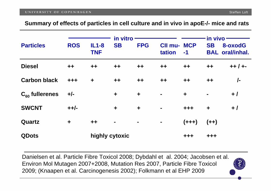
in vitro in vivoParticles ROS IL1-8 SB FPG CII mu- MCP SB 8-oxodG
TNF tation -1 BAL oral/inhal.
Diesel ++ ++ ++ ++ ++ ++ ++ ++ / +-
Carbon black +++ + ++ ++ ++ ++ ++ /-
C60 fullerenes +/- + + - + - + /
SWCNT ++/- + + - +++ + + /
Quartz + ++ - - - (+++) (++)
QDots highly cytoxic +++ +++
Summary of effects of particles in cell culture and in vivo in apoE-/- mice and rats
Danielsen et al. Particle Fibre Toxicol 2008; Dybdahl et al. 2004; Jacobsen et al. Environ Mol Mutagen 2007+2008, Mutation Res 2007, Particle Fibre Toxicol2009; (Knaapen et al. Carcinogenesis 2002); Folkmann et al EHP 2009
Steffen Loft
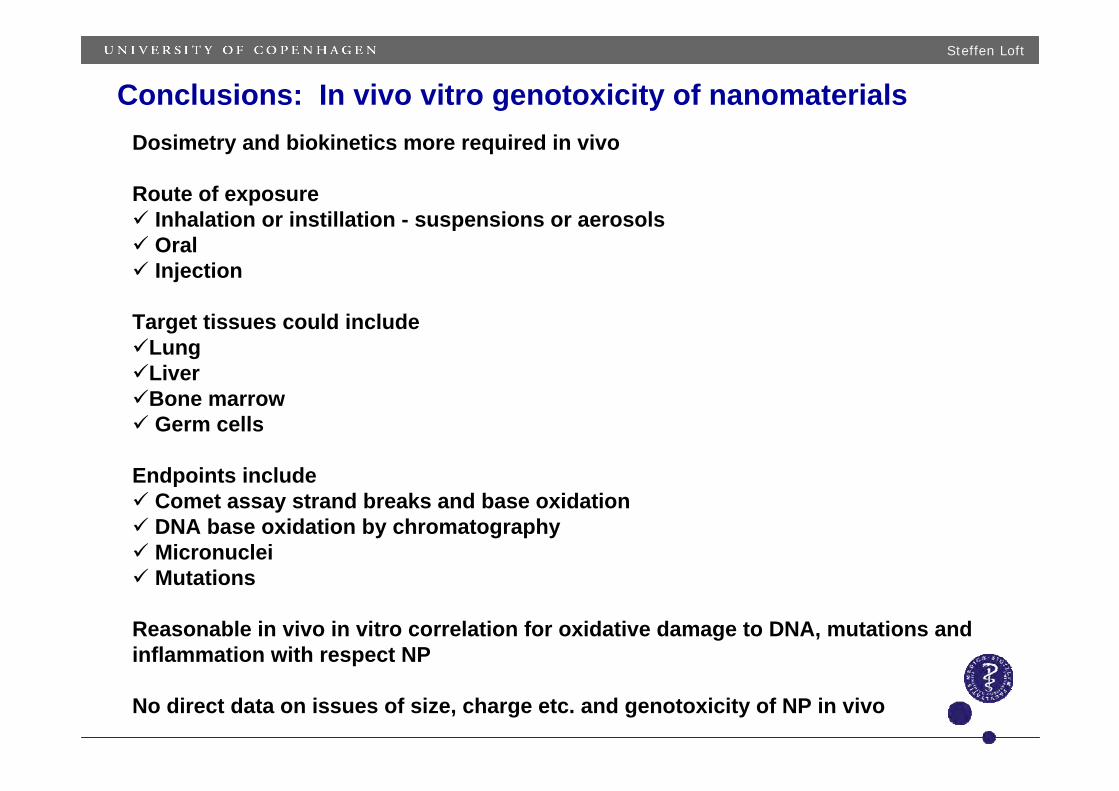
Conclusions: In vivo vitro genotoxicity of nanomaterialsDosimetry and biokinetics more required in vivo
Route of exposureInhalation or instillation - suspensions or aerosols OralInjection
Target tissues could includeLungLiverBone marrow Germ cells
Endpoints includeComet assay strand breaks and base oxidationDNA base oxidation by chromatography MicronucleiMutations
Reasonable in vivo in vitro correlation for oxidative damage to DNA, mutations and inflammation with respect NP
No direct data on issues of size, charge etc. and genotoxicity of NP in vivo

January 22, 2008: HESI Nanomaterial Environmental, Health, and Safety Committee Presentation. "HESI Nanomaterial Environmental, Health, and Safety (EHS)." Presented at the 2008 HESI Annual Meeting, San Juan, Puerto Rico. Presentation by Dr. Raymond David, BASF Corp.
HESI Nanomaterial Environmental,
Health, and Safety (EHS)
January 22, 2008
HESI Annual Meeting
San Juan, Puerto Rico
Outline
Brief introduction: Nanotechnology & EHS
Considerations
HESI International Nanomaterial EHS Safety
Consortium Goals
Consortium Activities
Consortium Steps
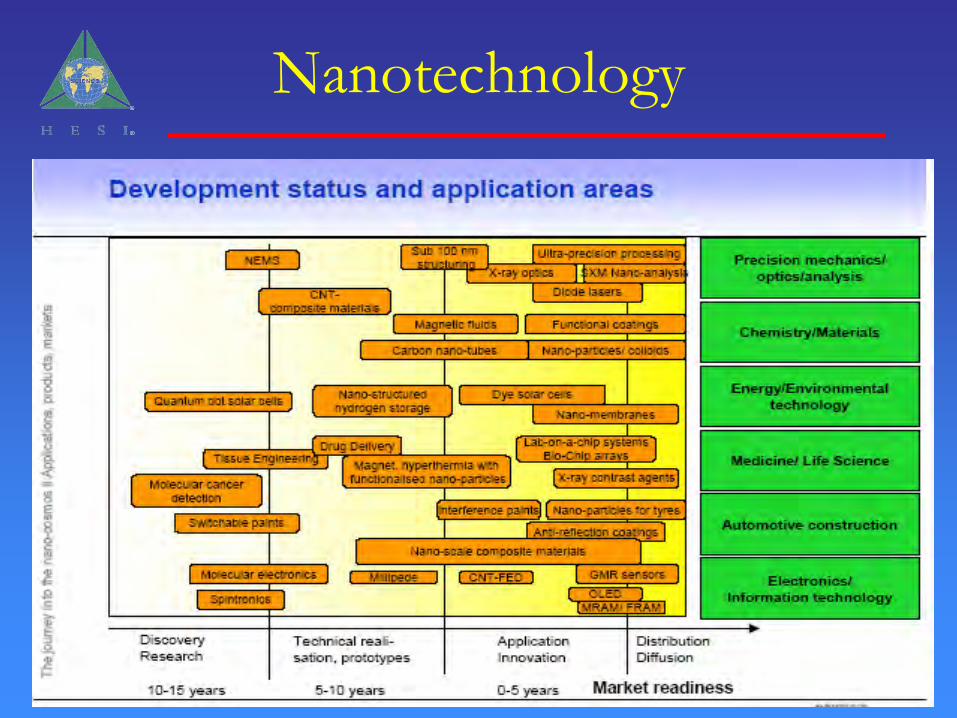
Nanotechnology
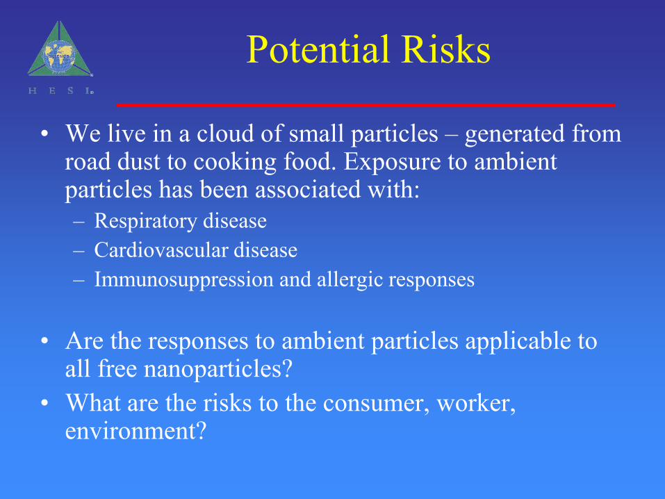
Potential Risks
• We live in a cloud of small particles – generated from road dust to cooking food. Exposure to ambient particles has been associated with:– Respiratory disease– Cardiovascular disease– Immunosuppression and allergic responses
• Are the responses to ambient particles applicable to all free nanoparticles?
• What are the risks to the consumer, worker, environment?
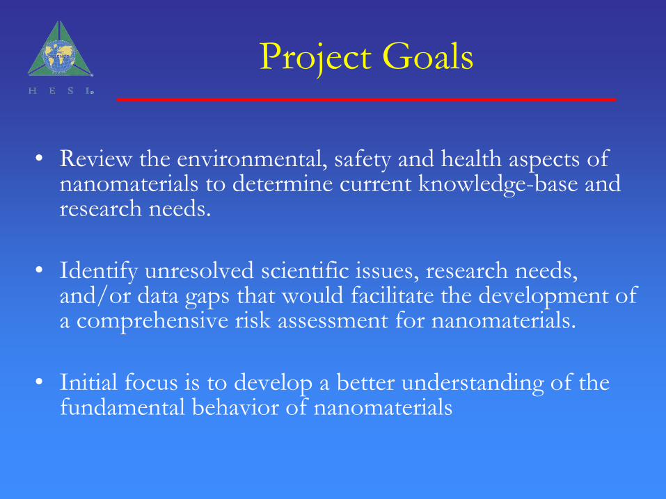
Project Goals
• Review the environmental, safety and health aspects of nanomaterials to determine current knowledge-base and research needs.
• Identify unresolved scientific issues, research needs, and/or data gaps that would facilitate the development of a comprehensive risk assessment for nanomaterials.
• Initial focus is to develop a better understanding of the fundamental behavior of nanomaterials
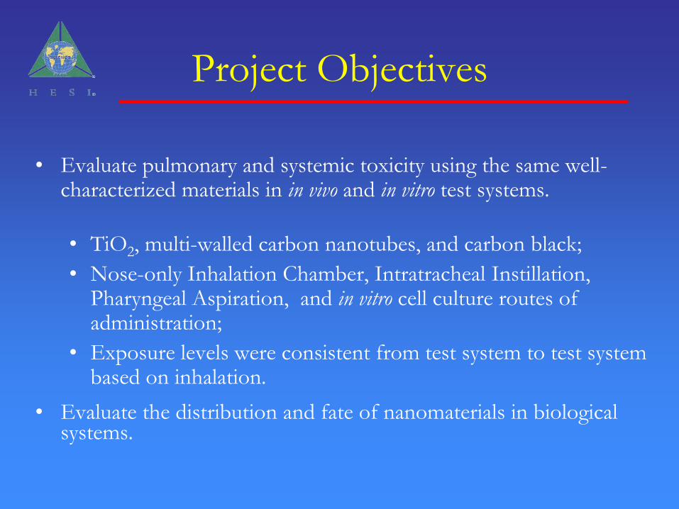
Project Objectives
• Evaluate pulmonary and systemic toxicity using the same well-characterized materials in in vivo and in vitro test systems.
• TiO2, multi-walled carbon nanotubes, and carbon black;
• Nose-only Inhalation Chamber, Intratracheal Instillation, Pharyngeal Aspiration, and in vitro cell culture routes of administration;
• Exposure levels were consistent from test system to test system based on inhalation.
• Evaluate the distribution and fate of nanomaterials in biological systems.
Pulmonary toxicity studiesNose-only inhalation chamber study in
rats
Studies conducted Dr. Karin Wiench, BASF-AG
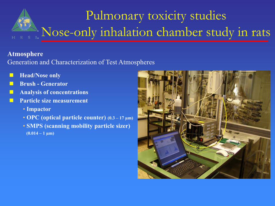
Head/Nose only Brush - Generator Analysis of concentrations Particle size measurement
• Impactor• OPC (optical particle counter) (0.3 – 17 µm)
• SMPS (scanning mobility particle sizer)(0.014 – 1 µm)
AtmosphereGeneration and Characterization of Test Atmospheres
Pulmonary toxicity studies
Nose-only inhalation chamber study in rats
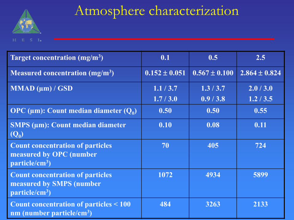
Atmosphere characterization
Target concentration (mg/m3) 0.1 0.5 2.5
Measured concentration (mg/m3) 0.152 0.051 0.567 0.100 2.864 0.824
MMAD (µm) / GSD 1.1 / 3.71.7 / 3.0
1.3 / 3.70.9 / 3.8
2.0 / 3.01.2 / 3.5
OPC (µm): Count median diameter (Q0) 0.50 0.50 0.55
SMPS (µm): Count median diameter (Q0)
0.10 0.08 0.11
Count concentration of particles measured by OPC (number particle/cm3)
70 405 724
Count concentration of particles measured by SMPS (number particle/cm3)
1072 4934 5899
Count concentration of particles < 100 nm (number particle/cm3)
484 3263 2133

• Male Wistar rats
Head-nose exposure to 6 hours a day on 5 consecutive days
– 2, 10, 50 mg/m3 TiO2 (P25)
– 0.1, 0.5, 2.5 mg/m3 MWCNT
– 0.5, 2.5, 10 mg/m3 Carbon Black
• Evaluations:
– Broncho alveolar lavage (BAL)
– Organ burden (lung, mediastinal lymph nodes, liver, kidney, spleen and basal brain with olfactory bulb)
– H & E based histopathology
– Cell proliferation and apoptosis
– SEM
– TEM
• Lavage immediately after the last exposure and post exposure days 3 and 16
• For other examination, sacrifice either directly after termination of treatment or after a recovery period of 16 days
Study Design
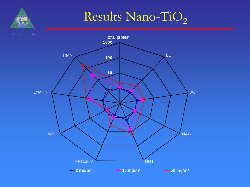
1
10
100
1000
total protein
LDH
ALP
NAG
GGTcell count
MPH
LYMPH
PMN
2 mg/m3 10 mg/m3 50 mg/m3
Results Nano-TiO2
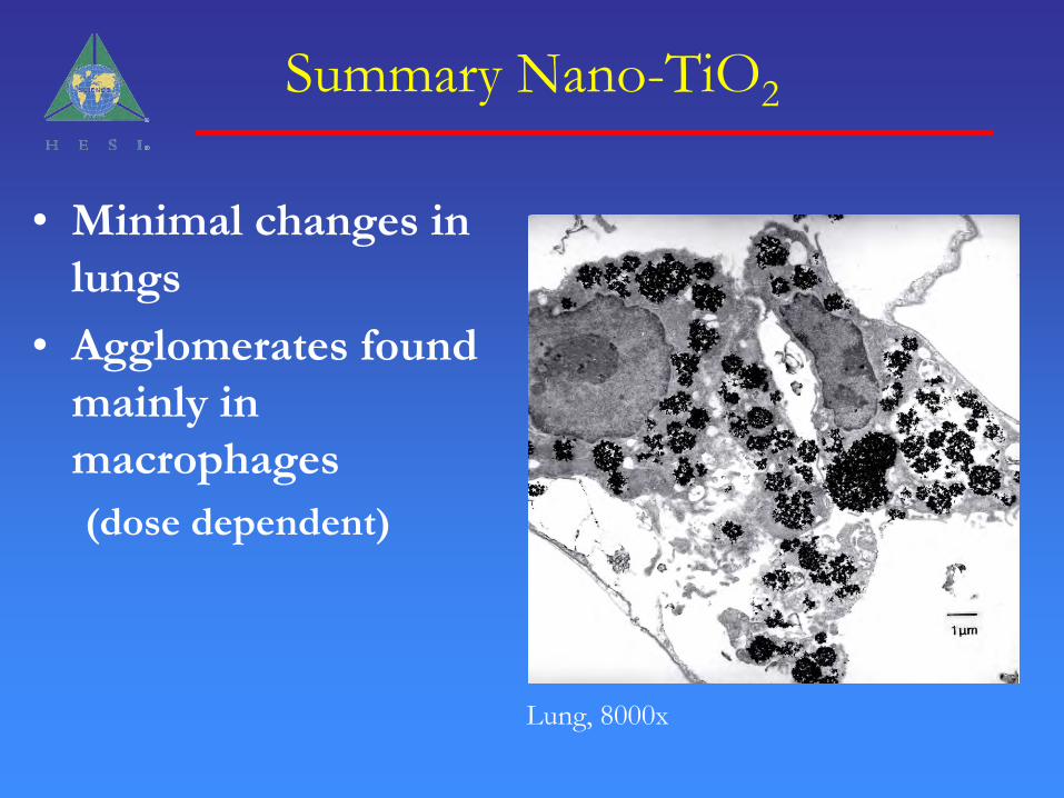
Summary Nano-TiO2
• Minimal changes in
lungs
• Agglomerates found
mainly in
macrophages
(dose dependent)
Lung, 8000x
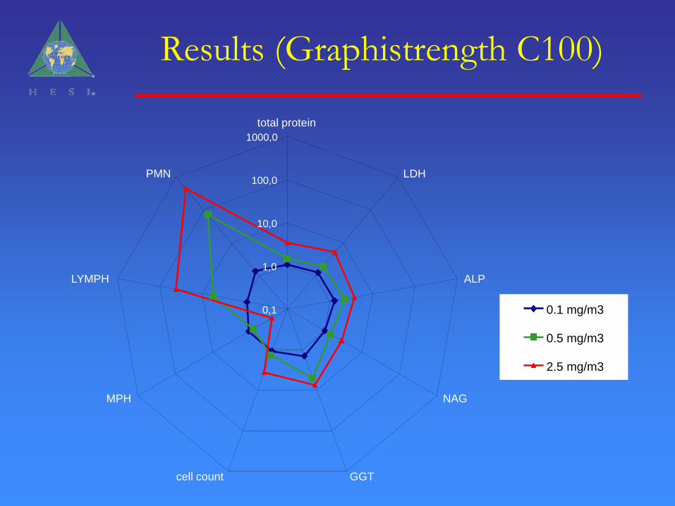
Results (Graphistrength C100)
0,1
1,0
10,0
100,0
1000,0
total protein
LDH
ALP
NAG
GGTcell count
MPH
LYMPH
PMN
0.1 mg/m3
0.5 mg/m3
2.5 mg/m3
Results (Graphistrength C100)
• 2.5 mg/m3
– Increased PMNs (abs. + rel.) in blood and BAL, even after 28 days of recovery
– Increased lymphocytes (rel.) in blood
– Increased absolute lung weight (11.5 %) immediately after the last exposure
– Increased relative lung weight (11.4 %) immediately after the last exposure
• 0.5 mg/m3
– Increased relative lung weight (10.5 %) immediately after the last exposure
– Increased PMNs (abs. + rel.) in blood and BAL, even after 28 days of recovery
• 0.1 mg/m3
– No findings
Results (Carbon Black)
• Clinical pathology:
– No findings
• Others:
– Discoloration of the lung at the end of the
exposure and after the recovery period
Completion of Comparative
Toxicity Evaluation
• Pharyngeal Aspiration Studies – Commitment from Vince Castranova at NIOSH
• Intratracheal Instillation Studies – In discussions with EPA; may consider alternative collaborators
• Target for Completion – 2008
• Will add ‘macro’sized material for comparison

In Vitro Evaluations
Studies conducted by Dr. James Gibson, East Carolina University
Study design
• Incubation of rat (L2), human lung cells (A549), rat alveolar macrophages, and CHO cells with TiO2 or MWCNT.
• Cytotoxicity and cell proliferation evaluated.
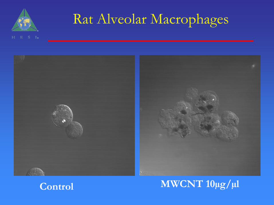
Rat Alveolar Macrophages
Control MWCNT 10µg/µl
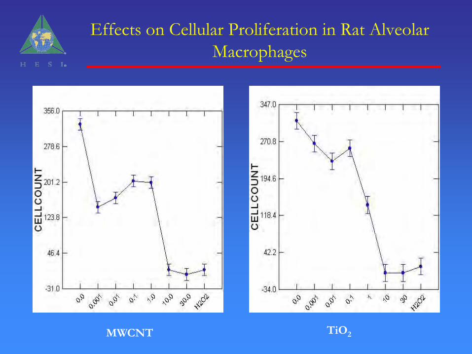
Effects on Cellular Proliferation in Rat Alveolar
Macrophages
MWCNT TiO2

Effects on Cell Proliferation in L2 Rat Lung Cells
MWCNT TiO2
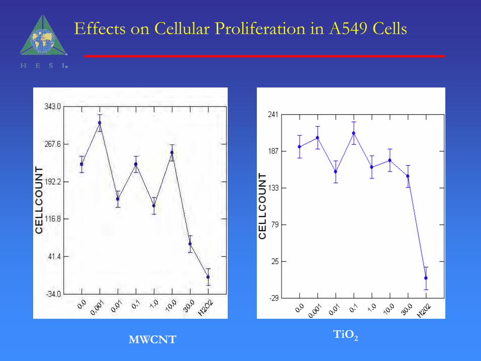
Effects on Cellular Proliferation in A549 Cells
MWCNT TiO2
Results and Next Steps
• There are differences in response sensitivity between rat and human cell lines. Next steps are to look at other cell lines.
• There may be differences in response to TiO2and MWCNT. Next steps to look at the time course of response.
Distribution of Polystyrene Nanospheres Following Systemic or Lung Exposure
Studies conducted by Dr. Kathy Sarlo, P&G

Focus of Program:
• Understand tissue distribution of different sized particles following different routes of exposure
• Female F-344 rat• Systemic vs. Airway Exposure• 20nm -- 1000nm fluorospheres
– commercially available– well characterized, longevity– required least amount of analytical resources
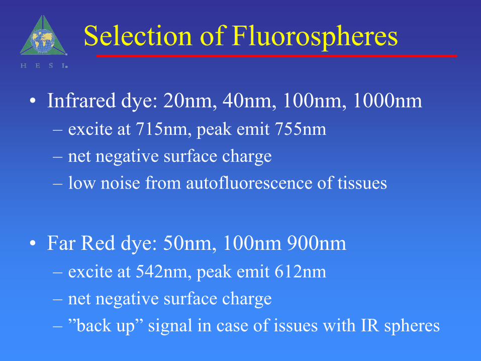
Selection of Fluorospheres
• Infrared dye: 20nm, 40nm, 100nm, 1000nm– excite at 715nm, peak emit 755nm– net negative surface charge – low noise from autofluorescence of tissues
• Far Red dye: 50nm, 100nm 900nm– excite at 542nm, peak emit 612nm– net negative surface charge– ”back up” signal in case of issues with IR spheres
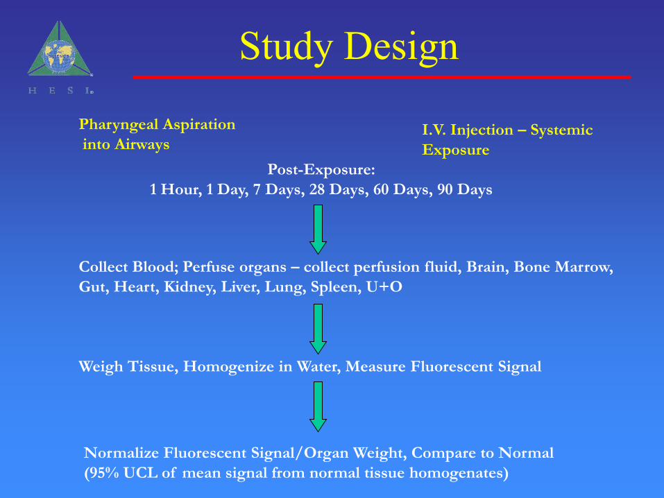
Pharyngeal Aspiration
into AirwaysI.V. Injection – Systemic
Exposure
Post-Exposure:
1 Hour, 1 Day, 7 Days, 28 Days, 60 Days, 90 Days
Collect Blood; Perfuse organs – collect perfusion fluid, Brain, Bone Marrow,
Gut, Heart, Kidney, Liver, Lung, Spleen, U+O
Weigh Tissue, Homogenize in Water, Measure Fluorescent Signal
Normalize Fluorescent Signal/Organ Weight, Compare to Normal
(95% UCL of mean signal from normal tissue homogenates)
Study Design
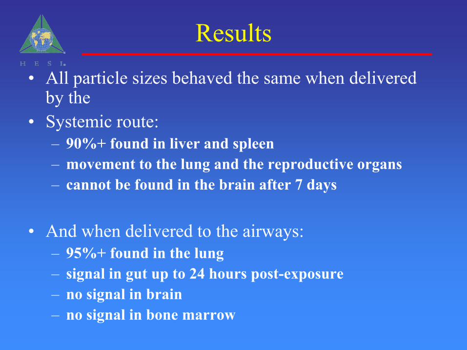
Results• All particle sizes behaved the same when delivered
by the• Systemic route:
– 90%+ found in liver and spleen– movement to the lung and the reproductive organs– cannot be found in the brain after 7 days
• And when delivered to the airways:– 95%+ found in the lung– signal in gut up to 24 hours post-exposure– no signal in brain– no signal in bone marrow
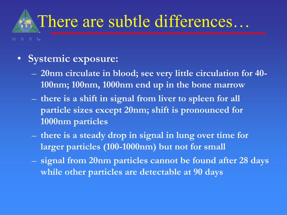
There are subtle differences…
• Systemic exposure:
– 20nm circulate in blood; see very little circulation for 40-
100nm; 100nm, 1000nm end up in the bone marrow
– there is a shift in signal from liver to spleen for all
particle sizes except 20nm; shift is pronounced for
1000nm particles
– there is a steady drop in signal in lung over time for
larger particles (100-1000nm) but not for small
– signal from 20nm particles cannot be found after 28 days
while other particles are detectable at 90 days
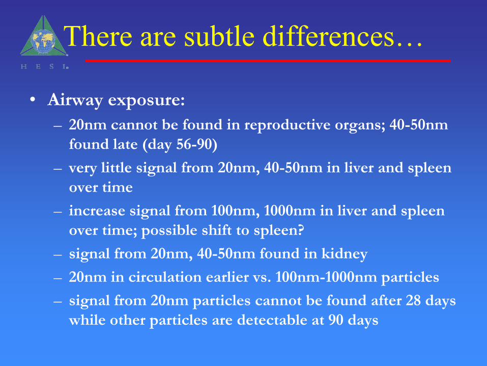
There are subtle differences…
• Airway exposure:
– 20nm cannot be found in reproductive organs; 40-50nm
found late (day 56-90)
– very little signal from 20nm, 40-50nm in liver and spleen
over time
– increase signal from 100nm, 1000nm in liver and spleen
over time; possible shift to spleen?
– signal from 20nm, 40-50nm found in kidney
– 20nm in circulation earlier vs. 100nm-1000nm particles
– signal from 20nm particles cannot be found after 28 days
while other particles are detectable at 90 days
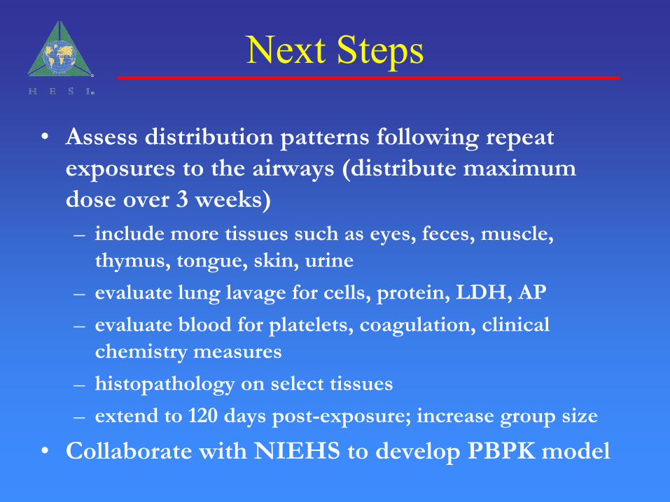
Next Steps
• Assess distribution patterns following repeat
exposures to the airways (distribute maximum
dose over 3 weeks)
– include more tissues such as eyes, feces, muscle,
thymus, tongue, skin, urine
– evaluate lung lavage for cells, protein, LDH, AP
– evaluate blood for platelets, coagulation, clinical
chemistry measures
– histopathology on select tissues
– extend to 120 days post-exposure; increase group size
• Collaborate with NIEHS to develop PBPK model
Additional 2008 Activities
Overwhelming response from participants was the forum for information exchange. Future activity is for a series of Workshops, Seminars, Webinars to Focus on (2 Topics Likely for 2008):
• Analytical Methods
• Exposure Evaluation
• Study designs for PK or other routes of exposure
• Life-Cycle Assessment
• Dissolution
Leadership
• Karluss Thomas, HESI• Raymond M. David, BASF Corp.• Hon-Wing Leung, Arkema Inc.• Timothy Landry, Dow Chemical

Consortium Participants
• 3M Corporation
• Arkema, Inc.
• BASF Corporation
• Duke University Medical Center
• East Carolina University
• L’Oreal Corporation
• National Institute of Environmental Health Sciences
• The Dow Chemical Company
• National Institute of Occupational Safety and Health
• The Procter & Gamble Company
• US Consumer Product Safety Commission
• US Environmental Protection Agency
• US Food and Drug Administration
October 3-6, 2005: HESI Nanomaterial EHS Project Committee Poster Presentation 2nd International Symposium on Nanotechnology and Occupational Health, Minneapolis, MN September 20-21, 2005: HESI Nanomaterial EHS Project Committee Poster Presentation Annual Meeting of the Japan Society of Toxicology, Tokyo, Japan. September 11-14, 2005: HESI Nanomaterial EHS Project Committee Poster Presentation 42nd Congress of the European Societies of Toxicology (Eurotox 2005), Krakow, Poland June 27, 2005: HESI Nanomaterial EHS Project Committee Presentation Chem-Bio Integrated Management Society (CBIMS) meeting, Tokyo, Japan March 6-10, 2005: HESI Nanomaterial EHS Project Committee Roundtable 44th Annual Meeting of the Society of Toxicology, New Orleans, LA.
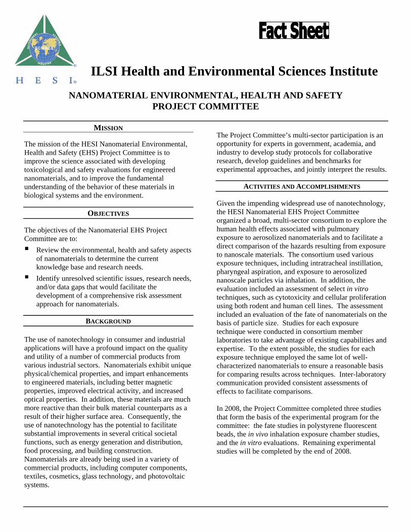
ILSI Health and Environmental Sciences Institute
NANOMATERIAL ENVIRONMENTAL, HEALTH AND SAFETY PROJECT COMMITTEE
MISSION
The mission of the HESI Nanomaterial Environmental, Health and Safety (EHS) Project Committee is to improve the science associated with developing toxicological and safety evaluations for engineered nanomaterials, and to improve the fundamental understanding of the behavior of these materials in biological systems and the environment. OBJECTIVES The objectives of the Nanomaterial EHS Project Committee are to: Review the environmental, health and safety aspects
of nanomaterials to determine the current knowledge base and research needs.
Identify unresolved scientific issues, research needs, and/or data gaps that would facilitate the development of a comprehensive risk assessment approach for nanomaterials.
BACKGROUND
The use of nanotechnology in consumer and industrial applications will have a profound impact on the quality and utility of a number of commercial products from various industrial sectors. Nanomaterials exhibit unique physical/chemical properties, and impart enhancements to engineered materials, including better magnetic properties, improved electrical activity, and increased optical properties. In addition, these materials are much more reactive than their bulk material counterparts as a result of their higher surface area. Consequently, the use of nanotechnology has the potential to facilitate substantial improvements in several critical societal functions, such as energy generation and distribution, food processing, and building construction. Nanomaterials are already being used in a variety of commercial products, including computer components, textiles, cosmetics, glass technology, and photovoltaic systems.
The Project Committee’s multi-sector participation is an opportunity for experts in government, academia, and industry to develop study protocols for collaborative research, develop guidelines and benchmarks for experimental approaches, and jointly interpret the results.
ACTIVITIES AND ACCOMPLISHMENTS Given the impending widespread use of nanotechnology, the HESI Nanomaterial EHS Project Committee organized a broad, multi-sector consortium to explore the human health effects associated with pulmonary exposure to aerosolized nanomaterials and to facilitate a direct comparison of the hazards resulting from exposure to nanoscale materials. The consortium used various exposure techniques, including intratracheal instillation, pharyngeal aspiration, and exposure to aerosolized nanoscale particles via inhalation. In addition, the evaluation included an assessment of select in vitro techniques, such as cytotoxicity and cellular proliferation using both rodent and human cell lines. The assessment included an evaluation of the fate of nanomaterials on the basis of particle size. Studies for each exposure technique were conducted in consortium member laboratories to take advantage of existing capabilities and expertise. To the extent possible, the studies for each exposure technique employed the same lot of well-characterized nanomaterials to ensure a reasonable basis for comparing results across techniques. Inter-laboratory communication provided consistent assessments of effects to facilitate comparisons. In 2008, the Project Committee completed three studies that form the basis of the experimental program for the committee: the fate studies in polystyrene fluorescent beads, the in vivo inhalation exposure chamber studies, and the in vitro evaluations. Remaining experimental studies will be completed by the end of 2008.
Nanomaterial EHS Project Committee Page 2 Fact Sheet
FUTURE ACTIVITIES In the first quarter of 2009, the Nanomaterial EHS Project Committee will conduct a webinar on “Genotoxicity of Nanomaterials.” The committee has elected to disband after the completion of the genotoxicity webinar. Interested parties are encouraged to submit future targeted proposals on nanomaterial safety and toxicity to HESI for consideration via the Emerging Issues survey process.
OUTREACH To share information about its scientific activities, the Nanomaterial EHS Project Committee has engaged in multiple outreach activities:
2008: 47th Annual Meeting of the Society of Toxicology,
Seattle, WA
2007: 11th International Congress of Toxicology,
Montreal, Canada 8th CSL/JIFSAN Joint Symposium on Food Safety
and Nutrition, Greenbelt, MD
2006: National Institute of Health Sciences, Tokyo, Japan 33rd Annual Meeting of the Japan Society of
Toxicology, Nagoya, Japan Food Products Association, Washington, DC American College of Toxicology Annual Meeting,
Palm Springs, CA
2005: 42nd Congress of the European Societies of
Toxicology (Eurotox), Krakow, Poland 44th Annual Meeting of the Society of Toxicology,
New Orleans, LA Chem-Bio Integrated Management Society
(CBIMS) meeting, Tokyo, Japan Annual Meeting of the Japan Society of Toxicology,
Tokyo, Japan
2nd International Symposium on Nanotechnology and Occupational Health, Minneapolis, MN
LEADERSHIP AND INFORMATION Chair ................................................ Dr. Raymond David
(BASF Corporation) HESI Staff ................................... Nancy G. Doerrer, MS
Ms. Cyndi Nobles
For more information, contact: Nancy G. Doerrer, MS
at 202-659-3306 or [email protected].
PROJECT COMMITTEE MEMBERSHIP
BASF Corporation The Cola-Cola Company
The Dow Chemical Company L’Oreal Corporation
The Procter & Gamble Company
PUBLIC PARTICIPATION
CDC National Institute of Occup. Safety and Health East Carolina University
NIH National Institute of Environmental Health Sciences North Carolina State University
University of Rochester US Consumer Product Safety Commission
US Environmental Protection Agency US Food and Drug Administration
PUBLICATIONS
Eight-part series in Toxicological Sciences (2005-2006): Thomas, K, and Sayre, P. 2005. Research strategies for safety evaluation of nanomaterials, Part I: evaluating the human health implications of exposure to nanoscale materials. Toxicol Sci. 87, 316-321. Holsapple, MP, Farland, WH, Landry, TD, Monteiro-Riviere, NA, Carter, JM, Walker, NJ, and Thomas, KV. 2005. Research strategies for safety evaluation of nanomaterials, Part II: toxicological and safety evaluation of nanomaterials, current challenges and data needs. Toxicol Sci. 88, 12-17. Balshaw, DM, Philbert, M, and Suk, WA. 2005. Research strategies for safety evaluation of
Nanomaterial EHS Project Committee Page 3 Fact Sheet
nanomaterials, Part III: nanoscale technologies for assessing risk and improving public health. Toxicol Sci. 88, 298-306. Tsuji, JS, Maynard, AD, Howard, PC, James, JT, Lam, C-W, Warheit, DB, and Santamaria, AB. 2006. Research strategies for safety evaluation of nanomaterials, Part IV: risk assessment of nanoparticles. Toxicol Sci. 89, 42-50. Borm, P, Klaessig, FC, Landry, TD, Moudgil, B, Pauluhn, J, Thomas, K, Trottier, R, and Wood, S. 2006. Research strategies for safety evaluation of nanomaterials, Part V: role of dissolution in biological fate and effects of nanoscale particles. Toxicol Sci. 90, 23-32. Powers, KW, Brown, SC, Krishna, VB, Wasdo, SC, Moudgil, BM, and Roberts, SM. 2006. Research strategies for safety evaluation of nanomaterials, Part VI: characterization of nanoscale particles for toxicological evaluation. Toxicol Sci. 90, 296-303. Thomas, T, Thomas, K, Sadrieh, N, Savage, N, Adair, P, and Bronaugh, R. 2006. Research strategies for safety evaluation of nanomaterials, Part VII: evaluating consumer exposure to nanoscale materials. Toxicol Sci. 91, 14-19. Thomas, K, Aguar, P, Kawasaki, H, Morris, J, Nakanishi, J, and Savage, N. 2006. Research strategies for safety evaluation of nanomaterials, Part VIII: international efforts to develop risk-based safety evaluations for nanomaterials. Toxicol Sci. 92, 23-32.
4/09


![Welcome!!!! [hesiglobal.org]hesiglobal.org/wp-content/uploads/sites/11/2016/06/... · HESI in 2009: Some highlights . . . ¾. Extensive portfolio of technical committees, project](https://static.fdocuments.net/doc/165x107/5f660efe0e22bc182037d8fd/welcome-hesi-in-2009-some-highlights-extensive-portfolio-of-technical.jpg)
![Welcome! [hesiglobal.org]hesiglobal.org/wp-content/uploads/sites/11/2016/06/HESI_ECETOC.pdf · •Biological significance of DNA adducts. ... ¾48 h embryo toxicity assay using Zebrafish,](https://static.fdocuments.net/doc/165x107/5a9d0b617f8b9a8a6a8b6809/welcome-biological-significance-of-dna-adducts-48-h-embryo-toxicity.jpg)