Nanocrystalline Hydroxyapatite and Zinc-doped Hydroxyapatite
14
ORIGINAL ARTICLE Nanocrystalline hydroxyapatite and zinc-doped hydroxyapatite as carrier material for controlled delivery of ciprofloxacin G. Devanand Venkatasubbu • S. Ramasamy • V. Ramakrishnan • J. Kumar Received: 26 May 2011 / Accepted: 4 August 2011 / Published online: 24 August 2011 Ó The Author(s) 2011. This article is published with open access at Springerlink.com Abstract In bone disorders infections are common. The concentration of majority of antibiotics is very low in the bone tissue. A high local dose can be obtained from the ciprofloxacin-loaded hydroxyapatite nanoparticles. The present study is aimed at developing the use of hydroxy- apatite and zinc-doped hydroxyapatite nanoparticles as a carrier for ciprofloxacin drug delivery system. The cipro- floxacin-loaded hydroxyapatite and zinc-doped hydroxy- apatite have a good antibacterial activity against Pseudomonas aeruginosa and Staphylococcus aureus. Hydroxyapatite and zinc-doped hydroxyapatite were pre- pared and characterized using X-ray diffraction, Trans- mission electron microscopy and inductively coupled plasma optical emission spectrometry. They were loaded with ciprofloxacin using optimized drug loading parame- ters. Drug loading, in vitro drug release and antimicrobial activity were analyzed. The influence of zinc on the con- trolled release of ciprofloxacin was analyzed. The results show that the presence of zinc increases the drug release percentage and that the drug was released in a controlled manner. Keywords Biomaterials Ciprofloxacin Controlled release Drug delivery Hydroxyapatite Pseudomonas aeruginosa Staphylococcus aureus Introduction Nanotechnology deals with the manipulation of material characteristics (Ochekpe et al. 2009). Due to the large surface to volume ratio they are advantageous to attach drug molecules and other compounds (De Jong and Borm 2008). This is the main factor behind the application of nanotechnology in medical field for the delivery of drugs, proteins or genes. The drugs are effectively delivered by nanoparticles because nanoparticles are effectively taken up by the cells. They penetrate deep into tissue through fine capillaries and cross the fenestration present in the epi- thelial lining. Drug delivery can be either localized or targeted to a particular diseased site (Panyam and Labha- setwar 2003). Controlled drug delivery is the technology by which the drug can be released at a predetermined rate for a long period of time into the blood or delivered at the target site (Szycher 1986). Unlike the traditional oral, intravenous drug delivery methods whereby the drug is distributed to both healthy and diseased tissue, in controlled local drug delivery high concentration of drug is achieved at the infected site. This leads to increase in therapeutic index and therapeutic efficacy and reduction in overall serum con- centration and deleterious side effects to other organs (De Jong and Borm 2008; Melville et al. 2008; Noel MS et al. 2008; De Gaetano et al. 2005). Drug stability, optimized drug absorption, treatment continuation in natural phase and improvement in pharmacokinetic characteristics of drug can be achieved by localized drug delivery (Somberg 1989). In controlled drug delivery system the carriers play a vital role since they incorporate the drug, retain it and release it progressively with time. So properties such as drug incorporation and release; formulation stability and shelf life; biocompatibility, biodistribution, and G. Devanand Venkatasubbu (&) S. Ramasamy J. Kumar Crystal Growth Centre, Anna University, Chennai 600025, Tamil Nadu, India e-mail: [email protected] V. Ramakrishnan Department of Laser Studies, School of Physics, Madurai Kamaraj University, Madurai, Tamil Nadu, India 123 3 Biotech (2011) 1:173–186 DOI 10.1007/s13205-011-0021-9
-
Upload
hajarul-massi -
Category
Documents
-
view
63 -
download
2
description
ha
Transcript of Nanocrystalline Hydroxyapatite and Zinc-doped Hydroxyapatite

ORIGINAL ARTICLE
Nanocrystalline hydroxyapatite and zinc-doped hydroxyapatiteas carrier material for controlled delivery of ciprofloxacin
G. Devanand Venkatasubbu • S. Ramasamy •
V. Ramakrishnan • J. Kumar
Received: 26 May 2011 / Accepted: 4 August 2011 / Published online: 24 August 2011
� The Author(s) 2011. This article is published with open access at Springerlink.com
Abstract In bone disorders infections are common. The
concentration of majority of antibiotics is very low in the
bone tissue. A high local dose can be obtained from
the ciprofloxacin-loaded hydroxyapatite nanoparticles. The
present study is aimed at developing the use of hydroxy-
apatite and zinc-doped hydroxyapatite nanoparticles as a
carrier for ciprofloxacin drug delivery system. The cipro-
floxacin-loaded hydroxyapatite and zinc-doped hydroxy-
apatite have a good antibacterial activity against
Pseudomonas aeruginosa and Staphylococcus aureus.
Hydroxyapatite and zinc-doped hydroxyapatite were pre-
pared and characterized using X-ray diffraction, Trans-
mission electron microscopy and inductively coupled
plasma optical emission spectrometry. They were loaded
with ciprofloxacin using optimized drug loading parame-
ters. Drug loading, in vitro drug release and antimicrobial
activity were analyzed. The influence of zinc on the con-
trolled release of ciprofloxacin was analyzed. The results
show that the presence of zinc increases the drug release
percentage and that the drug was released in a controlled
manner.
Keywords Biomaterials � Ciprofloxacin �Controlled release � Drug delivery � Hydroxyapatite �Pseudomonas aeruginosa � Staphylococcus aureus
Introduction
Nanotechnology deals with the manipulation of material
characteristics (Ochekpe et al. 2009). Due to the large
surface to volume ratio they are advantageous to attach
drug molecules and other compounds (De Jong and Borm
2008). This is the main factor behind the application of
nanotechnology in medical field for the delivery of drugs,
proteins or genes. The drugs are effectively delivered by
nanoparticles because nanoparticles are effectively taken
up by the cells. They penetrate deep into tissue through fine
capillaries and cross the fenestration present in the epi-
thelial lining. Drug delivery can be either localized or
targeted to a particular diseased site (Panyam and Labha-
setwar 2003).
Controlled drug delivery is the technology by which the
drug can be released at a predetermined rate for a long
period of time into the blood or delivered at the target site
(Szycher 1986). Unlike the traditional oral, intravenous
drug delivery methods whereby the drug is distributed to
both healthy and diseased tissue, in controlled local drug
delivery high concentration of drug is achieved at the
infected site. This leads to increase in therapeutic index and
therapeutic efficacy and reduction in overall serum con-
centration and deleterious side effects to other organs (De
Jong and Borm 2008; Melville et al. 2008; Noel MS et al.
2008; De Gaetano et al. 2005). Drug stability, optimized
drug absorption, treatment continuation in natural phase
and improvement in pharmacokinetic characteristics of
drug can be achieved by localized drug delivery (Somberg
1989). In controlled drug delivery system the carriers play
a vital role since they incorporate the drug, retain it
and release it progressively with time. So properties such
as drug incorporation and release; formulation stability
and shelf life; biocompatibility, biodistribution, and
G. Devanand Venkatasubbu (&) � S. Ramasamy � J. Kumar
Crystal Growth Centre, Anna University,
Chennai 600025, Tamil Nadu, India
e-mail: [email protected]
V. Ramakrishnan
Department of Laser Studies, School of Physics,
Madurai Kamaraj University, Madurai, Tamil Nadu, India
123
3 Biotech (2011) 1:173–186
DOI 10.1007/s13205-011-0021-9

functionality should be analyzed thoroughly when choos-
ing a carrier for delivery of drugs. The drug release from
any carrier depends upon solubility of drugs, microstruc-
ture of carrier, degradation of carrier and the bond between
the drug and the carrier (De Jong and Borm 2008; Ginebra
et al. 2006).
Bone diseases like osteomyelitis and osteoarticular
infections which are associated with bacterial infection are
highly complicated and involve operative debridement and
removal of all foreign bodies followed by antibiotic ther-
apy (Murugan and Ramakrishna 2006; Samit Kumar Nandi
et al. 2009; Gautier et al. 2001). The blood circulation in
these infected sites is limited and so the antibiotic distri-
bution is also poor. So growth factors and antimicrobials
should be supplied to the osseous site by site-specific drug
delivery (Hae-Won Kim et al. 2004). By this method the
drug action is localized and maintained for a long time. In
spite of high drug concentration at the infection site the
systemic side effects is low (Itokazu et al. 1999). By having
a localized drug delivery we can maintain a high concen-
tration of drug for a long time at the specific infected bone
site by controlling the release of drug. The drug concen-
tration can be controlled in a way that it neither reaches the
toxic level nor falls below the minimum effective level
(Ginebra et al. 2006). Even though high doses of antibiotics
are administered by systemic routes, effective drug con-
centration is not achieved at the site of infection because of
the impaired blood circulation, pharmacokinetic parame-
ters of the drug and high elimination rate (Pham et al. 2002;
Slosarczyk et al. 2000).
Staphylococcus aureus and Pseudomonas aeruginosa
are the most important pathogens causing bone and joint
infections, soft tissue and overwhelming sepsis. Staphylo-
coccus is a Gram-positive and Pseudomonas is a Gram-
negative bacterium. S. aureus expresses many surface
adhesions that promote attachment to plasma and extra
cellular matrix (ECM) proteins of the host cell. With the
use of new and high-dose antibacterial agents the fear of
new strain of S. aureus which is resistant to all available
antibacterial agents arises. S. aureus can evade the immune
response and antimicrobial therapy by another mechanism
known as ‘‘Small colony variants’’ (SCVs), a sub popula-
tion of S. aureus (Harris and Geoff Richards 2006; Uwe
Joosten et al. 2005).
Ciprofloxacin (CPX), 1-cyclopropyl 6-fluro-1, 4-dihy-
dro-4-oxo-7-(1-piperazinyl)-3-quinoline carbolitic acid is a
broad-spectrum fluroquinolone antibacterial agent used in
the treatment of both Gram-positive and Gram-negative
microorganisms. It stops cell division by inhibiting DNA
gyrase, a type II topoisomerase and topoisomerase IV. The
minimum inhibition concentration (MIC) of CPX is
0.25–2 lg/ml for pathogens like Staphylococcus aureus
and Pseudomonas aeruginosa which cause osteomyelitis.
With such a low MIC the dosage of CPX is 700 mg twice a
day for a period of 6 weeks to several months. The treat-
ment is extremely long and in spite of that the percentage
of cure is only 56%. This prolonged treatment and high
dose of CPX leads to toxicity and development of resistant
strains. So new sustained release systems have been pro-
posed as an alternative for systemic treatment (Patel et al.
2006; Zeiler 1985; Murugan and Panduranga Rao 2002).
Polymethymethacrylate (PMMA) beads have been used
initially to treat infectious bone diseases. But it has to be
removed surgically as they are non-biodegradable. Resorb-
able biomaterials like collagen, fibrinogen and PLA when
used do not replace bone grafting (Panyam and Labhasetwar
2003; Murugan and Ramakrishna 2006; Cheng and Kuhn
2007). Hydroxyapatite, a typical type of calcium phosphate
bioceramic, is used as a carrier for delivery of drugs, non
viral gene, antigen, enzyme and proteins because of their
biocompatibility, bioactivity and high affinity towards
drugs, proteins and DNA. Since Hydroxyapatite has a low
solubility in physiological condition it can be used as a
carrier for the local delivery of drugs both by surgical
placement and injection. The controlled localized drug
delivery from hydroxyapatite minimizes the toxicity to other
organs by minimizing the drug concentration in the blood. It
also avoids repeated dosage of drugs. Hydroxyapatite can
bind to both positive and negative molecules by simple
adsorption. So in the particulate form it is used for the
delivery of various kinds of molecules like antibiotics,
contraceptives, acetylsalicylic acid, hormones, insulin and
anticancer drugs (Paul and Sharma 1999; Madhana Sundar
et al. 2005; Szymura-Oleksiak et al. 2001). Hydroxyapatite
is used in the controlled drug delivery for bone cancer,
chronic osteomyelitis and in the delivery of agents with a
low bone penetration and short biological half life. The
concentration of drug depends upon the ability of the drug to
penetrate through the micro pores of hydroxyapatite, rate of
penetration and the pharmacokinetic profile of the drug.
Since hydroxyapatite has a micro pore structure and excel-
lent biological response to the physiological condition it can
ensure slow release of the drug (Slosarczyk et al. 2000;
Ingrid Russoni de Lima et al. 2006).
Zinc is an important trace element in all biological tis-
sues. More than 300 types of enzymes like alkaline phos-
phates take part in bone metabolism. Zinc participates in
the activity of these enzymes (Amit Bandyopadhyay et al.
2007). Zinc has a direct effect on the proliferative effect of
osteoblastic cells and inhibitory effect on osteoclastic bone
resorption (Tang et al. 2009). Besides these, zinc also takes
part in nucleic acid metabolism, maintenance of membrane
structure and its function, hormonal activity, biominerali-
zation and pathological calcification (Mason 2006). It acts
as an antioxidant. Many biochemical processes like car-
bohydrate metabolism, protein digestion and blood clotting
174 3 Biotech (2011) 1:173–186
123

require zinc. It plays an important role in immune system
also. So it would be desirable to obtain hydroxyapatite with
certain amount of zinc to enhance several biochemical
processes (Say Chye Joachim Loo et al. 2008).
In the present work hydroxyapatite and zinc-doped
hydroxyapatite were synthesized in nano form and char-
acterized. Ciprofloxacin was loaded to both of these
materials. The various parameters for drug loading were
optimized. The drug loading percentage and drug release
profile were analyzed and the influence of zinc on the
release of ciprofloxacin discussed. The interaction between
hydroxyapatite and drug was analyzed. Antimicrobial
activity of drug loaded hydroxyapatite was analyzed
against Pseudomonas aeruginosa and Staphylococcus
aureus.
Experimental procedure
Synthesis and characterization of hydroxyapatite
nanoparticles
Hydroxyapatite nanoparticles were synthesized by wet
chemical precipitation reaction:
10Ca OHð Þ2þ6 H3PO4 ! Ca10 PO4ð Þ6 OHð Þ2þ18H2O
Aqueous suspension o f calcium hydroxide (Ca (OH)2)
and orthophosphoric acid (H3PO4, 85%), both of analytical
grade, were used as reagents for the preparation. One liter
of an aqueous suspension of H3PO4 (0.6 M) was slowly
added drop by drop to one litre of an aqueous suspension of
Ca (OH)2 (1 M) while stirring for 2 h at room temperature
(Pataquiva Mateus et al. 2007). Concentrated NaOH was
added until a final pH of 11 was obtained. The white
powder obtained was washed using deionized water and
dried in an oven at 80 �C for 24 h (Edouard Jallot et al.
2005). Zinc-added hydroxyapatite was prepared by adding
Zn(NO3)2.6H2O to the solution. Four samples of
hydroxyapatite with zinc concentration of 2, 3, 4, 5 wt%
(Gibson et al. 1999) were obtained.
Powder X-ray Diffraction (XRD, Seifert, JSO-DE
BYEFLEX 2002, Germany) was utilized to identify the
crystalline phase composition. The morphology and crystal
structure of the product were observed by Transmission
Electron Microscopy (TEM). The instrument was JEOL
2000Fx-II operated at 200 kV, High Resolution, analytical
TEM with a W-source and a point–point resolution of 2 A.
The functional groups present in the hydroxyapatite were
analyzed by FTIR (FTIR, Perkin Elmer Spectrum One).
Raman measurements were carried out using a Horiba
Jobin–Yvon-HR 800 UV micro-Raman setup. The 325-nm
line of He-Cd laser was used as the excitation source with a
2400 grooves mm-1 grating in the backscattering
geometry. A 500-lm confocal pinhole was used to obtain
high-resolution Raman spectra. Energy Dispersive X-Ray
fluorescence (EDX) is done with Hitachi VP-SEM
S-3400 N. Micro Hardness was analyzed using Leitz
Wetzlar Miniload 2 hardness tester equipped with a Mic-
roduromat 4000 E fitted to the objective nosepiece (Reic-
hert-Jung Ltd). X-ray Photo electron spectroscopy (XPS)
was carried out by PHI 5000 Versa probe instrument. X-ray
Fluorescence Spectroscopy (XRF) was measured using
Rigaku ZSX Primus-II instrument.
Drug loading
In order to load drug on hydroxyapatite nanoparticles,
ciprofloxacin hydrochloride was dissolved in 100 ml of
distilled water at different concentrations (5, 10, 20%). 1 g
of hydroxyapatite nanoparticle was added to all the three
drug solutions and stirred using magnetic stirrer for various
time periods (20, 40, 80, 160 min) at various temperatures
(40, 50, 60, 70 �C). Then the solution was left undisturbed
overnight. The suspension was then centrifuged (2249g,
5 min) and the supernatant and precipitate were separated.
The amount of drug loaded was determined by finding the
difference in ciprofloxacin concentration in the aqueous
solution before and after loading. Percentage of drug
loading is calculated using the following equation:
Percentage of drug loading ¼ A�Bð Þ=A½ � � 100
where A and B represent the initial and final drug con-
centration of the aqueous drug solution.
Drug loading on zinc-doped Hydroxyapatite was done in
the same way where instead of hydroxyapatite zinc-doped
hydroxyapatite was used. One gram of zinc-doped
hydroxyapatite (2, 3, 4, 5 wt%) was added to 5% cipro-
floxacin solution and stirred at optimized temperature and
for optimized time.
Pellets of 100 mg, 8 mm in diameter pressed with
100 MPa of pure hydroxyapatite, zinc-added hydroxyapa-
tite, ciprofloxacin-loaded hydroxyapatite and ciprofloxacin
(control) were made and used in antimicrobial studies.
Drug release-in vitro study
In order to determine the drug release profile, 100 mg of
the drug-loaded hydroxyapatite and zinc-doped hydroxy-
apatite was introduced into a screw-capped glass bottle
containing 50 ml of phosphate-buffered saline (PBS)
medium at 37 �C and pH 7.4 under sterile condition.
In vitro drug release study was done in static mode. The
drug release study was done for a period of 600 h. 5 ml
samples were withdrawn by a pipette and centrifuged at
2249g and replaced immediately with 5 ml of fresh PBS
medium, which was accounted for when calculating the
3 Biotech (2011) 1:173–186 175
123
amount released. Ciprofloxacin concentration in the
supernatant was measured spectrophotometrically at a
wavelength of 274 nm. Drug release from zinc-doped
hydroxyapatite samples was also done in the same way.
Antibacterial activity
The antibacterial activity of pure hydroxyapatite, zinc-
doped hydroxyapatite nanoparticles and ciprofloxacin-loa-
ded hydroxyapatite and zinc-doped hydroxyapatite nano-
particles was tested by a quantitative diffusion disk test
using Staphylococcus aureus and Pseudomonas aerugin-
osa. They were cultured on Luria–Bertani (LB) agar plates.
Pellets of 100 mg, 8 mm in diameter pressed with 100 MPa
of pure hydroxyapatite, zinc-doped hydroxyapatite, cipro-
floxacin (control), ciprofloxacin-loaded hydroxyapatite and
ciprofloxacin-loaded hydroxyapatite zinc-doped hydroxy-
apatite nanoparticles were aseptically placed at the center of
the Petri plate. The plates were incubated for 24 h at 37 �C.
The microbial inhibition zone was observed under optical
microscope. The inhibition zone of the samples and control
for the two different bacterial cultures were measured.
Results and discussions
Figure 1 shows the XRD pattern of pure hydroxyapatite
and zinc-doped hydroxyapatite nanoparticles. It shows the
formation of single-phase hydroxyapatite and the spectrum
matches with the JCPDS values (09-0432). The major
peaks indicate the crystalline form. The ‘a’ and ‘c’ value of
hydroxyapatite nanoparticles are 9.416 and 6.886 A,
respectively, which are close to the lattice parameter of
stoichiometric hydroxyapatite (Powder diffraction file
ICDD 09-0432 a = 9.418 A and c = 6.884 A). The inter-
planar distance d values agree well with the values esti-
mated from TEM. Table 1 depicts these values. The crys-
talline size of hydroxyapatite nanoparticles can be
calculated by Scherrer formula as follows:
Xhkl ¼ Kk=b1=2Cos h
where Xhkl is the crystallite size (nm), k is the wavelength
of monochromatic X-ray beam (nm) (k = 0.15418 nm for
CuKa radiation), b � is the full width at half maximum for
the diffraction peak under consideration (rad), ‘h’ is the
diffraction angle (�), and ‘K’ is a constant varying with
crystal habit and chosen to be 0.9. In the XRD pattern, the
(211) peak has the most distinct reflection. So, the mean
crystalline size is calculated with the line broadening of the
(211) reflection. The crystalline size of the pure hydroxy-
apatite crystals is about 52.00 nm.
In zinc-doped hydroxyapatite the zinc concentration
[Zn/(Ca ? Zn)] is expressed as the weight percentage of
zinc. The peak in the XRD pattern of zinc-doped
hydroxyapatite is identical to the XRD pattern of pure
hydroxyapatite and no other crystalline phase is detected.
As zinc concentration increases, the XRD peak of the
samples become broader, indicating lower crystallinity due
to the addition of zinc. Crystallinity, Xc is defined as the
fraction of the cryslline phase in a sample volume. An
empirical relation between Xc and b1/2 is commonly
deduced according to the following equation:
Xc ¼ KA=b1=2
� �3
where KA is a constant set at 0.24 and b1/2 is the FWHM of
the (211) reflection (in degrees). (Table 2) shows the single
crystal size and crystallinity of pure hydroxyapatite and
Zinc doped hydroxyapatite. As the zinc content increased,
both the crystallinity and crystal size decreased gradually.
Similar observation has been made by Bonfield and his
group for silicon substitution (de Araujo et al. 2007). The
ionic radius is smaller for Zn2? (0.074 nm) than that of
Ca2? (0.099 nm), which might have distorted the crystal
structure of hydroxyapatite. The most important structure
parameters are the lattice parameters and unit cell volume,
which are shown in Table 3. The lattice parameter ‘a’
decreases with increasing Zn percentage. The lattice
parameter ‘c’ also decreases with increasing Zn fraction.
Such contraction in ‘a’ and ‘c’ due to the smaller ionic
20 30 40 50 60 70
Inte
nsity
(a.u
)
2θ(degree)
Pure Hydroxyapatite
2% zinc doped hydroxyapatite
3% zinc doped hydroxyapatite
4% zinc doped hydroxyapatite
5% zinc doped hydroxyapatite
Fig. 1 XRD image of pure hydroxyapatite and zinc-doped
hydroxyapatite
Table 1 The comparison of values of d as calculated from the
electron diffraction pattern and calculation from the XRD pattern
Plane (peak
indexed as
in XRD)
XRD d value
Wave length
k = 1.542 A
TEM d-value
(first 3 rings)
Camera
constant = 0.0251 A
002 3.48 3.44
211 2.79 2.77
212 2.25 2.24
176 3 Biotech (2011) 1:173–186
123

radius of Zn2? (0.074 nm) compared with that of Ca2?
(0.099 nm) is reflected in the smaller unit cell volume with
Zn2? addition.
The TEM image of pure hydroxyapatite nanoparticle is
presented in Fig. 2a. The picture confirms the formation of
nanocrystalline powder. The particles are uniform and their
sizes range from 10 to 20 nm due to agglomeration. They
show needle-like structure. Fine discrete particles of nano
range are clearly visible in the loosely agglomerated
powder. The hydroxyapatite nanoparticles are crystalline as
can be seen from XRD. The nanocrystals exhibit sharp
edges and corners. The rate of nucleus formation and the
rate of growth are related to the concentration of reactants,
super saturation, temperature, stirring rate etc. TEM
micrographs of different zinc concentration are shown in
Fig. 2bi, ii, iii, iv. TEM micrographs show the influence of
zinc addition. On zinc incorporation the morphology of
hydroxyapatite changes. As the concentration of zinc
increases, agglomeration of particles also increases. The
zinc addition in hydroxyapatite structure causes a decrease
in the hydroxyapatite crystalline nature which may be due
to both substitutional and interstitial site occupancy of zinc
as discussed in the interpretation of Raman spectra and
XRD. There are no additional selected area diffraction
rings with addition of zinc up to 5 wt%. This confirms the
structure as seen in XRD. The d-values as calculated from
the TEM electron diffraction rings exactly match with the
prominent peaks obtained from the XRD analysis (Fig. 1).
EDX analysis of Zinc-doped hydroxyapatite is given in
Fig. 3. It clearly showed the presence of Ca, P, Zn and O in
the zinc-doped hydroxyapatite structure. The decrease in
the Ca/P upon increase in the zinc concentration suggests
that Ca2? in crystal lattice of hydroxyapatite reduces with
the increase of Zn2?. The expected [Ca]/[P] wt ratio is
2.15, according to the stoichiometry of pure hydroxyapa-
tite. Table 4 indicates that the stoichiometry deviation of
the ([Ca] ? [Zn])/[P] wt ratios are higher than the [Ca]/
[P] ratios of the pure samples. In the doped samples, apart
from the incorporation of carbonate groups, other mecha-
nism can cause deviation from stoichiometry. In all the
samples the [Ca]/[P] wt ratios are higher than the expected
ones and they are around 2.4. It means that the samples are
about 14% P deficient, but these values are within the [Ca]/
[P] limits that the hydroxyapatite structure can accommo-
date. They are also compatible with the values found in the
mineral part of human bones. EDX measurements indi-
cated that the average [Ca]/[P] ratios in the pure sample is
2.83, 30% higher than the expected values of 2.15. This
difference can be explained by incorporation of carbonate
groups substituting the phosphate ones in the hydroxyap-
atite matrix. This is an advantage of the chemical route
used in the present work since the carbonate groups are
known to accelerate the re-absorption of hydroxyapatite
implants. In the Zn2?-doped samples, the EDX results
indicates the deviation from stoichiometry, with
([Ca] ? [Zn])/[P] ratios of about 2.24, 14% higher than the
2.15 expected value. This indicates that the dopant
enhances the incorporation of carbonate groups. Synthetic
hydroxyapatite is similar to the hydroxyapatite of human
bones and tooth and contains carbonate groups. The car-
bonate groups occupy phosphate sites leading to P defi-
ciency. The pure hydroxyapatite samples were prepared in
air and thus there is a reasonable chance that carbonates are
formed via reaction of the hydroxyl groups in the aqueous
suspension with the CO2 present in the atmosphere forming
H2CO3 that is easily dissolved in water. The carbonate
groups are then free to be incorporated in the hydroxyap-
atite nanoparticles occupying the phosphate sites in the
hydroxyapatite structure. This substitution, however, did
not cause appreciable change in the XRD pattern of
hydroxyapatite since the X-Ray scattering factors for
phosphates and carbonates are not very different and the
amount of phosphate substitution by carbonates is quite
low: around 30% and 14% in Zn-doped hydroxyapatite
according to the EDX results, below the detection limit of
XRD in this particular case. On the other hand, the
Table 2 Single crystal size and crystallinity of Zn-doped
hydroxyapatite
Sample Line
width
(211)
FWHM
(�)
Line width
(211)
FWHM
(rad)
Average
single
crystal size
D (A)
Crystallinity
(Xc)
Hydroxyapatite 0.156 0.002722 52 3.641
2% Zinc-doped
hydroxyapatite
0.161 0.00280945 50 3.312
3% Zinc-doped
hydroxyapatite
0.267 0.00465915 30 0.726
4% Zinc-doped
hydroxyapatite
0.292 0.0050954 27 0.555
5% Zinc-doped
hydroxyapatite
0.347 0.00605515 23 0.330
Table 3 Cell parameters of zinc-doped hydroxyapatite
Sample a (A) c (A) V (A)
Hydroxyapatite 9.4360 ± 0.0818 6.8928 ± 0.1273 531.500
2% Zinc-doped
hydroxyapatite
9.4255 ± 0.0582 6.8860 ± 0.1069 530.562
3% Zinc-doped
hydroxyapatite
9.3791 ± 0.0949 6.8590 ± 0.0152 522.531
4% Zinc-doped
hydroxyapatite
9.3516 ± 0.0022 6.8303 ± 0.0022 522.030
5% Zinc-doped
hydroxyapatite
9.3365 ± 0.0163 6.8275 ± 0.1656 521.211
3 Biotech (2011) 1:173–186 177
123

reabsorption of implants made with hydroxyapatite is faster
when carbonate groups are present in the hydroxyapatite
structure (Koort et al. 2008).
The zinc-doped hydroxyapatite samples were analyzed
by ICP-OES spectroscopy to find out the amount of zinc
present in the zinc-doped hydroxyapatite samples. Table 5
Fig. 2 a Bright field TEM image of pure hydroxyapatite. b TEM image of i 2 wt% zinc-doped hydroxyapatite, ii 3 wt% zinc-doped
hydroxyapatite, iii 4 wt% zinc-doped hydroxyapatite, iv 5 wt% zinc-doped hydroxyapatite
178 3 Biotech (2011) 1:173–186
123

Fig. 3 EDX spectrum of zinc-doped hydroxyapatite. a 2% Zinc-doped hydroxyapatite, b 3% zinc-doped hydroxyapatite, c 4% zinc-doped
hydroxyapatite, d 5% zinc-doped hydroxyapatite
Table 4 Calculation of weight concentration ratios of hydroxyapatite
and zinc-doped hydroxyapatite via EDX
Sample Ca/P (wt%) (Ca ? Zn)/P (wt%)
Hydroxyapatite 2.79 2.79
2% Zinc-doped hydroxyapatite 2.60 2.62
3% Zinc-doped hydroxyapatite 2.56 2.50
4% Zinc-doped hydroxyapatite 2.27 2.31
5% Zinc-doped hydroxyapatite 2.20 2.24
Table 5 ICP-OES values of weight percentage of zinc in zinc-doped
hydroxyapatite
S. No Sample Zinc concentration
(Weight %)
1 2% Zinc-doped Hap 0.4990
2 3% Zinc-doped Hap 0.7400
3 4% Zinc-doped Hap 0.9710
4 5% Zinc-doped Hap 1.3740
3 Biotech (2011) 1:173–186 179
123

gives the amount of zinc in the zinc-doped hydroxyapatite
samples. The amount of zinc increases with increase in the
amount of zinc added in the mother liquid.
The amount of zinc in the zinc-doped hydroxyapatite
was quantitatively confirmed by XRF. The relative
amounts of Ca, Zn and P in the nanoparticles are listed in
Table 6. The XRF results agree with the ICP-OES results
regarding the zinc concentration. The zinc content of the
samples is lower than those of corresponding amount of
starting material. This implies that some of the Zinc ions
remain in the mother solution after precipitation. Pure
hydroxyapatite has a theoretical composition of Ca/P wt
ratio, 2.15. The Ca/P wt ratios of pure and all Zinc-doped
hydroxyapatite powders were higher than that of the
stoichiometric hydroxyapatite. The addition of Zinc seems
to affect the stoichiometry of hydroxyapatite. The stoi-
chiometry of Zinc-doped hydroxyapatite is compared with
the (Ca ? Zn): P ratio because Zinc is likely to replace
some Ca ions. The increase in the amount of Zinc
decreased the (Ca ? Zn): P ratio. This might be due to
the generation of crystal defects from Zinc substitution.
The (Ca ? Zn): P ratio for 2 and 3% zinc-doped
hydroxyapatite is 2.67 and 2.64, respectively. The Ca/P
ratio for pure hydroxyapatite is 2.83. The (Ca ? Zn): P
ratio for 2 and 3% zinc-doped hydroxyapatite shows a
slight variation from the Ca/P ratio of pure hydroxyapa-
tite. For 4 and 5% Zinc-doped hydroxyapatite the
(Ca ? Zn): P ratio is 2.35 and 2.29, respectively, where
the variation is larger. This shows that in 2 and 3% zinc-
doped hydroxyapatite most of the Zn2? ions substitute the
position of Ca2? in the apatite lattice. However, in 4 and
5% zinc-doped hydroxyapatite it seems that not all the
Zn2? ions are in the substitutional position. This is con-
firmed from the XRD, FTIR and EDX analysis where
there is no existence of any noticeable diffraction peak
and band other than that of hydroxyapatite.
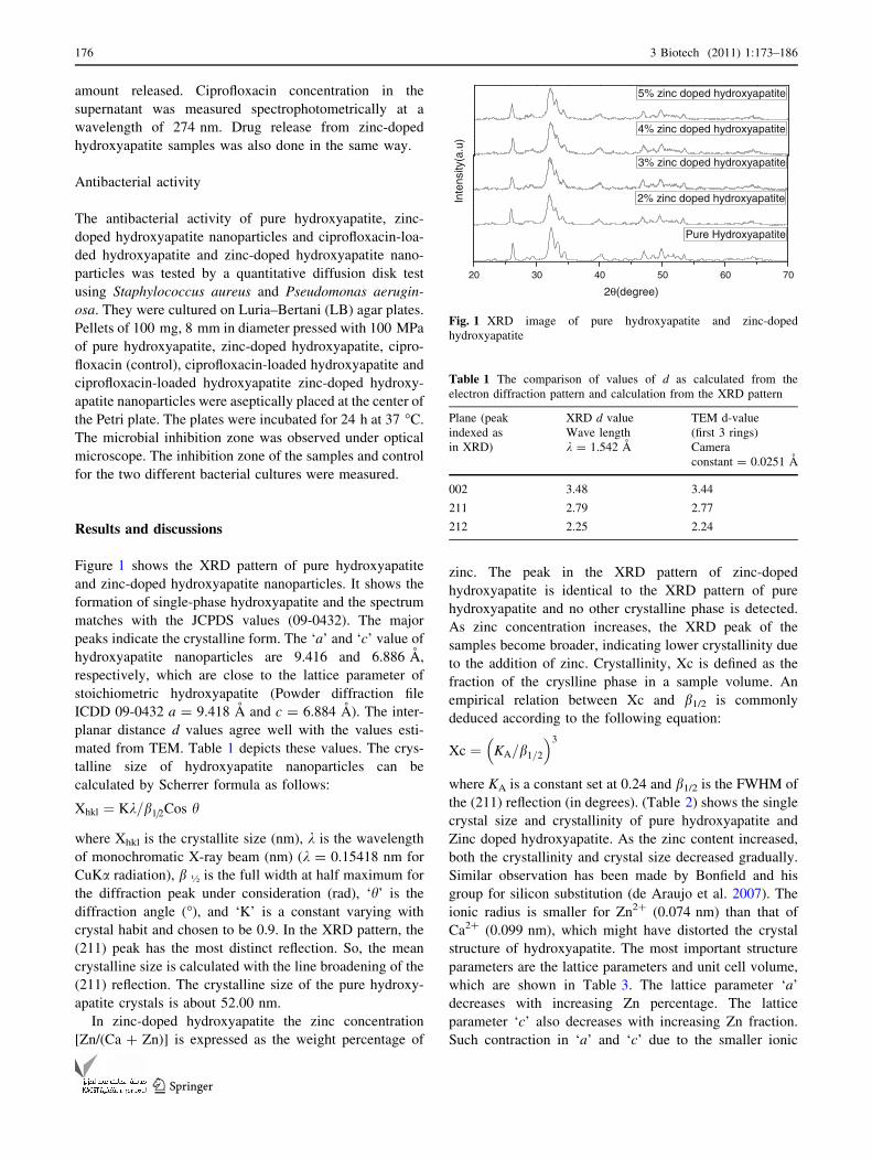
The structure of the ciprofloxacin-loaded hydroxyapa-
tite was analyzed using FTIR spectroscopy As Shown in
(Fig. 4). Characteristic structural bands of both hydroxy-
apatite and ciprofloxacin were observed for all hydroxy-
apatite/drug samples. The hydroxyapatite loaded with
drug represents mixed bands typical of hydroxyapatite
(P–O at 566, 602, 962, 1,091 and O–H at 632, 3,564) and
ciprofloxacin(C=C stretching at 1,608, CH2 bending at
1,468, mixed vibrations at 1,311, CH in plane bending at
1,272, CN stretching at 867). The corresponding band
intensities increase with increase in the drug loading
percentage. The drug-free hydroxyapatite shows little
difference confirming that the drug has no significant
effect on the structure.
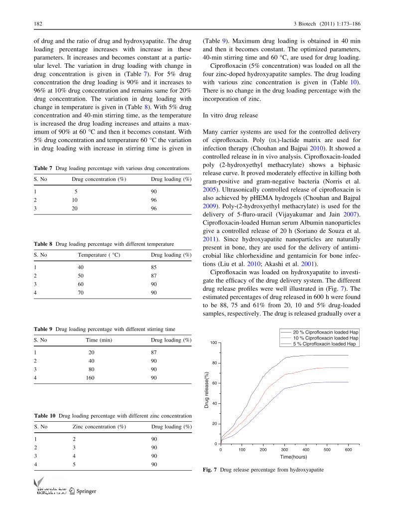
The structure of ciprofloxacin and the optimized
geometry deduced through Gaussian 03 W is given in
(Fig. 5). The Raman spectrum of ciprofloxacin-loaded
hydroxyapatite is shown in (Fig. 6). The observed Raman
frequencies agree quite well with the literature. The most
prominent peaks at 1,379 and 1,622 are associated with the
ring vibrations. The Raman spectrum of hydroxyapatite
loaded with various concentration of drug is given in
(Fig. 6). It is interesting to note that the above peaks are
more pronounced and it is highest for 5% and least for 20%
drug loading. The frequencies of vibrations of PO43- ion
are also shifted to lower wave number region. The ring
stretching vibration at 1,622 cm-1 for drug gets shifted to
lower wave number region on coating of the drug on
hydroxyapatite. It shifts to 1,605 cm-1 for 5% drug con-
centration, 1,597 cm-1 for 10% drug concentration and
1,591 cm-1 for 20% drug concentration. The shift is more
for 20% while considering the structure of the drug there
are three possible sites for molecular adsorption on
hydroxyapatite: (1) the aromatic ring (2) the carbonyl
groups and (3) lone pair electrons of the hydrogen atoms.
According to the charge transfer idea of SERS, the
charge transfer between the adsorbed molecule and the
substrate is controlled by molecular frame vibration. It is
modulated by the vibration of those adsorbed groups that
are linked directly to the surface. A perpendicular orien-
tation of the drug molecule would result through carbonyl
groups. In the present case the carbonyl stretching mode is
not observed or may be very week. This eliminates the
possible adsorption through carbonyl groups. The obser-
vation of enhancement in the intensity of ring vibrations
suggests that there will be charge transfer between the drug
and the hydroxyapatite through p bonds. The observation
of shifts in the frequencies of PO4 vibrations suggests that
Table 6 Chemical composition of hydroxyapatite and zinc-doped hydroxyapatite by XRF
Sample Ca (wt%) Zn (wt%) P (wt%) Zn: (Ca ? Zn)
(wt%)
Ca: P
(wt%)
(Ca ? Zn):
P (wt%)
Ca: (P ? Zn)
(wt%)
Zn: (P ? Zn)
(wt%)
Hydroxyapatite 73.940 0 26.060 0 2.83 2.83 2.83 0
2% Zinc-doped hydroxyapatite 72.230 0.533 27.237 0.00732 2.65 2.67 2.60 0.019
3% Zinc-doped hydroxyapatite 71.919 0.611 27.470 0.00842 2.61 2.64 2.56 0.021
4% Zinc-doped hydroxyapatite 69.229 0.921 29.850 0.01312 2.31 2.35 2.24 0.029
5% Zinc-doped hydroxyapatite 68.370 1.300 30.330 0.01865 2.25 2.29 2.16 0.041
180 3 Biotech (2011) 1:173–186
123
the drug molecules are very nearer to the PO4 groups.
Hence it can be concluded that the drug molecule has an
interaction with hydroxyapatite with a flat on orientation
with their p electron clouds.
Drug loading
Drug loading efficiency of hydroxyapatite was examined as
described previously. It is dependent on the concentration
500 1000 1500 2000 2500 3000 35000
20
40
60
80
100
0
20
40
60
80
100
0
20
40
60
80
100
0
20
40
60
80
100
0
20
40
60
80
100
Tra
nsm
itta
nce
(A
.U)
Wavenumbers (cm-1)
500 1000 1500 2000 2500 3000 3500
Wavenumbers (cm-1)
500 1000 1500 2000 2500 3000 3500
Wavenumbers (cm-1)
500 1000 1500 2000 2500 3000 3500
Wavenumbers (cm-1)
500 1000 1500 2000 2500 3000 3500
Wavenumbers (cm-1)
Hap
3564
1091
473
632
566
602
962
Tra
nsm
itta
nce
(A
.U)
3564
1091
566
602
632
473
1272 13
11
1468
1608
1447
722
769
867
10 % ciprofloxacin loaded Hap
Tra
nsm
itta
nce
(A
.U)
20 % Ciprofloxacin loaded Hap
3564
1091
473
566
602
632 962
1272
1311
1468
1608
1447
722 76
986
7
Tra
nsm
itta
nce
(A
.U)
5 % Ciprofloxacin loaded Hap
3564
1091
962632
566
602
473
1608
1272 13
11
1468
867
722
769
Tra
nsm
itta
nce
(A
.U)
Ciprofloxacin
1311
1272
1468
1608
1447
769
722
869
Fig. 4 FTIR spectrum of ciprofloxacin-loaded hydroxyapatite
Fig. 5 a Structure of ciprofloxacin, b optimized geometry of
ciprofloxacin deduced through Gaussian 03 W
500 1000 1500 2000 2500 3000 3500 4000
Hap
Ciprofloxacin
1622
Ram
an In
tens
ity
5 % Ciprofloxacin loaded Hap
1605
10 % Ciprofloxacin loaded Hap
1597
Raman Shift (cm-1)
20 % Ciprofloxacin loaded Hap1591
Fig. 6 Raman spectrum of ciprofloxacin-loaded hydroxyapatite
3 Biotech (2011) 1:173–186 181
123
of drug and the ratio of drug and hydroxyapatite. The drug
loading percentage increases with increase in these
parameters. It increases and becomes constant at a partic-
ular level. The variation in drug loading with change in
drug concentration is given in (Table 7). For 5% drug
concentration the drug loading is 90% and it increases to
96% at 10% drug concentration and remains same for 20%
drug concentration. The variation in drug loading with
change in temperature is given in (Table 8). With 5% drug
concentration and 40-min stirring time, as the temperature
is increased the drug loading increases and attains a max-
imum of 90% at 60 �C and then it becomes constant. With
5% drug concentration and temperature 60 �C the variation
in drug loading with increase in stirring time is given in
(Table 9). Maximum drug loading is obtained in 40 min
and then it becomes constant. The optimized parameters,
40-min stirring time and 60 �C, are used for drug loading.
Ciprofloxacin (5% concentration) was loaded on all the
four zinc-doped hydroxyapatite samples. The drug loading
with various zinc concentration is given in (Table 10).
There is no change in the drug loading percentage with the
incorporation of zinc.
In vitro drug release
Many carrier systems are used for the controlled delivery
of ciprofloxacin. Poly (DL)-lactide matrix are used for
infection therapy (Chouhan and Bajpai 2010). It showed a
controlled release in in vivo analysis. Ciprofloxacin-loaded
poly (2-hydroxyethyl methacrylate) shows a biphasic
release curve. It proved moderately effective in killing both
gram-positive and gram-negative bacteria (Norris et al.
2005). Ultrasonically controlled release of ciprofloxacin is
also achieved by pHEMA hydrogels (Chouhan and Bajpal
2009). Poly-(2-hydroxyethyl methacrylate) is used for the
delivery of 5-fluro-uracil (Vijayakumar and Jain 2007).
Ciprofloxacin-loaded Human serum Albumin nanoparticles
give a controlled release of 20 h (Soriano de Souza et al.
2011). Since hydroxyapatite nanoparticles are naturally
present in bone, they are used for the delivery of antimi-
crobial like chlorhexidine and gentamicin for bone infec-
tions (Liu et al. 2010; Akashi et al. 2001).
Ciprofloxacin was loaded on hydroxyapatite to investi-
gate the efficacy of the drug delivery system. The different
drug release profiles were well illustrated in (Fig. 7). The
estimated percentages of drug released in 600 h were found
to be 88, 75 and 61% from 20, 10 and 5% drug-loaded
samples, respectively. The drug is released gradually over a
Table 7 Drug loading percentage with various drug concentrations
S. No Drug concentration (%) Drug loading (%)
1 5 90
2 10 96
3 20 96
Table 8 Drug loading percentage with different temperature
S. No Temperature ( �C) Drug loading (%)
1 40 85
2 50 87
3 60 90
4 70 90
Table 9 Drug loading percentage with different stirring time
S. No Time (min) Drug loading (%)
1 20 87
2 40 90
3 80 90
4 160 90
Table 10 Drug loading percentage with different zinc concentration
S. No Zinc concentration (%) Drug loading (%)
1 2 90
2 3 90
3 4 90
4 5 90
0 100 200 300 400 500 6000
20
40
60
80
100
Dru
g re
leas
e(%
)
Time(hours)
20 % Ciprofloxacin loaded Hap10 % Ciprofloxacin loaded Hap5 % Ciprofloxacin loaded Hap
Fig. 7 Drug release percentage from hydroxyapatite
182 3 Biotech (2011) 1:173–186
123

period of time. This shows that the drug is released in a
controlled manner. The amount of drug released in 24 h of
time is well above the minimum inhibition concentration of
CPX which is 0.25–2 lg/ml for pathogens like S. aureus
and P. aeruginosa which cause bone infections. Such a
release profile observed could provide a rapid delivery of
drug to give antibacterial effects at the infected site and a
sustained release to aid long-term healing and avoid the
toxic and adverse systemic effects caused by high con-
centration of antibiotics. The slow release of ciprofloxacin
is due to the presence of trapped carboxylic group, which
reacts with Ca in the hydroxyapatite and zinc-doped
hydroxyapatite (Baker 1987). The plotted graphs revealed
that all the samples showed typical drug release pattern
with a lag time which was similar to the pattern for the
membrane diffusion process of the drug (Matsunaga et al.
2010). The release of ciprofloxacin from hydroxyapatite
nanoparticles exhibits a typical two-stage release mecha-
nism. The drug release is high during the initial time and
then it reduces and becomes constant. Ciprofloxacin release
during the initial burst stage is due to desorption of cip-
rofloxacin molecules that are located on the surface of the
particles. These particles do not strongly interact with the
hydroxyapatite nanoparticles. During the in vitro drug
release analysis the hydroxyapatite absorbs the surrounding
fluid into the nanoparticles. This leads to the dissolution
and exclusion of the loaded ciprofloxacin. When the large
particles break into smaller particles more ciprofloxacin
gets exposed to the fluid. When the loosely adsorbed cip-
rofloxacin had almost completely desorbed, the drug
release becomes slow. The slow release of ciprofloxacin
molecules is due to the incorporation of the drug
hydroxyapatite nanoparticles. This results from the disso-
lution of the hydroxyapatite nanoparticles.
Drug release profile from the zinc-doped hydroxyapatite
is given in (Fig. 8). The percentage of drug released
increases with increase in the zinc concentration. The per-
centages of drugs released in 600 h were estimated to be 85,
88, 92 and 15.60% from 2, 3, 4, 5% zinc-doped samples
respectively. There is a sudden outburst of drug in the initial
few hours and then the drug is released in a controlled
manner. The drug release percentage increases with increase
in the zinc percentage from 2 to 5%. The concentration of
zinc increases in the doped sample which is given by the ICP
analysis. But there is a slightly higher increase in the drug
release from 4 and 5% zinc-doped hydroxyapatite. The
release of zinc from zinc-doped hydroxyapatite along with
ciprofloxacin release may be the reason for the increase in
drug release. The mechanism by which zinc release induces
higher drug release is not known.
The incorporation of zinc into hydroxyapatite and the
mechanism of zinc incorporation have been studied
extensively by various researchers (Tang et al. 2009; Sogo
et al. 2004). The release of zinc from zinc-doped
hydroxyapatite and the increase in osteoblastic cell growth
by zinc-doped hydroxyapatite are well documented (Ito
et al. 2002; Vojislav Stanic et al. 2010). This simultaneous
release of zinc and ciprofloxacin will promote both osteo-
blastic cell growth and antimicrobial activity.
Antibacterial activity
The antibacterial activity of ciprofloxacin-loaded hydrox-
yapatite and zinc-doped hydroxyapatite against different
0 100 200 300 400 500 6000
20
40
60
80
100
Dru
g re
leas
e(%
)
Time (hours)
2% Zinc doped Hap 3% Zinc doped Hap 4% Zinc doped Hap 5% Zinc doped Hap
Fig. 8 Drug release percentage from the zinc-doped hydroxyapatite
Table 11 Zone of inhibition of as-synthesized ciprofloxacin-loaded
hydroxyapatite and ciprofloxacin-loaded hydroxyapatite zinc-doped
hydroxyapatite
S. No Sample Zone of Inhibition (cm)
Staphylococcusaureus
Pseudomonasaeruginosa
1 Ciprofloxacin (control) 2.0 2.4
2 5% Ciprofloxacin-loaded
hydroxyapatite
1.7 2.15
3 10% Ciprofloxacin-loaded
hydroxyapatite
2.0 2.6
4 20% Ciprofloxacin-loaded
hydroxyapatite
3.0 4.1
5 2 wt% Zinc-doped
hydroxyapatite ? 5%
ciprofloxacin
1.8 2.0
6 3 wt% Zinc-doped
hydroxyapatite ? 5%
ciprofloxacin
1.8 2.0
7 4 wt% Zinc-doped
hydroxyapatite ? 5%
ciprofloxacin
1.9 2.1
8 5 wt% Zinc-doped
hydroxyapatite ? 5%
ciprofloxacin
1.9 2.1
3 Biotech (2011) 1:173–186 183
123

micro-organism was monitored. Disk diffusion method was
used to determine the zone of inhibition. The results of
antibacterial activity of ciprofloxacin-loaded hydroxyapa-
tite and zinc-doped hydroxyapatite are given in (Table 11).
The zone of inhibition around the ciprofloxacin (control)
and ciprofloxacin-loaded hydroxyapatite, against Pseudo-
monas aeruginosa is given in Fig. 9a and against Staphy-
lococcus aureus in Fig. 9b, respectively. The zone of
inhibition around the ciprofloxacin-loaded zinc-doped
hydroxyapatite against Pseudomonas aeruginosa is given
in Fig. 10a and against Staphylococcus aureus in
Fig. 10b. The zone of inhibition increases with increase in
the ciprofloxacin concentration. This is relevant with the
apparent amount of drug release. When the amount of
drug release increases the zone of inhibition also increa-
ses. All the ciprofloxacin-loaded zinc-doped hydroxyapa-
tite nanoparticles have the greater antibacterial activity
than ciprofloxacin-loaded pure hydroxyapatite, and the
Fig. 9 a Antimicrobial activity of i Ciprofloxacin (control), ii 5%
ciprofloxacin-loaded hydroxyapatite, iii 10% ciprofloxacin-loaded
hydroxyapatite, iv 20% ciprofloxacin-loaded hydroxyapatite against
Pseudomonas aeruginosa. b Antimicrobial activity of i Ciprofloxacin
(control), ii 5% ciprofloxacin-loaded hydroxyapatite, iii 10%
ciprofloxacin-loaded hydroxyapatite, iv 20% ciprofloxacin-loaded
hydroxyapatite against Staphylococcus aureus
Fig. 10 a Antimicrobial activity of i ciprofloxacin-loaded 2 wt%
zinc-doped hydroxyapatite, ii ciprofloxacin-loaded 3 wt% zinc-doped
hydroxyapatite, iii ciprofloxacin-loaded 4 wt% zinc-doped hydroxy-
apatite, iv ciprofloxacin-loaded 5 wt% zinc-doped hydroxyapatite
against Pseudomonas aeruginosa. b Antimicrobial activity of
i ciprofloxacin-loaded 2 wt% zinc doped hydroxyapatite, ii cipro-
floxacin loaded 3 wt% zinc doped-hydroxyapatite, iii ciprofloxacin
loaded 4 wt% zinc doped hydroxyapatite, iv ciprofloxacin-loaded 5
wt% zinc-doped hydroxyapatite against Staphylococcus aureus
184 3 Biotech (2011) 1:173–186
123

ciprofloxacin-loaded antibacterial activity of zinc-doped
hydroxyapatite gets higher with the increase of zinc con-
centration. Since the crystallinity of hydroxyapatite
decreases with increase in wt% of Zn2?, the surface area of
the nano grains increases thereby forming bonds with the
microorganisms and causes cell death. The proposed
antibacterial mechanism of Zn2? is that the ions in the
crystal surface form strong bonds with thiole, imidazole,
amino and carboxyl groups of microorganism membrane
proteins, causing structural changes. A microorganism
membrane with structural changes exhibits a significant
increase in permeability, leaving the microorganism cells
incapable of properly regulating transport through the
plasma membrane and, finally, causing cell death (Vojislav
Stanic et al. 2010).
Conclusion
A drug delivery system with hydroxyapatite and zinc-
doped hydroxyapatite was developed for use in bone
infections. Both pure hydroxyapatite and zinc-doped
hydroxyapatite can be used as drug delivery system for the
controlled delivery of ciprofloxacin. Ciprofloxacin can be
loaded on hydroxyapatite to enhance wound healing as a
drug delivery system. Ciprofloxacin-loaded zinc-doped
hydroxyapatite can be used for drug delivery and induce
cell growth. The antimicrobial property increases with
increase in the drug concentration and the amount of zinc.
The drug loading depends on the concentration of drug,
temperature and stirring time. The drug loading percentage
increases with increase in drug concentration, temperature
and stirring time and then becomes constant. The drug
release from zinc-doped hydroxyapatite is higher than the
drug release from pure hydroxyapatite. The drug release
profile from both pure and zinc-doped hydroxyapatite
shows a controlled release. The higher release of cipro-
floxacin from zinc-doped hydroxyapatite is due to the
release of zinc from zinc-doped hydroxyapatite. Both zinc
and ciprofloxacin will be released when drug is loaded on
zinc-doped hydroxyapatite. This will induce both cell
growth and antimicrobial activity.
Acknowledgments Prof. S. Ramasamy, CSIR Emeritus Scientist
and Mr. G. Devanand Venkatasubbu, CSIR SRF acknowledge the
financial support given to them to carryout this work under CSIR
Emeritus Scientist Scheme number 21(0714)/08/EMR-II dated 28-04-
2008. The Authors are grateful to Dr. G. S. Avadhani, Principal
Research Scientist, IISc, India, for taking TEM images.
Open Access This article is distributed under the terms of the
Creative Commons Attribution License which permits any use, dis-
tribution and reproduction in any medium, provided the original
author(s) and source are credited.
References
Akashi A, Matsuya Y, Unemori M, Akamine A (2001) Release profile
of antimicrobial agents from a-tricalcium phosphate cement.
Biomaterials 22:1717–2713
Baker R (1987) Controlled release of biologically active agents.
Wiley-Interscience, New York, pp 27–29
Bandyopadhyay A, Withey EA, Susmita Bose J (2007) Influence of
Zno doping in Calcium phosphate ceramics. Mater Sci Eng C
27:14–17
Cheng X, Kuhn L (2007) Chemotherapy drug delivery from calcium
phosphate nanoparticles. Int J Nanomed 2(4):667–674
Chouhan R, Bajpai AK (2010) Release dynamics of ciprofloxacin
from swellable nanocarriers of poly(2-hydroxyethyl methacry-
late): an in vitro study. NanomedicineNBM 6:453–462
Chouhan R, Bajpal AK (2009) An in vitro release study of 5-fluro-
uracil (5-FU) from swellable poly-(2-hydroxyethyl methacry-
late) (PHEMA) nanoparticles. J Mater Sci Mater Med
20:1103–1114
de Araujo TS, Macedo ZS, de Oliveira PASC, Valerio MEG (2007)
Production and characterization of pure and Cr3?-doped
hydroxyapatite for biomedical applications as fluorescent probes.
J Mater Sci 42:2236–2243
De Gaetano F, Ambrosio L, Raucci MG, Marotta A, Catauro M
(2005) Sol-gel processing of drug delivery materials and release
kinetics. J Mater Sci Mater Med 16:261–265
De Jong WH, Borm PJA (2008) Drug delivery and nanoparticles:
applications and hazards. Int J Nanomed 3(2):133–149
Gautier H, Daculsi G, Merle C (2001) Association of vancomycin and
calcium phosphate by dynamic compaction: in vitro character-
ization and micribiological activity. Biomaterials 22:2481–2487
Gibson IR, Best SM, Bonfield W (1999) Chemical characterization of
silicon-substituted hydroxyapatite. J Biomed Mater Res
44:422–428
Ginebra MP, Traykova T, Planell JA (2006) Calcium phosphate
cements as bone drug delivery systems: a review. J Controlled
Release 113:102–110
Harris LG, Geoff Richards R (2006) Staphylococci and implant
surfaces: a review. Injury. Int J Care Injured 37:S3–S14
Ito A et al (2002) Zinc-releasing calcium phosphate for stimulating
bone formation. Mater Sci Eng C 22(1):21–25
Itokazu M, Esaki M, Yamamoto K, Tanemori T, Kasai T (1999)
Local drug delivery system using ceramics:vacuum method for
impregnating a chemotherapeutic agent into a porous hydroxy-
apatite block. J Mater Sci Mater Med 10:249–252
Jallot E, Nedelec J-M, Grimault AS, Chassot E, Grandjean-Laquer-
riere A, Laquerriere P, Laurent-Maquin D (2005) STEM and
EDXS characterisation of physico-chemical reactions at the
periphery of sol-gel derived Zn-substituted hydroxyapatites
during interaction with biological fluids, Colloids Surf Biointer-
faces 42:205–210
Joosten U, Joist A, Gosheger G, Liljenqvist U, Brandt B, von Eiff C
(2005) Effectiveness of hydroxyapatite-vancomucin bone
cement in the treatment of Staphylococcus aureus induced
chronic osteomyelitis. Biomaterials, 26:5251–5258
Kim H-W, Knowles JC, Kim H-E (2004) Hydroxyapatite/poly(e-carprolactone) composite coatings on hydroxyapatite porous
bone scaffold for drug delivery. Biomaterials 25:1279–1287
Koort JK, Makinen TJ, Suokas E, Veiranto M, Jalava J, Tormala P,
Aro HT (2008) Sustained release of ciprofloxacin from an
osteoconductive poly(DL)-lactide implant. Acta Orthop
79(2):295–301
Liu WC, Wong CT, Fong MK, Cheung WS, Kao RY, Luk KD, Lu
WW (2010) Gentamicin-loaded strontium-containing hydroxy-
apatite bioactive bone cement—an efficient bioactive antibiotic
3 Biotech (2011) 1:173–186 185
123

drug delivery system. J Biomed Mater Res B Appl Biomater
9:397–406
Loo SCJ, Siew YE, Ho S, Yin Chiang Boey F (2008) Synthesis and
hydrothermal treatment of nanostructured hydroxyapatite of
controllable sizes. J Mater Sci Mater Med 19:1389–1397
Mason P (2006) Physiological and medicinal zinc. Pharm J
276:271–274
Matsunaga K, Murata H, Mizoguchi T, Nakahira A (2010) Mecha-
nism of incorporation of zinc into hydroxyapatite. Acta Biomater
6(6):2289–2293
Melville AJ, Rodriguez-Lorenzo LM, Forsythe JS (2008) Effects of
calcinations temperature on the drug delivery behavior of
ibuprofen from hydroxyapatite powders. J Mater Sci Mater
Med 19:1187–1195
Murugan R, Panduranga Rao K (2002) Controlled release of
antibiotics from surface modified coralline hydroxyapatite.
Trends Biomat Artif Organs 16(1):43–45
Murugan R, Ramakrishna S (2006) Designing biological apatite
suitable for neomycin delivery. J Mater Sci 41(13):4343–4347
Nandi SK, Mukherjee P, Roy S, Kundu B, Kumar De D, Basu D
(2009) Local antibiotic delivery systems for the treatment of
osteomyelitis-A review. Mater Sci Eng C 29(8):2478–2485
Noel SP, Courtney H, Bumgardner JD, Haggard WO (2008) Chitosan
films a potential local drug delivery system for antibiotics. Clin
Orthopaed Related Res 466:1377–1382
Norris P, Noble M, Francolini I, Vinogradov AM, Stewart PS, Ratner
BD, Costerton JW, Stoodley P (2005) Ultrasonically controlled
release of ciprofloxacin from self-assembled Coatings on poly(2-
hydroxyethyl methacrylate) hydrogels for Pseudomonasaerugin-osa biofilm prevention. Antimicrob Agents Chemother
49:4272–4279
Ochekpe NA, Olorunfemi PO, Ngwuluka NC (2009) Nanotechnology
and drug delivery part 2: nanostructures for drug delivery. Trop J
Pharm Res 8(3):275–287
Panyam J, Labhasetwar V (2003) Biodegradable nanoparticles for
drug and gene delivery to cells and tissues. Adv Drug Deliv Rev
55:329–347
Pataquiva Mateus AY, Ferraz MP, Monteiro FJ (2007) Microspheres
based on hydroxyapatite nanoparticles aggregates for bone
regeneration. Key Eng Mater 330–332, 243–246
Patel SA, Patel NM, Patel MM (2006) Simultaneous spectrophoto-
metric estimation of ciprofloxacin and ornidazole in tablets.
Indian J Pharm Sci 68(5):665–667
Paul W, Sharma CP (1999) Development of porous spherical
hydroxyapatite granules: application towards protein delivery.
J Mater Sci Materiald Med 10:383–388
Pham HH, Luo P, Genin F, Dash AK (2002) Synthesis and
characterization of hydroxyapatite-ciprofloxacin delivery system
by precipitation and spray drying technique. AAPS PharmSci-
Tech 3(1):1–9
Russoni de Lima I, Machado Coasta A, Napoleao Bastos I (2006)
Development and characterization of 5% mol Zn bioceramic in
granular form. Mater Res 9:399–403
Slosarczyk A, Szymura-Oleksiak J, Mycek B (2000) The kinetics of
pentoxifylline from drug loaded hydroxyapatite implants. Bio-
materials 21:1215–1221
Sogo Y, Ito A, Fukasawa K, Sakurai T, Ichinose N (2004) Zinc
containing hydroxyapatite ceramics to promote osteoblastic cell
activity. Mater Sci Technol 20(9):1079–1083
Somberg JC (1989) Drug delivery system. J Clin Pharmacol 2:673
Soriano de Souza CA, Souto APRM, Silva-Boghossian CM,
Granjeiro JM, Alves GG, Rossi AM, Rocha-Leao MHM
(2011) Adsorption of chlorhexidine on synthetic hydroxyapatite
and in vitro biological activity. Colloid Surf B Biointerfaces
87:310–318
Stanic V, Dimitrijevic S, Antic-Stankovic J, Mitric M, Jokic B, Plecas
IB, Raicevic S (2010) Synthesis, characterization and antimi-
crobial activity of copper and zinc-doped hydroxyapatite nano-
powders. Appl Surf Sci 256:6083–6089
Sundar M, Ramesh Babu N, Prem Victor S, Ram Kumar K, Sampath
Kumar TS (2005) Biphasic calcium phosphate for antibiotic
release. Trends Biomat Artif Organs 18(2): 213–218
Szycher M (1986) Controlled drug delivery: a critical review.
J Biomater Appl 1:171–182
Szymura-Oleksiak J, Slosarczyk A, Cios A, Mycek B, Paszkiewicz Z,
Szklarczyk S, Stankiewicz D (2001) The kinetics of pentoxif-
ylline release in vivo from drug-loaded hydroxyapatite implants.
Ceram Int 27:767–772
Tang Y, Chappell HF, Dove MT, Reeder RJ, Lee YJ (2009a) Zinc
incorporation into hydroxyapatite. Biomaterials 30:2864–2872
Tang Y, Chappell HF, Dove MT, Reeder RJ, Lee YJ (2009b) Zinc
incorporation into hydroxyapatite. Biopmaterials 30:2864–2872
Vijayakumar P, Jain NK (2007) Suppression of agglomeration of
ciprofloxacin-loaded human serum albumin nanoparticles. AAPS
PharmSciTech 8:1–6
Zeiler H-J (1985) Evaluation of the in vitro bactericidal action of
ciprofloxacin on cells of escherichia coli in the logarithmic and
stationary phases of growth. Antimicrob Agents Chemother
28(4):524–527
186 3 Biotech (2011) 1:173–186
123