Nanocomposite Film Containing Fibrous Cellulose Scaffold ...
14
polymers Article Nanocomposite Film Containing Fibrous Cellulose Scaffold and Ag/TiO 2 Nanoparticles and Its Antibacterial Activity Yanxiang Li 1,2, *, Jessica Tian 2 , Chuanfang Yang 1 and Benjamin S. Hsiao 2, * 1 Key Laboratory of Green Process and Engineering, Institute of Process Engineering, Chinese Academy of Sciences, Beijing 100190, China; [email protected] 2 Department of Chemistry, Stony Brook University, Stony Brook, NY 11794, USA; [email protected] * Correspondence: [email protected] (Y.L.); [email protected] (B.S.H.); Tel.: +86-10-8254-5011 (Y.L.); +1-631-632-7793 (B.S.H.) Received: 10 August 2018; Accepted: 19 September 2018; Published: 20 September 2018 Abstract: Cellulose is a natural polymer that is widely used in daily life, but it is susceptible to microorganism growth. In this study, a simple sol–gel technique was utilized to incorporate the cellulose scaffold with Ag/TiO 2 nanoparticles. The morphology and crystal structure of the as-prepared Ag/TiO 2 /cellulose composite film were characterized using scanning electron microscopy (SEM) and X-ray diffraction (XRD) methods. Antibacterial tests involving the use of Escherichia coli (E. coli) were carried out under dark and UV-light conditions to evaluate the efficiency of the Ag/TiO 2 /cellulose composite film in comparison with pristine cellulose paper and TiO 2 /cellulose composite film. The results indicated that the antibacterial activity of the Ag/TiO 2 /cellulose composite film outperformed all other samples, where the Ag content of 0.030 wt% could inhibit more than 99% of E. coli. This study suggests that finely dispersed nanocale Ag/TiO 2 particles in the cellulose scaffold were effective at slowing down bacterial growth, and the mechanisms of this are also discussed. Keywords: cellulose; Ag/TiO 2 sol–gel; antibacterial activity; synergetic effect 1. Introduction Cellulose is the most abundant biopolymer on Earth, with over 150 billion tons of biomass produced every year [1]. From a structural perspective, cellulose is a carbohydrate polymer generated from the repeating unit of β-D-glucopyranose molecules that are covalently linked through β-1,4-glucan [2]. Cellulose has a large number of hydroxyl groups (three per anhydroglucose (AGU) unit) on the repeating unit, which leads to extensive hydrogen bond networks that make it insoluble in common solvents. The unique structure and abundance of cellulose makes them a source of material with fascinating properties, including hydrophilicity, renewability, biodegradability, and biocompatibility. As a result, cellulose products are widely used in our daily life, such as basic cloths, foods, papers, pharmaceutics, and healthcare. Recently, they have also been considered in many advanced environmental and energy applications, such as for water treatment [3–5], solar cells [6–8], and supercapacitors [9–11], just to name a few. Because of their unique chemical structure and properties, cellulose products are also ideal scaffolds for the growth of micro-organisms [12,13]. The reason why cellulose is more sensitive to bacterial colonization is because it is typically porous, hydrophilic, able to retain substantial water content, and also able to easily diffuse oxygen and nutrients throughout the scaffold, thus providing a perfect environment for bacterial growth. For practical applications, modification of cellulose with antibacterial properties is often necessary. Polymers 2018, 10, 1052; doi:10.3390/polym10101052 www.mdpi.com/journal/polymers
Transcript of Nanocomposite Film Containing Fibrous Cellulose Scaffold ...
polymers
Article
Nanocomposite Film Containing Fibrous CelluloseScaffold and Ag/TiO2 Nanoparticles and ItsAntibacterial Activity
Yanxiang Li 1,2,*, Jessica Tian 2, Chuanfang Yang 1 and Benjamin S. Hsiao 2,*1 Key Laboratory of Green Process and Engineering, Institute of Process Engineering,
Chinese Academy of Sciences, Beijing 100190, China; [email protected] Department of Chemistry, Stony Brook University, Stony Brook, NY 11794, USA; [email protected]* Correspondence: [email protected] (Y.L.); [email protected] (B.S.H.);
Tel.: +86-10-8254-5011 (Y.L.); +1-631-632-7793 (B.S.H.)
Received: 10 August 2018; Accepted: 19 September 2018; Published: 20 September 2018�����������������
Abstract: Cellulose is a natural polymer that is widely used in daily life, but it is susceptibleto microorganism growth. In this study, a simple sol–gel technique was utilized to incorporatethe cellulose scaffold with Ag/TiO2 nanoparticles. The morphology and crystal structure ofthe as-prepared Ag/TiO2/cellulose composite film were characterized using scanning electronmicroscopy (SEM) and X-ray diffraction (XRD) methods. Antibacterial tests involving the useof Escherichia coli (E. coli) were carried out under dark and UV-light conditions to evaluate theefficiency of the Ag/TiO2/cellulose composite film in comparison with pristine cellulose paperand TiO2/cellulose composite film. The results indicated that the antibacterial activity of theAg/TiO2/cellulose composite film outperformed all other samples, where the Ag content of 0.030 wt%could inhibit more than 99% of E. coli. This study suggests that finely dispersed nanocale Ag/TiO2
particles in the cellulose scaffold were effective at slowing down bacterial growth, and the mechanismsof this are also discussed.
Keywords: cellulose; Ag/TiO2 sol–gel; antibacterial activity; synergetic effect
1. Introduction
Cellulose is the most abundant biopolymer on Earth, with over 150 billion tons of biomassproduced every year [1]. From a structural perspective, cellulose is a carbohydrate polymergenerated from the repeating unit of β-D-glucopyranose molecules that are covalently linked throughβ-1,4-glucan [2]. Cellulose has a large number of hydroxyl groups (three per anhydroglucose(AGU) unit) on the repeating unit, which leads to extensive hydrogen bond networks that makeit insoluble in common solvents. The unique structure and abundance of cellulose makes them asource of material with fascinating properties, including hydrophilicity, renewability, biodegradability,and biocompatibility. As a result, cellulose products are widely used in our daily life, such as basiccloths, foods, papers, pharmaceutics, and healthcare. Recently, they have also been considered in manyadvanced environmental and energy applications, such as for water treatment [3–5], solar cells [6–8],and supercapacitors [9–11], just to name a few.
Because of their unique chemical structure and properties, cellulose products are also idealscaffolds for the growth of micro-organisms [12,13]. The reason why cellulose is more sensitive tobacterial colonization is because it is typically porous, hydrophilic, able to retain substantial watercontent, and also able to easily diffuse oxygen and nutrients throughout the scaffold, thus providing aperfect environment for bacterial growth. For practical applications, modification of cellulose withantibacterial properties is often necessary.
Polymers 2018, 10, 1052; doi:10.3390/polym10101052 www.mdpi.com/journal/polymers
Polymers 2018, 10, 1052 2 of 14
Silver ions (Ag+) and silver nanoparticles (Ag NPs) are well-known components for providingantibacterial activity [14–16], and many studies have reported the excellent antibacterial propertiesof polymer composites containing Ag NPs [14–19]. However, their relatively high manufacturingcosts often limit their practical application. Titanium dioxide (TiO2) has also been demonstrated tohave excellent antibacterial and photocatalytic properties under UV irradiation. This system hasbeen extensively investigated due to their low-cost, non-toxic, and stable chemical and physicalproperties [20–23]. In brief, under UV irradiation, TiO2 can exhibit biocidal properties resultingfrom the generation of reactive oxygen species (ROS) [24]. The antibacterial activity of TiO2 thusdepends on the rate of ROS formation with respect to the rate of recombination to the photo-inducedelectron-hole (H+/e−). Generally, the high recombination rate of photo-induced electron-holes and thewide band-gap energy can significantly limit the antibacterial and photocatalytic performance of TiO2.
The incorporation of Ag NPs was found to be able to narrow the band-gap energy of TiO2 andcreate some plasmonic processes at the surface of TiO2, thus enhancing its antibacterial activity [25–29].Typically, there are three routes to fabricating Ag/TiO2 composites: the hydrothermal [30–34],photoreduction [23,35,36], and sol–gel [26,27,37] methods. The hydrothermal method can avoidthe agglomeration of nanoparticles, but the required use of high temperature and high pressureto initiate the reaction often limits its value for industrial application. The photoreduction methodinvolves the use of UV irradiation to deposit Ag NPs onto the TiO2 scaffold (e.g., nanosponges) in silvernitrate (AgNO3) solutions. However, the large-scale usage of UV irradiation on an industrial levelcan result in air pollution problems. In a way, the sol–gel synthesis represents a simple and efficientapproach to fabricate nanoscale Ag/TiO2 composites as the method has been extensively demonstratedto produce uniform crystalline metal oxide thin films on the various substrates. For example, severalstudies dealing with the fabrication of TiO2 thin films on the cellulose substrates have been reportedusing the sol–gel method [20,38–41]. However, none has been reported to produce Ag/TiO2 compositenanoparticles directly from the cellulose scaffold, which was the purpose of this study.
In this work, we demonstrate a cost-efficient and environmentally-friendly method at roomtemperature to first prepare an Ag/TiO2 sol, followed by immobilization onto a cellulose scaffold (filterpaper). The approach combines the sol–gel and dip-coating processes to fabricate Ag/TiO2/cellulosecomposite films (or papers). This approach has many advantages, including how: (1) it is a simple,green, and easily scalable process; (2) the process provides reduced manufacturing costs comparedto the use of silver individually; (3) silver nanoparticles can be dispersed uniformly in the systemwithout agglomeration; and (4) the system offers a synergetic effect by combining both silver and TiO2
antibacterial activities.
2. Materials and Methods
2.1. Materials
Titanium (IV)-n-butoxide (97%), nitric acid (70%), and ascorbic acid were purchased from SigmaAldrich (St. Louis, MO, USA). Silver nitrate (AgNO3) was purchased from the Fisher ScientificCompany (Hampton, NH, USA). Cellulose filter paper (Grade No 131, 100% alpha cotton) waspurchased from Advantec (Taibei, Taiwan). All chemicals and cellulose substrate were used withoutfurther purification or pre-treatment.
2.2. Preparation of TiO2 Sol
In the typical procedure, 6 mL of Titanium (IV)-n-butoxide was first dissolved in 2 mL of isopropylalcohol, and the solution was subsequently added drop-wise to a solution containing 2.0 mL of nitricacid and 200 mL of distilled H2O and vigorously stirred at room temperature. White precipitate wasformed immediately upon each additional drop. The final mixture was vigorously stirred for 48 h,followed by aging for 48 h at room temperature to produce the TiO2 sol. The colloidal suspension ofthe TiO2 sol was a white-blue color, semi-transparent, but quite stable for over one month.

Polymers 2018, 10, 1052 3 of 14
2.3. Preparation of Ag/TiO2 Sol
For every 10.0 mL of TiO2 sol, 31, 156, and 780 µL of 0.1 M AgNO3 solutions were used toproduce theoretical 0.2%, 1.0%, and 5.0% Ag to TiO2 molar ratios of sols, respectively. The AgNO3
solution was also added drop-wise to the TiO2 sol in a dark container, while being vigorously stirredat room temperature. The suspension was stirred for 30 min before adding excess ascorbic acid (0.1 M)drop-wise, where the resulting sol was continuously stirred for another 30 min. The final Ag/TiO2 solwas orange-brown and semi-transparent.
2.4. Ag/TiO2/Cellulose Composite Film Fabrication
The cellulose substrate (commercial filter paper) was first submersed in the Ag/TiO2 sol for 30 s.The sol-saturated paper was then placed in a preheated oven at 65 ◦C for 5 min to remove somesolvents, and then cured at 95 ◦C for 5 min to form TiO2 particles. After that, the impregnated paperwas treated in boiling water for 2 h. During this process, the crystalline TiO2 particles became moreperfect, and the unattached TiO2 particles were removed. Finally, the recovered Ag/TiO2/cellulosecomposite film was dried at 40 ◦C.
2.5. Scanning Electron Microscope (SEM)
The surface morphology of the Ag/TiO2/cellulose composite film was analyzed by a scanningelectron microscope (SEM, FEG-SEM LEO 1550, Carl Zeiss, Germany) equipped with a Robinsonbackscattered electron detector and 10 eV Schottky field-emission gun. The instrument also containedan energy-dispersive spectroscopy (EDS) spectrometer (detector from EDAX and controller fromIridium Ultra software (iXRF)) to characterize the chemical composition.
2.6. Thermal Gravitational Analysis (TGA)
Thermal gravimetric analysis (TGA) was carried out on a TGA Q50 machine (TA, New Castle,DE, USA). The samples were run at a heating rate of 10 ◦C/min in the range of 20–700 ◦C under anair atmosphere.
2.7. X-ray Diffraction (XRD)
The X-ray diffraction (XRD) patterns were obtained using a D8 X-ray diffractometer (Bruker,Karlsruhe, Germany) with CuKα radiation. The chosen wavelength (λ) was 0.154 nm, which wasgenerated by CuKα radiation at 40 kV and 40 mA using a Ni filter. Data collection was carried outusing a flat holder in the Bragg-Brentano geometry (10◦–60◦, 5◦min−1).
2.8. Zeta-Potential
The zeta-potential of the TiO2 sol was measured in triplicate with a Zetaprobe AnalyzerTM
instrument (Colloidal Dynamics, St. Johns, FL, USA). This instrument consisted of a built-in titrationset-up equipped with a pH electrode and ESA sensor probe. Before analyzing the sample, the pHelectrode was calibrated using three different pH buffer standards (pH = 4.01, 7.01, and 10.01), followedby a standard titration solution. The ESA sensor was calibrated using the standard zeta probe polarsolution (KSiW solution). Upon completion of the calibration test, the TiO2 sol was filled in the sampleholder, where the ESA sensor was then introduced into the sample under magnetic stirring to analyzethe zeta potential.
2.9. Transmission Electron Microscopy (TEM)
A piece of TiO2/cellulose film was vigorously stirred in water and subsequently sonicated topeel off the TiO2 NPs. The suspension was deposited on a carbon-coated copper grid and dried inair. The specimens were observed using a JEM 2100F transmission electron microscope (TEM, JEOL,Japan), operated at 200 kV.
Polymers 2018, 10, 1052 4 of 14
2.10. Preparation of PBS
To prepare the E. coli sample for the antibacterial test, phosphate-buffered saline (PBS) buffersolution was prepared using the following procedure: NaCl (8.01 g), KCl (0.20 g), Na2HPO4 (1.14 g),and KH2PO4 (0.27 g) were mixed with 500 mL of distilled water in a beaker. The solution was thentransferred into a 1.0 L volumetric flask, where more distilled water was added until the solutionbecame 1.0 L. Finally, the PBS solution was stored in a large 1.0 L Pyrex jar and kept in a refrigerator.
2.11. Preparation of E. coli
E. coli was cultured in fresh lysogeny broth (LB), a nutritionally rich medium, overnight. The cellswere centrifuged at 10,000 rpm for 4 min. The supernatant was decanted, and the cell pellet wasre-suspended with PBS. The resulting cells were centrifuged and the supernatant was decanted again.This process was repeated one more time using PBS to separate the cells from the nutrient broth toprevent further cell growth. Once the supernatant was decanted for the third time, the cells, suspendedin PBS, were transferred to a larger container, and PBS was added until the volume of the suspensionwas 360 mL.
2.12. Preparation of LB/Agar Plates
Peptone (5.0 g), yeast extract (2.5 g), NaCl (5.0 g), and agar (7.5 g) were first mixed with 250 mL ofdistilled water to form a homogeneous solution, where the final solution was diluted to a total volumeof 500 mL. This solution was then transferred to a Pyrex jar, which was autoclaved at 121 ◦C in a liquidloading cycle. The final agar was cooled to approximately 55 ◦C before use.
To prepare the LB/agar plate, a layer of LB agar (~15 mL) was poured into a sterile petri dish.The plate was swirled in a circular motion to distribute the agar uniformly on the bottom of the dish.Each plate was cooled to room temperature, solidified (~20 min), and flipped to avoid condensationon the agar. To store the plates, parafilm was used to wrap around the edge between the plate andthe cover.
2.13. Antibacterial Test
In this test, two environmental conditions were evaluated: dark and ultra-violet (UV) light.The measurements were conducted in triplicate, using the procedure as follows: A 2.0 cm2 film couponwas soaked in 8.0 mL of bacterial solution for 2 h in a petri dish. Under the dark condition, the petridish was put in a small incubator, of which window was covered with aluminum foil. Under theUV-light condition, the petri dish was placed in a biosafety cabinet equipped with the UV setting atroom temperature. After each treatment, 1.0 mL of the tested bacterial solution was taken out, and five10-fold dilutions were carried out. 200 µL of the highest dilution was spread on an agar plate with acell spreader. The agar plates were incubated at 37 ◦C for 24 h in the dark, where the colony-formingunits (CFU) were counted afterward.
3. Results and Discussion
3.1. Preparation of Ag/TiO2/Cellulose Composite Films
A simple and green method based on colloid chemistry was demonstrated to prepareAg/TiO2/cellulose composite films at room temperature. In this method, ascorbic acid (i.e., vitaminC) was chosen as the reducing agent due to its non-toxic and mild reducing ability (in contrast withthe commonly used and more toxic NaBH4), which resulted in a homogeneous dispersion of Ag NPsamong the continuous TiO2 thin film deposited on the surface of cellulose paper. With this method,the zeta potential of the TiO2 colloidal suspension was found to be +36.9 mV. Such a large charge valueminimized the tendency of particle agglomeration due to electrostatic repulsion, thus promoting thestability of the TiO2 sol. The size distribution of the TiO2 hydrosol was determined by dynamic light

Polymers 2018, 10, 1052 5 of 14
scattering (DLS, Malvern Panalytical Ltd., Malvern, UK), where the average size was about 21 nm.As for the Ag/TiO2 sol with different Ag added, the zeta potentials were found to be +29.8, +28.6,and +26.4 mV, respectively. These results indicate that the addition of Ag reduced the stability of theTiO2 sol; however, the mixture was still relatively stable at least for 5 h at room temperature. It wasseen that DLS of the Ag/TiO2 sol exhibited a broader distribution with the increase of Ag concentration(Figure 1a). With the highest Ag concentration (5%), the Ag/TiO2 sol showed a bimodal distributiondue to the large Ag NPs aggregation (Figure 1b).
Polymers 2018, 10, x FOR PEER REVIEW 5 of 14
promoting the stability of the TiO2 sol. The size distribution of the TiO2 hydrosol was determined by dynamic light scattering (DLS, Malvern Panalytical Ltd, Malvern, UK), where the average size was about 21 nm. As for the Ag/TiO2 sol with different Ag added, the zeta potentials were found to be +29.8, +28.6, and +26.4 mV, respectively. These results indicate that the addition of Ag reduced the stability of the TiO2 sol; however, the mixture was still relatively stable at least for 5 h at room temperature. It was seen that DLS of the Ag/TiO2 sol exhibited a broader distribution with the increase of Ag concentration (Figure 1a). With the highest Ag concentration (5%), the Ag/TiO2 sol
showed a bimodal distribution due to the large Ag NPs aggregation (Figure 1b).
Figure 1. Dynamic light scattering (DLS) spectra of the TiO2 sol and TiO2/Ag sols with different Ag content.
3.2. Structure and Morphology Characterization of Ag/TiO2/cellulose Composite Films
Figure 1. Dynamic light scattering (DLS) spectra of the TiO2 sol and TiO2/Ag sols with differentAg content.

Polymers 2018, 10, 1052 6 of 14
3.2. Structure and Morphology Characterization of Ag/TiO2/cellulose Composite Films
Figure 2 shows the SEM images of cellulose paper (substrate), TiO2/cellulose composite film(without Ag), and EDS elemental analysis of the composite film, respectively. Compared to the basecellulose paper (Figure 2a,b), the TiO2/cellulose composite shows an evenly distributed and continuousTiO2 thin film layer (Figure 2c,d), while preserving the original microfiber structures of the filter paper.The higher magnification images show that most of the hierarchical fibers on the surface of the cellulosepaper are uniformly coated with the TiO2 layer (Figure 2d). The EDS analysis (Figure 2e) confirmedthe presence of the Ti element, with major peaks occurring at 4.51, 4.93, and 0.45 eV, indicating theformation of TiO2. Due to the formation of a continuous, dense film layer, it was difficult to observeindividual TiO2 nanoparticles. To reveal the structure of TiO2 NPs, a piece of TiO2/cellulose film wasvigorously stirred in water to peel off the TiO2 layer, which was subsequently sonicated. The TEMimage of the resulting TiO2 sample is shown in Figure 2f, which indicated that the size of the TiO2 NPswas in the range of 3–5 nm. High-resolution TEM imaging of this sample was also carried out, wherethe lattice space of 0.35 nm indicated that the TiO2 NPs had the anatase phase (the inset of Figure 2f).
Polymers 2018, 10, x FOR PEER REVIEW 6 of 14
Figure 2 shows the SEM images of cellulose paper (substrate), TiO2/cellulose composite film (without Ag), and EDS elemental analysis of the composite film, respectively. Compared to the base cellulose paper (Figure 2a,b), the TiO2/cellulose composite shows an evenly distributed and continuous TiO2 thin film layer (Figure 2c,d), while preserving the original microfiber structures of the filter paper. The higher magnification images show that most of the hierarchical fibers on the surface of the cellulose paper are uniformly coated with the TiO2 layer (Figure 2d). The EDS analysis (Figure 2e) confirmed the presence of the Ti element, with major peaks occurring at 4.51, 4.93, and 0.45 eV, indicating the formation of TiO2. Due to the formation of a continuous, dense film layer, it was difficult to observe individual TiO2 nanoparticles. To reveal the structure of TiO2 NPs, a piece of TiO2/cellulose film was vigorously stirred in water to peel off the TiO2 layer, which was subsequently sonicated. The TEM image of the resulting TiO2 sample is shown in Figure 2f, which indicated that the size of the TiO2 NPs was in the range of 3–5 nm. High-resolution TEM imaging of this sample was also carried out, where the lattice space of 0.35 nm indicated that the TiO2 NPs had the anatase phase (the inset of Figure 2f).
Figure 2. Scanning electron microscope (SEM) images of cellulose paper (substrate) at lower magnification (a) and higher magnification (b); the TiO2/cellulose composite film at lower magnification (c) and higher magnification (d); energy-dispersive spectroscopy (EDS) elemental analysis of TiO2/cellulose composite film (e); transmission electron microscope (TEM) of TiO2 particles peeled off from TiO2/cellulose film by vigorous stirring, followed by sonication (f).
Figure 2. Scanning electron microscope (SEM) images of cellulose paper (substrate) at lowermagnification (a) and higher magnification (b); the TiO2/cellulose composite film at lowermagnification (c) and higher magnification (d); energy-dispersive spectroscopy (EDS) elementalanalysis of TiO2/cellulose composite film (e); transmission electron microscope (TEM) of TiO2 particlespeeled off from TiO2/cellulose film by vigorous stirring, followed by sonication (f).

Polymers 2018, 10, 1052 7 of 14
The TGA analysis was conducted to determine the TiO2 content in the TiO2/cellulose compositefilm. The results are illustrated in Figure 3. It was found that the TiO2/cellulose film underwent threeweight-loss stages. The first stage appeared below 100 ◦C, which could be attributed to the loss ofphysically adsorbed water. The second stage appeared in the range of 250–350 ◦C, which could beattributed to the carbonization process by dehydration, depolymerization, and decomposition of thecellulose substrate. The third stage appeared between 350 and 700 ◦C, which could be attributed to thecomplete decomposition of the cellulose substrate, where the residue was the inorganic componentof TiO2. Based on this technique, the content of TiO2 determined by TGA was found to be 1.74 wt %(Figure 3).
Polymers 2018, 10, x FOR PEER REVIEW 7 of 14
The TGA analysis was conducted to determine the TiO2 content in the TiO2/cellulose composite film. The results are illustrated in Figure 3. It was found that the TiO2/cellulose film underwent three weight-loss stages. The first stage appeared below 100 °C, which could be attributed to the loss of physically adsorbed water. The second stage appeared in the range of 250–350 °C, which could be attributed to the carbonization process by dehydration, depolymerization, and decomposition of the cellulose substrate. The third stage appeared between 350 and 700 °C, which could be attributed to the complete decomposition of the cellulose substrate, where the residue was the inorganic component of TiO2. Based on this technique, the content of TiO2 determined by TGA was found to be 1.74 wt % (Figure 3).
For the Ag/TiO2/cellulose composite films, the Ag content could not be determined from the TGA technique because both Ag and TiO2 would remain after cellulose decomposition. To overcome this problem, the Ag+ concentration was determined by the inductively coupled plasma-optical emission spectrometry (ICP-OES, Thermo Icap 6300, Thermo Scientific, Waltham, MA, USA) method through acid digestion of the Ag/TiO2/cellulose composite films. As a result, the Ag weight percentage in the Ag/TiO2/cellulose films were calculated to be 0.003, 0.009, and 0.030 wt %, corresponding to the theoretical addition of 0.2%, 1.0%, and 5% molar ratio of Ag to TiO2.
Figure 3. Thermal gravimetric analysis (TGA) of cellulose paper and TiO2/cellulose composite film.
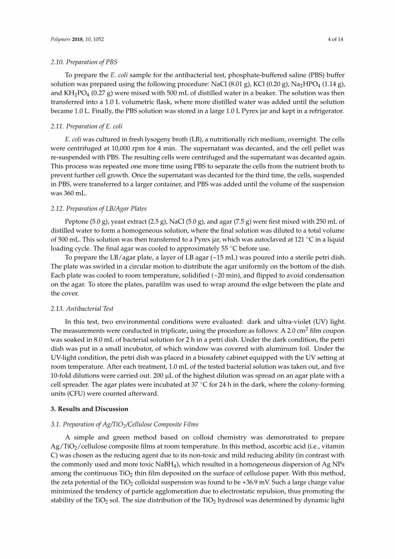
Figure 4 shows the surface morphology of the varying Ag/TiO2/cellulose composite films. It was found that Ag NPs were dispersed uniformly in the TiO2 layer without aggregation. Interestingly, Ag NPs exhibited the form of nanowire (the diameter was between 30–50 nm), which were clearly seen in Figure 4b,d at the lower ratios of Ag (i.e., 0.2 and 1.0 mol %). At the higher ratio of Ag (5.0 mol %), the diameter of Ag NPs was found to increase quite substantially, and were in the range of 100–200 nm. Perhaps this indicates that at low Ag concentration, the NP was dominated by the 1D crystal growth, leading to a nanowire morphology; where at higher Ag concentration (e.g., 5 mol %), the NP possessed the 3D crystal growth, leading to the greatly increased diameter. It has been noted that the ascorbic acid could act not only as a reducing agent, but also as a stabilizing agent in this sol–gel process [42].
Figure 3. Thermal gravimetric analysis (TGA) of cellulose paper and TiO2/cellulose composite film.
For the Ag/TiO2/cellulose composite films, the Ag content could not be determined from the TGAtechnique because both Ag and TiO2 would remain after cellulose decomposition. To overcome thisproblem, the Ag+ concentration was determined by the inductively coupled plasma-optical emissionspectrometry (ICP-OES, Thermo Icap 6300, Thermo Scientific, Waltham, MA, USA) method throughacid digestion of the Ag/TiO2/cellulose composite films. As a result, the Ag weight percentage inthe Ag/TiO2/cellulose films were calculated to be 0.003, 0.009, and 0.030 wt %, corresponding to thetheoretical addition of 0.2%, 1.0%, and 5% molar ratio of Ag to TiO2.
Figure 4 shows the surface morphology of the varying Ag/TiO2/cellulose composite films. It wasfound that Ag NPs were dispersed uniformly in the TiO2 layer without aggregation. Interestingly, AgNPs exhibited the form of nanowire (the diameter was between 30–50 nm), which were clearly seen inFigure 4b,d at the lower ratios of Ag (i.e., 0.2 and 1.0 mol %). At the higher ratio of Ag (5.0 mol %),the diameter of Ag NPs was found to increase quite substantially, and were in the range of 100–200 nm.Perhaps this indicates that at low Ag concentration, the NP was dominated by the 1D crystal growth,leading to a nanowire morphology; where at higher Ag concentration (e.g., 5 mol %), the NP possessedthe 3D crystal growth, leading to the greatly increased diameter. It has been noted that the ascorbicacid could act not only as a reducing agent, but also as a stabilizing agent in this sol–gel process [42].
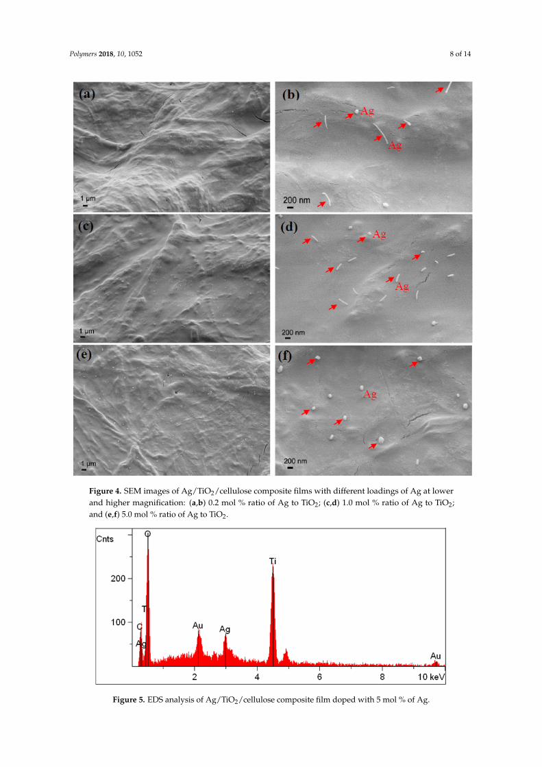
The EDS spectra did not reveal any Ag signals in the Ag/TiO2/cellulose composite films at thelower molar ratios of Ag (0.2–1.0 mol %) because the Ag content was very low. However, for the samplewith 5 mol % of Ag/TiO2, the Ag signal occurring around 3.0 keV was clearly observed, as shown inFigure 5.
Polymers 2018, 10, 1052 8 of 14Polymers 2018, 10, x FOR PEER REVIEW 8 of 14
Figure 4. SEM images of Ag/TiO2/cellulose composite films with different loadings of Ag at lower and higher magnification: (a,b) 0.2 mol % ratio of Ag to TiO2; (c,d) 1.0 mol % ratio of Ag to TiO2; and (e,f) 5.0 mol % ratio of Ag to TiO2.
The EDS spectra did not reveal any Ag signals in the Ag/TiO2/cellulose composite films at the lower molar ratios of Ag (0.2–1.0 mol %) because the Ag content was very low. However, for the sample with 5 mol % of Ag/TiO2, the Ag signal occurring around 3.0 keV was clearly observed, as shown in Figure 5.
Figure 4. SEM images of Ag/TiO2/cellulose composite films with different loadings of Ag at lowerand higher magnification: (a,b) 0.2 mol % ratio of Ag to TiO2; (c,d) 1.0 mol % ratio of Ag to TiO2;and (e,f) 5.0 mol % ratio of Ag to TiO2.Polymers 2018, 10, x FOR PEER REVIEW 9 of 14
Figure 5. EDS analysis of Ag/TiO2/cellulose composite film doped with 5 mol % of Ag.
The XRD patterns of cellulose, TiO2/cellulose, and Ag/TiO2/cellulose composite films and TiO2 powder are shown in Figure 6. In Figure 6a, it can be seen that all three diffraction profiles are similar, dominated by the cellulose diffraction peaks. This is reasonable because of the low content of TiO2 and Ag in the composite films. To determine the crystal structure of TiO2 NPs, Ag/TiO2 powder was peeled off from the cellulose substrate by vigorously stirring the Ag/TiO2/cellulose film in water. In Figure 6b, it can be seen that the peeled Ag/TiO2 powder shows a distinct crystalline phase of TiO2 with no signs of Ag crystals due to its very small doping amount. The diffraction peaks for the TiO2 powder located at 2θ = 25.2°, 37.8°, 48.1°, and 54.2° could be indexed by the (101), (004), (200), and (105) diffraction peaks of the TiO2 anatase phase, respectively, which are consistent with the TEM results. We note that anatase is generally recognized to be the most active among the common crystal phases of TiO2 [43–45].
Figure 6. X-ray diffraction (XRD) pattern for (a) cellulose paper, TiO2/cellulose, and Ag/TiO2/cellulose composite film; (b) the Ag/TiO2 powder peeled off the composite showing the distinct crystalline phase of TiO2.
3.3 Antibacterial Activities
The antibacterial activities of TiO2/cellulose and Ag/TiO2/cellulose composite films were evaluated against E. coli under dark and UV conditions. For comparison, pristine cellulose filter paper was also tested under the same conditions, and the results are shown in Figure 7. It can be seen from Figure 7b1,b2 that the number of CFU increased by approximately 20% in the dark condition, when compared to the control group without the addition of cellulose. This indicates that the cellulose substrate is prone to bacteria growth. TiO2/cellulose composite films were found to have little antibacterial effects under either of the dark or UV conditions (Figure 7c1,c2). It was seen that Ag/TiO2/cellulose composites containing 5 mol % Ag/TiO2 displayed significant antibacterial
Figure 5. EDS analysis of Ag/TiO2/cellulose composite film doped with 5 mol % of Ag.

Polymers 2018, 10, 1052 9 of 14
The XRD patterns of cellulose, TiO2/cellulose, and Ag/TiO2/cellulose composite films and TiO2
powder are shown in Figure 6. In Figure 6a, it can be seen that all three diffraction profiles are similar,dominated by the cellulose diffraction peaks. This is reasonable because of the low content of TiO2
and Ag in the composite films. To determine the crystal structure of TiO2 NPs, Ag/TiO2 powder waspeeled off from the cellulose substrate by vigorously stirring the Ag/TiO2/cellulose film in water.In Figure 6b, it can be seen that the peeled Ag/TiO2 powder shows a distinct crystalline phase ofTiO2 with no signs of Ag crystals due to its very small doping amount. The diffraction peaks for theTiO2 powder located at 2θ = 25.2◦, 37.8◦, 48.1◦, and 54.2◦ could be indexed by the (101), (004), (200),and (105) diffraction peaks of the TiO2 anatase phase, respectively, which are consistent with the TEMresults. We note that anatase is generally recognized to be the most active among the common crystalphases of TiO2 [43–45].
Polymers 2018, 10, x FOR PEER REVIEW 9 of 14
Figure 5. EDS analysis of Ag/TiO2/cellulose composite film doped with 5 mol % of Ag.
The XRD patterns of cellulose, TiO2/cellulose, and Ag/TiO2/cellulose composite films and TiO2 powder are shown in Figure 6. In Figure 6a, it can be seen that all three diffraction profiles are similar, dominated by the cellulose diffraction peaks. This is reasonable because of the low content of TiO2 and Ag in the composite films. To determine the crystal structure of TiO2 NPs, Ag/TiO2 powder was peeled off from the cellulose substrate by vigorously stirring the Ag/TiO2/cellulose film in water. In Figure 6b, it can be seen that the peeled Ag/TiO2 powder shows a distinct crystalline phase of TiO2 with no signs of Ag crystals due to its very small doping amount. The diffraction peaks for the TiO2 powder located at 2θ = 25.2°, 37.8°, 48.1°, and 54.2° could be indexed by the (101), (004), (200), and (105) diffraction peaks of the TiO2 anatase phase, respectively, which are consistent with the TEM results. We note that anatase is generally recognized to be the most active among the common crystal phases of TiO2 [43–45].
Figure 6. X-ray diffraction (XRD) pattern for (a) cellulose paper, TiO2/cellulose, and Ag/TiO2/cellulose composite film; (b) the Ag/TiO2 powder peeled off the composite showing the distinct crystalline phase of TiO2.
3.3 Antibacterial Activities
The antibacterial activities of TiO2/cellulose and Ag/TiO2/cellulose composite films were evaluated against E. coli under dark and UV conditions. For comparison, pristine cellulose filter paper was also tested under the same conditions, and the results are shown in Figure 7. It can be seen from Figure 7b1,b2 that the number of CFU increased by approximately 20% in the dark condition, when compared to the control group without the addition of cellulose. This indicates that the cellulose substrate is prone to bacteria growth. TiO2/cellulose composite films were found to have little antibacterial effects under either of the dark or UV conditions (Figure 7c1,c2). It was seen that Ag/TiO2/cellulose composites containing 5 mol % Ag/TiO2 displayed significant antibacterial
Figure 6. X-ray diffraction (XRD) pattern for (a) cellulose paper, TiO2/cellulose, and Ag/TiO2/cellulosecomposite film; (b) the Ag/TiO2 powder peeled off the composite showing the distinct crystallinephase of TiO2.
3.3. Antibacterial Activities
The antibacterial activities of TiO2/cellulose and Ag/TiO2/cellulose composite films wereevaluated against E. coli under dark and UV conditions. For comparison, pristine cellulose filterpaper was also tested under the same conditions, and the results are shown in Figure 7. It canbe seen from Figure 7b1,b2 that the number of CFU increased by approximately 20% in the darkcondition, when compared to the control group without the addition of cellulose. This indicates thatthe cellulose substrate is prone to bacteria growth. TiO2/cellulose composite films were found tohave little antibacterial effects under either of the dark or UV conditions (Figure 7c1,c2). It was seenthat Ag/TiO2/cellulose composites containing 5 mol % Ag/TiO2 displayed significant antibacterialactivity against E. coli, where almost all E. coli were inhibited under the UV condition (Figure 7d1,d2).Compared to the TiO2/cellulose composite film, Ag/TiO2/cellulose composite film exhibited superiorantibacterial performance against E. coli due to the synergetic effect of silver and anatase TiO2, whichcan be explained as follows. Under UV irradiation, TiO2 nanocrystals can effectively generate ROS,such as hydroxyl radicals (OH) and other reactive oxygen species, including superoxide anion (O2
−)and hydrogen peroxide (H2O2). The ROS can interact with the cell wall through chemical binding, thusinactivating the phosphorus species and eventually causing bacterial death [46]. With the additionaldoping of Ag NPs, Ag NPs act as electron traps, and the electron transferring from TiO2 to Ag canfurther inhibit the recombination of photon-generated electron/hole pairs, as confirmed by the redshift of light adsorption in UV-vis diffuse reflectance spectra (DRS) and its estimated decreased bandgap (as shown in Figure 8), which promoted the formation of more ROS. As a result, the antibacterialactivity of Ag/TiO2/cellulose was significantly improved. In addition, the good dispersion of AgNPs could enhance the surface area to the mass ratio that might favor the direct transfer from the

Polymers 2018, 10, 1052 10 of 14
chemisorbed silver ions in the Ag/TiO2/cellulose to the bacteria upon contact, thus further enhancingthe biocidal effect [19].
Polymers 2018, 10, x FOR PEER REVIEW 10 of 14
activity against E. coli, where almost all E. coli were inhibited under the UV condition (Figure 7d1,d2). Compared to the TiO2/cellulose composite film, Ag/TiO2/cellulose composite film exhibited superior antibacterial performance against E. coli due to the synergetic effect of silver and anatase TiO2, which can be explained as follows. Under UV irradiation, TiO2 nanocrystals can effectively generate ROS, such as hydroxyl radicals (OH) and other reactive oxygen species, including superoxide anion (O2−) and hydrogen peroxide (H2O2). The ROS can interact with the cell wall through chemical binding, thus inactivating the phosphorus species and eventually causing bacterial death [46]. With the additional doping of Ag NPs, Ag NPs act as electron traps, and the electron transferring from TiO2 to Ag can further inhibit the recombination of photon-generated electron/hole pairs, as confirmed by the red shift of light adsorption in UV-vis diffuse reflectance spectra (DRS) and its estimated decreased band gap (as shown in Figure 8), which promoted the formation of more ROS. As a result, the antibacterial activity of Ag/TiO2/cellulose was significantly improved. In addition, the good dispersion of Ag NPs could enhance the surface area to the mass ratio that might favor the direct transfer from the chemisorbed silver ions in the Ag/TiO2/cellulose to the bacteria upon contact, thus further enhancing the biocidal effect [19].
Figure 7. Dark-condition antibacterial results for (a1) control; (b1) cellulose; (c1) TiO2/cellulose composite film; and (d1) Ag/TiO2/cellulose composite film (Ag/TiO2 molar ratios of 5.0%). Ultra-violet (UV)-condition antibacterial results for (a2) control; (b2) cellulose; (c2) TiO2/cellulose composite film; and (d2) Ag/TiO2/cellulose composite film (Ag/TiO2 molar ratios of 5.0%).
Figure 8. (a) UV-vis diffuse reflectance spectra (DRS) of cellulose filter paper; TiO2/cellulose; and Ag/TiO2/cellulose film. (b) The band gap of TiO2/cellulose and Ag/TiO2/cellulose film.
Figure 7. Dark-condition antibacterial results for (a1) control; (b1) cellulose; (c1) TiO2/cellulosecomposite film; and (d1) Ag/TiO2/cellulose composite film (Ag/TiO2 molar ratios of 5.0%). Ultra-violet(UV)-condition antibacterial results for (a2) control; (b2) cellulose; (c2) TiO2/cellulose composite film;and (d2) Ag/TiO2/cellulose composite film (Ag/TiO2 molar ratios of 5.0%).
Polymers 2018, 10, x FOR PEER REVIEW 10 of 14
activity against E. coli, where almost all E. coli were inhibited under the UV condition (Figure 7d1,d2). Compared to the TiO2/cellulose composite film, Ag/TiO2/cellulose composite film exhibited superior antibacterial performance against E. coli due to the synergetic effect of silver and anatase TiO2, which can be explained as follows. Under UV irradiation, TiO2 nanocrystals can effectively generate ROS, such as hydroxyl radicals (OH) and other reactive oxygen species, including superoxide anion (O2−) and hydrogen peroxide (H2O2). The ROS can interact with the cell wall through chemical binding, thus inactivating the phosphorus species and eventually causing bacterial death [46]. With the additional doping of Ag NPs, Ag NPs act as electron traps, and the electron transferring from TiO2 to Ag can further inhibit the recombination of photon-generated electron/hole pairs, as confirmed by the red shift of light adsorption in UV-vis diffuse reflectance spectra (DRS) and its estimated decreased band gap (as shown in Figure 8), which promoted the formation of more ROS. As a result, the antibacterial activity of Ag/TiO2/cellulose was significantly improved. In addition, the good dispersion of Ag NPs could enhance the surface area to the mass ratio that might favor the direct transfer from the chemisorbed silver ions in the Ag/TiO2/cellulose to the bacteria upon contact, thus further enhancing the biocidal effect [19].
Figure 7. Dark-condition antibacterial results for (a1) control; (b1) cellulose; (c1) TiO2/cellulose composite film; and (d1) Ag/TiO2/cellulose composite film (Ag/TiO2 molar ratios of 5.0%). Ultra-violet (UV)-condition antibacterial results for (a2) control; (b2) cellulose; (c2) TiO2/cellulose composite film; and (d2) Ag/TiO2/cellulose composite film (Ag/TiO2 molar ratios of 5.0%).
Figure 8. (a) UV-vis diffuse reflectance spectra (DRS) of cellulose filter paper; TiO2/cellulose; and Ag/TiO2/cellulose film. (b) The band gap of TiO2/cellulose and Ag/TiO2/cellulose film. Figure 8. (a) UV-vis diffuse reflectance spectra (DRS) of cellulose filter paper; TiO2/cellulose; andAg/TiO2/cellulose film. (b) The band gap of TiO2/cellulose and Ag/TiO2/cellulose film.
Figure 9 illustrates the antibacterial results under dark and UV conditions for Ag/TiO2/cellulosecomposite films with Ag/TiO2 molar ratios of (Figure 9a1) 0.2%, (Figure 9b1) 1.0%, and (Figure 9c1)5.0%, respectively. It was found that the antibacterial activity against E. coli was greatly enhanced withan increase in the Ag doping content. The incorporation of 5 mol % Ag/TiO2 nanocomposites ontocellulose filter paper appeared to inhibit almost all bacteria colonies under the UV condition. The CFUwere counted from both Figures 7 and 9, where the results, in the form of a bar chart, are illustratedin Figure 10. It was apparent that the antibacterial activity of the Ag/TiO2/cellulose composite filmoutperformed all other samples, where 5 mol % Ag/TiO2 was able to inhibit more than 99% of E. coliunder the UV condition.

Polymers 2018, 10, 1052 11 of 14
Polymers 2018, 10, x FOR PEER REVIEW 11 of 14
Figure 9 illustrates the antibacterial results under dark and UV conditions for Ag/TiO2/cellulose composite films with Ag/TiO2 molar ratios of (Figure 9a1) 0.2%, (Figure 9b1) 1.0%, and (Figure 9c1) 5.0%, respectively. It was found that the antibacterial activity against E. coli was greatly enhanced with an increase in the Ag doping content. The incorporation of 5 mol % Ag/TiO2 nanocomposites onto cellulose filter paper appeared to inhibit almost all bacteria colonies under the UV condition. The CFU were counted from both Figures 7 and 9, where the results, in the form of a bar chart, are illustrated in Figure 10. It was apparent that the antibacterial activity of the Ag/TiO2/cellulose composite film outperformed all other samples, where 5 mol % Ag/TiO2 was able to inhibit more than 99% of E. coli under the UV condition.
Figure 9. Antibacterial results under the dark condition for Ag/TiO2/cellulose composite films with Ag/TiO2 molar ratios of (a1) 0.2%; (b1) 1.0%; and (c1) 5.0%. Antibacterial results under the UV condition for Ag/TiO2/cellulose composite films with Ag/TiO2 molar ratios of (a2) 0.2%; (b2) 1.0%; and (c2) 5.0%.
Figure 10. Bar chart of the antibacterial effect of cellulose; TiO2/cellulose; Ag/TiO2/cellulose composite films with Ag/TiO2 molar ratios of 0.2%; 1%; and 5% under dark and UV light.
4. Conclusions
Figure 9. Antibacterial results under the dark condition for Ag/TiO2/cellulose composite films withAg/TiO2 molar ratios of (a1) 0.2%; (b1) 1.0%; and (c1) 5.0%. Antibacterial results under the UVcondition for Ag/TiO2/cellulose composite films with Ag/TiO2 molar ratios of (a2) 0.2%; (b2) 1.0%;and (c2) 5.0%.
Polymers 2018, 10, x FOR PEER REVIEW 11 of 14
Figure 9 illustrates the antibacterial results under dark and UV conditions for Ag/TiO2/cellulose composite films with Ag/TiO2 molar ratios of (Figure 9a1) 0.2%, (Figure 9b1) 1.0%, and (Figure 9c1) 5.0%, respectively. It was found that the antibacterial activity against E. coli was greatly enhanced with an increase in the Ag doping content. The incorporation of 5 mol % Ag/TiO2 nanocomposites onto cellulose filter paper appeared to inhibit almost all bacteria colonies under the UV condition. The CFU were counted from both Figures 7 and 9, where the results, in the form of a bar chart, are illustrated in Figure 10. It was apparent that the antibacterial activity of the Ag/TiO2/cellulose composite film outperformed all other samples, where 5 mol % Ag/TiO2 was able to inhibit more than 99% of E. coli under the UV condition.
Figure 9. Antibacterial results under the dark condition for Ag/TiO2/cellulose composite films with Ag/TiO2 molar ratios of (a1) 0.2%; (b1) 1.0%; and (c1) 5.0%. Antibacterial results under the UV condition for Ag/TiO2/cellulose composite films with Ag/TiO2 molar ratios of (a2) 0.2%; (b2) 1.0%; and (c2) 5.0%.
Figure 10. Bar chart of the antibacterial effect of cellulose; TiO2/cellulose; Ag/TiO2/cellulose composite films with Ag/TiO2 molar ratios of 0.2%; 1%; and 5% under dark and UV light.
4. Conclusions
Figure 10. Bar chart of the antibacterial effect of cellulose; TiO2/cellulose; Ag/TiO2/cellulose compositefilms with Ag/TiO2 molar ratios of 0.2%; 1%; and 5% under dark and UV light.
4. Conclusions
Ag/TiO2/cellulose nanocomposite films were fabricated by a sol–gel method at room temperature.In this method, AgNO3 was first added into a TiO2 sol, and Ag nanocrystals were generated in situby ascorbic acid (i.e., vitamin C). The method is green, simple, and easy to scale up. The synergisticeffects of the uniform coating of anatase TiO2 nanocrystals (in the form of granules with a diameterranging from 3–5 nm) and the incorporated, well-dispersed Ag nanocrystals enhanced the antibacterialactivity of the resulting Ag/TiO2/cellulose nanocomposite films. The inclusion of 5% molar ratios ofAg/TiO2 in these composite films exhibited the best antibacterial performance against E. coli, wheremore than 99% of E. coli were inhibited under the UV condition. The demonstrated Ag/TiO2/cellulose
Polymers 2018, 10, 1052 12 of 14
composite system has great potential for practical antibacterial applications in both healthcare andwater purification industries.
Author Contributions: Y.L. conceived and designed the experiments under the guidance of B.S.H.; J.T. performedthe experiments under the supervision of Y.L.; Y.L. and J.T. analyzed the data; J.T. wrote the original draft underthe supervision of Y.L.; C.Y. reviewed the manuscript; B.S.H. reviewed and improved the final paper.
Funding: This research was funded by the National Science Foundation (DMR-1808690) in U.S.A. and a Fellowshipto YX Li from the Li Foundation Inc., USA.
Acknowledgments: The authors would like to thank Xinwei Mao for her assistance of the antibacterial testing.
Conflicts of Interest: The authors declare no conflict of interest.
References
1. Klemm, D.; Heublein, B.; Fink, H.P.; Bohn, A. Cellulose: Fascinating biopolymer and sustainable rawmaterial. Angew. Chem. Int. Ed. 2005, 44, 3358–3393. [CrossRef] [PubMed]
2. Moon, R.J.; Martini, A.; Nairn, J.; Simonsen, J.; Youngblood, J. Cellulose nanomaterials review: Structure,properties and nanocomposites. Chem. Soc. Rev. 2011, 40, 3941–3994. [CrossRef] [PubMed]
3. Ma, H.Y.; Burger, C.; Hsiao, B.S.; Chu, B. Ultrafine Polysaccharide Nanofibrous Membranes for WaterPurification. Biomacromolecules 2011, 12, 970–976. [CrossRef] [PubMed]
4. Mohammed, N.; Grishkewich, N.; Tam, K.C. Cellulose nanomaterials: Promising sustainable nanomaterialsfor application in water/wastewater treatment processes. Environ. Sci. Nano 2018, 5, 623–658. [CrossRef]
5. Carpenter, A.W.; de Lannoy, C.F.; Wiesner, M.R. Cellulose Nanomaterials in Water Treatment Technologies.Environ. Sci. Technol. 2015, 49, 5277–5287. [CrossRef] [PubMed]
6. Cheng, Q.Y.; Ye, D.D.; Yang, W.T.; Zhang, S.H.; Chen, H.Z.; Chang, C.Y.; Zhang, L.N. Construction ofTransparent Cellulose-Based Nanocomposite Papers and Potential Application in Flexible Solar Cells.ACS Sustain. Chem. Eng. 2018, 6, 8040–8047. [CrossRef]
7. Shchipunov, Y.; Postnova, I. Cellulose Mineralization as a Route for Novel Functional Materials.Adv. Funct. Mater. 2018, 28, 28. [CrossRef]
8. Jia, C.; Li, T.; Chen, C.J.; Dai, J.Q.; Kierzewski, I.M.; Song, J.W.; Li, Y.J.; Yang, C.P.; Wang, C.W.; Hu, L.B.Scalable, anisotropic transparent paper directly from wood for light management in solar cells. Nano Energy2017, 36, 366–373. [CrossRef]
9. Wang, X.D.; Yao, C.H.; Wang, F.; Li, Z.D. Cellulose-Based Nanomaterials for Energy Applications. Small2017, 13, 1–19. [CrossRef] [PubMed]
10. Peng, Z.Y.; Zou, Y.B.; Xu, S.Q.; Zhong, W.B.; Yang, W.T. High-Performance Biomass-Based Flexible Solid-StateSupercapacitor Constructed of Pressure-Sensitive Lignin-Based and Cellulose Hydrogels. ACS Appl.Mater. Interf. 2018, 10, 22190–22200. [CrossRef] [PubMed]
11. Bu, Y.; Cao, M.L.; Jiang, Y.Y.; Gao, L.; Shi, Z.J.; Xiao, X.; Wang, M.K.; Yang, G.; Zhou, Y.H.; Shen, Y. Ultra-thinbacterial cellulose/poly(ethylenedioxythiophene) nanofibers paper electrodes for all-solid-state flexiblesupercapacitors. Electrochim. Acta 2018, 271, 624–631. [CrossRef]
12. Neely, A.N.; Maley, M.P. Survival of enterococci and staphylococci on hospital fabrics and plastic.J. Clin. Microbiol. 2000, 38, 724–726. [PubMed]
13. Iyigundogdu, Z.U.; Demir, O.; Asutay, A.B.; Sahin, F. Developing Novel Antimicrobial and Antiviral TextileProducts. Appl. Biochem. Biotechnol. 2017, 181, 1155–1166. [CrossRef] [PubMed]
14. Rai, M.; Yadav, A.; Gade, A. Silver nanoparticles as a new generation of antimicrobials. Biotechnol. Adv. 2009,27, 76–83. [CrossRef] [PubMed]
15. Morones, J.R.; Elechiguerra, J.L.; Camacho, A.; Holt, K.; Kouri, J.B.; Ramirez, J.T.; Yacaman, M.J.The bactericidal effect of silver nanoparticles. Nanotechnology 2005, 16, 2346–2353. [CrossRef] [PubMed]
16. Richard, L.D.; Samuel, F.E. The development and functions of silver in water purification and disease control.Catal. Today 1997, 36, 107–114.
17. Dankovich, T.A.; Gray, D.G. Bactericidal paper impregnated with silver nanoparticles for point-of-use watertreatment. Environ. Sci. Technol. 2011, 45, 1992–1998. [CrossRef] [PubMed]
Polymers 2018, 10, 1052 13 of 14
18. Park, S.-H.; Ko, Y.-S.; Park, S.-J.; Lee, J.S.; Cho, J.; Baek, K.-Y.; Kim, I.T.; Woo, K.; Lee, J.-H. Immobilization ofsilver nanoparticle-decorated silica particles on polyamide thin film composite membranes for antibacterialproperties. J. Membr. Sci. 2016, 499, 80–91. [CrossRef]
19. Regiel, A.; Irusta, S.; Kyziol, A.; Arruebo, M.; Santamaris, J. Preparation and characterization ofchitosan-silver nanocomposite films and their antibacterial activity against staphylococcus aureus.Nanotechnology 2013, 24, 1–13. [CrossRef] [PubMed]
20. Daoud, W.A.; Xin, J.H.; Zhang, Y.-H. Surface functionalization of cellulose fibers with titanium dioxidenanoparticles and their combined bactericidal activities. Surf. Sci. 2005, 599, 69–75. [CrossRef]
21. Chauhan, I.; Mohanty, P. In situ decoration of TiO2 nanoparticles on the surface of cellulose fibers and studyof their photocatalytic and antibacterial activities. Cellulose 2014, 22, 507–519. [CrossRef]
22. Abdel Rehim, M.H.; El-Samahy, M.A.; Badawy, A.A.; Mohram, M.E. Photocatalytic activity and antimicrobialproperties of paper sheets modified with TiO2/Sodium alginate nanocomposites. Carbohydr. Polym. 2016,148, 194–199. [CrossRef] [PubMed]
23. Luo, Y.; Huang, J.G. Hierarchical-Structured Anatase-Titania/Cellulose Composite Sheet with HighPhotocatalytic Performance and Antibacterial Activity. Chemistry 2015, 21, 2568–2575. [CrossRef] [PubMed]
24. Perkas, N.; Lipovsky, A.; Amirian, G.; Nitzan, Y.; Gedanken, A. Biocidal properties of TiO2 powder modifiedwith Ag nanoparticles. J. Mater. Chem. B 2013, 1, 5309–5316. [CrossRef]
25. Pan, X.; Medina-Ramirez, I.; Mernaugh, R.; Liu, J. Nanocharacterization and bactericidal performance ofsilver modified titania photocatalyst. Colloid Surf. B 2010, 77, 82–89. [CrossRef] [PubMed]
26. Abdel-Fatah, W.I.; Gobara, M.M.; Mustafa, S.F.M.; Ali, G.W.; Guirguis, O.W. Role of silver nanoparticles inimparting antimicrobial activity of titanium dioxide. Mater. Lett. 2016, 179, 190–193. [CrossRef]
27. Thiel, J.; Pakstis, L.; Buzby, S.; Raffi, M.; Ni, C.; Pochan, D.J.; Shah, S.I. Antibacterial properties of silver-dopedtitania. Small 2007, 3, 799–803. [CrossRef] [PubMed]
28. Cacciato, G.; Bayle, M.; Pugliara, A.; Bonafos, C.; Zimbone, M.; Privitera, V.; Grimaldi, M.G.; Carles, R.Enhancing carrier generation in TiO2 by a synergistic effect between plasmon resonance in Ag nanoparticlesand optical interference. Nanoscale 2015, 7, 13468–13476. [CrossRef] [PubMed]
29. Ali, T.; Ahmed, A.; Alam, U.; Uddin, I.; Tripathi, P.; Muneer, M. Enhanced photocatalytic and antibacterialactivities of Ag-doped TiO2 nanoparticles under visible light. Mater. Chem. Phys. 2018, 212, 325–335.[CrossRef]
30. Hussain, M.; Tariq, S.; Ahmad, M.; Sun, H.Y.; Maaz, K.; Ali, G.; Hussain, S.Z.; Iqbal, M.; Karim, S.; Nisar, A.Ag-TiO2 nanocomposite for environmental and sensing applications. Mater. Chem. Phys. 2016, 181, 194–203.[CrossRef]
31. Liu, H.; Dong, X.N.; Nan, L.; Ma, H.X.; Chen, X.J.; Zhu, Z.F. A novel fabrication of silver-modified TiO2
colloidal-assembled microstructures and enhanced visible photocatalytic activities. Mater. Lett. 2015, 159,142–145. [CrossRef]
32. Xu, H.F.; Li, G.; Liu, N.; Zhu, K.R.; Zhu, G.; Jin, S.W. Ag @ hierarchical TiO2 core-shell nanostructures forenhanced photocatalysis. Mater. Lett. 2015, 142, 324–327. [CrossRef]
33. Zhang, F.L.; Cheng, Z.Q.; Cui, L.Y.; Duan, T.T.; Anan, A.; Zhang, C.F.; Kang, L.J. Controllable synthesis ofAg@TiO2 heterostructures with enhanced photocatalytic activities under UV and visible excitation. RSC Adv.2016, 6, 1844–1850. [CrossRef]
34. Wang, D.; Zhou, Z.-H.; Yang, H.; Shen, K.-B.; Huang, Y.; Shen, S. Preparation of TiO2 loaded with crystallinenano Ag by a one-step low-temperature hydrothermal method. J. Mater. Chem. 2012, 22, 16306–16311.[CrossRef]
35. Yu, D.H.; Yu, X.; Wang, C.; Liu, X.C.; Xing, Y. Synthesis of natural cellulose-templated TiO2/Ag nanospongecomposites and photocatalytic properties. ACS Appl. Mater. Interfaces 2012, 4, 2781–2787. [CrossRef][PubMed]
36. Ginter, J.; Kisielewska, A.; Spilarewicz-Stanek, K.; Cichomski, M.; Batory, D.; Piwonski, I. Tuning of thephotocatalytic activity of thin titanium dioxide coatings by highly ordered structure and silver nanoparticles.Microporous Mesoporous Mater. 2016, 225, 580–589. [CrossRef]
37. Mahy, J.G.; Lambert, S.D.; Leonard, G.L.M.; Zubiaur, A.; Olu, P.Y.; Mahmoud, A.; Boschini, F.; Heinrichs, B.Towards a large scale aqueous sol–gel synthesis of doped TiO2: Study of various metallic dopings for thephotocatalytic degradation of p-nitrophenol. J. Photochem. Photobiol. A 2016, 329, 189–202. [CrossRef]

Polymers 2018, 10, 1052 14 of 14
38. Li, S.; Huang, J.G. Cellulose-Rich Nanofiber-Based Functional Nanoarchitectures. Adv. Mater. 2016, 28,1143–1158. [CrossRef] [PubMed]
39. Cai, H.; Mu, W.; Liu, W.; Zhang, X.; Deng, Y. Sol–gel synthesis highly porous titanium dioxide microsphereswith cellulose nanofibrils-based aerogel templates. Inorg. Chem. Commun. 2015, 51, 71–74. [CrossRef]
40. Galkina, O.L.; Sycheva, A.; Blagodatskiy, A.; Kaptay, G.; Katanaev, V.L.; Seisenbaeva, G.A.; Kessler, V.G.;Agafonov, A.V. The sol–gel synthesis of cotton/TiO2 composites and their antibacterial properties.Surf. Coat. Technol. 2014, 253, 171–179. [CrossRef]
41. Daoud, W.A.; Xin, J.H. Nucleation and Growth of Anatase Crystallites on Cotton Fabrics at low temperature.J. Am. Ceram. Soc. 2004, 87, 953–955. [CrossRef]
42. Ludivine, M.; Rémi, D.; Ryan, J.M.; Lawrence, A.H.; Bertrand, D.; Christopher, B.M. One-step green synthesisof gold and silver nanoparticles with ascorbic acid and their versatile surface post-functionalization. RSC Adv.2016, 6, 33092–33100.
43. Pantaroto, H.N.; Ricomini, A.P.; Bertolini, M.M.; da Silva, J.H.D.; Neto, N.F.A.; Sukotjo, C.; Rangel, E.C.;Barao, V.A.R. Antibacterial photocatalytic activity of different crystalline TiO2 phases in oral multispeciesbiofilm. Dent. Mater. 2018, 34, E182–E195. [CrossRef] [PubMed]
44. Li, W.; Bai, Y.; Liu, C.; Yang, Z.; Feng, X.; Lu, X.; van der Laak, N.K.; Chan, K.-Y. Highly Thermal Stable andHighly Crystalline Anatase TiO2 for Photocatalysis. Environ. Sci. Technol. 2009, 43, 5423–5428. [CrossRef][PubMed]
45. Tanaka, K.; Capule, M.F.V.; Hisanaga, T. Effect of crystallinity of TiO2 on its photocatalytic action.Chem. Phys. Lett. 1991, 187, 73–76. [CrossRef]
46. Shuai, C.; Shuai, C.; Feng, P.; Gao, C.; Peng, S.; Yang, Y. Antibacterial Capability, Physicochemical Properties,and Biocompatibility of nTiO2 Incorporated Polymeric Scaffolds. Polymers 2018, 10, 328. [CrossRef]
© 2018 by the authors. Licensee MDPI, Basel, Switzerland. This article is an open accessarticle distributed under the terms and conditions of the Creative Commons Attribution(CC BY) license (http://creativecommons.org/licenses/by/4.0/).