Dobutamine Myocardial Perfusion Imaging - Journal of Nuclear

Myocardial stress imaging:
a clinical tool has come of age
Arend F.L. Schinkel
CIP-GEGEVENS KONINKLIJKE BIBLIOTHEEK, DEN HAAG
Schinkel, Arend F .L.
Mycardial stress imaging: a clinical tool has come of age Thesis Rotterdam. -With ref. -With summary in Dutch.
ISBN 9090161805
© A.F.L. Schinkel, 2002, Rotterdam, The Netherlands. All rights reserved. No part of this publication may be reproduced, stored in a retrieval system of any nature, or transmitted in any form by any means, electronic, mechanical, photocopying, recording, or otherwise, without the prior written permission of the author.
No responsibility is assumed by the author for any injury and/or damage to persons or property as a matter of products liability, negligence or otherwise, or from any use or operation of any methods, products, instructions or ideas contained in the material herein. No suggested test or procedure should be carried out unless, in the reader's judgment, its risk is justified. Because of rapid advances in the medical sciences, independent verification of diagnoses and/or drug dosages should be made.
Publication of this thesis was financially supported by: Alaris Medical Systems; Amersham Health; Bayer; amg the stent company; Boehringer Ingelheim; Biomedic/Aloka; BristolMyers Squibb; Cardialysis; Cyclotron VU; Fysicon Groep; GlaxoSmithKline; J-J Cordis; Kensey Nash Corporation; 3M Pharma; Medis Medical Imaging Systems; Merck Sharp & Dohme; Orbus; Pfizer; Philips Medical Systems; Prous Science; Roche Diagnostics; Sanofi Synth6labo; Servier Nederland; Siemens; Tramedico; Viatris; Zambon.
My cardia! stress imaging: a clinical tool has come of age
Beeldvormend cardiaal stress onderzoek: een ldinische test is volwassen geworden
Proefschrift
ter verkrijging van de graad van Doctor aan de Erasmus Universiteit Rotterdam
op gezag van de Rector Magnificus Prof.dr.ir. J.H. van Bemmel
en volgens besluit van het College voor Promoties.
De openbare verdediging zal plaatsvinden op woensdag 27 november 2002 om 13.45 uur
door
Arnoldus Franciscus Leonardus Schinkel geboren te Leiderdorp

Promotiecommissie
Promotor: Prof.dr. J.R.T.C. Roelandt
Overige Leden: Prof.dr.ir. A.F.W. van der Steen
Prof.dr. P.J. De Feijter
Prof.dr. E.P. Krenning
Copromoter: Dr. D. Poldermans
Financial support by the Netherlands Heart Foundation for the publication of this thesis is gratefully acknowledged.

Aan mijn ouders


Contents
Preface 11
Part 1: Myocardial viability
Chapter 1 Noninvasive evaluation of ischemic heart disease: myocardial 15 perfusion imaging or stress echocardiography? Schinkel AFL, Bax JJ, Geleijnse ML, Boersma E, Elhendy A, Roelandt JRTC, Poldermans D. Eur Heart J, in press
Chapter 2 How many patients with ischemic cardiomyopathy exhibit viable 29 myocardium? Schinkel AFL, Bax JJ, Boersma E, Elhendy A, Roeland! JR TC, Poldermans D. Am J Cardio/2001;88:561-564
Chapter 3 Prevalence of myocardial viability assessed by single-photon emission 35 computed tomography in patients with chronic ischemic left ventricular dysfunction Schinkel AFL, Bax JJ, Sozzi FB, Boersma E, Valkema R, Elhendy A, Roeland! JRTC, Poldermans D. Heart 2002;88:125-130
Chapter 4 Residual myocardial viability on dobutamine stress echocardiography 43 in regions with chronic electrocardiographic Q-wave infarction Schinkel AFL, Bax JJ, Boersma E, Elhendy A, Vourvouri EC, Roelandt JRTC, Poldermans D. Am Heart J, in press
Chapter 5 Assessment of viable tissue in Q-wave regions by metabolic imaging 53 using single-photon emission computed tomography in ischemic cardiomyopathy Schinkel AFL, Bax JJ, Elhendy A, Boersma E, Vourvouri EC, Sozzi FB, Valkema R, Roelandt JRTC, Poldermans D. Am J Cardio/2002;89:1171-1175
Chapter 6 Dobutamine-induced contractile reserve in stunned, hibernating, and 61 scarred myocardium in patients with ischemic cardiomyopathy Schinkel AFL, Bax JJ, Elhendy A, Valkema R, van Domburg RT, Vourvouri EC, Sozzi FB, Roelandt JRTC, Poldermans D. J Nucl Med, in press
7

Chapter 7
Chapter 8
Effect of diabetes mellitus on myocardiai18F -lluorodeoxyglucose single~photon emission computed tomography for the assessment of myocardial viability Schinkel AFL, Bax JJ, Valkema R, Elhendy A, van Domburg RT, Vourvouri EC, Bountioukos M, Roeland! JRTC, Poldermans D.
Perfusion and contractile reserve in chronic dysfunctional myocardium: Relation to functional outcome after surgical revascularization Bax JJ, Poldermans D, Schinkel AFL, Boersma E, Elhendy A, Maat A, Valkema R, Krenning EP, Roeland! JRTC. Circulation 2002;106:1!4-118
Part 2: Prognosis
75
87
Chapter 9 Long-term prognostic value of dobutamine stress technetium-99m- 95 sestamibi SPECT: A single-center experience with 8-year follow-up Schinkel AFL, Elhendy A, van Domburg R T, Bax JJ, Valkema R, Roeland! JRTC, Poldermans D. Radiology. in press
Chapter 10 Prognostic value of dobutamine-atropine stress 99mTc-tetrofosmin 107 myocardial perfusion SPECT in patients with known or suspected coronary artery disease Schinkel AFL, Elhendy A, van Domburg RT, Bax JJ, Roeland! JRTC, Poldermans D. J Nucl Med 2002;43:767-772
Chapter 11 Prognostic value of dobutamine-atropine stress myocardial perfusion 115 imaging in patients with diabetes Schinkel AFL, Elhendy A, van Domburg RT, Bax JJ, Vourvouri EC, Sozzi FB, Valkema R, Roeland! JRTC, Poldermans D. Diabetes Care 2002;25:1637-1643
Chapter 12 Incremental value of exercise 99mTc-tetrofosmin myocardial 125 perfusion single-photon emission computed tomography for the prediction of cardiac events Schinkel AFL, Elhendy A, van Domburg RT, Bax JJ, Vourvouri EC, Bountionkos M, Rizzello V, Agricola E, Valkema R, Roeland! JRTC, Poldermans D. Am J Cardia!, in press
Chapter 13 Long-term prognosis after a normal exercise stress technetium-99m- 135 sestamibi SPECT study Elhendy A, Schinkel AFL, van Domburg RT, Bax JJ, Valkema R, Roeland! JRTC, Poldermans D.
Chapter 14 Prognostic significance of silent ischemia assessed by dobutamine 145
8
stress 99-m technetium sestamibi SPECT imaging Elhendy A, Schinkel AFL, van Domburg RT, Bax JJ, Valkema R, Roeland! JRTC, Poldermans D. Am J Cardia/, in press

Part 3: New techniques
Chapter 15 The influence of left ventricular myocardial contractile reserve on 155 atrial natriuretic peptide and brain natriuretic peptide Schinkel AFL, Vourvouri EC, Bax JJ, Boomsma F, Bountioukos M, Rizzello V, Roeland! JRTC, Poldermans D.
Chapter 16 Left ventricular hypertrophy screening using a hand-held ultrasound 165 device Vourvouri EC, Poldermans D, Schinkel AFL, Koroleva LY, Sozzi FB, Parharidis GE, Bax JJ, Roeland! JRTC. Eur Heart }2002;23:1516-1521
Chapter 17 Screening for left ventricular dysfunction using a hand-carried 173 cardiac ultrasound device Vourvouri EC, Schinkel AFL, Roeland! JRTC, Boomsma F, Sianos G, Bountioukos M, Sozzi FB, Rizzello V, Bax JJ, Poldermans D.
Summary and conclusions
Samenvatting en conclusies
Dankwoord
Curriculum Vitae
List of publications
185
191
197
199
201
9


Preface
Coronary artery disease is a major cause of morbidity and mortality in the western world (1 ). Depending on the progression and severity of coronary artery disease and the myocardial response this may result in angina pectoris, myocardial infarction, chronic ischemic heart disease and cardiac death. Several techniques have been developed to evaluate patients with known or suspected coronary artery disease.
In 1973, Strauss and Zaret and coworkers (2,3) hypothesized that exercise should be used to maximize differences in relative perfusion bet\veen normal and abnormal coronary vascular beds during myocardial perfusion imaging. This is a safe and simple noninvasive way of assessing myocardial perfusion at rest and to detect myocardial ischemia. In 1979, Wann and colleagues (4) demonstrated that the mechanical consequences of ischemia can be detected noninvasively by real-time two-dimensional stress echocardiography. Since then, advances in exercise and pharmacological stress protocols, developments in nuclear cardiology, and significant improvements in echocardiographic equipment have provided the foundation for the growth of myocardial stress imaging (5-11 ).
Myocardial stress imaging has seen little to parallel its rapid development. Currently, noninvasive imaging of the heart using radionuclide tracers under stress and resting conditions and dobutamine stress echocardiography are established techniques for the evaluation of patients with known or suspected coronary artery disease. Myocardial stress imaging can be used for the detection, localization and determination of the functional significance of coronary heart disease, preoperative risk stratification, and assessment of prognosis (5-11). Furthermore, myocardial viability can be evaluated with dual-isotope nuclear imaging and dobutamine stress echocardiography (12,13). As a result, myocardial stress imaging has become the workhorse of cardiologists for the evaluation of patients with (suspected) ischemic heart disease. This thesis deals with myocardial stress imaging, and focuses on both nuclear imaging and dobutamine stress echocardiography.
Outline of the thesis
Part 1: Myocardial viability
Recently, an epidemic of patients with heart failure due to coronary artery disease has been reported (14,16). Coronary revascularization can be an alternative treatment in selected patients (16, 17). Surgery in this category of patients is however associated with a higher morbidity and mortality, and thus a careful selection of patients who may benefit from revascularization is necessary (12,13). It has been demonstrated that in the presence of viable myocardium revascularization may improve left ventricular function, heart failure symptoms and prognosis (12,13,17). The first part of this thesis deals with the assessment of myocardial viability using dual-isotope nuclear imaging and dobutamine stress echocardiography.
11
Chapter 1 is a systematic review evaluating the value of the two modalities in the detection of coronary artery disease, assessment of prognosis, prediction of functional recovery after myocardial infarction, and prediction of recovery of function in patients with ischemic cardiomyopathy. In this pooled analysis, only direct comparative studies on nuclear imaging and stress echocardiography in the same patients were included.
In chapter 2 the prevalence of myocardial viability in 83 patients with ischemic cardiomyopathy is assessed. To evaluate myocardial viability all patients underwent dobutamine stress echocardiography.
Subsequently, chapter 3 evaluates the prevalence of myocardial viability in 104 patients using dual-isotope nuclear imaging. Myocardial perfusion as the only criterion of viability was compared with combined perfusion and metabolic imaging.
Next, a consecutive series of 150 patients with ischemic cardiomyopathy with chronic electrocardiographic Q wave infarction was studied. Myocardial viability was assessed by the end-diastolic wall thickness at resting echocardiography, and additional testing using stress echocardiography (chapter 4).
Chapter 5 evaluates myocardial viability in Q-wave regions in consecutive series of 148 patients with ischemic cardiomyopathy, who had heart failure as the predominant symptom. All 148 patients underwent echocardiography at rest to identify dysfunctional myocardial tissue and dual-isotope nuclear imaging to assess myocardial glucose utilization and metabolism.
In chapter 6, the presence of contractile reserve in response to dobutamine infusion was studied in patients with stunned and hibernating myocardium. A total of 198 patients underwent both stress echocardiography to assess myocardial contractile reserve and dualisotope nuclear perfusion imaging.
Chapter 7 describes the feasibility and image quality of dual-isotope nuclear perfusion imaging using acipimox in patients with diabetes mellitus. The study population consisted of 70 patients, subsets of patients with insulin-dependent diabetes mellitus and with non-insulin dependent diabetes mellitus were studied.
Our aim in chapter 8 was to assess the clinical implications of segments with intact perfusion without contractile reserve. A total of 114 patients with ischemic cardiomyopathy undergoing surgical revascularization were evaluated using nuclear perfusion imaging and low-dose dobutamine stress echocardiography. The findings were subsequently related to functional outcome, assessed 9-12 months after coronary revascularization.
Part 2: Prognosis
Previous studies have shown that 201Tl myocardial perfusion variables have incremental value for the prediction of cardiac events over clinical and exercise test information alone (5,6). The new technetium-99m (99mTc) labeled perfusion tracers provide an improved image quality, and have a much shorter half-life compared to 201 Tl (7,8). This thesis assesses the prognostic value of99mTc myocardial perfusion imaging.
Chapter 9 reports on the long-term prognostic value of 99mTc-sestamibi myocardial perfusion imaging. The study population comprised 531 patients with limited exercise capacity. These patients were followed during a 8-year period after nuclear testing.
In chapter 10 the prognostic value of dobutamine stress 99mTc-tetrofosmin myocardial perfusion imaging is assessed in 721 patients with known or suspected coronary artery disease.
12

Exercise capacity in patients with diabetes mellitus is often impaired because of noncardiac disease, as claudication or polyneuropathy. Chapter 11 describes the prognostic value of 99mTc myocardial perfusion imaging in 207 patients with diabetes mellitus unable to perform an exercise test.
As described in chapter 12 our aim was to assess the incremental value of exercise 99mTc-tetrofosmin myocardial perfusion imaging for the prediction of cardiac events. A total of 655 patients performed 9mTc-tetrofosmin imaging and were followed for 4 years.
Patients with a normal exercise 99mTc-sestamibi myocardial perfusion images were shown to have a favorable outcome at intermediate follow-up. Our aim was to evaluate the incidence and predictors of mortality during long-term follow-up after a normal exercise 99mTc-sestarnibi study in 218 patients.
Subsequently, we assessed the prognostic significance of reversible perfusion abnormalities in patients without angina during dobutamine stress 99mTc-sestamibi imaging (chapter 14). The study reports on cardiac events in 224 patients with completely or partially reversible perfusion abnormalities during 7-year follow-up.
Part 3: New techniques
Recently, new techniques have been proposed for non-invasive evaluation of the heart. In this thesis the value of hand-held ultrasound devices and the cardiac markers atrial natriuretic peptide and brain natriuretic peptide are evaluated.
In chapter 15, our objective was to evaluate the influence of left ventricular myocardial contractile reserve on atrial natriuretic peptide and brain natriuretic peptide. Dobutamine stress echocardiography and plasma natriuretic peptide concentrations were assessed in 66 patients with a varying degree of heart failure.
As described in chapter 16, we tested the diagnostic potential of a hand-held ultrasound device for screening for left ventricular hypertrophy. Hand-held echocardiography was performed in 100 patients with hypertension.
Chapter 17 deals with screening for left ventricular dysfunction. The screening potential of an hand-carried ultrasound device for LV dysfunction was assessed.
I. Hoyer! DL, Arias E, Smith BL, Murphy SL, Kochanek KD. Deaths: Final Data for 1999. National vital statistics reports; vol 49 no. 8. Hyattsville, Maryland: National Center for Health Statistics. 2001.
2. Strauss HW, Zaret BL, Martin ND, Wells HP, Flamm MD. Noninvasive evaluation of regional myocardial perfusion with potassium 43. Radiology. 1973;108:85-90.
3. Zaret BL, Strauss HW, Martin ND, Wells HP, Flamm MD. Nonivasive regional myocardial perfusion with radioactive potassium. N Eng! J Med. 1973;288:809-812.
4. Wann LS, Faris N, Childress RH, Dillon JC, Weyman AE, Feigenbaum H. Exercise cross-sectional echocardiography in ischemic heart disease. Circulation. 1979;60:1300-1306.
5. Brown KA. Prognostic value ofthallium-201 myocardial perfusion imaging: a diagnostic tool comes of age. Circulation. 199!;83:363-380.
6. Brown KA. Prognostic value of myocardial perfusion imaging: state of the art and new developments. J Nucl Cardiol. 1996;3:516-53 7.
7. Beller GA, Zaret BL. Contributions of nuclear cardiology to diagnosis and prognosis of patients with coronary artery disease. Circulation. 2000; I 0 I: 1465-1478.
13

8. Iskander S, Iskandrian AE. Risk assessment using single-photon emission computed tomographic technetium-99m sestamibi imaging. JAm Col! Cardiel. 1995;26:639-647.
9. Poldermans D, Fioretti PM, Boersma E, Bax JJ, Thomson IR, Roeland! JRTC, Simoons ML. Long-term prognostic value of dobutamine-atropine stress echocardiography in 1737 patients with knovm or suspected coronary artery disease: A single-center experience. Circulation. 1999;99:757-762.
10. Elhendy A, van Domburg RT, Poldermans D, Bax JJ, Nierop PR, Geleijnse ML, Roeland! JR TC. Safety and feasibility of dobutamine-atropine stress echocardiography for the diagnosis of coronary artery disease in diabetic patients unable to perform an exercise stress test. Diabetes Care. 1998;21: 1797-1802.
II. Geleijnse ML, Fioretti PM, Roeland! JRTC. Methodology, feasibility, safety and diagnostic accuracy of dobutamine stress echocardiography. J Am Coll Cardiol. 1997;30:595-606.
12. Bax JJ, Cornel JH, Visser FC, Fioretti PM, van Lingen A, Huitink JM, Kamp 0, Nijland F, Roelandt JRTC, Visser CA. Prediction of improvement of contractile function in patients with ischemic ventricular dysfunction after revascularization by fluorine-18 fluorodeoxyglucose single-photon emission computed tomography. J Am Coil CardieL 1997;30:377-383.
13. Bax JJ, Poldermans D, Elhendy A, Cornel JH, Boersma E, Rambaldi R, Roeland! JRTC, Fioretti PM. Improvement ofleft ventricular ejection fraction, heart failure symptoms and prognosis after revascularization in patients with chronic coronary artery disease and viable myocardium detected by dobutamine stress echocardiography. J Am Coli CardieL 1999;34: 163-169.
14. Ho KK, Pinsky JL, Kannel WB, Levy D. The epidemiology of heart failure: the Framingham Study. JAm Col! Cardiel. 1993;22:6A-13A.
15. McCullough PA, Philbin EF, Spertus JA, Kaatz S, Sandberg KR, Weaver WD. Confirmation of a heart failure epidemic: findings from the Resource Utilization Among Congestive Heart Failure (REACH) study. JAm Col! Cardiol2002;39:60-69.
16. Gheorghiade M, Borrow RO. Chronic heart failure in the United States- a manifestation of coronary artery disease. Circulation. 1998;97:282-289.
17. Allman KC, Shaw LJ, Hachamovitch R, Udelson JE. Myocardial viability testing and impact of revascularization on prognosis in patients with coronary artery disease and left ventricular dysfunction: a meta-analysis. JAm Coli Cardiel. 2002;39: 1151-1158.
14

Chapter 1
Noninvasive evaluation of ischemic heart
disease: myocardial perfusion imaging or
stress echocardiography?
Schinkel AFL, Bax JJ, Geleijnse ML,
Boersma E, Elhendy A,
Roelandt JRTC, Poldermans D
Eur Heart J, in press
15

Review Article
Noninvasive evaluation of ischaemic heart disease: myocardial perfusion imaging or stress
echocardiography?
A. F. L. Schinkel', J. J. Bax2, M. L. Geleijnse1
, E. Boersma', A. Elhendy1,
J. R. T. C. Roeland!1 and D. Poldermans1
1 Department of Cardiology, Thoraxcenter, Erasmus MC, Rotterdam, The Netherlands; 2Department of Cardiology, Leiden University Medical Center, Leiden, The Netherlands
Introduction
Stress echocardiography and myocardial perfusion im· aging are commonly used noninvasive imaging modalities for the evaluation of ischaemic heart disease. Both modalities have proved clinically useful in the entire spectrum of coronary artery disease11
-29l. Both tech
niques can detect coronary artery disease and provide prognostic informationP-ZIJ_ Both techniques can identify low-risk and high-risk subsets among patients with known or suspected coronary artery disease and thus guide patient management decisions[ls-zq_ In patients with acute myocardial infarction, both techniques have been used to identify residual viable tissue and predict improvement of function over timel22
"261. In patients
with chronic ischaemicleft ventricular (LV) dysfunction, viability assessment with either modality can be used to predict improvement of function after revascularization and thus guide patient treatrnentl27~291.
Hence, the use of noninvasive cardiac imaging can help guide management and potentially reduce healthcare costsP0l. The question remains what is the optimal noninvasive cardiac imaging method in which setting? This article evaluates the value of the two modalities in: (I) the detection of coronary artery disease, (2) the prognosis of coronary artery disease in patients with known or suspected coronary artery disease, (3) prediction of functional recovery following acute myocardial infarction and ( 4) prediction of functional recovery after revascularization in patients with chronic ischaemic LV dysfunction. To provide the most objective information, only direct comparative studies on stress echocardiogra-
Revision submitted 27 August 2002, and accepted 28 August 2002.
Correspondence: Don Poldermans, MD, PhD, Thoraxcenter Room Ba 300. Department of Cardiology, Erasmus Medical Center, Dr. Molewaterplein 40, 3015 GD Rotterdam, The Netherlands.
phy and perfusion imaging in the same patients are included and pooled analysis of the data was performed.
Methods
The available studies were identified by MEDLINE searches using the following key words: noninvasive imaging, stress echocardiography, dobutamine, dipyridamole, adenosine, myocardial perfusion imaging, technetium-99m sestamibi, technetium-99m tetrofosmin and thallium-201. In addition, a manual search of eight cardiology and nuclear medicine journals (American Heart Journal, American Journal of Cardiology, Circulation, European Heart Journal, Heart, Journal of the American College of Cardiology, Journal of Nuclear Cardiology, Journal of Nuclear Medicine) from January 1975 to 2001 was carried out. Only studies that performed a head-to-head comparison between stress echocardiography and some form of nuclear imaging were selected. From these articles the sensitivity and specificity of the techniques were compared. Studies that did not provide this information were excluded. From the pooled data, weighted sensitivities and specificities were calculated. Comparison of sensitivities and specificities was performed using McNemar testing; a P-value <0·05 was considered significant.
Results
Detection of coronary artery disease
Seventeen direct comparisons (1405 patients) with different stressors (five exercise, two adenosine, one dipyridamole, eight dobutamine, and one combined adenosine and dobutamine) were identified (Table !). Pooling of the data showed a slightly higher overall
17

~
00
Table 1 Myocardial perfusion imaging ~·s stress ec/wcardiography ilt tile diagnosis of coronary arte1y disease
Author Year Pt• Definition of significant CAD Sensitivity
Stress Tracer MPT Echocardiography
Maurerlll 1981 36 ~50% stenosis Exercise TI-201 74%(17/23) 83% {19/23) Nguyen12J 1990 25 :2: 50% stenosis Adenosine TI-201 90% (18/20) 60% (12120) Galantilll 1991 53 ;;::: 70% stenosis Exercise 11-201 IOO'Yo (27/27) 53%(25/27) PozzoJil4 l 1991 75 :2:50% stenosis Exercise Tc-99m 84%(41/49) 71%{35/49) Quinones!>~ 1992 ll2 :2: 50% stenosis Exercise TI-201 76% (65/86) 74%(64/86) Salustri16J 1992 44 >50% stenosis Exercise Tc-99m 77% (23/30) 67%(20/30) Gunalpl7J 1993 27 > 50"/o stenosis Dobutaminc Tc-99m 94%(17/18) 83%(15/18) AmanuUah!8l 1993 40 ;;:, 50% stenosis Adenosine Tc-99m 94% (32/34) 74'Yn (25/34) Marwickl9l 1993 97 > 50"/o stenosis Adenosine Te-99m 86% (51/59) 58% (34/59) Marwickl~l 1993 97 >50% stenosis Dobutaminc Tc-99m 80"/o(47/59) 85% (50{59) Marwickl10l 1993 217 >50% stenosis Dobutamine Tc-99m 76% (108/142) 72% (1 02/142) Forsterf1 lJ 1993 21 >50% stenosis Dobutamine Tc-99m 83%(10/12) 75% (9/12) Scniorf12l 1994 61 > 50"/o stenosis Dobutaminc Tc-99m 95%(42/44) 93% (41144) Holl3J 1995 54 ;:>::50% stenosis Dobutamine Tl-201 98% (42/43) 93% (40/43) Kisacikl14l 1996 69 > 50"/o stenosis Dobutamine Tc-99m 96% (45/47) 94%(44/47) Huangl15J 1997 93 ;;:, 50% stenosis Dobutaminc 11-201 90% (60/67) 93% (62/67) Parodil16l 1999 101 ;;:, 50% stenosis Dipyridamole Tc-99m 79% (63/80) 78% (62/80) Smart[ 17] 2000 18.1 ;;:, 50% stenosis Dobutamine Tc-99m 80% (95/119) 87%(104/119) Pooled analysis 84'Yo (803/959) 80% (765/959)
MPI-myocardial perfusion imaging; Tc-99m=Technetium-99m; Tl-201 =Thallium-201 chloride.
Specificity
MP! Echocardiography
92% (121!3) 92% (12/13) 100% (5/5) 100%(5/5) 92% (24/26) 96% (25/26) 88% (23/26) 96% (25/26) 8l'Yo(21/26) 88% (23/26) 86%(12/14) 71%(10114) 88% (8/9) 88% (8/9)
100%(6/6) 100%(6/6) 71% (27/38) 87% (33/38) 74% (28/38) 82%(31f38) 67'!/o (50175) 83% {62175) 89% (8/9) 89% (8/9) 71%(12/17) 94% (16117) 7.1%(8/11) 73% (8/11) 64% (14/22) 86% (19/22) 81%(21/26) 77% (20/26) 90%(19/21) 76% (16/21) 73% (47/64) 91%(58/64) 77% (345/446) 86% (385/446)

r angina
ECG changes
~ 1 ./ systolic dysfunct;on
diastolic dysfunctiOn
hypoperfusion
flow maldistribution
Time from onset of ischemia ---+ Figure 1 The ischacmic cascade represents a sequence of pathophysiologic events caused by coronary artery disease. Nuclear imaging probes an earlier event (hypoperfusion) in the ischaemic cascade than stress echocardiography does (systolic dysfunction).
sensitivity for myocardial perfusion imaging as compared to stress echocardiography (84% vs 80%, P<0·05). This finding is in line with the ischaemic cascade (Fig. 1), since perlusion abnormalities (detected by perfusion imaging) proceed systolic dysfunction (detected by stress echocardiography)l3 1l.
On the other hand, stress echocardiography was more specific compared to perfusion imaging (86% vs 77%, P=O·OOJ). Figure 2 demonstrates the differences in sensitivity and specificity of the two modalities. It should be noted that the gold standard for the presence/absence of coronary artery disease was angiography in these studies, which may affect specificity of the tests. In the majority of the studies ~50% stenosis was used as the definition of significant coronary artery disease. In the study of Marwick et a!. [9J, results were also analysed with a cutoff of >70% stenosis. The results were altered little using this cutoff, as only four patients had a stenosis severity of 50-70%.
Pharmacological stress can be a useful alternative to exercise stress protocols in patients who are unable to exercise because of neurological, orthopedic, or peripheral vascular disease. Because wall motion abnormalities are a consequence of myocardial ischaemia, dobutamine may be more effective than vasodilator (adenosine or dipyridamole) stress echocardiographyP2l. In line with this, combined data from seven direct comparative studies demonstrated that dobutamine stress echocardiography had a higher sensitivity for the diagnosis of coronary artery disease than vasodilator stress echocardiography, while specificity was similar. When dobutamine stress echocardiography was compared to dobutamine or vasodilator perfusion scintigraphy, dobutamine echocardiography was equally sensitive but slightly more specific than perfusion scintigraphyl33l.
Two subgroups of patients were analysed separately: hypertensive patients and female patients. In patients with hypertension, abnormal thallium perfusion results
lo Echocardiography 1111 Nuclear I 100 -
75
~ 50
25
0 Sensitivity Specificity
Figure 2 Sensitivity and specificity of stress echocardiography and nuclear imaging for the detection of coronary artery disease (data based on referencesl1- 171). o = echocardiograpby; Ill =nuclear.
have been demonstrated in the absence of obstructive coronary artery disease[34· 3~1; this may lower specificity. Summarized data from two studies (Table 2, 286 patients) have demonstrated a somewhat higher sensitivity for perfusion imaging compared to stress echocardiography (87% vs 74%, P<0·005), and confirmed the lower specificity for perfusion imaging (44% vs 85%, P<O·OOl)l36
·37l.
The diagnosis of coronary artery disease in women may be more challenging due to the lower prevalence of coronary artery disease. In addition, single-vessel disease is a common finding in women[38
•39l. The accuracy of
perfusion imaging appears to be decreased in women with breast tissue attenuationP9l, and the smaller LV chamber size in women[40l. Pooled data from three direct comparisons[41
-43
J revealed a comparable sensitivity of the two techniques (71% vs 80%, P=ns) (175 patients, Table 2) with a higher specificity of stress echocardiography for the detection of coronary artery disease (72% vs 89%, P<O·Ol). Comparative studies on adenosine stress imaging in patients with hypertension and women are not available. Further research on the relative value of adenosine stress echocardiography and nuclear perfusion imaging in these subgroups is needed.
Prognosis in coronary artery disease
Noninvasive cardiac imaging is frequently used for risk stratification of patients with known or suspected coronary artery disease. There are two direct comparisons available on the prognostic value of myocardial perfusion imaging and stress echocardiography. Geleijnse et a[.P 8l studied 220 patients with chest pain \vith dobutamine-atropine stress echocardiography and simultaneous technetium-99m sestamibi single photon emission computed tomography (SPECT) imaging. During follow-up of 31 ± 15 months, 24 patients had hard cardiac events (nonfatal myocardial infarction or
19

Table 2 J.l1yocardial perfusion imaging vs stress echocardiography in special patient subsets
Definition of Sensitivity Specificity Author Year Pts significant Stress Tracer
CAD MPI Echocardiography MPI Echocardiography
Hypertension ElhendyP6l 1998 84 ;;:>:50% stenosis Dobutamine Tc-99m 67% (44/66) 73% (48/66) 83% (151l8) 83% (15/18) Fragassol37l 1999 101 >50% stenosis Dipyridamole Tc-99rn 98% (56/57) 61% (35/57) 36% (16/44) 91%(40/44) Fragassol'7l 1999 101 >50% stenosis Dobutamine Tc-99m 98% (56/57) 88% (50/57) 36% (16/44) 80% (35144)
Pooled analysis 87% (156/180) 74'Yo (1331180) 44% (471106) 85% (90/106) Women
Takcuchi141 l \996 61 ;;:>:50% stenosis Dobutamine Tl-201 78% (14/18) 72% (13/18) 70'Yo (30/43) 91%(39/43) Elhendyi"21 1998 70 ~50% stenosis Dobutamine Tc-99m 64% (29/45) 78% (35/45) 72% (18/25) 92% (23125) Ho1431 1998 44 ;;:>:50% stenosis Dobutamine Tl-201 79% (19124) 92% (22/24) 75% (15120) 80% (16/20) Pooled analysis 71%(62/87) 80% (70/87) 72% (63/88) 89% (78/88)
MPio:=myocardial perfusion imaging; Tc-99m=Technetium-99m; Tl-201 =Thallium-201 chloride.
cardiac death). A normal test was related to a good prognosis, with a low annual cardiac event rate of 0·4% by echocardiography and 0·5% by perfusion imaging. In that study, stress echocardiography and technetium-99m sestamibi SPECT provided comparable prognostic information.
Olmos et a!Y 9l studied 248 patients who underwent exercise echocardiography simultaneously with thallium-201 SPECT. During follow-up (obtained in 225 patients with a mean follow-up of 3·7 ± 2·0 years), 64 cardiac events occurred (eight nonfatal infarctions and seven cardiac deaths). A significant difference was observed between patients with normal and abnormal tests for all end points, including death alone, for both modalities. Overall cardiac event rate in patients with normal test results was comparable for both exercise echocardiography and thallium-201 SPECT (1·05% vs H3%, ns). Annual cardiac death rate was favorably low for both normal echocardiography and normal SPECT imaging (0·08% vs 0·08%, ns). Since only two direct comparisons are available, more large studies are required to fully elucidate the relative prognostic value of myocardial perfusion imaging and stress echocardiography.
Assessment of myocardial viability
The hallmark of viability on dobutamine echocardiography is the improvement of wall motion during the infusion of low-dose dobutamine {5-10 ).lg. kg- 1
min- 1). More recent studies have employed a low-high
dose protocol (with dosages up to 40 ).lg. kg- 1 min- 1,
with the addition of atropine). This protocol allows assessment of both viability (response during low-dose dobutamine) and stress-induced ischaemia (response during high-dose dobutamine). For nuclear imaging, different techniques are available. Thallium-201 imaging can be used to evaluate perfuSion and cell membrane integrity. Two protocols are used mainly: restredistribution imaging and stress-redistributionreinjection imagingl27- 29l. While rest-redistribution imaging provides only information on myocardial
20
viability, the reinjection protocol allows assessment of stress-induced ischaemia and viability.
Technetium-99m sestamibi can be used to assess perfusion and intact mitochondria. Sestamibi imaging is performed under resting conditions; in the absence of a stress study, this protocol only provides information on viability. It has been demonstrated that the addition of nitrates before tracer administration enhances viability detectionl441.
Glucose utilization can be evaluated with F18-fiuorodeoxyglucose (FDG). FDG imaging can nowadays be performed with positron emission tomography and SPECT. The introduction of SPECT imaging has contributed to a more widespread use of FDGl45l. Generally, cardiac FDG uptake is compa1ed with regional perfusion. Viability is defined when perfusion/FDG uptake is normal, or when perf us ion is reduced with enhanced FDG uptake.
Prediction of functional recovery after acute myocardial infarction The phenomenon of reversible dysfunction after myocardial infarction, known as stunning has been well establishedl46
.47l. The presence of viable, but stunned myocardium has been used to predict functional recovery. Five studiesl2l-l6J with 209 patients and 958 dyssynergic myocardial segments, compared perfusion imaging with stress echocardiography in the same patient population after acute myocardial infarction and aimed at the prediction of recovery of function (Table 3). All of these studies used dobutamine stress, in most studies a low dose dobutamine protocol was employed for echocardiography. Nuclear imaging tended to have a higher sensitivity (although not significant), whereas stress echocardiography was more specific in the prediction of recovery of function (Fig. 3).
Prediction of functional recovery after revascularization in chronic ischaemic LV dysfunction Table 4 shows the accuracy of different viability techniques for the prediction of improvement of function
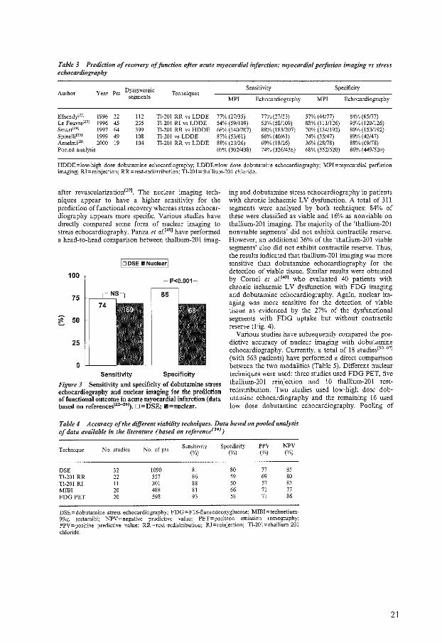
Table 3 Prediction of recovery of function afteJ" acute myocardial i11jarctiou: myocardial perfusion imaging vs stress echocardiography
Dyssynergic Sensitivity Specificity Author Year Pts Techniques
segments MPI Echocardiography MPI Echocardiography
ElhendyP"I 1996 32 !12 Tl-201 RR vs LDDE 77% (27/35) 77% (27/35) 57% (44177) 84% {65!77) Le Feuvrel231 1996 45 235 Tl-201 RI vs LDDE 54% (59/109) 53% (58/109) 88%(111/126) 95% (120/126) Smart1241 1997 64 399 Tl-201 RR vs HDDE 68% (140/207) 88% (183/207) 70% (1341192) 80% (1531192) Spinc1Jil25J 1999 49 108 Tl-201 vs LDDE 87% (53/61) 66% (40/61) 74% (35/47) 89% (42/47) Anselmil261 2000 19 104 Tl-201 RR vs LDDE 88% (23/26) 69%(18/26) 36% (28178) 88% (69178) Pooled analysis 69% (302/438) 74% (326/438) 68% (352/520) 86% (449/520)
HDDE=low-high dose dobutamine echocardiography; LDDE=low dose dobutamine echocardiography; MPI:::myocardial perfusion imaging; RI=reinjection; RR=rest-redistribution; Tl-201=thallium-201 chloride.
after revascularization129l. The nuclear imaging techniques appear to have a higher sensitivity for the prediction of functional recovery whereas stress echocardiography appears more specific. Various studies have directly compared some form of nuclear imaging to stress echocardiography. Panza et af.148l have performed a head-to-head comparison between thallium-201 imag-
I :::J DSE II Nuclear I
100
75
c 50
25
0 Sensitivity Specificity
Figure 3 Sensithity and specificity of dobutamine stress echocard.iography and nuclear imaging for the prediction of functional outcome in acute myocardial infarction (data based on referencesll2-Z6l). O=DSE; llll=nuclear.
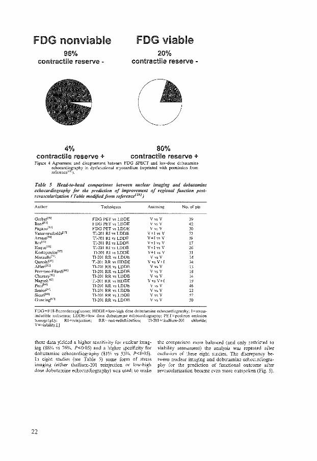
ing and dobutamine stress echocardiography in patients with chronic ischaemic LV dysfunction. A total of 311 segments were analysed by both techniques; 84% of these were classified as viable and 16% as nonviable on thallium-201 imaging. The majority of the 'thallium-201 nonviable segments' did not exhibit contractile reserve. However, an additional 36% of the 'thallium-201 viable segments' also did not exhibit contractile reserve. Thus, the results indicated that thallium-201 imaging was more sensitive than dobutamine echocardiography for the detection of viable tissue. Similar results were obtained by Cornel et af.l491 who evaluated 40 patients with chronic ischaemic LV dysfunction with FDG imaging and dobutamine echocardiography. Again, nuclear imaging was more sensitive for the detection of viable tissue as evidenced by the 27% of the dysfunctional segments -with FDG uptake but without contractile reserve (Fig. 4).
Various studies have subsequently compared the predictive accuracy of nuclear imaging with dobutamine echocardiography. Currently, a total of 18 studiesi>o-o7) (with 563 patients) have performed a direct comparison between the two modalities (Table 5). Different nuclear techniques were used: three studies used FDG PET, five thallium-201 reinjection and 10 thallium-201 restredistribution. Two studies used low-high dose dobutamine echocardiography and the remaining 16 used low dose dobutamine echocardiography. Pooling of
Table 4 Accuracy of tile different viability techniques. Data based on pooled analysis of data available in the literature (based on refel"ellcef291
)
Technique No. studies No. of pls Sensitivity Specificity PPV ]',"PV
(%) (%) (%) (%)
DSE 32 1090 81 80 77 85
Tl-201 RR 22 557 86 59 69 80 Tl-201 Rl 11 301 88 50 57 83
MIBI 20 488 81 66 71 77
FOG PET 20 598 93 58 71 86
DSE=dobutamine stress echocardiography; FDG=F18~fiuorodeoxyglucose; MIBI=technetium-99m sestamibi; NPV::onegativc predictive value: PET=positron emission tomography; PPV=positive predictive value: RR=rcst~redistribution; Rl=reinjection; Tl-201=thallium-201 chloride.
21
FOG nonviable 96%
contractile reserve
4% contractile reserve +
FOG viable 20%
contractile reserve -
80% contractile reserve +
Figure 4 Agreement and disagreement between FOG SPECT and low-dose dobutamine echocardiography in dysfunctional myocardium (reprinted with permission from rcferencel491).
Tahle 5 Head-to-head comparisons between nuclear imaging and dohutamine eclwcardiography for t!Je prediction of improvement of regional function postrevasculadzation (Table modified from referenC£f291)
Author Techniques Assessing No. of pts
Gerber[501 FDG PET vs LODE Vvs V 39 Baerl5'1 FDG PET vs LDDE V VS V 42 Paganol52l FDG PET vs LODE Vvs V 30 Vanoverschelctel031 Tl-201 RI vs LDDE V+l vs V 73 Arnesel541 Tl-201 RI vs LODE V+Ivs V 38 Baxl551 Tl-201 RI vs LDDE V+l vs V 17 Haquc! 5
"1 TI-201 Rl vs LODE V+l vs V 26 Kos\opou!osl'71 Tl-201 RI vs LDDE V+J vs V 31 Marzullo150l Tl-201 RR vs LDDE Vvs V 14 Qureshil59l Tl-201 RR vs HDDE VvsV+I 34 Alfieri1601 Tl-201 RR vs LDDE Vvs V 13 Perrrone-Filardi161 l Tl-201 RR vs LDDE Vvs V 18 Chameyi62l Tl-201 RR vs LDDE Vvs V 14 Naguehi6.1l Tl-201 RR vs HDDE VvsV+I 19 Pacel641 Tl-201 RR vs LDDE Vvs V 46 SeniorW'J Tl-201 RR vs LDDE V V$ V 22 Sicari1661 Tl-201 RR vs LDDE v vs v 57 Gunning1671 Tl-201 RR vs LDDE V VS V 30
FDG=FlS-fluorodeoxyglucose; HDDE=low-high dose dobutamine echocardiography: l=stressinducible ischaemia; LDDE=low dose dobutamine echocardiography; PET=positron emission tomography: RI ::=reinjection; RR=rest-redistribution; Tl-201 :::thallium-201 chloride; V=viability.[,J
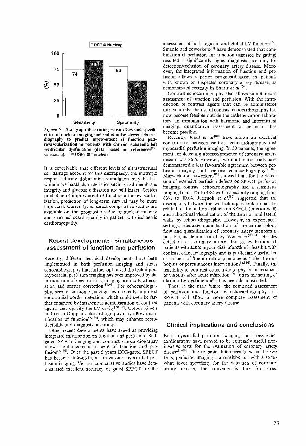
these data yielded a higher sensitivity for nuclear imaging (88% vs 76%, P<O·OS) and a higher specificity for dobutamine echocardiography (81% vs 53%, P<0·05). In eight studies (see Table 5) some form of stress imaging (either thallium-201 reinjection or low-high dose dobutamine echocardiography) was used; to make
22
the comparison more balanced (and only restricted to viability assessment) the analysis was repeated after exclusion of these eight studies. The discrepancy between nuclear imaging and dobutamine echocardiography for the prediction of functional outcome after revascularization became even more outspoken (Fig. 5).
lo DSE II Nuclear I 100
75
c 50
25
0 Sensitivity Specificity
Figure 5 Bar graph illustrating sensitivities and specificities of nuclear imaging and dobutamiue stress echocardiography to predict improvement of function postrevascularization in patients with chronic ischaemic left ventricular dysfunction (data based on references15(1-
sz,s8,6(0-62J). D = DSE; 1111! =nuclear.
It is conceivable that different levels of ultrastructural cell damage account for this discrepancy: the inotropic response during dobutamine stimulation may be lost while more basal characteristics such as cell membrane integrity and glucose utilization are still intact. Besides prediction of improvement of function after revascularization, prediction of long-tenn survival may be more important. Currently, no direct comparative studies are available on the prognostic value of nuclear imaging and stress echocardiography in patients with ischaemic cardiomyopathy.
Recent developments: simultaneous assessment of function and perfusion
Recently, different technical developments have been implemented in both perfusion imaging and stress echocardiography that further optimized the techniques. Myocardial perfusion imaging has been improved by the introduction of new cameras, imaging protocols, attenuation and scatter correction[68
'691. For echocardiogra
phy, second harmonic imaging has markedly improved endocardial border detection, which could even be further enhanced by intravenous administration of contrast agents that opacify the LV cavity[70-nJ. Colour kinesis and tissue Doppler echocardiography may allow quantification of function!73·
741, which may enhance reproducibility and diagnostic accuracy.
Other recent developments have aimed at providing integrated information on function and perfusion. Both gated SPECT imaging and contrast echocardiography al!O\v simultaneous assessment of function and perfusionF5·761. Over the past 5 years ECG-gated SPECT has become state-of-the-art in cardiac myocardial perfusion imaging. Various comparative studies have demonstrated excellent accuracy of gated SPECT for the
assessment of both regional and global LV function[77J. Smanio and coworkers!78
l have demonstrated that combination of perfusion and function (assessed by gating) resulted in significantly higher diagnostic accuracy for detection/exclusion of coronary artery disease. Moreover, the integrated infonnation of function and perfusion allows superior prognostification in patients with known or suspected coronary artery disease, as demonstrated recently by Sharir e/ a!P91.
Contrast echocardiography also allows simultaneous assessment of function and perfusion. With the introduction of contrast agents that can be administered intravenously, the use of contrast echocardiography has now become feasible outside the catheterization laboratory. In combination with hannonic and intermittent imaging, quantitative assessment of perfusion has become possible.
Recently, Kaul et af_[SOJ have shown an excellent concordance between contrast echocardiography and myocardial perfusion imaging. In 30 patients, the agreement for detecting absence/presence of coronary artery disease was 86%. However. two multicentre trials have demonstrated a less favourable agreement between perfusion imaging and contrast echocardiography[Bl.H2J.
Marwick and coworkersiBlJ showed that. for the detection of extensive perfusion defects on SPECT perfusion imaging, contrast echocardiography had a sensitivity ranging from 13% to 48% with a specificity ranging from 63% to 100%. Jucquois et al.[82l suggested that the discrepancy between the two techniques could in part be related to attenuation artifacts on SPECT (inferior wall) and suboptimal visualization of the anterior and lateral walls by echocardiography. However, in experienced settings, adequate quantification of myocardial blood flow and quantification of coronary artery stenoses is possible, as demonstrated by Wei et af.!8H
41. Besides detection of coronary artery disease, evaluation of patients with acute myocardial infarction is feasible with contrast echocardiography and is particularly useful for assessment of 'the no-refiow phenomenon' after thrombolysis or percutaneous interventions[85
·86l. Finally, the
feasibility of contrast echocardiog:raphy for assessment of viability after acute infarction[87l and in the setting of chronic LV dysfunction[SBJ has been demonstrated.
Thus, in the near future, the combined assessment of perfusion and function by echocardiography and SPECT will allow a more complete assessment of patients with coronary artery disease.
Clinical implications and conclusions
Both myocardial perfusion imaging and stress echocardiography have proved to be extremely useful noninvasive tests for the evaluation of coronary artery disease[l-·291. Due to basic differences between the two tests, perfusion imaging is a sensitive test \\lith a somewhat lower specificity for the detection of coronary artery disease; the converse is true for stress
23

echocardiography[l-171_ This systematic review focuses on direct comparative studies on stress echocardiography and perfusion imaging in order to provide the most objective information. Nevertheless, a potential risk of pooling data from different studies is to mix patients with different clinical characteristics and risk profile.
Two available direct head-to-head comparative studies demonstrated a similar prognostic value of perfusion imaging and stress echocardiography. Larger comparisons are needed to draw further conclusions.
For the assessment of myocardial viability after acute infarction the modalities seem to be equally sensitive, whereas stress echocardiography is the more specific test[22
-26l. Hence, for the early assessment of viability
stress echocardiography may be preferable. However, specificity is determined by segments that are nonviable that do not improve in function. A lower specificity suggests that a substantial percentage of segments that are viable do not recover in function. Most studies have evaluated recovery of function at a rather short timeinterval after infarction ( <3 months) and longer follow-up may be needed.
In patients with chronic ischaemic ventricular dysfunction, nuclear imaging has a high sensitivity for the detection of viable myocardium and a low specificity, whereas the converse is true for stress echocardiography[S0·-671_ The lower specificity of nuclear imaging can again be an issue of duration of follow-up. Recent data have demonstrated that a substantial percentage of segments need longer time after revascularization to (fully) recover in functionls 9J_ In addition, large direct comparative studies are required to evaluate the prognostic value of nuclear imaging and stress echocardiography in patients with chronic ischaemic left ventricular dysfunction.
In conclusion, the current analysis demonstrated that both techniques are useful in the evaluation of patients with coronary artery disease, although small differences between accuracies exist in different settings. The most important factor for using a test remains the local expertise and availability of these imaging modalities. In addition, patient characteristics (habitus, acoustic window, pregnancy) may influence the choice of the technique, and finally the studies discussed generally reflect the experience in university centers and many of these studies may be influenced by selection and referral bias, which limits application of the results to the general population.
References
[1] Maurer G, Nanda NC. Two-dimensional echocardiographic evaluation of exercise induced left and right ventricular asynergy: correlation with thallium scanning. Am 1 Cardiol1981; 48: 720-7.
[2] Nguyen T, Hco J, Ogilby JD, Iskandrian AS. Single photon emission computed tomography with thallium-201 during adenosine-induced coronary hyperemia: correlation with coronary arteriography, exercise thallium imaging and twodimensional cchocardiography. JAm Coli Cardiel 1990; 16: 1375-83.
24
[3] Galanti G. Sciagra R, Comeglio M eta/. Diagnostic accuracy of peak exercise echocardiography in coronary artery disease: comparison with thallium-201 myocardial scintigraphy. Am Heart J 1991; 122: 1609-16.
[4] Pezzoli MMA, Fioretti PM, Salustri A, Reijs AE, Roelandl JR TC. Exercise echocardiography and technetium 99m MIBI single photon emission computed tomography in the detection of coronary artery disease. Am J Cardioll991; 67: 350-5.
[5] Quiiiones MA, Verani MS, Haichin RM, Mahmarian JJ. Suarez J, Zoghbi WA. Exercise echocardiography versus thallium-201 single-photon emission computed tomography in evaluation of coronary artery disease. Circulation 1992; 85: 1026-31.
[6] Salustri A. Pezzoli M:MA, Hermans W et a!. Relationship between exercise echocardiography and perfusion singlephoton emission computed tomography in patients with single-vessel coronary artery disease. Am Heart 1 1992; 124: 75-83.
[7] Glinalp B, Dokumaci B, Uyan C eta/. Value of dobutamine technetium-99m-sestamibi SPECT and echocardiography in the detection of coronary artery disease compared with coronary angiography. J Nucl Med 1993; 34: 889-94.
[8] Amanullah AM, Bevegard S, Lindvall K, Aasa M. Assessment of left ventricular wall motion in angina pectoris by two-dimensional echocardiography and myocardial perfusion by technetium-99m scstamibi tomography during adenosineinduced coronary vasodilatation and comparison with coronary angiography. Am J Cardiol!993; 72:983-9.
[9] Marwick T, Willemart B, D'Hondt AM eta!. Selection of the optimal nonexercise stress for the evaluation of ischemic regional myocardial dysfunction and malperfusion. Comparison of dobutamine and adenosine using echocardiography and 99mTc-MIBJ single photon emission computed tomography. Circulation 1993; 87: 345-54.
[10] Marwick T, D'Hondt AM, Baudhin T eta/. Optimal use of dobutamine stress for the detection and evaluation of coronary artery disease: combination with echocaroiography or scintigraphy, or both? JAm Col! Cardiol 1993; 22: 159--67.
[11] Forster T, McNeill AJ, Salustri A eta!. Simultaneous dobutamine stress echocardiography and tcchnctium-99m isonitrile single-photon emission computed tomography in patients with suspected coronary artery disease. J Am Coli Cardiol !993; 21: 1591--6.
[12] Senior R, Sridhara BS, Anagnostou E, Handler C, Raftery EB, Lahiri A. Synergistic value of simultaneous stress dobutamine sestamibi single-photon emission computerized tomography and cchocardiography in the detection of coronary artery disease. Am Heart J 1994; 128: 71J.-8.
[13] Ho FM, Huang P1, Liau CS eta/. Dobutamine stress echocardiography compared with dipyridamole thallium-201 singlephoton emission computed tomography in detecting coronary artery disease. Eur Heart J 1995; 16: 570-5.
[14] Kisacik HL, 6zdemir K, Al!inyay E et a!. Comparison of exercise testing with simultaneous dobutamine stress echocardiography and technetium-99m isotrinile single-photon emission computerized tomography for diagnosis of coronary artery disease. Eur Heart J 1996; 17: 113-9.
[15] Huang P1, Ho YL, Wu CC eta/. Simultaneous dobutaminc stress echocardiography and thallium-201 perfusion imaging for the detection of coronary artery disease. Cardiology 1997; 88: 556--<52.
[16] Parodi G, Picano E, Marcassa C e/ a/. High dose dipyridamole myocardial imaging: simultaneous sestamibi scintigraphy and two-dimensional echocardiography in the detection and evaluation of coronary artery disease. Italian Group of Nuclear Cardiology. Coron Artery Dis 1999; 10: 177-84.
[17] Smart SC, Bhatia A, Hellman Ret a/. Dobutamine-atropine stress echocardiography and dipyridamole scstamibi scintigraphy for the detection of coronary artery disease: limitations and concordance. JAm Coli Cardiel 2000: 36: 1265-73.
[18] Geleijnse ML, Elhendy A, Van Domburg RT ez a!, Cardiac imaging for risk stratification with dobutamine atropine stress

testing in patients with chest pain. Circulation 1997; 96: 137-47.
[19] Olmos LI, Dakik H, Gordon R eta!. Long-term prognostic value of exercise echocardiography compared with exercise 201TJ, ECG, and clinical variables in patients evaluated for coronary artery disease. Circulation 1998; 98: 2679-86.
(20] Brown KA. Do stress echocardiography and myocardial perfusion imaging have the same ability to identify the lowrisk patient with known or suspected coronary artery disease? Am J Cardio11998; 81: 1050-3_
[21] Brown K.A. Prognostic value of thallium-201 myocardial perfusion imaging: a diagnostic tool comes of age. Circulation 1991; 83: 363-81.
[22] Elhendy A, Trocino G, Salustri A cl al. Low-dose dobutamine echocardiography and rest-redistribution thallium-201 tomography in the assessment of spontaneous recovery of left ventricular function after recent myocardial infarction. Am Heart J 1996; 131: 1088-96.
[23] Le Feuvre C, Baubion N, Aubry N, Metzger JP, de Vernejoul P, Vacher on A. Assessment of reversible dyssynergic segments after acute myocardial infarction: dobutamine echocardiography versus thallium-201 single photon emission computed tomography. Am Heart J 1996; 131: 668-75.
[24] Smart SC, Stoiber T, Hellman Ret al. Low dose dobutamine echocardiography is more predictive of reversible dysfunction after acute myocardial infarction than resting single photon emission computed tomographic thallium-201 scintigraphy. Am Heart J 1997; 133: 822-34.
[25] Spinelli L, Petretta M, Cuocolo A el a/. Prediction of recovery of left ventricular dysfunction after acute myocardial infarction: comparison between 99mTc-sestamibi cardiac tomography and low-dose dobutamine cchocardiography. J Nucl Med 1999; 40: 1683--92.
[26] Anselmi M, Golia 0, Maines M eta/. Comparison between low-dose dobutamine echocardiography and thallium-201 scintigraphy in the detection of myocardial viability in patients with recent myocardial infarction. Int J Ca.rdiol 2000: 73: 213-23.
[27] Wijns W, Vatner SF, Camici PO. Hibernating myocardium. N Eng! J Med 1998; 339: 173-81.
[28] Beller GA. Noninvasive assessment of myocardial viability. N Eng! J Med 2000; 343: 1488-90.
[29] Bax JJ, Poldermans 0, Elhendy A, Boersma E, Rahimtoola SH. Sensitivity, specificity, and predictive accuracies of various non-invasive techniques for detecting hibernating myocardium. Curr Probl Cardiel 2001; 26: 141---86.
[30] Underwood SR, Godman B, Salyani S, Ogle JR, Ell PJ. Economics of myocardial perfusion imaging in Europe- the EMPIRE Study. Eur Heart J 20: 157-66.
[31] Neslo RW, Kowalchuk GJ. The ischemic cascade: temporal sequence of hemodynamic, electrocardiographic and symptomatic expressions of ischemia. Am J Ca.rdiol 1987; 57: 23C-30C.
[32] Fung A Y, Gallagher KP, Buda AJ. The physiologic basis of dobutamine as compared with dipyridamole stress interventions in the assessment of critical coronary artery stenosis. Circulation 1997; 76:943--51.
[33] Geleijnse ML. Marwick TH, Boersma E, Deckers JW, Melin JA, Fioretti PM. Optimal pharmacological stress testing for the diagnosis of coronary artery disease: a probabilistic approach. Eur Heart 11995; \6 (Suppl M}: 3,.10.
[34] Houghton JL, Frank MJ, Car AA, von Dohlen TW, Prisant LM. Relations among impaired coronary flow reserve, left ventricular hypertrophy and thallium perfusion defects in hypertensive patients without obstructive coronary artery disease. JAm Coll Cardiel 1990; 15:43-51.
[35] DePeuy EG, Guertler-Krawczynska E, Perkins JV, Robbins WL, Whelchel JD, Clements SO. Alterations in myocardial thallium-201 distribution in patients with chronic systemic hypertension undergoing 201 single-photon emission computed tomography. Am J Cardiol1988; 62:234---8.
[36] Elhendy A, Gelcijnse ML, van Domburg RT et al. Comparison of dobutamine stress echocardiography and technetium-
99m sestarnibi single-photon emission tomography for the diagnosis of coronary artery disease in hypertensive patients with and without left ventricular hypertrophy. Eur J Nucl Med 1998; 25: 69---78.
[37] Fragasso G, Lu C, Dabrowski P, Pagnotta P, Sheiban I, Chierchia SL. Comparison of stress/rest myocardial perfusion tomography, dipyridamole and dobutamine stress echocardiography for the detection of coronary disease in hypertensive patients with chest pain and positive exercise test. J Am Co\1 Cardiel 1999; 34: 441-7.
[38] Wenger NK, Speroff L. Packard B. Cardiovascular health and disease in women. N Eng! 1 Med 1993: 329: 247-56.
[39] Mosca L, Manson JE, Sutherland SE, Langer RD, Manolio T, Barrett-Conner E. Cardiovascular disease in women. A statement for bcalthcare professionals from the American Heart Association. Circulation 1997; 96: 2468-82.
[40] Hansen CL, CrabbeD, Rnbin S. Lower diagnostic accuracy of thallium-201 SPECT myocardial perfusion imaging in women: an effect of smaller chamber size. JAm Coil Cardia! 1996; 28: 1214-9.
[41] Takeuchi M, Sonoda S, Miura Y, Kuroiwa A. Comparative diagnostic value of dobutamine stress echocardiography and stress thallium-201 single-photon emission computed tomography for detecting coronary artery disease in women. Coron Art Dis 1996: 7: 831-5.
[42] Elhendy A, van Domburg RT, Bax JJ era!. Noninvasive diagnosis of coronary artery stenosis in women with limited exercise capacity: comparison of dobulamine stress echocardiography and 99mTc sestamibi single-photon emission CT. Chest 1998; 114: 1097~104.
[43] Ho YL, Wu CC, Huang PJ el al. Assessment of coronary artery disease in women by dobutamine stress echocardiography: comparison with stress thallium-201 single·photon emission computed tomography and exercise electrocardiography. Am Heart J 1998; 135: 655-62.
[44] BisiG, Sciagra R, Santoro GM, Fazzini PF. Rest technetium-99m sestamibi tomography in combination with short-term administration of nitrates: feasibility and reliability for prediction of postrevascularization outcome of asynergic territories. JAm Coll Cardiel 1994: 24: 1282-9.
[45] Bax JJ, Patton JA, Poldermans D. Elhendy A, Sandler MP. 18-Fluorodeoxyglucose imaging with PET and SPECT: Cardiac applications. Semin Nucl Med 2000; 30:281---8.
[46] Braunwald E, Rutherford JD. Reversible ischemic left ventricular dysfunction: evidence for the hibernating myocardium. JAm Coli Cardiel 1986; 8: 1467-70.
[47] Rahimtoola SH. The hibernating myocardium. Am Heart 1 1989; 117: 211-21.
[48] Panza JA, Dilsizian V, Laurienzo JM, Curiel RV, Katsiyiannis PT. Relation between thallium uptake and contractile response to dobutamlne. Implications regarding myocardial viability in patients with chronic coronary artery disease and left ventricular dysfunction. Circulation 1995; 91:990-8.
[49] Cornel JH. Bax JJ, Elhendy A eta!. Agreement and disagreement between 'metabolic viability' and 'contractile reserve· in akinetic myocardium. J Nucl Cardiel 1999: 6: 383-8.
[50] Gerber BL, Vanoverschelde J-LJ, Bol A et a/. Myocardial blood flow, glucose uptake and recruitment of inotropic reserve in chronic left ventricular ischemic dysfunction. Implications for the pathophysiology of chronic hibernation. Circulation 1996: 94: 651-9.
[51] Baer FM, Voth E, Deutsch HJ era!. Predictive value of low dose dobutaminc transesophagca\ echocardiography and fiuorine-18 fluorodeoxyglueose positron emission tomography for recovery of regional left ventricular function after successful revascularization. JAm Coll Cardio11996; 28: 60---9.
[52] Pagano D, Bonser RS, Townend JN, Ordoubadi F, Lorenzoni R, Carnici PG. Predictive value of dobutamine echocardiography and positron emission tomography in identifying hibernating myocardium in patients with postischacmic heart failure. Heart 1998; 79: 281-8.
[53] Vanoverschelde J-LJ, D'Hondt A-M, Marwick Tel a/. Headto-head comparison of exercise-redistribution-reinjection
25
thallium single-photon emission computed tomography and low dose dobutamine echocardiography for prediction of reversibility of chronic left ventricular ischemic dysfunction. J Am Coli Cardiel 1996: 28: 432-42.
[54] Arnese M. Cornel JH, Salustri A et a/. Prediction of improvement of regional left ventricular function after surgical revascularization: A comparison of low-dose dobutamine echocardiography with 201-TL SPECT. Circulation 1995; 91: 2748--52.
[55] Bax JJ, Cornel JH, Visser FC eta/. Prediction of recovery of myocardial dysfunction following revascularization; Comparison of F18-fluorodeoxyglucose/thallium-201 single photon emission computed tomography, thallium-201 stressreinjection single photon emission computed tomography and doblltaminc cchocardiography. J Am Coli Cardiel 1996; 28: 558-64
[56] Haque T, Furukawa T, Takahashi M, Kinoshita M. Identification of hibernating myocardium by dobutamine stress echocardiography: Comparison with thallium-201 reinjection imaging. Am Heart J 1995; 130: 553-63.
[57] Kostopoulos KG, Kranidis AI, Bouki KP eta/. Detection of myocardial viability in the prediction of improvement of left ventricular function after successful coronary revascularization by using dobutamine stress echocardiography and quantitative SPECT rest-redistribution-reinjection 201Tl imaging after dipyridamole infusion. Angiology 1996; 47: 1039-46.
[58] Marzullo P, Parodi 0, Reiscnhofcr B et a/. Value of rest thallium-201/technetium-99m Sestamibi and dobutamine echocardiography for detecting myocardial viability. Am J Cardiel 1993; 71: 166-72.
[59] Qureshi U, Nagueh SF, Afridi 1 era/. Dobutamine echocardiography and quantitative rest-redistribution 201Tl tomography in myocardial hibernation. Relation of contractile reserve to 201TI uptake and comparative prediction of recovery of function. Circulation 1997; 95: 626-35.
[60] Alfieri 0, La Canna G, Giubinni R, Pardini A, Zogno M, Fucci C. Recovery of myocardial function. Eur 1 CardioThorac Surg 1993; 7: 325-30.
[61] Perrone-Filardi P, Pace L, Prastaro M et a/. Assessment of myocardial viability in patients with chronic coronary artery disease. Rest-4-hour-24-hour 201Tl tomography versus dobutamine echocardiography. Circulation 1996; 94: 2712~9.
[62] Charney R. Schwinger ME, Chun 1 et al. Dobutamine cchocardiography and resting-redistribution thallium-201 scintigraphy predicts recovery of hibernating myocardium after coronary revascularization. Am Heart J 1994; 128: 864--9.
[63] Nagueh SF, Vaduganathan P, Ali N e1 a/. Identification of hibernating myocardium: Comparative accuracy of myocardial contrast echocardiography, rest-redistribution thallium-201 tomography and dobutaminc cchocardiography. J Am Coli Cardiel 1997; 29: 985-93.
[64] Pace L, Perrone-Filardi P, Mainenti P et a/. Combined evaluation of rest-redistribution thallium-201 tomography and low-dose dobutamine echocardiography enhances the identification of viable myocardium in patients with chronic coronary artery disease. Eur J Nucl Mcd 1998; 25: 744- 50.
[65] Senior R, Glenville B, Basu S e/ a!. Dobutamine echocardiography and thallium-201 imaging predict functional improvement after revascularization in severe ischaemic left ventricular dysfunction. Br Heart J 1995; 74: 358-64.
[66] Sicari R, Varga A, Picano E eta!. Comparison of combination of dipyridamole and dobutamine during echocardiography with thallium scintigraphy to improve viability detection. Am J Cardiel 1999; 83: 6-10.
[67] Gunning MG, Anagnostopoulos C, Knight CJ eta!. Comparison of 201TL 99mTc-tetrofosmin, and dobutamine magnetic resonance imaging for identifying hibernating myocardium. Circulation 1998:98: 1869-74.
[68] Corbett JR, Ficaro EP. Attenuation corrected cardiac perfusion SPECT. Curr Opin Cardiel 2000; 15: 330-36.
[69] Germano G. Technical aspects of myoca.rdial SPECT imaging. 1 Nucl Med 2001:42: 1499-507.
26
[70] Caidahl K, Kazzam E, Lidberg 1 et al. New concept in echocardiography: harmonic imaging of tissue without use of contrast agent. Lancet 1998; 352: 1264-70.
[71] Sozzi FB. Poldermans D, Bax JJ et a!. Second harmonic imaging improves sensitivity of dobutamine stress echocardiography for the diagnosis of coronary artery disease. Am Heart J 2001: 142: 153-9.
[72] Hundley WG, Kizilbash AM, Afridi I, Franco F, Peshock RM, Grayburn PA. Administration of an intravenous perfluorocarbon contrast agent improves echocardiographic determination of left ventricular volumes and ejection fraction: comparison with cine magnetic resonance imaging. J Am Col! Cardiol 1998; 32: 1426-32.
{73] Mor·Avi V, Lang RM. Recent advances in echocardiographic evaluation of left ventricular anatomy, perfusion, and function. Cardiel Rev 2001; 9: 146-59.
[74] Waggoner AD, Bierig SM. Tissue Doppler imaging: a useful echocardiographic method for the cardiac sonographer to assess systolic and diastolic ventricular function. J Am Soc Echocardiogr 2001; 14: 1143-52.
[75] Kaul S. Myocardial contrast echocardiography: basic principles. Progr Cardiovasc Dis 2001: 44: 1-11.
[76] Germano G, Berman DS. Quantitative gated SPECT. 1 Nucl Med 2001; 42: 528-9.
[77] Germano G. Automatic analysis of ventricular function by nuclear imaging. Curr Opin Cardiel 1998; 13: 425-9.
[78] Smanio PE, Watson DD. Segalla DL Vinson EL, Smith WH, Beller GA. Value of gating of technetium-99m sestamibi single-photon emission computed tomographic imaging. J Am Coil Cardiel 1997; 30: 1687-92.
[79] Sharir T, Germano G, Kavanagh PB el a/. Incremental prognostic value of post-stress left ventricular ejection fraction and volume by gated myocardial perfusion single photon emission computed tomography. Circulation 1999; 100: !035-42.
[80] K.aul S, Senior R, Dittrich H, Raval U, Khattar R, Lahiri A. Detection of coronary artery disease with myocardial contrast echocardiography: comparison with 99mTc-sestamibi singlephoton emission computed tomography. Circulation 1997; 96: 785-92.
[81] Marwick TH, Brunken R, Meland N eta!. Accuracy and feasibility of contrast echocardiography for detection of perfusion defects in routine practice: comparison with wall motion and technetium-99m sestamibi single-photon emission computed tomography. The Nycomed NC100100 investigators. JAm Col! Cardiel 1998: 32: 1260-9.
[82] 1ucquois L Nihoyannopoulos P, D'Hondt AM et a!. Comparison of myocardial contrast echocardiography with NCJOOIOO and (99m)Tc sestambi SPECT for detection of resting myocardial perfusion abnormalities in patients with previous myocardial infarction. Heart 2000; 83: 518-24.
[83] Wei K, Jayawcera AR, Firoozan S, Linka A, Skyba DM. Kaul S. Quantification of myocardial blood flow with ultrasound-induced destruction of micro bubbles administered as a constant venous infusion. Circulation 1998: 97: 473-83.
[84] Wei K. Detection and quantification of coronary stenosis severity with myocardial contrast cchocardiography. Prog Cardiovasc Dis 2001: 44: 81-100.
[85] Lepper W, Hoffmann R, Kamp 0 et a!. Assessment of myocardial reperfusion by intravenous myocardial contrast echocardiography and coronary flow reserve after primary percutaneous transluminal coronary angioplasty [correction of angiography] in patients with acute myocardial infarction. Circulation 2000; 101:2368--74.
[86] Ito H. Tomooka T, Sakai N et a/. Lack of myocardial perfusion immediately after successful thrombolysis. A predictor of poor recovery of left ventricular function in anterior myocardial infarction. Circulation 1992: 85: 1699-85.
[87] Main ML, Magalski A, Chcc NK, Coen MM, Skolnick DG, Good TH. Full-motion pulse inversion power Doppler contrast echocardiography differentiates stunning from necrosis

and predicts recovery of left ventricular function after acute myocardial infarction. JAm Col! Cardiol2001; 38: 1390-4.
[88] Nagueh SF, Vaduganathan P, Ali N el a!. Identification of hibernating myocardium: comparative accuracy of myocardial contrast echocardiography, rest-redistribution thallium-
201 tomography and dobutamine echocardiography. J Am Coli Cardia! 1997; 29: 985-93.
[89) Bax JJ. Visser FC, Poldermans D el a!. Time course of functional recovery of stunned and hibernating segments after surgical revascularization. Circulation 2001; 104: 1314-8.
27

Chapter 2
How many patients with ischemic
cardiomyopathy exhibit viable
myocardium?
Schinkel AFL, Bax JJ, Boersma E,
Elhendy A, Roelandt JRTC, Poldermans D
Am J Cardio/2001;88:561-564
29

How Many Patients With Ischemic Cardiomyopathy Exhibit Viable Myocardium?
Arend F.L. Schinkel, MD, Jeroen J. Box, MD, Eric Boersma, PhD, Abdou Elhendy, MD,
Jos R.T.C. Roelandt, MD, and Don Poldermans, MD
S evere left ventricular (LV) dysfunction secondary to chronic coronary artery disease is a major prob
lem in clinical cardiology. 1 Incidence is increasing and the severity of LV dysfunction is related to clinical outcome. L 2 Current options of treatment include medical therapy and heart transplantation. Transplantation is associated with excellent survival, but the number of donor hearts is limited, and medical therapy is still suboptimaL 1-·3 Coronary revascularization can be an alternative treatment in selected patients. However, surgery in these patients is associated with higher morbidity and mortality, and thus a careful selection of patients who may benefit from revascularization is necessary to offset this higher risk. It has been demonstrated that in the presence of viable myocardium, revascularization may improve LV function, heart failure symptoms, and prognosis.4 •5 In view of the rapidly increasing number of patients with ischemic cardiomyopathy and the suboptimal therapeutic options, it is of interest to know how many patients may be eligible (based on viability assessment) for coronary revascularization. This information is currently not available. To establish the prevalence of
From ~he Departmen~ of Cardiology, Tharaxcenter, Erasmus Medical Ce:1ter Ro;terda'TI, Rot;erdam; and the Department of Cardiology, LeiOen University Medical Center, Leider, The Netherlands. Dr Schinkel's address is: Department of Cardiology, Thoraxcenler Room BA 302, Erasmus Medical Cel'ter Rot:erdom, Dr. Molewolerplein 40, 3015 GD Ro~erdam, The Netherlands. E--mail: arenaschin\el@ hetne.nl. Manuscript received Janoary 1 1, 2001 , revised manuscript received and accep~ed Apr:l 2, 2001.
myocardial viability, consecutive patients who had ischemic cardiomyopathy were studied with debutamine stress echocardiography.
The study population comprised 83 consecutive patients with chronic coronary artery disease and a LV ejection fraction (EF) of :<:;35%, who presented with heart failure symptoms. Patients with primary cardiomyopathy or significant valvular heart disease were not included. All patients underwent echocardiography at rest to identify regional dysfunction, followed by dobutamine stress to detect residual viability in these regions. The local medical ethics committee approved the protocol and all patients gave informed consent.
All echocardiograms were performed with a Sonos 5500 imaging system (Andover, Massachusetts) with a 1.8-:MHz transducer using second harmonic imaging to optimize endocardial border visualization. After baseline echocardiography, dobutamine was administered, starting at a dose of 5 p,glkg based on body weight per minute for 5 minutes, followed by a 10 p..g/kg/min dose for 5 minutes (low dose). Incremental dobutamine doses of 10 p..gikg/min were then given at 3-minute intervals up to a dose of 40 p,g/kg/min, and atropine was added, if necessary. Test end points were: target heart rate, extensive new wall motion abnormalities, ST-segment depression 2=2 mm, severe angina, a decrease in systolic blood pressure >40 mm Hg, blood pressure >2401120 mm Hg, and significant (supra)ventricular arrhythmia. The echocardiograms were digitized on optical disks and scored by 2 experienced reviewers using a 16-segment model.6 Re-
31

-TABLE I Clinical Characteristics of 83 Patients Who Underwent Dobutomine Stress Echocordiogrophy
Clinical Features No. of Patients
Age (yrs ::':: SD) 59± 10 Moo 70 (84%) Previous Ml 78(94%)
Q-wove Ml 66(80%) Anterior Ml 57(69%) Septal Ml 15 (18%) lateral Ml 18 (22%) Inferior/posterior Ml 27 (33%)
Previous CABG 13 (16%) Previous PTCA 7 (8%) Diabetes mellitus 13 (16%) Smoking previous/current 29 (35%)/30 (36%) Hypertension previous/current 5 (6%)/11 (13%) Multivessel disease 71 (66%) LBBB 9 (11%) RBBB 3 (4%) LVEF s:20% 19 (23%) LVEF >20% and s:JO% 41 (49%) LVEF >30% and s:35% 23 (28%)
Dolo ore presented'" numbers[%). There were no patient. with o Ml, PTCA,
or CABG <3 months before viability testing.
CABG = corMory artery bypo" graft surgery; LBSS ~ left bundle bronch block; Ml ~ myocordiol infarction; PTCA ~ percutoneou• tr<lnoluminol core-
nory ongioplosty; RBBB ~ right bundle bronch block.
panent was cJassmeu as v1at>1e m me presence or 2:4
dysfunctional viable segments.7
L VEF was assessed by radionuclide ventriculography at rest in all patients. A small field-of-view gamma camera system (Orbiter, Siemens, Erlangen, Germany) was used; after injection of technetium-99 m (740 MBq), radionuclide ventriculography was performed at rest. L VEF was calculated by standard methods (Odyssey VP, Picker, Cleveland, Ohio).7
All continuous data are expressed as mean ± SD, percentages are rounded. Continuous variables were compared using the Student's t test for unpaired samples. Differences betvleen proportions were compared using the chi-square test. If the distribution was not normal, the nonpararnetric Wilcoxon test and the Spearman correlation analysis was used. A p value of <0.05 was considered statistically significant.
The clinical characteristics of the study population are listed in Table 1. Heart failure was the principal clinical presentation in all patients; New York Hearl Association (NYHA) functional class was an average of2.8 ± 0.6 (with 80% of the patients in NYHA class III or IV). L VEF was severely depressed (mean 25 ± 7%, range 10% to 35%). Medication consisted of aspirin and/or oral anticoagulants (92%), angiotensin-
converting enzyme inhibitors (84%), f3-blockers (52%), digoxin
Normal Dysfunctional (27%), diuretics (63%), and nitrates (70%).
Heart rate increased from 78 ±
hypokinetic 36%
normal
35%
severe ~hypokinesia
13 beats/min at rest to 118 ± 17 beats/min at peak stress (p <0.0001). Overall, systolic blood pressure did not significantly change during dobutarnine infusion. Test end points were target hearl rate (n = 76, 92%), severe ischemia on electrocardiography (n = 4, 5%). severe angina (n = 2, 2%), or a decrease in systolic blood pressure of>40mmHg(n= 1, 1%).Apeak
akinesia dobutamine infusion dose of 40 Jl-gl kg/min was used in 59 patients (71%); 37 patients received atropine. Echocardiographic analysis was performed on 1,328 segments and revealed 143 normal and 226 mild hypokinetic segments. Of 959
FIGURE 1. Incidence of 5 po:ttems of conlro:ctile function in 1,328 LV segments; 959 segments (72%) were dysfunctional, ond 369 segments (28%) exhibited normal conlro:ctile function.
gional wall motion and systolic wall thickening were scored on a 5-point scale: 1 = normal, 2 = mildly hypokinetic, 3 = severely hypokinetic, 4 = akinetic, and 5 = dyskinetic. Myocardial segments were considered normal if the regional wall motion was normal or mildly hypokinetic. Only dysfunctional segments (severe hypokinesia, akinesia, or dyskinesia at echocardiography at rest) were evaluated for myocardial viability. Segments with an improvement, worsening, or a biphasic wall motion response during stress echocardiography were considered viable. Segments with unchanged wall motion were considered nonviable. A
32
dysfunctional segments (72%), 484 segments exhibited severe hypoki
nesia, 460 segments had akinesia, and 15 had dyskinesia (Figure 1). Patients had an average of 11.6 ± 3.9 dysfunctional segments. During dobutamine stress echocardiography, 205 dysfunctional segments exhibited sustained improvement, 47 worsening, and 86 segments had a biphasic response. Thus, a total of338 dysfunctional segments (35%) were considered viable (Figure 2). Moreover, 39% of these segments demonstrated a limited coronary flow reserve (showing direct worsening during dobutamine infusion or a biphasic response). Patients had an average of 4.1 ± 3.2 dysfunctional but viable segments; those with a L VEF

Nonviable Viable rate.7•11- 15 Thus, (preoperative) viability testing may guide patient management. The exact prevalence of viability among patients with heart fail-
biphasic ure, secondary to poor LV function in the presence of chronic coronary artery disease, is unknown. In the present study, this topic was addressed and a consecutive cohort of patients was evaluated for viability. On a segmental basis, 35% of the dysfunctional segments was considered viable. Applying the cut-off value of 2::4 dysfunctional but viable segments, 57% of the patients had
sustained improvement
viable segments; they had an average of7.5::!:: 2.9 dysfunctional but viable segments.
FIGURE 2. Prevalence of vioble ond nonviable tissue; 35% of dysfunctional myocardium wos viable, whereas 65% was nonviable myocardium. Twenty-one percent of dysfunctional myocardium showed sustained improvement, 5% worsening, and 9"/o a biphosic woll motion paffern.
In the only other study addressing the issue of the prevalence of viability in patients with ischemic cardiomyopathy, Auerbach et aJl 6 demonstrated that 156 of 283 patients had
:520% had more dysfunctional segments compared with patients with a LVEF >20% and :530% (14.3 :!: 1.9 vs 11.1 :!: 4.1; p <0.005) or a LVEF >30% (14.3 :!: 1.9 vs 10.1 :!: 3.8; p <0.005). The number of dysfunctional segments was not significantly different between patients with a L VEF > 30% and those with aLVEF >20%and :530%(10.1::!:: 3.8vs 11.1:!: 4.1; p = 0.3). Forty-seven patients (57%) had clinically significant viability (an average of 7.5 :!: 2.9 segments) and 36 patients (43%) had predominantly nonviable myocardium (an average of 1.6 :!: 1.2 viable segments). Clinical features were similar in the patients with and without viability. Moreover, there was no correlation between LVEF and the number of viable dysfunctional segments (r = 0.03, n = 83, p = NS; see Figure 3).
Heart failure is becoming the most comprehensive problem in clinical cardiology, in terms of affected patients. It has been shown that dysfunctional myocardium does not always represent irreversibly damaged myocardium; some patients improve in LV function and clinical outcome after revascularization.4 ·5•8
However, revascularization procedures are associated with a higher risk in these patients.4 •5•9 Therefore, a careful evaluation is mandatory for optimal patient management and risk stratification. To explain the postoperative improvement in LV performance and prognosis, the concept of viability was proposed.4 •5
Patients with viable myocardium are likely to benefit from revascularization, whereas patients without viable myocardium will not benefit.4 •5 Moreover, preoperative viability testing results in improved risk stratification.'o Finally, viable myocardium in patients with ischemic cardiomyopathy who were treated med~ ically was associated with an extremely high event
viable myocardium on positron emission tomography using F18-fluorode
oxyglucose. Thus, our study and Auerbach et al's work demonstrate that 57% and 55%, respectively, of the patients with ischemic cardiomyopathy had significant viable tissue. This does not necessarily mean that all of these patients are suitable candidates for revascularization; other factors including comorbidity, inadequate target vessels, and severe LV dilation also influence the decision as to whether patients should undergo revascularization. Moreover, it is unclear whether these findings apply to the general population, because the patients in this and Auerbach's study were evaluated in tertiary referral centers. An important observation in the present study, in line with Auerbach et al's study,16 is that the clinical characteristics were similar between patients with and without viability; this implies that specific testing for viability is mandatory. Finally, the prevalence of viability was equally distributed among patients with different degrees of LV dysfunction; thus, viability assessment should not be reserved for patients with milder (or more severe) forms of LV dysfunction.
In the present study, dobutamine echocardiography was used to detect viability. The hallmark of viable myocardium on dobutamine stress echocardiography is improvement of contractile function during the infusion of low-dose dobutamine (5 to 10 p..g!kg/min) (contractile reserve).5 The protocol has been expanded to low- to high-dose dobutamine echocardiography, using a maximum dosage of 40 p..glkg/min with the addition of atropine if necessary.7 The low- to highdose dobutamine protocol is safe 17 and has the advantage that it allows assessment of viability and limited coronary flow reserve; from a patient standpoint, both issues are important for the decision to proceed with revascularization. Still, recent studies have indicated
33

16
" c 14 • E
"' 12 • • " 10 c 0 8 u c 2 6 • ~ ~ 4 • :c 2 rn > 0
0 5 10 15 20 25
LV ejection fraction
30 35
4. Beller GA. Assessment of myocardial viabiliry. Curr Opinion Cordio/1997;12:459-467. 5. Wijns W, Vatner SF, Ca.mici PG. Hibematins myocardium. N Eng/ J Med 1998:339:173-181 6. Bourdillon PD. Broderik TM, Sawada SG, Armstrong WF, Ryan T, Dillon JC, Fineberg NS, Feigenbaum H. Regional wall motion index for infarct and non-infarct region after rcperli.J,;ion in acute myocardial infarction: comparison with global wall motion index. JAm Soc Echocardiogr 1989;2:398-407 7. B(l)( JJ, Poldcrrnans D, Elhcndy A, Cornel ffi, Boersma E, Rambaldi R, Roeland! JRTC, Fioretti PM . Improvement of left ventricular ejection frnction, heart failure symptotm and prognosis after revascularization in patients with chronic coronary artery disease and viable myocardium detected by dobutamine stress echocardiograpby. JAm Col/ Cardia/ 1999;34:163-169. 8. Rahimtoola SH. From coronary artery dis<:ase to heart failure: role of the hibernating myocardium. Am J Cardio/1995;75:16E"·22E. 9. Argenziano M, Spotnitz ILlvl, Whang W, Bigger IT
FIGURE 3. Scaiterplot indicating no significant correlation between LVEF and the number of viable dysfunctionol segmenb.
Jr, Parides M, Rose EA. Rlsk stratification for coronary bypa;s surgery in patients with left ventricular dysfunction: analysis of the coronary artery bypass grafting patch trial database. Circulation 1999;HlO(suppl. 19)· llll9-124
that nuclear imaging techniques may be more sensitive for the detection of myocardial viability. 18•19 In the present study, only 35% of the dysfunctional segments were classified as viable. In previous studies with nuclear imaging techniques, this percentage was usually higher.18 However, in the study by Auerbach et al16 and this study, the prevalence of viability on a patient basis was comparable.
In conclusion, dobutamine stress echocardiography demonstrated viability in 35% of the dysfunctional segments. When the cut-off value of 2:!4 dysfunctional but viable segments was applied to classify a patient as viable, 57% of the patients were considered to have substantial viability. Thus, in 57% of the patients with ischemic cardiomyopathy and heart failure, a clinically significant amount of viable myocardium is present and coronary revascularization may be considered as therapy for these patients.
1, Gbeorghiade M, Bonow RO. Chronic heart failure in the United States. A manifestation of coronary artery disease. Circulation 1998;97:282-289. 2. Challapalli S, Bonow RO, Gheorghiade M. Medical management of heart failure s•cond.ary to coronary artery disease. CoronArl Disease 1998;9:659-674 3. Eva:os RW, Manninen DL, Garrison LP, Maier AM. Donor availability as the primary determinant of the future of heart lrnnsplantation. JAMA 1986:255:1892-1898.
34
10. Haas F, Haehnel CJ, Picker W, Nekolla S, Mortinoff S, Meisner H, Schwaiger M. Preoperative
positron cmisoion tomographic viability assessment and preiopcrative and post· operative risk in patients with advanced ischemic heart disease. J Am Coil Cardio/1997;30:1693-1700. 11. Eitzman D, Al-Aouar ZR, Kanter HL, vom Dahl J, Kirsh M, Dceb GM, Schwaiger M. Clinical outcome of patients with advanced coronary artery disease Mter viability studies with positron emission tomography. JAm Coli Cordiol 1992;20:559-565. 12. Di Carli MF, Davidson M, Little R, Khanna S, Mody FV, Brunken RC, Czemin J, Rokhsar S, Stevenson LW, Laks H, et al. Value of metabolic imaging with positron emission tomography for evaluating prognosis in patients with coronary artery disease and ldl ventricular dysfunction. Am J Cardio/ 1994;73· 527-533. 13. Pasquet A, Robert A, D'Hondt AM, Dion R, Melin JA. Vanoverschcldc JL Prognostic value of myocardial ischemia and viability in patients with chronic left ventricular ischemic dysfunction. Circulation 1999;100:141-148. 14. Fagley PR, Beller GA, Watson DD, Gimplc LW, Ragostn. M. Improved outcome after coronary bypass surgery in patients with ischemic cardiomyopathy and residual myocardial viability. Circulation 1997;96:793-800. 15. LeeKS, MarwickTH, Cook SA, Go RT, Fix JS, James KB, Sapp SK, Macintyre WJ, Thomas JD. Prognosis of patients with left ventricular dy•funcrion, with and without vffib)e myocardium after myocardial infarction. Relative efficacy of medical therapy ond revasculari.zation. Circulation 1994;90:2687-2694. 16. Auerbach MA, Sch1ider H, Hoh C, G~mbhir SS, Yaghoubi S, Sayre JW, Silverman D, Phelps ME, Schelbert HR, Czernin J. Prevalence of myocardial viability :<S detected by positron emission tomography in patients with ischemic cardiomyopathy. Circulation 1999;99:2921-2926. 17. Polderman.s D, Rambaldi R, Bax JJ, Come! ill, Thomson IR, Valkema R, Boersma E, Fior.,rti PM, Breburda CS, Roelandt JRTC. Safety and utility of atropine addition during dobutamine stress echocardiography for the assessm.ont of viable myocardium in patients with severe left ventricular dy<fl!llction. Eur Hearl J 1998;19:1712-1718. 18. Panza JA, Dilsizian V. Lauricnzo JM, Curiel RV, Katsiyiannis PT. Relation between thallium uptake ond contractile response to dobutamine. Implications regarding myocardial viability in patients with chronic coronary artery disease and left ventricula; dysfunction. Circulation 1995;91 :990-998 19. Cornel JH, BaxJJ, Elhcndy A, Visser FC, Boersma F~ Poldcrmans D, SloofGW, Fioretti PM. Agn:emcnt ond disagreement between "metabolic viability'" ond 'contractile reserve" in akinetic myocardium. J Nucl Cardio/1999;6:383--388
Chapter 3
Prevalence of myocardial viability assessed
by single photon emission computed
tomography in patients with chronic
ischemic dysfunction
Schinkel AFL, Bax JJ, Sozzi FB, Boersma E, Valkema R,
Elhendy A, Roelandt JRTC, Poldennans D
Heart 2002;88:125-130
35


Prevalence of myocardial viability assessed by single photon emission computed tomography in patients with chronic ischaemic left ventricular dysfunction
A F L Schinkel, J J Box, F B Sozzi, E Boersma, R Valkema, A Elhendy, J R T C Roelandt, D Poldermans
Heort2002;88: 125-130
Objective: To assess the prevalence of myocardial viability by lechnetium-99m (Tc-99m)-tetrofosmin/ fluorine--18-fluorodeoxyglucose (FDG) single photon emission computed tomography (SPECT) in patients with ischoemic cardiomyopathy. Design: A retrospective observational study. Setting: Thoroxcenler Rotterdam (a tertiary referral centre). Patients: 104 patients with chronic coronary artery disease and severely depressed left ventricular function presenting with heart failure symptoms.
See end of orticle for outhors' offiliotions
Main outcome measures: Prevalence of myocardial viability as evaluated by Tc-99m-tetrofosmin/ FDG SPECT imaging. Two strategies for assessing viability in dysfunctional myocardium were used: perfusion imaging alone, and the combination of perfusion and metabolic imaging.
Correspondence to: Dr Don Poldermons, Thoroxcenter, Deportment of Cordiology, Erosmus Medicol Center Rotterdom, Dr Molewoterplein 40, 3015 GD Rotterdom, Netherlonds; [email protected]
Results: On perfusion imaging alone, 56 patients (54%) hod o signiftcont amount of viable myocardium, whereas 48 patients (46%) did not. Among the 48 patients with no significant viability by perfusion imaging alone, seven additional patients (15%) hod significantly viable myocardium on combined perfusion and metabolic imaging. Thus with o combination of perfusion and metabolic imaging, 63 patients (61 %) hod viable myocardium and 41 (39%) did not.
Accepted 10 April 2002
Conclusions: On the basis of the presence of viable dysfunc:tionol myocardium, 61% of patients with chronic coronary artery disease and depressed lefl ventricular ejection fraction presenting with heart failure symptoms may be considered for coronary revasculorisotion. The combination of perfusion and metabolic imaging identified more patients with sign"tficant viability than myocardial perfusion imaging alone.
Chronic coronary anery disease is lhe most important cause of left ventricular dysfunction leading to heart failure. When this occurs it has an extremely poor
prognosis.' ' In nearly 70% of the patients in 1:, major heart failure trials, coronary artery disease was the underlying cause of the hean failure.' The management of these patients remains difficult, while both the incidence and the prevalence of chronic heart failure have been increasing in recent years.' Medical treatment is still suboptimal in these patients, although significant improvements in survival have been achieved with angiotensin convening enzyme (ACE) inhibition, blockade of aldosterone receptors by spironolactone, and ~ adrenergic blockade.'-' Other therapeutic options are heart transplantation and coronary revascularisarion. The possibilities of heart transplantation arc limited by the availability of donor heans, but coronary revascularisation could be an effective alternative. In a subset of patients vvith heart failure secondary to chronic coronary artery disease, revascularisation has been shovvn to improve left ventricular ejection fraction (LVEF), hean failure symptoms, and survival.'' However, revascularisation in such patients is also associated with increased periproccdu.ral morbidity and mortality.' '
The concept of myocardial viability was proposed to explain the improvement of LVEF and heart failure symptoms after revascularisation." ''Dysfunctional but viable myocardium is likely to regain contranile function after coronary revascularisation." On the other hand, dysfunctional nonviable myocardium (scar tissue) clearly will not improve. Bon ow estimated that between 25---40% of patients with heart failure caused by ischaemic heart disease have the potential for functional improvement after revascularisation." Quantifi-
cation of myocardial viability in patients with chronic coronary artery disease, depressed left ventricular function, and heart failure is at present limited, but this information is important as such patients may benefit from revascularisation.14
" Our aim in this study was therefore to evaluate myocardial viability in a consecutive series of 104 patients with chronic coronary artery disease and depressed LVEF presenting with heart failure symptoms.
METHODS Patient population, study protocol The study population consisted of 104 consecutive patients with ischaemic cardiomyopathy (chronic coronary artery disease as assessed by angiography and an LVEF of :s: 35%), who presented with heart failure as the predominant symptOm and were referred for evaluation of myocardial viability. Patients with primary cardiomyopathy or concomitant significant valvar disease were not included.
All patients underwent resting echocardiography to identify dysfunctional myocardial tissue, and dual isotope simultaneous acquisition myocardial single photon emission computed tomography (SPECT), including Tc-99m tetrofosmin and fluorodeoxyglucose (FDG) to assess myocardial perfusion and
Abbreviations: ACE, ongiotensin conver~ing enzyme; FDG, Auorodeoxyglucose; LVEf, left ventriculor eiection froction; PET, pos'1tron emission lomogrophy; ROC, receiver opero!ing chorocteristic; SPECT, single photon emission computed !omogrophy
37
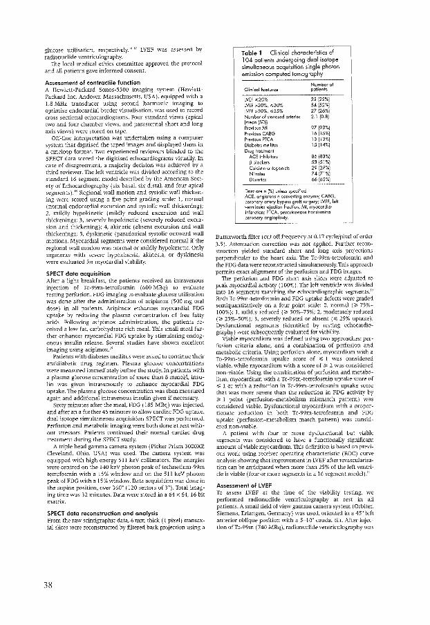
glucose utilisation, respectively."" LVEF was assessed by radionuclide ventriculography.
The local medical ethics committee approved the protocol and all patients gave informed consent.
Assessment of contractile function A Hewlett-Packard Sonos-5500 imaging system (HewlettPackard Inc, Andover. Massachusetts, USA), equipped with a 1.8 MHz uansducer using second harmonic imaging to optimise endocardial border visualisation, was used to record cross sectional echocardiograms. Four standard views (apical two and four chamber views, and parasternal short and long axis views) were stored on tape.
Off-line interpretation was undertaken using a computer >ystem that digitised the taped images and displayed them in a cincloop format. Two experienced reviewers blinded to the SPECT data scored the digitised echocardiograms visually. In case of disagreement. a majority decision was achieved by a third reviewer. The left ventricle was divided according to the standard 16 segment model described by the American Society of Echocardiography (six basal. six distal, and four apical segments)." Regional wall motion and systolic wall thickening were scored using a five point grading scale: L normal (normal endocardial excursion and systolic wall thickening); 2. mildly hypokinetic (mildly reduced excursion and wall thickening); 3, severely hypokinetic {severely reduced excursion and thickening); 4, akinetic (absent excursion and wall thickening); 5, dyskinetic (paradoxical systolic outward wall morion). Myocardial segments were comidered normal if the regional wall motion was normal or mildly hypokinetic. Only segments with severe hypokinesia, akinesia, or dyskinesia were evaluated for myocardial viability.
SPECT data acquisition After a light breakfast, the patients received an intravenous injection of Tc-99m-tetrofosmin (600 MBq) to evaluate resting perfusion. FDG imaging to evaluate glucose utilisation was done after the administration of acipimox (500 mg oral dose) in all patients. Acipimox enhances myocardial FOG uptake by reducing the plasma concentration of free fatty acids. Following acipimox administration, the patiems received a low fat, carbohydrate rich meal. This >mall meal fur· ther enhances myocardial FDG uptake by stimulating endog· cnous insulin release. Several studies have shown excellent imaging using acipimox."
Patients with diabetes mellitus were asked to continue their antidiabetic drug regimen. Plasma glucose concentrations were measured immediately before the study. In patients with a plasma glucose concentration of more than 8 mmol/1, insulin was given intravenously to enhance myocardial FDG uptake. The plasma glucose concemration was then measmed again and additional intravenous insulin given if necessary.
Sixty minutes after the meal, FOG { 185 MBq) was injected, and after an a further 45 minutes to allow cardiac FOG uptake, dual isotope simultaneous acquisition SPECT was performed. Perfusion and metabolic imaging were both done at restvv:ithout stressors. Patients continued their normal cardiac drug treatment during the SPECT study.
A triple head gamma camera system (Picker Prism 3000XP, Cleveland. Ohio. USA) was used. The camera system was equipped with high energy 511 keV collimators. The energies were centred on the 140 keV phmon peak of technetiillll-99m tetrofosmin with a 15% window and on the 511 keV photon peak of FDG with a 15% window. Data acquisition was done in the supine position. over 360° { 120 sectors of 3"). Total imaging time was 32 minutes. Data were stored in a 64 x 64, 16 bit matrix.
SPECT data reconstruction and analysis From the raw scintigraphic data, 6 mm thick ( l pixel) transaxial slices were reconstructed by filtered back projeCtion using a
38
IT~ble 1 Clinical characteristic;~ 'I 104 patients undergoing dual isotope I
simultaneous acquisition single photon .
I
emission computed tomogr:~::.,, of I
Clinicallealures patients
LVH ,;;20% 23 [22%] LVEF >20'7",, ,;;30% 54 (52'7"o] tVEF >30%, .;35% 27[26%) Number of s~nosed arteries 2. I {0.8) {mean (SD)) Previous Ml Previous CABG Previcu• PTCA Diabetes mellitus Drug treatment
ACE inhibiter. ~ Blocker. Calcium ontogonists Nitrates Diuretics
Doto ore n [%] unless specified.
97 {93%) 16(15%] 13 [13%] 15 [14%]
86 {83%) 53 {51%) 29 [28'Yo) 74 [71%) 66 [63%]
ACE, angiotensin converting enzyme; CABG, coronary artery bypa» graft surgery; LVEF, leh ventricular ejedion fraction; Ml, myocardial infarction; PTCA, percutaneous tronsluminal coronary ongioplosly.
Butterworth filter (cut off frequency at 0.17 cycle;pixcl of order 35). Attenuation correction was nor applied. Further reconstruction yielded standard short and long axis projections perpendicular to the heart axis. The Tc·99m-tetrofosmin and the FDG data were reconstructed ~imultaneously. This approach permits exact alignment of the perfusion and FOG images.
The perfusion and FOG short axis slices were adjusted to peak myocardial acrivity (100%). The left ventricle was divided into 16 segments matching the echocardiographic segments." Both Tc-99m-tetrofosmin and FDG uptake defects were graded semiquantitatively on a four point scale: 0, normal (;;<. 75%-100%); J, mildly reduced(;?; 50o/a--75%; 1. moderately reduced (;;<. 25%-50%): 3, severely reduced or absent (>S 25% uptake). Dysfunctional segments (identified by resting echocardiography) were subsequently evaluated for viability.
Viable myocardium was defined using two approaches: perfusion criteria alone, and a combination of perfusion and metabolic criteria. Using perfusion alone, myocardium ·with a To-99m-tetrofosmin uptake score of ,; I was considered viable, while myocardium with a >core of;:;. 2 was considered non"viable. Using the combination of perfusion and metabo· ]ism, myocardium with a Tc-99m-tetrofosmin uptake score of ,; 1 or with a reduction in Tc-99m-tetrofosmin uptake score that was more severe than the reduction in FDG activity by ;:;. 1 point (perfusion-metabolism mismatch pattern) was considered viable. Dysfunctional myocardium with a proportionate reduction in both Tc-99m-tetrofosmin and FDG uptake (perfusion-metabolism match pattern) was considered non-viable.
A patient with fom or more dysfunctional but viable segments was considered to have a functionally significant amount of viable myocardium. Tills definition is based on previous work using receiver operating characteristic (ROC) curve analysis showing that improvement in LVEF afterrevascularisation can be anticipated when more rhan 25% of the left ventricle is viable (four or more segments in a 16 segment model)."
Assessment of LVEF To assess LVEF ar the time of the viability testing, we performed radionudide ventriculography at rest in all patients_ A small field of view gamma can1era system (Orbiter, Siemens, Erlangen, Germany) was used, oriented in a 45' left anterior oblique position with a 5-10" caudal tilt. After injection of To-99m (740 MBq), radionuclide ventriculography was

~
I 0 Viobl' I ' Ill Non-viable :
ii ! m
' 30 g
~ 20 .2 g- w
Myocordiol perfusion
Figure 1 Myocardial perfusion in viable and non-viable tissue. In viable myocardium, perfusion was normal in 57% of dysfunctional segments, mildly reduced in 32%, moderately reduced in 10%, and severely reduced or absent in 1 %. Non-viable tissue hod moderately reduced perfusion in 42% of dysfunctional segments and severely reduced or absent perfusion in 56%.
done at rest with the patient in the supine position. The LVEF was calculated by standard methods (Odyssey VP. Picker, Cleveland, Ohio. USA).
Statistical analysis All continuous data are expressed as mean (SD). Percentages are rounded. Comparisons were made using the Student t test for unpaired samples. A probability value of p < 0.05 was considered significant.
RESULTS Patient characteristics Clinical characteristics of the 104 patients (87 men, !7 women; mean (SD) age. 60 (9) yean) are summarised in table !. All patients presented with symptoms of heart failure. The mean New York Heart Association (l\'YHA) functional class was 2.5 (0.9); 67 patients were in NYHA class III or IV. The majority (93%) of the patients had suffered a previous myocardial infarn (all occurring more than one month before viability assessment). LVEF assessed by radionuclideventriculography averaged 25 (7)% (range 9-35%). In 23 patients the LVEF was 20% or less, in 54 it was between 21% and 30%, and in 27 it was more than 30%.
Segmental analysis Contractile function Cross sectional echocardiographic analysis of regional left ventricular function was done in 1664 segments, of which 189 were normal and 297 mild hypokinetic. Of 1178 dysfunctional segments (71%), 584 showed severe hypokinesia, 578 akinesia, and 16 dyskinesia. The mean (SD) number of dysfunctional segments per patient was 11.3 (4.2).
Table 2 Comparison of segment viability in three groups divided by left ventricular eiection fraction
Charccteristics of lVEF ~20% LVEF 21-30% LVEF >30% segments (23 patients) (54 patients) (27 pctientsl
Dysfllnctioncl 14.1 [2.0) 10.9(4.5)! 9.7 {3.9) Viable by Tc-99m 6.4 (3.71 4.6 [3.8)* 3.8 [3.2) Viable byTc-99m 7.2 {4.0) 5.1 [4.1)* 4.2 [3.2) +FDG
•p-<0.05; tp<0.005 vLVEF ~20%. FDG, Auorodeoxyglucose; LVEF, left ventricular ejection fraction
~ A
" " N 60
-"
" ~ ' ~ ~
-~
ii ~ ~
!':
" ~ -"
" 0
~ ~
-~
~ j
~
" N
-"
" g ' ~ ~
-~ ., 11 0% ~ 8-11 12-16
Number of viable segments
Figure 2 (A) Amount of viable myocardium indicated by the number of viable segments in 23 patients with o left ventricular eiection fraction (LVEf) .;; 20%. Using perfusion as the only criterion of viability, 18 patients (78%) hod significant viability (e. 4 viable segments) ond live (22%) did not hove signilicontviobility. (B) Amount of viable myocardium indicated by the number of viable segments in 54 patients with on LVEF > 20% but,:;; 30%. Using perfusion as the only criter<on of viability, 27 patients (50%) hod significant viability (e. 4 viable segments). ond 27(50%) did not hove significant viability. (C) Amount of viable myocardium indicated by the number of viable segments in 27 patients with on lVEF > 30% but.;; 35%. Using perfusion os the only criterion of viability, 11 patients (41 %) had significant viability (;, 4 viable segments) and 16 (59%) did not hove significant viability.
Perfusion imaging alone Using myocardial perfusion as the only criterion of viability, 497 dysfunctional segments ( 42%) were classified as viable. Three hundred and sb:teen dysfunctional segments (27%) had normal Tc-99m-tetrofosmin uptake, and 181 (15%) had a mildly reduced uptake. The remaining 681 dysfunctional segments (58%) had a Tc-99m tetrofosmin uptake score of 2 or 3 and were classified as non-viable.
Combined perfusion ond metabolic imaging Using perfusion imaging in conjunction with FDG SPECT, 558 of the dysfunctional myocardial segments (47%) were classified as viable. Of these viable dysfunctional segments,
39

g A
"' ::~ N
.s 50 ~
~ ~
" ~ ~ • ~
g B
'l " 0 00
.s 50
* 44% 0
:,II ~ 35%
I " -E 13%
' "" • ~ 1111111 ~ 0 0-3 4-7 8 11 12-16
~ c
70c ~ oo~ " .s
* g '
" 1 " ' £
Number of viable segmen~s
Figure 3 (A) A moun~ of viable myocardium indicated by the number of viable segments in 23 patients with a left ventricular ejection fraction (LVEF) ,;; 20%. Using perfusion in combination with metabolic criteria for viability, 19 patients {83%) hod significant viability {"' 4 viable segments) and four {17%) did not. {B) Amount of viable myocardium indicated by the number of viable segments in 54 patients with on LVEF > 20% but eS 30%. Using perfusion in combination with metabolic criteria for viability, 30 patients (56%) hod significant viability(;, 4 viable segments) and 24 (44%) did not. {C) Amount of viable myocardium indicated by the number of viable segments in 27 patients on LVEF > 30% but,;; 35%. Using perfusion in combination with metabolic criteria for viability, 14 patients (52%) hod significant viability(;, 4 viable segments) and 13 {48%) did not.
497 ( 42%) had normal or mildly reduced perfusion and 61 additional segments (5%) had a perfusion-metabolism mismatch pattern. Conversely, 620 dysfunctional segments (53%) were classified as non-viable (with a matching pattern of perfusion and metabolism). Figure 1 shows the myocardial perlusion in the dysfuncrional segments that were classified as viable and non-viable by combined perfusion and metabolic imaging.
Patient analysis Contractile function Patients with an LVEF of 20% or less had more dysfunctional segments than those with an LVEF of 21-30% ( 14.1 (2.0) v 10.9 {4.5), p < 0.005) or those with an LVEF of more than 30% (14.1 (2.0) v 9.7 (3.9), p < 0.0001). The number of dysfunc-
40
tiona! segments was not significantly different between patients with an LVEF of more than 30% and those with an LVEF of 21-30% (9.7 (3.9) v 10.9 {4.5); p = 0.2) (table2).
Perfusion imaging alone All patients had on average 4.8 (3.7} (range 0-14) dysfunctional but viable segments. When myocardial perfusion was used as the only criterion of viability, 56 patients (54%) had a significant amount of viable myocardium (on average 7.5 (2.9) segments) and 48 patients { 46%) did not have significant viability (1.6 (1.2) segments). Figure 2 shows the number of viable segments per patient with perlusion imaging alone for the three LVEF groups.
Combined perfusion and metabolic imaging Among the 48 patients with no significant viability by perfusion imaging alone. seven additional patients ( 15%) had viable myocardium when assessed using a combination of perfusion and metabolic imaging. All 56 patients with significant viability when assessed by perfusion alone also had significant viability with combined perfusion and metabolic imaging (on average, 8.2 (3.2) viable segments). Thus perfusion imaging in combination with metabolic imaging showed that 63 patients (61%) had a significant amount of viable tissue (7.8 (3.2) segments). Forty one patients (39%) did not have significant residual viable dysfunctional tissue (on average, 1.6 (1.2) viable segments). OveralL the patients had 5.4 (4.0) viable dysfunctional segments. Figure 3 shows the number of viable dysfunctional segments for patients in the three different LVEF categories.
DISCUSSION Viable myocardium Heart failure is becoming a major problem in clinical cardiology. In recent years the incidence of patients with heart failure has increased despite progress in prevention and treaunent.' Chronic heart failure is predominantly caused by coronary artery disease and has a poor prognosis.' In a subset of patients with heart failure resulting from coronary artery disease, coronary revascularisation may improve left ventricular function, heart failure symptoms, and survival,'' but the risks of revascularisation in such patients are substantial. Also, apart from the need to assess clinical variables such as target vessels and comorbid factors, it is important to evaluate myocardial viability as part of the complex selection process in candidates for revascularisation.
Previous estimates suggest that between 25-40% of the patients with heart failure caused by coronary artery disease may show useful functional improvement after revascularisation." In our hospital, patients with coronary artery disease, a depressed LVEF, and heart failure are always referred for assessment of myocardial viability. This approach allows improved risk stratification and results in optimal treatment for the individual patient. In the present study, we examined myocardial viability in a consecutive series of patients from our heart failure registry.
A few previous studies have examined myocardial viability on an individual patient basis. Using FDG positron emission tomography (PET), Pasquet and colleagues found that among 66 patients with severe left ventricular dysfunction, 28 (47%) had an improvement in LVEF of at least 5% after coronary revascularisation, while eight (14%) had an improvement of more than 10%.22 Al-Mohammad and colleagues studied the prevalence of hibernating myocardiwn in 27 patients using FDG PET." Fourteen of these had significant areas of hibernating myocardiwn on PET. Auerbach and associates, also using FDG PET. studied 283 patients with ischaemic cardiomyopathy and found rhat 156 (55%) had viable myocardium." In the present study; 54% of the patients showed significant viability when myocardial perfusion was
used as the only index, while perfusion imaging in combination with metabolic imaging identified a further 7%; thus 61% of the patients in all had a significant amount of viable tissue. These observations are in close agreement with those of Al-Mohammad and Auerbach using FDG PET.''"
In the present study patients with an LVEF of 20% or less had more dysfunctional but viable segments than those with an LVEF of21-30% or with an LVEF of more than 30%. In line with our results, Fath-Ordoubadi and colleagues reported that in 47 patients with coronary artery disease and chronic left ventricular dysfunction, those with an LVEF of 30% or less had more dysfunctional but viable segments than those with an LVEF of more than 30%." Moreover. after revascularisation the LVEF improved in patients with an initial LVEF of 30% or less, whereas it remained unchanged in those with an LVEF of more than 30%. It seems that patients with the most severe left ventricular dysfunction benefit more from revascularisation than those with less severe dysfunction. These findings may have important clinical implications in relation to revascularisation, as severe left ventricular dysfunction has a very poor prognosis when treated medically.'
How much viable tissue is required to be clinically relevant? In the present study a patient was considered to have a significant amount of viable myocardium if there were at least four or more dysfunctional viable segments in a 16 segment model. This definition was based on a previous study using ROC curve analysis showing that improvement in LVEF after revascularisation can be anticipated when 25% or more of the left ventricle is viable." Di Carli and colleagues reported that in patients with coronary artery disease and left ventricular dysfunction and with more than 5% viable tissue, the survival rate of revascularised patients was higher than in those who were treated medically (88% v 50%)." These data suggest that revascularisation in patients with 5-25% of viable myocardial tissue, while being unlikely to result in functional recovery, may still improve prognosis. Moreover, five prognostic FDG PET studies have reported a high event rate (42%) in patients with viable tissue treated medically," though these studies were of retrospective design without randornised treatment. Nevertheless it appears that for functional improvement to occur more viable myocardium is needed than for improvement in prognosis. How much viable myocardium is needed to be clinically relevant remains to be establi>hed in randomised prospective studies focusing on both functional improvement and prognosis.
Perfusion alone versus perfusion and metabolism Detection of reduced myocardial perfusion (without any additional information about metabolism) has been proposed as a method for differentiating viable from non-viable myocardium. Thallium-201 or technetium-99m labelled tracers have been used in various studies to assess myocardial viability.'' However, it appears that the diagnostic accuracy of perfusion imaging for detecting viable tissue is less than that of perfusion imaging combined with FDG metabolic imaging. Three studies have shovvn that metabolic evidence of viability may be present while perfusion is absent. Sawada and colleagues studied 20 patients with a previous myocardial infarction and reported that evidence of viability obtained by FDG PET was still present in 50% of segments which had technetium-99m activity of less than 40%." Soufer and associates, in a study of 37 patients with coronary artery disease, found that FDG PET accurately predicted functional improvement after revascularisation in segments that were non-viable with technetium-99m imaging but viable with FDG metabolic imaging." Altchoefer and colleagues studied 111 patients ~vith coronary artery disease and showed that 5-11% of segments with technetium-99m activity of 30% or less were viable according to FDG imaging."
Our study supports those findings. YVhen only perfusion was used to indicate viable myocardium, 56 patiems (54%) showed significant viability, while a combination of perfusion and FOG imaging identified 63 patients (61%) with significantly viable myocardium. Hence, of the 48 patients with no significant viability by perfusion imaging alone, seven additional patients ( 15%} had significantly viable myocardium when the information on metabolic imaging was added. This is of clinical relevance, as about one in seven patients initially thought not to have a significant amount of viable tissue on perfusion imaging were found to be eligible for revascularisation when the combined lest was used. It thus appears that perfusion imaging alone underestimates viability, and the combination of perfusion imaging and FDG metabolic imaging is a better way of discriminating between viable and non-viable myocardium. Furthermore, combined imaging has been shown to provide better discrimination between segments with a relatively high and a relatively low likelihood of recovery of function after revascularisation."
Study limitations The centre where this study took place is a tertiary referral centre and thus a pre-existing patient selection bias may be present.
The SPECT imaging protocol did not include administration of nitrates before tracer injection. Nitrate administration may enhance the detection of myocardial viability with To-99mtetrofosmin SPECT in patients >vith coronary artery disease and left ventricular dysfunction."
During SPECT data reconstruction, no attenuation correction was applied and this may have influenced the accuracy of the results. Matsunari and colleagues reported that the use of attenuation corrected Tc-99m-tetrofosmin SPECT improved the detection of viable myocardium, mainly by decreasing the underestimation of viability in the inferior septal region."
The findings in our study did not include the results of recovery of function after revascularisation. However, that was not a goal of the study. Our aim was specifically to quantify the presence of significant myocardial viability in patients with chronic coronary artery disease, depressed LVEF, and heart failure symptoms.
Conclusions On the basis of the presence of viable dysfunctional myocardium, 61% of patients with chronic coronary artery disease and a depressed left ventricular ejection fraction presenting with heart failure symptoms may be considered for coronary rcvascularisation. The combination of perfusion and metabolic imaging identified more patients with viable myocardium than perfusion imaging alone.
Authors' affiliations A F l Schinkel, F B Sazzi, E Boersma, A Elhendy, J R T C Roeland!, D Poldermans, Deportment of Cardiology, Erasmus Medical Center, RoHerdom, Netherlands R Vo;:~lkeme>, Nudear Medicine, Erosmus Medical Centre J J Ba>rr Department of Cardiology. Lei den University Medico I Center, leiden, Netherlands
REFERENCES Bourassa MG, Gume 0, Bongdiwala Sl, el a/. Natural hiotory and pa~ems of current practice in heart failure. The o!udies of left ventricular dysfunction {SOlVD) investigator<. JAm Coli Cordial l993;2:2[suppl):l4-19A.
2 Harner AW, Tokoyomo M, Abraham M, el a/. End-systolic volume and long-term survival alter coronary artery bypass graft surgery in patients with impaired left ventricular function. Circvlotion 1994;90:2899-904
3 Gheorghiade M, Bon ow RO. Chronic heart failure in the United Slate•c mcnifestotion of coroncry artery di>ecse. Circvlofion 1998;97;282-9
4 Ghali JK, Cooper R, Ford E. Trends in hospitoli;z;otion rotes for heart failure in the Uniled Stoles, 1973-1986: evidence for increasing population prevalence. Arch lnlern Med 1990;150:769-73.
41
5 ffeffer MA, Brounwold E, Moye LA, el al, on behalf of the SAVE inve,tigators. Effect of coptopril on mortality and morbidity in patients with left ventricular dysfunction after myocardial infarction: result> of the survivol and ventricular enlargement trial. N Eng/ J Med 1 992;327:669-77.
6 Pitt B, Zonnad F, Remme WJ, et o/, lor the randomized oldadone evaluation siudy investigators. The effect of spironolactone on morbidity and mortolily in patients with severe heort failure. N Eng/ J Med 1999;341:709-17
7 CIEIIS-1! lnve..tigators. The cardiac insufficiency bisoprolol study II (CIBIS-11]: a rondomised trial. lancet 1999;353:9-13.
8 Boker OW, Jones R, Hodge• J, eta/. Management of heart failure. Ill. The role of revosculodzotion in the tre<otment of patients with moderate or severe left ven!riculor systolic dysfunction. JAMA 1994;272: 1528-34.
9 Elefteriodes JA, Talis G, levi E, et ci. Coronary artery bypaS> grafting in severe left ventricular dysfundon: excellent survival with improved ejection froction and functionol stole. JAm Coli Cardia! 1993;22:1411-17.
I 0 Rohimlooh:> SH. The hibernating myocardium. Am Heart) 1989;117:211-21.
11 Diamond GA. Hibernating myocardium. Am Heart) 1989;118: 1361. 12 Box JJ, Cornel JH, ViS>er FC, e/ ol. F18-Auorcdescxyglucose
single-photon emission computed tomography predicts functional outcome of dyssynergic myocardium a~er surgkal revo.oulorizotion.) Nucl Cordiol1997;4:302-8.
13 Bonow RO. Identification of vioble myocardium. Circulolion 1996;94:267 4--80.
14 AI-Mohommod A, Mohy IR, Norton MY, et ol. Prevalence of hibernating myocardium in patients with severely impaired ischoemic left ventricles. Heor/1998;80:559--64
15 A..,erbach MA, SchOder H, Hoh C, st oJ. Prevolence of myocardiol viobility as detected by posilron emission tomogrophy in potients with ischemic cardiomyopothy. Grculotion 1999;99:2921--6.
16 Sondler MP, Videlefsky S, Delbeke D, el o/. Evolualion of myocardial ischemic using o rest metoboli>m/siTeS> perfusion protocol with Auorine-18 deoxyglucose/technetium-99m MIBI and dual-isotope simultaneous-acquisition single-photon emission computed tomography.) Am Coli Cardio/1995;26:870-aO.
17 Sandler MP, BaxlJ, Potion JA, el a/. Fluorin.,..18-fluorodeoxygluco"' cardiac imaging using a modified sdntillotion camera. J Nvcl Med 1998;39:2035-43
18 Bo..,cdillon PO, Broderik TM, Sawodo SG, eta!. Regional well motion index for infarct ond nor~-infarct region after reperfusion in acute myocardial inforction: comporison with global well motion index.) Am Soc Edocordiogr 1989;2:398-407.
19 Nv..,tilo P, Knuuti J, Raitokari M, el ol. Effect of ontilipoly>is on heart and skeletal muscle glucose up toke in overnight fosted humans. Am J Phy.siol 1994;267:E941-6.
20 Rombaldi R, Poldennons D, BoxJJ, el ol. Dobutamine stress echocordiogrophy and technetium-99m-letrofosmin/fluorine _16-fluorodeoxygluco"' single.photon emiosion computed tomo,_rophy and 1nBuence of resting eject•on fraction to assess myocardial viobi 1ty in
42
patients with severe left ventricular dysfunction and healed myocardial infarction. Am) Cardiel 1999;84: 130-4.
21 Box JJ, Poldermons D, Elhendy A, e/ o/. Improvement of left ventriculor ejection froction, heart failure symptoms and prognosis o~er revoscularization in patients with chronic coronary artery disease ond viable myocardium detected by dobutomine stress echocardiogrophy. J Am Coli Cardio/1999;34: 163-9.
22 Pasq"et A, Lauer MS, Williams MJ, e/ ol. Prediction of global left venlriculor function ofter bypo>S surgery in potients with severe le~ venlriculor dysfunction. lmpod of pre-operative myocardial function, perfusion, and metabolism. fur Heart) 2000;21: 125-36.
23 folh-Ordoubadi F, Pagano D, Marinho NVS, eta/. Coronary revasculorization in the treotmenl of moderate and severe postischemic le~ ventricular dysfunction. Am J Cordial 1998;82:26--31.
24 Di Carli MF, Davidson M, LittleR, el ol. Value of metobolic imaging with positron emission tomography for evaluating prognosis in patients with coronary artery disease and left ventricular dysfunction. Am) Cordioi 1994;73:527-33.
25 Box JJ, Wijns W. Fluorodeoxyglucose imaging to aS>ess myocardial viobility: PET, SPECTor gemma camera coincidence imaging?) Nucl Med 1999;40:1893-5.
26 Box JJ, Wijns W, Cornel JH, el al. Accuracy of currently ovoiloble techniques lor prediction of functional recovery after revascularizotion in potients with left ventricular dysfunction due to chronic coronary artery disease: comparison of pooled do to.) Am Coil Cardiol 1997;30: 1451--60
27 Sawada SG, Allmon KC, Muzik 0, et ol. Positron emi>Sion tomogrophy detects evidence of viability in rest technetium-99m se.tamibi defects. J Am Coli Cordio/1994;23:92-8
28 So..,fer R, Dey HM, Ng CK, et o/. Comparison oloeotomibi single-photon emission computed tomography with positron emission tomography for esHmoting left ventricular myocardial viability. Am) Cardiol 1995;75:1214-19.
29 Allehaefer C, Vom Dahl J, Biedermann M, el oi. Significance of defect severity in technelium.99m-M181 SPECT at rest to a"ess myocardial viability: comparison with fluorine-18-FDG PET. J Nuci Med 1994;35:569-7 4.
30 BaxJJ, Visser FC, Elhendy A, el o/. Prediction of improvement of regional left ventricular function after revascularizalion using different perfusion-metabolism criterio.) Nucl Med 1999;40: 1866--73.
31 Flo!ols A, Corrie I, Estorch M, et ol. Nitrate odminislrotion to enhance the detection of myocardial viability by technetium.99m tetrofosmin single-photon emission tomogrophy. fur J Nud Med 1997;24:767-73.
32 Mo!s..,nari I, Boning G, Ziegler Sl, el ol. Attenuation-corrected 99mT c-tetroloomin >ingle-photon emission computed tomography in the detection of vioble myocardium: comporison with positron emission lomogrophy using 18F-fluorodeoxyglucose.) Am Coli Cardiel 1998;32:926--35.
Chapter 4
Residual myocardial viability on
dobutamine stress echocardiography in
regions with chronic electrocardiographic
Q wave infarction
Schinkel AFL, Bax JJ, Boersma E,
Elhendy A, Vourvouri EC,
Roelandt JRTC, Poldermans D
Am Heart J, in press
43
Abstract
Background: Q waves on the ECG are often considered to be reflective of irreversibly scarred myocardium, as a result of antecedent transmural myocardial infarction. However, there are some indications that residual viable tissue may be present in Q wave infarcted regions. It is clinically relevant to know how many Q wave regions contain viable tissue, since these patients may benefit from revascularization in terms of improvement of function and long-term survival.
Methods: Patients (n=l50) with chronic electrocardiographic Q wave infarction, heart failure symptoms and chronic coronary artery disease underwent dobutamine-atropine stress echocardiography to assess myocardial viability. Residual viability in regions with Q wave infarction was considered present when the end-diastolic wall thickness (EDWT) was >6 mm and the response during dobutamine infusion indicated viable tissue.
Results: Baseline echocardiography revealed 517 dysfunctional myocardial regions, 202 of the dysfunctional regions were related to Q waves on the ECG, the other 315 dysfunctional regions were not. EDWT was $6 mm in 13 regions with a Q wave on the ECG, with only 1 region exhibiting viable tissue during dobutamine stress echocardiography. EDWT was >6 mm in 189 regions with a Q wave with 118 (62%) having viable tissue on dobutamine stress echocardiography. In 6 dysfunctional regions without a Q wave EDWT was ::;6 mm with all being nonviable on dobutamine stress echocardiography; of the 309 regions without a Q wave and EDWT >6 mm, 204 (66%) exhibited viability on dobutamine stress echocardiography.
Conclusions: 58% of dysfunctional regions related to chronic Q waves was viable according to the combined information of EDWT and dobutamine stress echocardiography. EDWT :>6 mm virtually excludes viability; regions with EDWT >6 mm need additional testing to detect/exclude viability.
44
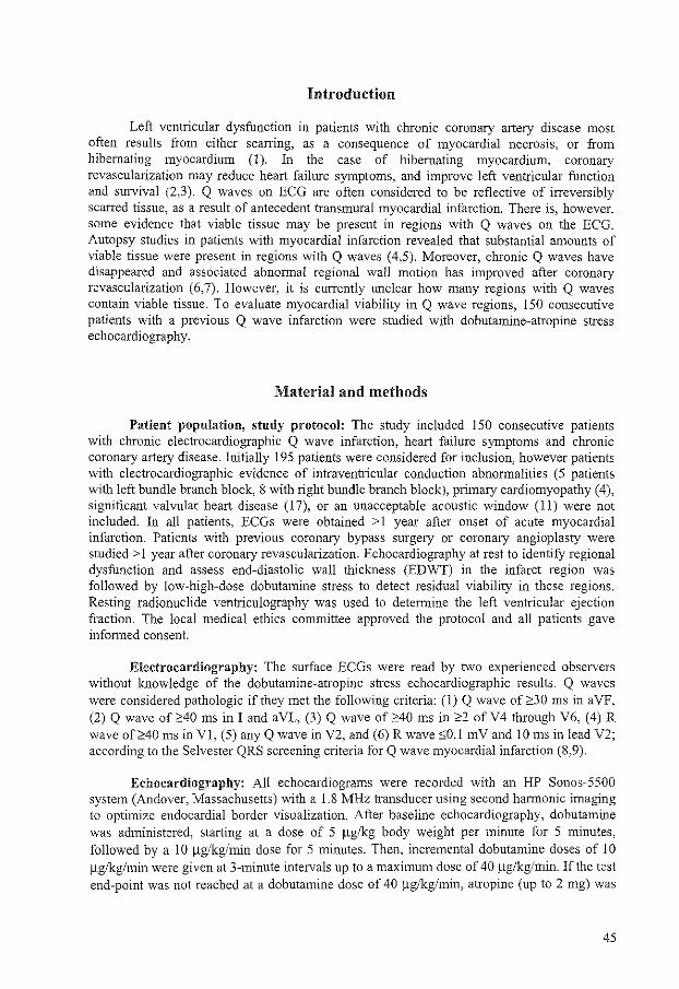
Introduction
Left ventricular dysfunction in patients with chronic coronary artery disease most often results from either scarring, as a consequence of myocardial necrosis, or from hibernating myocardium (1). In the case of hibernating myocardium, coronary revascularization may reduce heart failure symptoms, and improve left ventricular function and survival (2,3). Q waves on ECG are often considered to be reflective of irreversibly scarred tissue, as a result of antecedent transmural myocardial infarction. There is, however, some evidence that viable tissue may be present in regions with Q waves on the ECG. Autopsy studies in patients with myocardial infarction revealed that substantial amounts of viable tissue were present in regions with Q waves ( 4,5). Moreover, chronic Q waves have disappeared and associated abnormal regional wall motion has improved after coronary revascularization (6,7). However, it is currently unclear how many regions with Q waves contain viable tissue. To evaluate myocardial viability in Q wave regions, 150 consecutive patients with a previous Q wave infarction were studied with dobutamine-atropine stress echocardiography.
Material and methods
Patient population, study protocol: The study included 150 consecutive patients with chronic electrocardiographic Q wave infarction, heart failure symptoms and chronic coronary artery disease. Initially 195 patients were considered for inclusion, however patients with electrocardiographic evidence of intraventricular conduction abnormalities (5 patients with left bundle branch block, 8 with right bundle branch block), primary cardiomyopathy (4), significant valvular heart disease (17), or an unacceptable acoustic window (11) were not included. In all patients, ECGs were obtained > 1 year after onset of acute myocardial infarction. Patients with previous coronary bypass surgery or coronary angioplasty were studied > 1 year after coronary revascularization. Echocardiography at rest to identify regional dysfunction and assess end-diastolic wall thickness (EDWT) in the infarct region was followed by low-high-dose dobutamine stress to detect residual viability in these regions. Resting radionuclide ventriculography was used to determine the left ventricular ejection fraction. The local medical ethics committee approved the protocol and all patients gave informed consent.
Electrocardiography: The surface ECGs were read by two experienced observers without knowledge of the dobutamine-atropine stress echocardiographic results. Q waves were considered pathologic if they met the following criteria: (1) Q wave of2:30 ms in aVF, (2) Q wave of240 ms in I and aVL, (3) Q wave of240 ms in 22 ofV4 through V6, (4) R wave of240 ms in Vl, (5) any Q wave in V2, and (6) R wave o;O,l mV and 10 ms in lead V2; according to the Selvester QRS screening criteria for Q wave myocardial infarction (8,9).
Echocardiography: All echocardiograms were recorded with an HP Sonos-5500 system (Andover, Massachusetts) with a 1.8 MHz transducer using second harmonic imaging to optimize endocardial border visualization. After baseline echocardiography, dobutamine was administered, starting at a dose of 5 f.Lglkg body weight per minute for 5 minutes, followed by a l 0 ~g/kg/min dose for 5 minutes, Then, incremental dobutamine doses of l 0 ~g/kg/min were given at 3-minute intervals up to a maximum dose of 40 ~g/kg/min. If the test end-point was not reached at a dobutamine dose of 40 f.Lglkg/min, atropine (up to 2 mg) was
45

given intravenously. Test end-points were: achievement of target heart rate, extensive new wall motion abnormalities, ST -segment depression (2:2mm compared with baseline), severe angina, systolic blood pressure fall >40 uuu Hg, blood pressure >2401\20 mm Hg, significant arrhythmia.
Echocardiographic analysis: Two experienced reviewers unaware of the electrocardiographic data scored the echocardiograms using a standard 16-segment model (10). To relate the echocardiographic data to the Q waves on ECG, the left ventricle was divided into four major regions: anterior, septal, lateral, and inferoposterior. Measurement of EDWT and the scoring of regional function and response to dobutamine infusion were performed by two observers, blinded to the ECG data.
1. End-diastolic wall thickness: The resting echocardiographic images were analyzed off-line using standard parasternal and apical views. EDWT was assessed as previously described (ll, 12). The frames that provided best possible visualization of the endocardial and epicardial borders were selected. The EDWT was assessed at the center of each myocardial region from the leading endocardial edge to the leading epicardial edge and calculated as the mean value of three measurements. According to the EDWT, viability was considered absent when EDWT was s6 mm, and present when EDWT was >6 uuu (11,12).
2. Regional function and response to dobutamine: Regional wall motion and systolic wall thickening were scored on a 5-point scale: (l) normal, (2) mild hypokinetic, (3) severe hypokinetic, (4) akinetic and (5) dyskinetic. Myocardial segments were considered normal if the regional wall motion was normal or mild hypokinetic. Only dysfunctional segments (segments with severe hypokinesia, akinesia or dyskinesia at resting echocardiography) were evaluated for myocardial viability. Segments with improvement, worsening or a biphasic wall motion response during dobutamine stress echocardiography were considered viable (13). Segments with unchanged wall motion were considered nonviable (\3). For each patient a wall motion index score (WMSI) was calculated (total wall motion score divided by the number of analyzed segments) at rest, at low-dose, and at peak stress.
Assessment of the ejection fraction: The left ventricular ejection fraction was assessed by radionuclide ventriculography at rest in all patients. A small field-of-view gamma camera system (Orbiter, Siemens, Erlangen, Germany) was used, oriented in a 45° left anterior oblique position with a 5 to 10° caudal tilt. After injection of Tc-99m (740 MBq), radionuclide ventriculography was performed at rest with the patient in supine position. The left ventricular ejection fraction was calculated using standard methods (Odyssey VP, Picker, Cleveland, Ohio) (13).
Statistical analysis: All continuous data are expressed as mean ± SD, percentages are rounded. Continuous variables were compared using the student t-test for unpaired samples. Differences between proportions were compared using the chi-square test. A value of p<0.05 was considered statistically significant.
46

Results
Patient population, electrocardiographic results: All patients presented with heart failure symptoms, New York Heart Association (NYHA) functional class was on average 2.8 ± 0.7, while 72% of the patients was in NYHA functional class Ill or IV. The left ventricular function was severely impaired in all patients, the ejection fraction averaged 31% ± 12%. Importantly, 40 patients (27%) had a left ventricular ejection fraction <25%. The clinical characteristics of the study population are presented in Table 1.
All 150 patients had a previous Q wave myocardial infarction at least one year before the study (mean 7.2 ± 5.9 years before). Pathological Q waves on the echocardiogram were present in 220 myocardial regions: 101 anterior, 30 septal, 30 lateral, and 59 infero-posterior regions exhibited Q waves. On average patients had 1.5 ± 0.6 regions with a Q wave pattern.
Table 1. Demographics of 150 patients undergoing dobutamine stress echocardiography
Age (yrs) Male Previous CABG > 1 year Previous PTCA > 1 year Previous cerebrovascular disease Diabetes mellitus Family history Smoking previous/current Multivessel disease NYHA functional class LVEF (%)
59± 10 125 (83%) 22 (15%) 22 (15%) 2 (1%) 23 (15%) 91 (61%) 58 (39%) I 55 (35%) 137 (91 %) 2.8 ± 0.7 31 ± 12
Data are presented as mean± SD or number(%) of patients; CABG =coronary artery bypass graft surgery; LVEF ~left ventricular ejection fraction; MI ~myocardial infarction; NYHA = New York Heart Association; PTCA =percutaneous transluminal coronary angioplasty.
Resting echocardiography: Echocardiography at rest revealed 517 dysfunctional myocardial regions, 202 of the dysfunctional regions were related to Q waves on the ECG, the other 315 dysfunctional regions were not. Thus, 202 out of 220 regions (92%) with Q waves were dysfunctional at resting echocardiography. In addition, 315 regions not related to Q waves on the ECG revealed abnormal resting wall motion.
The EDWT was preserved (>6 mm) in 498 of 517 (96%) dysfunctional regions. Figures 1A and lB demonstrate the obtained data in regions without and with chronic Q waves, respectively. An EDWT ::::;6 mm was more frequently observed in Q wave regions than in dysfunctional regions without Q waves (13 of202 versus 6 of315, p<0.05).
47
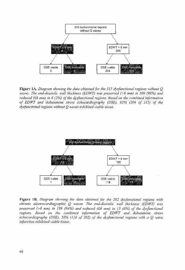
315 dysfunctional regions without Q waves
DSE viable 204
Figure lA. Diagram showing the data obtained for the 315 dysfunctional regions without Q waves. The end-diastolic wall thickness (EDWT} was preserved (>6 mm) in 309 (98%) and reduced (~6 mm) in 6 (2%) of the dysfunctional regions. Based on the combined information of EDWT and dobutamine stress echocardiography (DSE), 65% (204 of 315) of the dysfunctional regions without Q waves exhibited viable tissue.
Figure lB. Diagram showing the data obtained for the 202 dysfimctional regions with chronic electrocardiographic Q waves. The end-diastolic wall thickness (EDWT) was preserved (>6 mm) in 189 (94%) and reduced (:>6 mm) in 13 (6%) of the dysfimctional regions. Based on the combined information of ED WT and dobutamine stress echocardiography (DSE), 58% (118 of 202) of the dysfimctional regions with a Q wave infarction exhibited viable tissue.
48

Dobutamine stress echocardiography: Table 2 shows the hemodynamic and wall motion changes during dobutamine stress. There was a significant increase in heart rate at baseline compared to the heart rate at peak stress (74 ± 16 vs. 130 ± 18 beats/min, p<O.OOO 1 ). Overall, diastolic blood pressure decreased (75 ± 14 to 72 ± 15 mm Hg, p<0.05) and systolic blood pressure did not significantly change during dobutamine infusion. During low-dose dobutamine infusion, wall motion improved significantly, (baseline WMSI 2.81 ± 0.71 vs. low-dose WMSI 2.50 ± 0.66, p<O.OOOl). At peak stress, WMSI worsened compared to the WMSI at the low-dose stage (2.74 ± 0.69 vs. 2.50 ± 0.66, p<0.005). Regions with EDWT :06 mm virtually never exhibited viability on dobutamine stress echocardiography, as indicated in Figures lA and lB. In regions with EDWT >6 mm, the response to dobutamine varied. In regions with a Q wave infarction, 62% exhibited viability on stress echocardiography, whereas 38% of the regions demonstrated scar tissue. In regions without a Q wave, 66% exhibited viable tissue and 34% scar tissue on dobutamine stress echocardiography.
Table 2. Dobutamine stress echocardiographic data.
Heart rate (beats/min) Systolic BP (mm Hg) Diastolic BP (mm Hg) WMSI
Baseline
74 ± 16 126 ± 26 75 ± 14 2.81 ± 0.71
Low-dose
76 ± 18 125 ± 28 74 ± 15 2.50 ± 0.66
Peak
130 ± 18 126 ± 28 72 ± 15 2.74 ± 0.69
Data are presented as mean± SD or number (%) of patients. BP =blood pressure; WMSI =
wall motion score index.
Qwave n=202
0 Viable II Nonviable
No Qwave n=315
Figure 2. Pie chart showing the incidence of viable myocardium in 517 dysfunctional regions; combined information of end-diastolic wall thickness and dobutamine stress echocardiography demonstrated residual viability in 58% (118 of 202) of the dysfunctional regions with a Q wave pattern, and in 65% (204 of 315) dysfunctional regions without Q waves on the ECG (p~0.2).
49
Based on the combination of the EDWT and the response to dobutamine, 58% ( 118 of 202) of the regions with a Q wave infarction exhibited viable tissue. In addition, in 65% (204 of 315) of the dysfunctional regions not related to Q waves on the ECG, viable tissue was present. The incidence of viable tissue in dysfunctional regions with a Q wave infarction was not different from the incidence of dysfunctional regions without a Q wave infarction (Figure 2).
Discussion
The findings in the present study demonstrate that chronic Q waves on the ECG do not exclude the presence of viable myocardial tissue in patients with severely depressed left ventricular function. An EDWT 56 rnm, indicating scar tissue, was more frequently observed in Q wave regions than in dysfunctional regions without Q waves. In general, regions with an EDWT o£6 mm were scarce. Only 19 of 517 regions had an EDWT o£6 mm. La Canna et al (12), comparably showed that only 13% of dysfunctional regions in patients with severely depressed left ventricular function had an EDWT <5 mm. In line with these previous studies, regions with an EDWT :56 mm virtually never exhibited viable tissue on dobutamine echocardiography. Cwajg et al (11) showed that only 19% of the regions with an EDWT o£6 mm was viable during dobutamine stimulation. Baer et al (14) used magnetic resonance imaging and demonstrated that regions with EDWT <5 mm never improved in contractile function after revascularization, indicating that regions with very thin walls do not contain viable tissue and do not improve in function after revascularization. In regions with an EDWT >5.5 mm however, the response to dobutamine varied significantly. In our study, only 62% of the Q wave regions with an EDWT >6 rnm was viable on dobutamine stress echocardiography, whereas 38% of these regions exhibited scar tissue on dobutamine echocardiography. Similarly, La Canna et al (12) demonstrated that 42% of the dysnmctional segments with a preserved EDWT (>5 mm) was nonviable on dobutamine stress echocardiography. Moreover, Baer et al. (14) demonstrated that 38% of the segments with an EDWT >5.5 mm did not improve in function post-revascularization. Based on these results it can be concluded that assessment of EDWT can be used as an initial screening technique for assessment of viability. It can safely be concluded that regions with an EDWT :56 rnm do not contain viable myocardium and do not improve in function after revascularization. However, in segments with an EDWT >6 mm, additional testing is needed, since approximately 40% of these regions do not contain viable myocardium and will not improve in function postrevascularization.
The aim of the current study was to evaluate the incidence of viable tissue in regions with Q wave infarctions. Previous studies in small subsets of patients have already indicated that some patients with Q wave infarctions have viable myocardium. Brunken et al (15) showed in 20 patients that positron emission tomography using Fl8-fluorodeoxyglucose revealed residual glucose metabolism in 21 of 31 ( 68%) Q wave regions. Similarly, Tillisch et al (16) evaluated 17 patients with positron emission tomography in combination with F18-fluorodeoxyglucose and showed that 16 of 28 (57%) regions with Q waves on the ECG improved in contractile function after revascularization. Besides these 2 studies, no studies have focused on the incidence of viable myocardium in patients with Q wave infarctions. In the present study, a combination of resting echocardiography (to assess EDWT) and dobutamine stress echocardiography was used to assess viability, in a large cohort of patients. These echocardiographic techniques are more widely available than positron emission tomography. Based on the combined information of EDWT and the findings during dobutamine infusion, the incidence of viable myocardium in 150 patients with Q wave infarctions was 58%, in line with the previous studies. Hence, electrocardiographic evidence
50
of "transmural" infarction (Q waves) together with regional dysfunction at rest does not exclude the presence of viable myocardium. In particular, the incidence of viable myocardium in regions with Q wave infarction was comparable to the incidence of viable myocardium in regions without a Q wave (Figure 2). This finding is of clinical importance, since many patients present with Q wave infarctions, depressed LV function and heart failure symptoms. In the presence of viable myocardium, these patients may benefit from revascularization in terms of improvement of left ventricular function and longevity (17-20). As evidenced by the high incidence of viable myocardium in regions with Q wave infarctions in this large cohort of patients, viability testing should be performed and Q waves in combination with contractile dysfunction per se does not rule out the presence of viable myocardium.
Limitations: In the current study myocardial viability was assessed by dobutamineatropine stress echocardiography. This is a safe and widely available imaging method, with a good sensitivity and specificity for the evaluation of myocardial viability (17). Still, functional improvement of contractile dysfunction after coronary revascularization is considered the final proof of myocardial viability. In this study functional improvement after coronary revascularization was not assessed.
Patients who underwent previous coronary revascularization were included in the study. Coronary revascularization might have influenced the presence and the number of Q waves as well as the presence of viable myocardium. However, the time-interval between revascularization and the current study was > 1 year in all patients.
Conclusions: Chronic Q waves on electrocardiography do not exclude the presence of viable myocardium; in 58% of the Q wave regions in 150 patients, viable tissue was present. Thus, patients presenting with a previous Q wave infarction, severely depressed left ventricular function and heart failure should be referred for viability testing. Initial evaluation by resting echocardiography may already exclude the presence of viable tissue when EDWT is 6 mm or less. However, in regions with Q waves and an EDWT >6 rnrn, additional testing is needed since 38% of these regions did not show viability during dobutamine stress echocardiography
References
1. Beller GA. Noninvasive assessment of myocardial viability. N Eng J Med 2000;343: 1488-90.
2. Rahimtoola SH. From coronary artery disease to heart failure: role of the hibemating myocardium. Am J Cardioll995;75:16E-22E.
3. Wijns W, Vatner SF, Camici PG. Hibernating myocardium. N Eng J Med 1998;339:173-81.
4. Savage Rlv1, Wagner GS, Ideker RE, et al. Correlation of postmortem anatomic findings with electrocardiographic changes in patients with myocardial infarction: retrospective study of patients with typical anterior and posterior infarcts. Circulation 1977;55:279-85.
5. Sullivan W, Vlodaver Z, Tuna N, et al. Correlations of echocardiographic and pathologic findings in healed myocardial infarction. Am J Cardioll978;42:724-32.
6. Zeft HJ, Friedberg HD, King JF, et al. Reappearance of anterior QRS forces after coronary bypass surgery. An Electrovectorcardiographic study. Am J Cardiel 1975;36:163-8.
7. Conde CA, Meller J, Espinoza J, et al. Disappearance of abnonnal Q waves after aortocoronary bypass surgery. Am J Cardioll975;36 :889-93.
51

8. Anderson WD, Wagner NB, Lee KL, et al. Evaluation of a QRS scoring system for estimating myocardial infarct size, VI: identification of screening criteria for non-acute myocardial infarcts. Am J Cardio\1988;61:729-33.
9. Sevilla D, Wagner N, Anderson WD, et al. Sensitivity of a set of myocardial infarction screening criteria in patients with anatomically documented single and multiple infarcts. Am J Cardio\1990;66:792-5.
10. Bourdillon PD, Broderik TM, Sawada SG, et al. Regional wall motion index for infarct and non-infarct region after reperfusion in acute myocardial infarction: comparison with global wall motion index. JAm Soc Echo !989;2:398-407.
11. Cwajg JM, Cwajg E, Nagueh SF, et al. End-diastolic wall thickness as a predictor of recovery of function in myocardial hibernation: relation to rest-redistribution Tl-201 tomography and dobutamine stress echocardiography. J Am Col! Cardia! 2000;35: !152-61.
12. La Canna G, Rahimtoola SH, Visioli 0, et al. Sensitivity, specificity, and predictive accuracies of non-invasive tests, singly and in combination, for diagnosis of hibernating myocardium. Eur Heart J 2000;21: !358-67.
13. Bax JJ, Polderrnans D, Elhendy A, et al. Improvement ofleft ventricular ejection fraction, heart failure symptoms and prognosis after revascularization in patients with chronic coronary artery disease and viable myocardium detected by dobutamine stress echocardiography. JAm Col! Cardioll999;34:163-9.
14. Baer FM, Theissen P, Schneider CA, et al. Dobutamine magnetic resonance imaging predicts contractile recovery of chronically dysfunctional myocardium after successful revascularization. J Am Col! Cardia! !998;3!: 1040-8.
15. Brunken R, Tillisch J, Schwaiger M, et al. Regional perfusion, glucose metabolism, and wall motion in patients with chronic electrocardiographic Q wave infarctions: evidence for persistence of viable tissue in some infarct regions by positron emission tomography. Circulation !986;73:95!-63.
!6. Tillisch J, Brunken R, Marshall R, et a!. Reversibility of cardiac wall-motion abnormalities predicted by positron tomography. New Eng! J Med 1986;314:884-8.
!7. Bax JJ, Wijns W, Cornel JH, et a!. Accuracy of currently available techniques for prediction of functional recovery after revascularization in patients with left ventricular dysfunction due to chronic coronary artery disease: comparison of pooled data. J Am Coll Cardiol1997;30:!45!-60.
18. Pasquet A, Robert A, D'Hondt Al\11, et al. Prognostic value of myocardial ischemia and viability in patients with chronic left ventricular ischemic dysfunction. Circulation !999; 100: !4!-8.
19. Cigarroa CG, deFilippi CR, Brickner E, et al. Dobutamine stress echocardiography identifies hibernating myocardium and predicts recovery of left ventricular function after coronary revascularization. Circulation 1993;88:430-6.
20. La Canna G, Alfieri 0, Guibbini R, et a!. Echocardiography during infusion of dobutamine for identification of reversible dysfunction in patients with coronary artery disease. JAm Col! Cardio\1994;23:617-26.
52
Chapter 5
Assessment of viable tissue in Q-wave
regions by metabolic imaging using single
photon emission computed tomography in
ischemic cardiomyopathy
Schinkel AFL, Bax JJ, Elhendy A, Boersma E, Vourvouri EC,
Sozzi FB, Valkema R, Roelandt JRTC, Poldermans D
Am J Cardiol2002;89:ll7l-ll75
53

Assessment of Viable Tissue in Q-Wave Regions !by MetalboUe Imaging Using
Single-Photon Emission Computed Tomography illn Ischemic Cardiomyopathy
Arend F.L. Schinkel, MD, Jeroen J. Box, MD, Abdou Elhendy, MD, Eric Boersma, PhD, Eleni C. Vourvouri, MD, Fa biola B. Sozzi, MD, Roelf Valkema, MD,
Jos R.T.C. Roelandt, MD, and Don Poldermans, MD
Chronic electrocardiographic Q waves ore often believed to reflect irreversibly scarred, transmurally infarcted myocardium. The aim of this study was to evaluate whether residual viable tissue persists in dysfunctional myocardial regions related to chronic Q
waves on the surface electrocardiogram. A total of 148 patients with healed myocardial infarction and impaired left ventricular (LV) function with heart failure symptoms undetwent electrocardiography and metabolic imaging using technetium {Tc-99m) tetrofosmin/F 18-fluorodeoxyglucose (FDG) single-photon emission computed tomography {SPECn. The left ventricle was divided into 4 major regions to compare myocardial viability in re-o gions with and without chronic Q waves on surface electrocardiography. According to FOG SPECT metabolic
t raditionally, left ventricular (LV) dysfunction due to chronic coronary artery disease was considered
irreversible and amenable only to medical therapy. 1
However, it has become clear that chronic LV dysfunction in patients with chronic coronary artery disease is not always irreversible.2
'3 If jeopardized but
viable myocardium is present, coronary revascularization may lead to improved regional and global function.4-7 Moreover, improvement of functional status and better survival may be expected if patients with a substantial amount of dysfunctional but -viable myocardium are revascularized.4-7 Chronic electrocardiographic Q waves are often believed to reflect irre-versibly scarred, transmurally infarcted myocardium. There is, however, some evidence that residual viable myocardium may be present in dysfunctional regions with chronic Q waves on electrocardiography. Pathologic studies in patients with myocardial infarction suggested that substantial amounts of hibernating tissue may persist in regions with Q waves. 8'
9 In addition, disappearance of chronic Q waves and improve~
from the Deparimerts of Cardiology and Nuclear Medicine, Erasmus Medical Center, Ro~erdam; onci the Department of Cardiology, Lei· den Univers;ty Medical Center, leiden, The Netherlands. ManuscripT received October 31, 2001; revised manuscript received and ac· cepted February 6, 2002.
Address for reprints: Don Polder'T.ans, MD, PhD, Tharaxcenter, Roam Ba 300, Deportment of Cardiology, Erasmus Medical Center, Dr. Molewaterplein 40, 3015 GD Ro~e'dom, The Netherlands. Email: [email protected].
imaging, residual viable tissue persisted in a high proportion (61%) of dysfunctional myocardial regions with chronic Q waves. Regions with chronic Q waves were more often dysfunctional than regions without Q waves. Moreover, dysfunctional regions with chronic Q waves were less frequently viable compared with dysfunctional regions without Q waves on the electrocardiogram. This study demonstrates that chronic Q waves on eiectrocar· diography do not necessarily imply irreversib-ly scarred myocardium. Residual viable tissue persists in a hig~ proportion of dysfunctional ventricular regions according to FDG SPECT metabolic imaging. ©2002 by Excerpta Medica, Inc.
(Am J Card;ol 2002;89:1171-1175)
ment of LV function and contractility have been observed after coronary revascularization. 10
·11 It is
currently not clear in how many ventricular regions with Q waves viable myocardium persists. Accordingly, this study evaluates myocardial viability in Qwave regions in a consecutive series of patients with depressed LV ejection fraction (EF) and heart failure.
METHODS Patient popukltion and study protocol: The study
population consisted of 148 consecutive patients with chronic coronary artery disease and an impaired LV function, who had heart failure as the predominant symptom. All patients had a previous myocardial infarction >6 months before viability assessment Patients with left bundle branch block, primary cardiomyopathy, or concomitant significant valvular disease were not included. Patients with previous coronary revascularization were studied > 1 year after the revascularization procedure. All patients underwent echocardiography at rest to identify dysfunctional myocardial tissue and dual-isotope simultaneous acquisition myocardial single-photon emission computed tomography (SPECT), including technetium (Tc-99m) tetrofosmin and Fl8-ftuorodeoxyglucose (FDG) to assess myocardial perfusion and glucose utilization, respectively. The L VEF was assessed by radionuclide ventriculography. Systemic hypertension was defined as a blood pressure 2::140/90 mrn Hg, ot treatment with antihypertensive medication. Hyper-
55

cholesterolemia was defined as a total cholesterol ~6.4 mmol!L, or treatment with lipid-lowering medication. The hospital medical ethics committee approved the protocol and all patients gave informed consent.
Electrocardiographic analysis: Two experienced observers, illlaware of any other data, read the surface electrocardiograms. According to the Selvester QRS screening criteria for Q-wave myocardial infarction, Q waves were classified as pathologic if there was: (1) a Q wave of ~30 ms in lead a VF; (2) a Q wave of ~40 ms in leads I and aVL; (3) a Q wave of~40 ms in ~2 of leads V 4 to V6 ;
(4) a R wave of ~40 ms in lead V 1; (5) any Q wave in lead V2; and (6) R wave ::S0.1 mV and ~10 ms in lead V 2.
12•13 Accordingly, pathologic Q waves were assigned to
4 LV regions: anterior (preserved R wave in lead V1 and pathologic Q wave in ~1 ofleads V2 to V5), septal (pathologic Q waves in leads V1, V2, and/or V3), lateral (pathologic Q wave in > 1 of leads I, a VL, or V 6), or inferoposterior (pathologic Q wave in > 1 of leads II, lli, or a VF).14
Assessment of wall motion abnormalities: A Hewlett Packard Sonos-5500 imaging system (Andover, Massachusetts), equipped with a 1.8-MHz transducer using second harmonic imaging to optimize endocardial border visualization, was used to record 2-dimensional echocardiograms. Four standard views were recorded, and 2 experienced reviewers blinded to the SPECT data visually scored the digitized echocardiograms. Regional wall motion and systolic wall thickening were scored using a 16-segment model (Figure 1) and a 5-point grading scale: 1 = normal, 2 = mildly hypokinetic, 3 = severely hypokinetic, 4 = akinetic, and 5 = dyskinetic. 15
SPECT data acquisition: Patients received, after a light breakfast, an intravenous injection of Tc-99mtetrofosmin ( 600 MBq) to evaluate perfusion at rest. FDG imaging, to evaluate glucose utilization, was performed following administration of Acipimox (Byk, Zwanenburg, The Netherlands; 500 mg, oral dose) in aU patients. Acipimox enhances myocardial FDG uptake by reducing the plasma level of free-fatty acids. Following Acipimox administration, the patients received a low-fat, carbohydrate-rich meal. This small meal further enhances myocardial FDG uptake, by stimulating endogenous insulin release. Several studies have demonstrated very good image quality using Acipimox. 16
-18 Sixty minutes after the meal,
FDG (185 MBq) was injected, and after an additional 45 minutes to allow cardiac FDG uptake, dual-isotope simultaneous acquisition SPECT was performed. Perfusion and metabolic imaging were performed at rest without stressors. A triple-head gamma camera system (Picker Prism 3000XP, Cleveland, Ohio) was used. The camera system was equipped with commercially available high-energy 511-keV collimators. 19
The energies were centered on the 140-ke V photon peak of Tc-99m-tetrofosmin with a 15% window and on the 511-keV photon peak of FDG with a 15% window. Data acquisition was performed in the supine position, over 360° (120 sectors of3°). Total imaging time was 32 minutes. Data were stored in a 64 X 64, 16-bit matrix.
56
PS LAX PSSAX
AP4CH AP2CH
RA LA LA
FIGURE I. LV wall segments in the 16-segmenl model. Four ma· ior regions were considered: the anterior region comprised segments 2, 7, and 12.; the septal region consisted of segmenis 1, 5, 6, 10, and 11; the lateral region comprised segments 3, 8, and 13; and the inferoposterior region consisted of segmenis 4, 9, 14, 15, and 16. AO =aorta; AP2CH = apical2-chamber view; AP4CH = apical4-chamber view; lA = left atrium; LV= left ventricle; PSLAX = poraslernallong·oxis view; PSSAX = poraslemal short-oxis view; RA = right atrium; RV = right ven· tride.
SPECT data reconstruction and analysis: From the raw scintigraphic data, 6-mm-thick (1 pixel) transaxial slices were reconstructed by filtered backprojection using a Butterworth filter (cut-off frequency at 0.17 cycle/pixel of order 3.5). Attenuation correction was not applied. Further reconstruction yielded standard short- and long-axis projections perpendicular to the heart axis. The Tc-99m-tetrofosmin and the FDG data were reconstructed simultaneously, to obtain an exact alignment of the perfusion and metabolic images. The perfusion and FDG studies were adjusted to peak myocardial activity (100%). A similar 16-segment model as for the echo data was used (Figure 1). Only segments with severe hypokinesia, akinesia, or dyskinesia were evaluated for myocardial viability. Both T c-99m-tetrofosmin and FDG uptake defects were semiquantitatively graded on a 4-point scale: 0 = normal, 1 = mildly reduced, 2 = moderately reduced, and 3 = severely reduced or absent. Myocardium with a Tc-99m-tetrofosmin uptake score ::s1 or a reduction in Tc-99m-tetrofosmin uptake score more severe than the reduction in FDG activity by ~1 point (mismatch pattern) was considered viable. Dysfunctional myocardium with concordantly reduced Tc-99m-tetrofosmin and FDG uptake (match pattern) was considered nonviable.

TABlE 1 Clinical Chorocteristics of the 148 Study Patients
Men/women 125(84%)/23 {16%) Age (yrs) 60 :':: 9 Systemic hypertension 20 {14%) Diabetes mellitus 24 {16%) Hypercholesterolemia 67 {45%) Smoking history
Previous 58 {39%) Current 54 {36%)
family history of coronary artery disease 90 {61%) Previous cerebrovascular disease 2 {1%) Previous coronary revascularization 39 {26%) Multivessel coronary disease 134(91%) LVEF {%) 31:!: 11, range 10-43
Dolo ere presented os number [%1 of patients or mean value :t SD See Method, far definition of hyperlen•ion and hyperchobterolemio
Comparison of electrocardiography, echocardiography, and SPECT: Q waves were ascribed to 4 regions: anterior, septal, lateral, and inferoposterior. Accordingly, the corresponding SPECT and echo segments (both using the same 16-segment model, Figure I) were grouped according to these 4 regions. A region was considered dysfunctional if ~ 1 segment in the region was severely hypokinetic, akinetic, or dyskinetic on echocardiography. A region was considered viable when ~ 1 segment in the region was viable on SPECT (see the previously mentioned criteria).
Assessment of the left ventricular ejection fraction: To assess the L VEF at the time of the viability testing, radionuclide ventriculography at rest was performed in all patients. A small field-of-view gamma camera system (Orbiter, Siemens, Erlangen, Germany) was used, oriented in a 45° left anterior oblique position with a 5° to 10° caudal tilt. After injection of Tc-99m (740 MBq), radionuclide ventriculography was performed at rest with the patient in a supine position. The LVEF was calculated by standard methods (Odyssey VP, Picker, Cleveland, Ohio).20
Statistical analysis: All continuous data are expressed as mean ::±:: SD, percentages are rounded. Continuous variables were compared using the Student's t test for unpaired samples. Differences between proportions were compared using the chisquare test. A p value of <0.05 was considered statistically significant.
RESULTS The clinical characteristics of the patients are listed
in Table l. Electrocardiographic results: An abnonnal Q-wave
pattern was present in 216 myocardial regions (101 anterior, 29 septal, 28 lateral, and 58 inferoposterior). The patients had on average 1.5 :±: 0.6 regions associated with a Q-wave pattern on the electrocardiogram.
Wall motion: Of a total of216 Q-wave regions, 200 (93%) had an abnormal wall motion pattern. Most of the regions without Q waves also exhibited an abnormal wall motion pattern (326 of 376 regions, 87%). Hence, most of the regions had an abnormal wall
motion pattern, but regions with Q waves were more often dysfunctional compared with regions without Q waves (p <0.05). Regions with abnonnal wall motion had an average of2.7 :±: 1.1 dysfunctional segments: 1.5 ::±:: 0.8 severely hypokinetic, 1.2 .::':: 1.0 akinetic, and 0.03 :±: 0.21 dyskinetic segments. Overall, there were 526 dysfunctional regions, with 200 related to chronic Q waves on the electrocardiogram.
FOG SPECT: According to metabolic imaging with FDG SPECT, 365 of the 526 dysfunctional myocardial regions (69%) were classified as viable. There was a significant difference between the proportion of dysfunctional but viable regions between regions with and without Q waves. However, in a high proportion of regions with an abnormal wall motion and pathologic Q waves on the electrocardiogram, persistent viable myocardium was identified. FDG SPECT metabolic imaging demonstrated persistent viable myocardial tissue in 122 of the 200 dysfunctional regions (61%) with a Q-wave pattern, and in 243 of 326 dysfunctional regions (75%) without Q waves on the electrocardiogram (p <0.005). There were on average 1.4 .::':: 1.0 viable segments in a region. Figure 2 shows the incidence of viable myocardium in dysfunctional regions with and without chronic Q waves.
DISCUSSION Chronic electrocardiographic Q waves are often
believed to reflect scarred tissue. However, there is evidence that viable tissue may be present in dysfunctional myocardium related to chronic Q waves on the electrocardiogram. 8 -
11 The present study demonstrates that chronic Q waves on electrocardiography do not necessarily imply irreversible myocardial damage. According to FDG SPECT metabolic imaging, viable tissue persisted in a high proportion (61%) of dysfunctional myocardial regions with chronic Q waves. The persistence of viable tissue in Q-wave infarcted regions has important implications for clinical decision making, because multiple studies4
-720
have demonstrated that residual myocardial viability is an important dctcnninant of outcome after coronary revascularization. In the presence of persistent viable myocardium, revascularization can improve LV function, reduce heart failure symptoms, and improve perioperative and late surviva\.4
-7
"20 Moreover, pa
tients with LV dysfunction as a result of jeopardized but viable myocardium have a better prognosis following coronary revascularization than after medical therapy.21 The current findings suggest that, in patients with LV dysfunction as a result of Q-wave infarcted myocardium, viability testing should not be omitted simply because chronic Q waves are present on the electrocardiogram. FDG SPECT can be a useful noninvasive tool that enables physicians to identify those patients who will benefit most from coronary revascularization.
In the present study, residual viable tissue was present in a large proportion of dysfunctional regions with Q waves. Thus far, data on myocardial viability in Q-wave infarction are limited. Hashimoto et af2
studied 22 patients with FDG and positron emission
57

Qwave n=200
No Q wave n=326
FDG imaging using SPECT cameras equipped with high-energy collimators was developed. Several studies have compared FDG SPECT to FDG positron emission tomography, showing excellent agreement between the 2 techniques, whereas
D Viable 1111 Nonviable
243 (75%)
other studies have shown the accuracy of FDG SPECT to predict functional recovery.25
-29 The availability
of FOG SPECT may contribute to a more widespread use of FDG for the assessment of viability in patients with LV dysfunction.
Study limitations: This study has several limitations. The first limitation is the lack of quantitative information on myocardial perfusion and metabolism. Second, the correlation between
FIGURE 2. The proportion of viable myooordium in 526 dysfunctional ventricular regions according to FDG SPECT imaging. Viable tissue persisted in 122 of the 200 dysfunctional regions (61%) with chronic electrocarcliogrophic Q waves, ond in 243 of 326 dysfunctional regions (75%) without a Q-wave pattern (p <0.005).
electrocardiographic Q waves and the infarct location may not be exact. Third, coronary revascularization may have influenced the presence and number of Q waves as well as the detection
tomography. Myocardial viability in the infarcted region was observed in 36% of the patients with a Q-wave infarction. Tillisch et af3 studied 15 patients with LV dysfunction and Q waves using FDG positron emission tomography. In 54% of dysfunctional regions with a Q wave on the electrocardiogram, metabolic imaging demonstrated viable myocardium. Brunken et al14 studied 20 patients with FDG positron emission tomography and showed viable myocardium in 68% of the Q-wave regions. The present findings using FDG SPECT imaging are consistent with the previous observations with FDG positron emission tomography and revealed viable myocardium in 61% of the regions with a Q-wave infarction.
Assessment of viability by FOG SPECT: Since the introduction of the concept of myocardial viability, 2•
3
several noninvasive methods have been developed to identify jeopardized but viable myocardium. Various nuclear imaging techniques can be used to assess whether characteristics of viable myocardium, cell membrane integrity, or residual metabolic activity, are present in dysfunctional myocardium. Many studies have reported on the high diagnostic accuracy of FDG imaging with positron emission tomography to assess myocardial viability. Preserved myocardial uptake of FDG in combination with reduced myocardial perfusion (flow-metabolism mismatch) is indicative of viable myocardium. Matched defects are considered nonviable scar tissue. In patients with severe LV dysfunction, preoperative viability assessment using FDG positron emission tomography is a well-established method to predict functional recovery after coronary revascularization?4 However, positron emission tomography is not widely available, and the number of patients with LV dysfunction who require viability testing is growing. Therefore, as a widely available alternative to positron emission tomographic imaging,
58
of viable myocardium. Finally, functional outcome after coronary revascularization was not studied.
1. Wijru; W. Vatner SF, Camici PG. Hibemating myocardium. N Eng! J Med 1998;339:173-181. 2. Diamond GA. For=;ter JS. dcLuz PL. Wyatt HL, Swan HJC. Postextrasystolic pot~ntiation of ischemic myocardium by atrial stimulation. Am Heart J 1978;95: 204-209. 3. Rahimtoola Sli. A perspective on the three large multicenter randomized clinical trials of coronary bypass surgery for cflronic stable angina. Circulmion 1985;72(suppl \'}:123-135. 4. Eitzman D. Al·Aouar Z, Kanter H, vom Dahl J. Kirsh M, Dceb GM, Schwai!]cr M. Clinical outcome of patients with advanced coronary artery disoasc after viability studies with positron tomography. JAm Call Cardio/1992;20:559-565. 5. Lee KS, Marwick TH, Cook SA. Go RT. Fix JS. James KB, Sapp SK, Macintyre WJ, Thomas m. Prognosis of patients with left ventricular dysfunction, with and without viable myocardium after myocardial infarction: rolativc efficacy of medical therapy and revasculari:r..ation. Circulation 1994;90:2687 2694. 6. Di Carli YIF, Asganadic F. Schelbert HR. Brunken RC, Laks H, Phelps ME. Maddahi J. Quantitative relations between myocardial viability and improvement in heart failure symptoms after "'vascularizaiion in patients with ischemic cardiomyopathy. Circulaiion 1995;92:3436-3444. 7. Paglcy PR. Boller GA, Watson DD, Ginple LW, Ragosta M. Improved outcomes after coronary bypass mrgery in patients with ischemic cardiomyopathy and residual myocardial viability. Circulation 1997;96:793-800. S. Savage RM, Wagncr GS. Jdcker RE. Podolsky SA. Hackel DB. Correlation of postmortem anatomic findings with electrocardiographic changes in patients with myocardial infarction: relrospective study of patients with typical anterior and posterior infarcts. Circulation 1977;55:279-285 9. Sullivan w. Vlodaver z. Tuna K. Long L, Edward JE. Correlations of cchocardiographic and pathologic findings in healed myocardial infarction. Am J Cardio/1978;42:724--732. 10. Comic CA, Meller J, Espinoza J, Donoso E, Dack S. Disappcanmce of abnormal Q waves after aortocoronacy bypass surgery. Am J Cardia/ 1975;36· 8B9-89J. 11. Zeft HJ, Friedberg HD. King JF. Manley JC, Huston JH, Johruon WD. Rcappcarancc of anterior QRS forces after coronary bypass surgery. An dectroveclorcardiographic study. Am J Cardio/1975;36:163-168 12. Anderson WD. Wagner NB, Lee KL, White RD. Yuschak J, Behar VS. Selvcster Rli, ldekcr RE. Wagner GS. Evaluation of a QRS scoring system for estimating myocardial infarct size. Vl: identification of screening criteria for non·acute myocardial infarets. Am J Cardio/1988;61:729-733 13. Sevilla D. WagnerN, Anderson WD, ldeker R. Reimer K. Mibt EM, Hackel D, Sclvcster RH, Wagner GS. s~"Tl>ilivity of a set of myocardial infarction screening critoria in patients 'With anatomically documented single and multiple infarcts. Am J Cardio/1990;66:192-195. 14. Brunken R, Tillisch J. Schwaiger M, Child JS, Marshall R, Mandclkem M.
Phelps ME, Schelbert HR Regional perfusion, glucose metabolism, and wall motion in patients with chronic electrocardiographic Q wave infarctions: evidence forp<>rsistcncc of viable tissue in some infarct regiom; by positron emission tomography. Circulation 1986:73:951-963. 15. Bourdillon PD, Brodcrik TM, Sawada SG, Armstrong WF, Ryan T. Dillon JC, Fincb<>rg NS, Feigenbaum H. Regional wall motion index for infarct and non-infarct region after reperfusion in acute myocardial infarction: comparison with global wall motion index. JAm Soc EdJOcardiogr 1989;2:398-407. 16. Nuutila P. Knuuti MJ, Raitakari M. Ruotsalainen U, Terns M. Voipio-Pulkki LM. Hoaparnnta M, Solin 0. Wegelius U, Yki-JJrvinen H. Effect of antilipo1ysis on heart and skeletal muscle glucose uptake in overnight fasted humans. Am J P!Jysioll994;267:E941-E946. 17. Knuuti MJ, Ykii-Jarvinen H, Voipio-Plllkki LM, Maki M, Ruotsalainon U, Harkoncn R. Teras M, Haaparanta M, Bergman J, HartialaJ, Wcgelius U, Nuutila P. Enhanc~"Tllcnt of myocardial [fluorinc-l8]fluorodcoxyglucose uptake by a nicotinic acid derivative. J Nucl Med 1994;35:989-998. 18. Bax JJ, Veening M.A., Visser FC Van Lingcn A, Heine RJ, Cornel JH, Visser CA. Optimal metabolic conditions during fluorinc-18 fluorodcoxyglucosc imaging; a comparative study using different protocols. Eur J Nucl Med 1997:24:35-
" 19. Van Lingcn A, Huijgcns PC. Visser FC, Ossenkcppclc GJ, Hocks!ni OS, Martens HJM:, Huitink FC, Herschcid KDM, Green MV. Teule GJJ. Perfon:nance characteristics of a 511-kcV collimator for imaging positron cmitt= with a standard gamma-camera. Eur J Nucl Med 1992:19:315-321. 20. Bax JJ, Come! JH, Visser FC, Fioretti PM, Van Lingen A ... Huitink Jlvl. Kamp 0, Nijland F, Roeland! JRTC. Visser CA. Prediction of improvement of contractile function in patients with ischemic ventricular dysfunction after revoscularization by fluorine-! 8 fluorodeoxyglucose single-photon emission computed tomography. JAm Coli Cardio/1997;30:377-383. 21. Afridi l. Graybum PA, Panza JA, Oh JK, Zcghbi WA, Marwick TH. Myocardial viability during dobutamine echocardiography predicts survival in patients with coronary artery disease and severe left ventricular systolic dysfunction. JAm Coil Cardiol 1998;32:921-926
21. Hashimoto T, Kambara H, Fudo T, Hayashi M, Tornaki S, Tokunaga S. Tornaki N, Yonekura Y, Konishi J, Kawni C. Non-Q wave vei'Slls Q wave myocardial infarction: regional myocardial metabolism and blood flow assessed by positron emission tomography. JAm Coli Cardio/1988:12:88-93. 23. Tillisch J. Brunken R, Marshall R, Schwaiger M. Mandelkern M, Phelps M, Schelbert H. Reversibility of oardiac wall-motion abnormalities predicted by positron tomography. N Eng/ J Med 1986;314:884-888. 24. Bax JJ, Wijns W, Cornel lli. Visser FC. Boersma E, Fioretti PM. Accuracy of currently available techniques for prediction of functional recovery after rcvascularization in patients with left ventricular dysfunction due to chronic coronary artery disease: comparison of pooled data. JAm Col/ Cardio/1997;30· 1451-1460. 25. Sandler MP, Videlefsky S, Delbekc D, Patton JA, Meyerowitz C, Martin WH. Ohana I. Evaluation of myocardial ischemia using a rest metabolism/stress perfusion protocol with fluorine-IS deoxyglucoscJtcchnetium-99m MIBI and dual-isotope simultaneous-acquisition single-photon emission computed tomography. JAm Col/ Cardio/1995;26:870--880. 26. Bax JJ. Cornel lli, Visser FC. Fioretti PM. Huitink JM:, van Lingen A, Sloof GW, Visser CA_ Fl8-tluorodeoxyglucose single-photon emission computed tomography predicts functional Clltcome of dyssynergic myocardium after surgical rcvoscularization. J N~cl Cardio/1997;4:302-308. 27. Sandler MP, Bax JJ, Patton JA, Visser FC, Martin WH, Wijns W_ Fluorinc-18-fluorodcoxyglucosc cardiac imaging using a modified scintillation camera J Nucl Med 1998;39:2035--2043. 28. Martin V.'H, Dclbeke D. Patton JA, Hendrix B, Weinfeld Z, Ohana I, Kessler RM, Sandler MP. FDG-SPECT: correlation with FDG-PET. J N~cl Med 1995; 36:988-995. 29. Srinivasan G, Kitsiou AN, Bacharach SL. Bartlett ML, Miller-Davis C, Dilsizian V. ['"F]Fluorodcoxyglucosc single photon emission compu!cd tomography: can it replace PET and thallium SPECT for the assessment of myocardial viability? Circ~lation 1998;97:843-850.
59

Chapter 6
Dobutamine-induced contractile reserve
in stunned, hibernating, and scarred
myocardium in patients with
ischemic cardiomyopathy
Schinkel AFL, Bax JJ, Elhendy A,
Valkema R, van Domburg RT, Vourvouri EC,
Sozzi FB, Roelandt JRTC, Poldennans D
J Nucl Med, in press
61

Abstract
Background: Due to chronic exhaustion of cardiomyocytes and damage of the contractile apparatus, contractile reserve may be observed less frequent in hibernating than in stunned myocardium. The aim of this study was to assess the presence of contractile reserve in response to dobutarnine infusion in a large group of patients with stunned and hibernating myocardium.
Methods: A total of 198 consecutive patients with ischemic cardiomyopathy (L VEF:540%), underwent resting 2D echo to assess regional contractile dysfunction. Based on assessment of perfusion (with Tc-99m-tetrofosmin SPECT) and glucose utilization (with Fl8-fluorodeoxyglucose SPECT), dysfunctional segments were classified as stunned, hibernating or scar tissue (divided into transmural and nontransmural scars). Contractile reserve was evaluated by dobutamine stress echocardiography.
Results: Dobutamine-induced contractile reserve was more frequently found in stunned than in hibernating myocardium (61% versus 51%, respectively, p<O.Ol). Only 14% of the scarred segments improved in wall motion during dobutamine infusion, significantly less than stunned or hibernating myocardium (p<O.OOI). Nontransmural scars exhibited contractile reserve more frequently as compared to transmural scars.
Conclusion: The progressive reduction of contractile reserve in stunned, hibernating, and scar tissue, supports the hypothesis that stunning, hibernation, and scar are not circumscript pathophysiological entities but represent gradual ultrastructural damage on the myocyte level.
62
Introduction
In patients with ischemic cardiomyopathy, distinguishing between hibernating, stunned and scarred myocardium may have important implications for clinical management and outcome. If hibernating and/or stunned myocardium is present, coronary revascularization may not only reverse regional wall motion abnormalities but also improve global function, heart failure symptoms, and survival (1-3). Hibernating myocardium was defined as a chronic state of contractile dysfunction with reduced blood flow at rest (4,5). Frequently however, regional perfusion in chronic dysfunctional myocardium was (near-)normal and has been referred to as (repetitive) stunning ( 6). Instead of two different entities, it is likely that stunning and hibernation form a continuum and that a gradual reduction of flow is accompanied with more ultrastructural damage on the myocyte leveL Due to chronic exhaustion of the myocytes and damage of the contractile apparatus, contractile reserve may be observed less frequent in hibernating myocardium as compared to stunned myocardium. However, the presence/absence of contractile reserve in these different entities has not been studied extensively. Therefore, aim of this study was to assess the presence/absence of contractile reserve in response to dobutamine infusion in a large group of patients with stunned and hibernating myocardium.
Material and methods
Study population: A total of 198 consecutive patients in clinically cardiac stable condition, with angiographically proven chronic coronary artery disease, a depressed left ventricular ejection fraction (LVEFS.40%), and heart failure symptoms were studied at least six months fl-om previous myocardial infarction. Patients with primary cardiomyopathy, significant valvular heart disease, or an inadequate acoustic window were not included in the study protocol.
The study protocol was as follows. First, L VEF was assessed by radionuclide ventriculography. Next, dysfunctional myocardium was identified by resting 2D echo-cardiography; the dysfunctional myocardium was then evaluated for the presence of stunning or hibernation by assessing perfusion and glucose metabolism using single-photon emission computed tomography (SPECT) imaging. Finally, the presence of contractile reserve was evaluated by dobutamine stress echocardiography. All patients gave their informed consent; the local Ethics Committee approved the study protocol.
Radionuclide ventriculography, assessment of LVEF: After injection of Tc-99m (740 MBq), radionuclide ventriculography was performed at rest with the patient in supine position. A small field-of-view gamma camera system (Orbiter, Siemens, Erlangen, Germany) was used, oriented in a 45° left anterior oblique position with a 5 to 10° caudal tilt. The LVEF was calculated by standard methods (3).
2D echocardiography, assessment of regional dysfunction and contractile reserve: A commercially available imaging system (Hewlett Packard Sonos 5500, Andover, Mass.) and a 1. 8 MHz transducer using second harmonic imaging to optimize endocardial border visualization were used (9). Two-dimensional imaging was performed with the patient in the left lateral position; standard views were recorded on optical disk (cine loops).
To assess the contractile reserve in dysfunctional myocardium, dobutamine stress echocardiography was performed as described previously (9). Beta-blocker therapy was not routinely discontinued during dobutamine stress echocardiography. Two patients were in atrial fibrillation. After the resting echocardiographic study, dobutamine was administered
63

intravenously, starting at a dose of 5 ~glkg body weight per minute for 5 minutes, followed by a 10 ~g/kg/min dose for 5 minutes (low-dose). Incremental doses of 10 ~g/kg/min
dobutamine were given at 3-minute intervals up to a dose of 40 ~g/kg/min, and atropine was added if necessary.
Echocardiographic analysis: Two experienced observers, unaware of the clinical data (and the presence/absence of the SPECT results), scored the digitized echocardiograms offline. In case of disagreement, a majority decision was achieved by a third observer. The left ventricle was divided into 16 segments according to the American Society of Echocardiography (1 0). Regional wall motion and systolic wall thickening were scored using a 5-point grading scale: !=normal, 2o=mildly hypokinetic, 3~severely hypokinetic, 4~akinetic, 5=dyskinetic. Segments with hypokinesia, akinesia or dyskinesia were considered abnormal; segments with mild hypokinesia were considered as normal. Contractile reserve was defined as an improvement of segmental wall motion score by ;:::I grade following infusion of lowdose dobutamine. Wall motion score index was determined as the sum of the segmental scores of the 16 segments, divided by 16.
SPECT imaging, assessment of stunning/hibernation: All patients underwent dualisotope simultaneous acquisition SPECT. Resting Tc-99m-tetrofosmin SPECT ( 600 MBq) was used to assess regional perfusion. Myocardial glucose utilization was evaluated by F 18-fluorodeoxyglucose (FDG) SPECT (185 MBq). To optimize and standardize the metabolic conditions during FDG SPECT, Acipimox (500 mg orally, Byk, The Netherlands) was administered in all patients (11,12). Patients with diabetes mellitus were instructed to continue antidiabetic medication. Plasma glucose levels were measured immediately before the study. In patients with a plasma glucose level >8 mmoVL, insulin was administered intravenously to enhance myocardial FDG uptake. The plasma glucose level was measured again, and additional insulin was given if necessary. With this regimen, the image quality was good in all patients studied. A triple-head gamma camera system (Picker Prism 3000XP, Cleveland, Ohio) was used. The camera system was equipped with commercially available high-energy 511 keV collimators (13). The energies were centered on the 140 keV photon peak ofTc-99m-tetrofosmin with a 15% window and on the 511 keV photon peak of FDG with a 15% window. Data acquisition was performed over 360° (120 sectors of 3°), with the patient in the supine position. Total imaging time was 32 minutes. Data were stored in a 64 X 64, 16-bit matrix. The image data were reconstructed by filtered backprojection using a Butterworth filter (cutoff frequency 0.17 cycles/pixel); 6 mm thick (1 pixel) transaxial slices were obtained. Subsequently, standard short- and long-axis projections perpendicular to the heart axis were reconstructed.
Scintigraphic analysis: The left ventricle was divided into 16 segments identical to the echocardiographic segmentation (10). Both Tc-99m-tetrofosmin and FDG studies were analyzed quantitatively (segments normalized to maximum tracer uptake). Dysfunctional segments (identified by resting echocardiography) were evaluated for viability (14-16). Dysfunctional segments with normal perfusion (according to the normalized Tc-99m tetrofosmin uptake being >80%) were classified as stunned. Dysfunctional segments with a perfusion defect (normalized Tc-99m-tetrofosmin uptake <80%) were classified as hibernating when a perfusion-FDG mismatch was present (FDG uptake ;:::IO% increased as compared to Tc-99m-tetrofosmin uptake). Dysfunctional segments with a perfusion defect were classified as scar tissue when a perfusion-FDG match was present (<10% difference in tracer activities); these segments were subdivided into nontransmural and transmural scars. Segments with Tc-99m tetrofosmin and FDG uptake :2:50%, but without mismatch were
64

classified as nontransmural scars, whereas segments with tracer activities <50% were classified as transmural scars (14-16).
Statistical Analysis: Values are expressed as mean ± SD, when appropriate, percentages are rounded. Continuous variables were compared using the Student t-test for unpaired samples. Differences between proportions were compared using the Chi-square test. A value ofp<0.05 was considered statistically significant.
Results
Patient characteristics: The clinical characteristics of the 198 patients are summarized in Table 1. All patients had heart failure symptoms, New York Heart Association (NYHA) functional class was on average 2.5 ± 0.9 and the majority of the patients (120, 61%) was in NYHA class III or IV. Ventricular function was severely impaired in all patients; the LVEF averaged 30 ± 12%.
Table 1. Clinical characteristics of the 198 study patients
Male/Female Age (years) Previous MI Previous CABG Previous PTCA Previous CV A Hypertension Hypercholesterolemia Diabetes mellitus Smoking history/present Number of coronary arteries narrowed >50% Angina Medication
Aspirin/oral anticoagulants ACE inhibitors Diuretics Nitrates Beta blockers Calcium antagonists Digoxin
167 (84%) I 31(16%) 60 ± 9 182 (92%) 37 (19%) 30 (15%) 2 (1%) 25 (13%) 98 (49%) 31 (16%) 74 (37%) I 65 (33%) 2.5 ± 0.7 129 (65%)
175 (88%) 144 (73%) 103 (52%) 151 (76%) 114(58%) 73 (37%) 41 (21 %)
Data presented are number(%) of patients. ACE= angiotensin-converting enzyme; CABG = coronary artery bypass graft surgery; CV A = cerebrovascular accident; MI = myocardial infarction; PTCA =percutaneous transluminal coronary angioplasty.
65

2D echocardiography, assessment of regional dysfunction: Echocardiography at rest was performed in 3168 segments, 1016 (32%) segments were normal or mildly hypokinetic and not considered for myocardial viability. Of the 2152 dysfunctional segments, 1119 were severely hypokinetic, 1014 were akinetic, and 19 dyskinetic.
SPECT, tissue characterization: Dual isotope SPECT imaging demonstrated that 666 (31 %) of the 2152 dysfunctional segments had a normal perfusion and glucose utilization, these segments were considered stunned. A reduced perfusion and relatively preserved glucose utilization (hibernating myocardium) was seen in 221 (10%) of the dysfunctional segments. The majority of the dysfunctional segments (1265, 59%) had concordantly reduced perfusion and metabolism and were considered scar tissue. There were 248 (20%) nontransmural and 1017 (80%) transmural scars. The prevalence of stunned myocardium was higher in patients with mild heart failure symptoms (NYHA I/11) than in patients with severe heart failure symptoms (NYHA III/IV), 255 of 715 segments (36%) versus 411 of 1437 segments (29%), P=O.OOl. Stunned myocardium was more frequently present in patients with angina, 450 of 1349 segments (33%) than in patients without angina, 216 of 803 segments (27%), P<0.005.
Dobutamine echocardiography, assessment of contractile reserve, hemodynamic changes during dobutamine stimulation: The hemodynamic variables at baseline and during low-dose dobutamine infusion (5 11glkg/min and 10 11g/kg/min stages) are shown in Table 2. During low-dose dobutamine infusion both heart rate and the systolic heart ratesystolic blood pressure product increased significantly. The administration of dobutamine was well tolerated in all patients; the protocol was completed in all patients and no serious sideeffects occurred.
Table 2. Hemodynamic Changes during Dobutamine Infusion
Baseline 5 11gikg/min
Heart rate (beats/min) 74 ± 15 77 ± 16 Systolic BP (mmHg) 125 ± 23 126 ± 22 Diastolic BP (mmHg) 75 ± 12 74 ± 12 Rate pressure product 9243 ± 2479 9721 ± 2728 Wall motion score index 2.8 ± 0.7 2.5 ± 0.7 *
10 1-lg/kg/min
85±19*t 127 ± 22 70 ± 12 * t 10810 ± 3220 * t 2.5 ± 0.7 *
* p<0.05 versus baseline. t p<0.05 versus 5 Jlg/kg/min dobutamine infusion dose. Data presented are mean value± SD. BP =blood pressure.
Contractile reserve: Generally, wall motion score increased significantly in response to low-dose dobutamine (see Table 2). Overall, 696 (32%) ofall2152 dysfunctional segments exhibited contractile reserve during low-dose dobutamine stimulation. The prevalence of contractile reserve was higher in patients with mild heart failure symptoms (NYHA I/II) compared to patients with severe heart failure symptoms (NYHA III/IV), 269 of 715 segments (38%) versus 427 of 1437 segments (30%), respectively, P<O.OO!. In patients with and without angina, contractile reserve was present in 469 of 1349 (35%) and in 227 of 803 segments (28%), respectively, P<0.005.
66

100%
• 75% > • • ~ 52% • 50%
" e " 0 25% ()
0% 0%
3 4 5
Wall motion score
Figure 1. Contractile reserve according to baseline wall motion (3= severely hypokinetic, 4= akinetic, 5 = dyskinetic) in all dysfunctional segments.
100%
~ 75% 73%
~ ~ • ~ ~ 50% u ~ c 0 25% u
0%
0 2 3 Perfusion
Figure 2. The presence of contractile reserve according to resting Tc-99m-tetrofosmin perfusion (O~normal (100% 2: uptake> 80%), I~mildly reduced (80% 2: uptake> 50%), ]=moderately reduced (50%;;::: uptake > 25%), ]=severely reduced or absent (25%;;::: uptake 2: 0%).
Contractile reserve and baseline wall motion: Contractile reserve in relation to severity of wall motion abnormalities is demonstrated in Figure 1. Severely hypokinetic segments more often exhibited contractile reserve compared to akinetic segments (582 of 1119 (52%) versus 114 of 1014 (11%) respectively, p<O.OOl). In contrast, dobutamineinduced contractile reserve was absent in all 19 segments that were dyskinetic at baseline echocardiography.
67

Contractile reserve versus myocardial perfusion: Figure 2 demonstrates the relation behveen myocardial perfusion as measured by Tc-99m-tetrofosmin and the presence of contractile reserve. Contractile reserve was present in the majority (73%) of the segments with normal perfusion whereas it was virtually absent in segments with severely reduced or absent perfusion.
Contractile reserve in stunned, hibernating and scar tissue: Dobutamine-induced contractile reserve was observed in 519 out of 887 (59%) of the dysfunctional segments that were viable (either stunned or hibernating) according to FDG metabolic imaging. However, contractile reserve more frequently found in stunned than in hibernating myocardium ( 407 of 666, 61% versus 112 of 221, 51%, respectively, p<O.O!). Only 177 of the 1265 scarred segments (14%) improved in wall motion during dobutamine infusion, this was considerably less often compared to the contractile reserve seen in stunned or hibernating myocardium (p<O.OO!, see Figure 3).
Contractile reserve versus the extent of scar: Residual contractile reserve was related to the extent of scar tissue. Figure 4 shows that a contractile response to dobutamine was more frequently observed in non transmural than in transmural scar tissue.
100%
75%
50%
61% 25% 51%
0%
Stunned Hibernating Scar
Figure 3: Segments with (open portions) and without (solid portions) contractile reserve in response to dobutamine infusion in stunned, hibernating, and scarred myocardium. Contractile reserve was more frequently found in stunned than in hibernating myocardium (407 of 666, 61% versus 112 of 221. 51%, respectively, p<O.OJ). Only 177 of the 1,265 scarred segments (14%) exhibited contractile reserve, this was considerably less than in stunned or hibernating myocardium (p<O.OOJ).
68

50%
p < 0.001
" 40%
> ~
" 30% <I)
" 30% ~
~ :;::
" 20% " ~ -c: 0 10% (.)
10%
0%
Nontransmural scar Transmural scar
Figure 4. Contractile reserve in nontransmural and transmural scar tissue. Contractile reserve was morefj-equently observed in nontransmural than in transmural scar (p<O.OOJ)
Dobutamine echocardiography, assessment of ischemia: During low-high-dose dobutamine echocardiography, 345 (16%) segments showed sustained improvement, 351 (16%) a biphasic response, 131 (6%) worsening, and 345 (16%) no change of wall motion pattern. Hence, myocardial ischemia was present in 482 (22%) of the 2152 dysfunctional segments. Table 3 demonstrates the patterns of functional response to low-high- dose dobutamine infusion in stunned and hibernating myocardium. The prevalence of the biphasic response and worsening (patterns indicating myocardial ischemia) were similar between stunned and hibernating segments. A sustained improvement of wall motion during dobutamine echocardiography was more frequently observed in stunned than in hibernating myocardium, 217 (33%) versus 52 (24%) segments, P<0.005. Hibernating segments more often showed no change during dobutamine infusion compared to stunned segments, 94 (43%) versus 206 (31%) segments, P<0.05.
Table 3. Functional response during low-high-dose dobutamine infusion
Sustained improvement Biphasic response Worsening No change
Stunned segments (n~666)
217 (33%) 190 (29%) 53 (8%) 206 (31%)
Data presented are number of segments(%).
P-value
<0.05 NS NS <0.005
Hibernating segments (n~221)
52 (24%) 60 (27%) 15 (7%) 94 (43%)
69

Discussion
In the present study, in 198 patients with ischemic cardiomyopathy, dobutamineinduced contractile reserve was observed in 32% of all dysfunctional segments. It was more frequently observed in stunned myocardium than in hibernating myocardium ( 61% versus 51%, P<O.Ol). Moreover, patients with more severe heart failure less frequently showed contractile reserve, as compared to patients with mild heart failure. Contractile reserve was absent in the majority of scar segments; only 14% of the scar segments exhibited contractile reserve. Moreover, residual contractile reserve was also related to the extent of scar tissue; nontransmural scar tissue more often exhibited a dobutamine-induced contractile response than transmural scars.
Contractile reserve in chronic dysfunctional myocardium: In the current study, 32% of all dysfunctional segments exhibited contractile reserve. This percentage is in line with previous studies: in a recent meta-analysis (17) of 16 studies involving 448 patients with chronic ischemic LV dysfunction, 1128 of 2268 (50%) of dysfunctional segments had contractile reserve. The presence or absence of contractile reserve was related to the baseline wall motion abnormalities: contractile reserve occurred more frequently in severely hypokinetic segments as compared to akinetic segments, and in dyskinetic segments contractile reserve was never observed (Figure 1). Thus, the more severe the wall motion abnormalities, the lower the incidence of contractile reserve. The presence/absence of contractile reserve was also related to resting perfusion: the majority of segments with normal perfusion exhibited contractile reserve, whereas the incidence of contractile reserve was inversely related to the severity of the reduction in resting perfusion (Figure 2). Similar findings were reported by Panza et al, showing less contractile reserve in segments with more severely reduced perfusion evaluated by positron emission tomography (18). These observations suggest that more damaged segments (with more severe wall motion abnonnalities and lower levels of perfusion) less frequently exhibited contractile reserve.
Contractile reserve in stunning and hibernation: In the current study, segments were divided into stunned and hibernating, based on the combined assessment of perfusion and glucose utilization (3, 5-8). According to recent studies, stunned myocardium is thought to have normal resting perfusion, whereas hibernating myocardium may have reduced resting perfusion (5-8). Therefore, in the current study and in other recent studies (3, 5-8), chronic dysfunctional myocardium with normal resting perfusion is considered stunned, whereas segments with a reduced resting perfusion (but with preserved FDG uptake) are considered hibernating. More recently, Fallavolita and Canty (7,8) have shown in a pig model of chronic hibernation, that in dysfunctional myocardium blood flow was normal at 1-2 months (indicative of chronic stunning), but was reduced at 3-4 months (hibernation). These observations suggest a temporal progression of chronic stunning, (characterized by normal flow), to hibernation (with a reduced resting flow). It is conceivable that longer duration of myocardial dysfunction in combination with more severe reductions in myocardial blood flow lead to more extensive myocyte damage. Studies in patients with ischemic cardiomyopathy have shown structural dedifferentiation of cardiac myocytes in biopsy samples obtained at the time of coronary revascularization (6,19,20). Hibernating myocardium showed a loss of contractile filaments (sarcomeres), an accumulation of glycogen in the spaces previously occupied by the myofilaments, nuclei with uniformly distributed chromatin, small mitochondria, and a nearly absent sarcoplasmatic reticulum (6,19,20). For these reasons, it is anticipated that contractile reserve may be less frequently present in hibernating myocardium as compared to stunned myocardium. Indeed, studies in small groups of patients have indicated that contractile
70

reserve was less frequently preserved in hibernating myocardium as compared to stunned myocardium (21,22). In the current study, with a large group of patients, contractile reserve was present in 61% of the stunned segments as compared to 51% of the hibernating segments. Moreover, patients with severe heart failure less frequently exhibited contractile reserve as compared to patients with mild heart failure. These observations provide further evidence that more severe damage results in loss of contractile reserve. In these segments, viability may still be maintained (as evidenced by the preserved glucose utilization in some of these segments).
Clinical relevance and future studies: From a clinical point-of-view the most important question is the potential of improvement of contractile function in segments with hibernation but without contractile reserve (the segments that are thought to be more severely damaged). These segments are indeed damaged more severely as demonstrated by Pagano et al. recently (23). These authors studied 22 patients with ischemic cardiomyopathy with FDG PET and low-dose dobutamine echocardiography. Transmural biopsies were obtained during bypass surgery and the histological findings were related to the FDG PET and dobutamine echocardiographic data. Segments with preserved FDG uptake but without contractile reserve appeared more damaged (higher extent of myofibrillar loss) as compared to segments with preserved FDG uptake and contractile reserve. Segments with hibernation but without contractile reserve are frequently observed in patients with ischemic cardiomyopathy (up to 30-40%) (24,25). Currently, there are no data available on the recovery of function of these segments. However, we have recently demonstrated that recovery of function of hibernating myocardium may take longer time as compared to stunned myocardium (26). Thus, more severely damaged myocardium is likely to take longer time to recover in function. If there is a point-of-no-return (when damage is too much) needs further study. It is important to realize however that more severely damaged myocardium less frequently exhibits contractile reserve, but that recovery of function should not be excluded and therefore these patients may be still candidates for revascularization.
Contractile reserve in scar tissue: In the current study, the presence of contractile reserve was also evaluated in scar tissue. In all segments with scar tissue (as defined by combined perfusion-FDG imaging), contractile reserve was only present in 14%. In segments with transmural scars it was virtually never observed, while a substantial percentage of the segments with nontransmural scars did exhibit contractile reserve. This finding is not surprising, since segments with non transmural scar contain a substantial amount of normal, viable tissue (located epicardially). This tissue responds well to dobutamine infusion and explains the observed improved contraction. The resolution of echocardiography does not allow discrimination of these endocardial and epicardial layers, but recent studies with magnetic resonance imaging have demonstrated the superior resolution of this technique (27), enabling the detection of the "epicardial" contractile reserve in subendocardial scars (28). Although these segments are not likely to improve in contractile function postrevascularization, they may be important in the prevention of remodeling, and thus long-term prognosis.
Conclusions: LV dysfunction in patlents with ischemic cardiomyopathy is often caused by myocardial stunning, hibernation, and scar tissue. The results of this study show that dobutamine-induced contractile reserve was more often found in stunned than in hibernating myocardium. In contrast, contractile reserve was virtually absent in scarred myocardium. Nontransmural scar more often exhibited contractile reserve than transmural scar tissue. This progressive reduction of contractile reserve in stunned, hibernating, nontransmural and transmural scar tissue, supports the hypothesis that stunning, hibernation,
71

and scar are not circumscript pathophysiological entities but represent gradual ultrastructural damage on the myocyte leveL Further studies are needed to determine the clinical relevance (in terms of effect on long-term prognosis) of these different segments.
References
1. Lee KS, Marwick TH, Cook SA, et al. Prognosis of patients with left ventricular dysfunction, with and without viable myocardium after myocardial infarction. Relative efficacy of medical therapy and revascularization. Circulation. 1994;90:2687-2694.
2. Marwick TH, Zuchowski C, Lauer MS, Secknus MA, Williams J, Lytle BW. Functional status and quality of life in patients with heart failure undergoing coronary bypass surgery after assessment of myocardial viability. JAm Col! Cardia!. 1999;33:750-758.
3. Bax JJ, Visser FC, Poldermans D, et al. Relationship betv.reen preoperative viability and postoperative improvement in L VEF and heart failure symptoms. 1 Nucl Med. 2001 ;42:79-86.
4. Diamond GA, Forrester JS, deLuz PL, Wyatt HL, Swan HJ. Post-extrasystolic potentiation of ischemic myocardium by atrial stimulation. Am Heart J. 1978;95:204-209.
5. Rahimtoola SH. A perspective on the three large multicenter randomized clinical trials of coronary bypass surgery for chronic stable angina. Circulation. 1985;72:Vl23-Vl35.
6. Vanoverschelde JL, Wijns W, Depre C, et al. Mechanisms of chronic regional postischemic dysfunction in humans. New insights from the study of noninfarcted collateral-dependent myocardium. Circulation. 1993;87: 1513-1523.
7. Fallavollita JA, Perry BJ, Canty JM. 18F-2-deoxyglucose deposition and regional flow in pigs with chronically dysfunctional myocardium: evidence for transmural variations in chronic hibernating myocardium. Circulation. 1997;95: 1900-1909
8. Fallavollita JA, Canty JM. Differentiall8F-2-deoxyglucose uptake in viable dysfunctional myocardium with normal resting perfusion: evidence for chronic stunning in pigs. Circulation. 1999;99:2798-2805.
9. Sozzi FB, Poldermans D, Boersma E, et al. Does second harmonic imaging improve left ventricular endocardial border identification at higher heart rates during dobutamine stress echocardiography? JAm Soc Echo. 2000;13:1019-1024.
10. Bourdillon PD, Broderik TM, Sawada SG, et al. Regional wall motion index for infarct and non-infarct region after reperfusion in acute myocardial infarction: comparison with global wall motion index. JAm Soc Echo. 1989;2:398-407.
11. Nuutila P, Knuuti M1, Raitakari M, et al. Effect of antilipolysis on heart and skeletal muscle glucose uptake in overnight fasted humans. Am J Physiol. 1994;267:E941-E946.
12. Knuuti MJ, Ykii-Jarvinen H, Voipio-Pulkki LM, et al. Enhancement of myocardial (fluorine-18)fluorodeoxyglucose uptake by a nicotinic acid derivative. J Nucl Med. 1994; 35:989-998.
13. Van Lingen A, Huijgens PC, Visser FC, et al. Performance characteristics of a 511-keV collimator for imaging positron emitters with a standard gamma-camera. Eur 1 Nucl Med. 1992; 19:315-321.
14. Di Carli M, Shermann T, Khanna S, et al. Myocardial viability in asynergic regions subtended by occluded coronary arteries: relation to the status of collateral flow in patients with chronic coronary artery disease. JAm Col! Cardia!. 1994;23:860-868.
15. Bax JJ, Cornel JH, Visser FC, et al. Prediction of improvement of contractile function in patients with ischemic ventricular dysfunction after revascularization by F18-fluorodeoxyglucose SPECT. JAm Coli Cardiol. 1997;30:377-383.
16. Schelbert HR. Measurements of myocardial metabolism in patients with ischemic heart disease. Am J Cardia!. 1998;82:61K-67K.
72

17. Bax JJ, Wijns W, Cornel ill, Visser FC, Boersma E, Fioretti PM. Accuracy of currently available techniques for prediction of functional recovery after revascularization in patients with left ventricular dysfunction due to chronic coronary artery disease: comparison of pooled data. JAm Col! Cardia!. 1997;30:1451-1460.
18. Panza JA, Dilsizian V, Curiel RV, Unger EF, Laurienzo JM, Kitsiou AN. Myocardial blood flow at rest and contractile reserve in patients with chronic coronary artery disease and left ventricular dysfunction. J Nucl Cardiol. 1999;6:487-494.
19. Maes A, Flameng W, Nuyts J, et al. Histological alterations in chronically hypoperfused myocardium. Correlation with PET findings. Circulation. 1994;90:735-745.
20. Depre C, Vanoverschelde JL, Melin JA, et al. Structural and metabolic correlates of the reversibility of chronic left ventricular ischemic dysfunction in humans. Am J Physiol. 1995;268:Hl265-Hl275.
21. Sambuceti G, Giorgetti A, Corsiglia L, et al. Perfusion-contraction mismatch during inotropic stimulation in hibernating myocardium. J Nucl Med. 1998;39:396-402.
22. Sawada S, Elsner G, Segar DS, et al. Evaluation of perfusion and metabolism in dobutamine-responsive myocardium. JAm Col! Cardiol. 1997;29:55-61.
23. Pagano D, Townend JN, Parums DV, Bonser RS, Camici PG. Hibernating myocardium: Morphological correlates of inotropic stimulation and glucose uptake. Heart. 2000;83:456-461.
24. Panza JA, Dilsizian V, Laurienzo JM, Curiel RV, Katsiyiannis PT. Relation between thallium uptake and contractile response to dobutamine: implications regarding myocardial viability in patients with chronic coronary artery disease and left ventricular dysfunction. Circulation. 1995;91 :990-998.
25. Cornel JH, Bax JJ, Elhendy A, et al. Agreement and disagreement between "metabolic viability" and "contractile reserve" in akinetic myocardium. J Nucl Cardiel. 1999;6:383-388.
26. Bax JJ, Visser FC, Poldermans D, et al. Time course of functional recovery of stunned and hibernating segments after surgical revascularization. Circulation. 2001; 104:1 -314-I-318.
27. Kim RJ, Wu E, Rafael A, et al. The use of contrast-enhanced magnetic resonance imaging to identify reversible myocardial dysfunction. N Eng J Med. 2000;343: 1445-1453.
28. Garot J, Bluemke DA, Osman NF, et al. Transmural contractile reserve after reperfused myocardial infarction in dogs. JAm Col! Cardiol. 2000;36:2339-2346.
73

Chapter 7
Effect of diabetes mellitus on myocardial
18F -fluorodeoxyglucose single-photon
emission computed tomography for the
assessment of myocardial viability
Schinkel AFL, Bax JJ, Valkema R,
Elhendy A, van Domburg RT, V ourvouri EC,
Bountioukos M, Roelandt JRTC, Poldennans D
Submitted for publication
75

Abstract
Background: During the noninvasive assessment of myocardial viability with 18Ffluorodeoxyglucose (FDG) metabolic imaging, adequate regulation of metabolic conditions is needed to ensure optimal image quality. Preliminary data suggest a good image quality of FDG imaging following nicotinic acid derivatives (Acipimox) administration. However, the image quality of this protocol in patients with diabetes is unclear.
Methods: Seventy patients with ischemic cardiomyopathy undenvent FDG singlephoton emission computed tomography (SPECT) using Acipimox for the assessment of myocardial viability, followed by resting 2D echocardiography to identifY dysfunctional myocardial tissue. The image quality was scored visually and quantitatively; the myocardium to background (M/B) ratio was determined by region-of-interest analysis. The plasma concentrations of glucose and free fatty acids were determined to evaluate the metabolic conditions before and during FDG imaging.
Results: A total of 34 patients had diabetes mellitus; of these 34 patients, 12 had insulin-dependent diabetes mellitus and 22 had non-insulin dependent diabetes mellitus. The remaining 36 patients had no diabetes. During FDG SPECT no severe side effects occmTed. Acipimox significantly lowered plasma levels of free fatty acids in both groups. Fifteen of 34 patients with diabetes had a plasma glucose level >9 mmol/L, which was lowered successfully in all patients with additional insulin. Visual evaluation of the FDG images showed a good, moderate, and poor image quality in 27, 5, and 2 patients with diabetes mellitus respectively, and in 32, 4, and 0 patients without diabetes respectively (P~NS). The M/B ratio of FDG SPECT images was comparable in patients with and without diabetes mellitus (3.1 ± 1.0 versus 3.5 ± 0.9, P~NS). The type of diabetes had no influence on FDG image quality
Conclusion: FDG SPECT metabolic imaging following Acipimox is safe and practical for routine assessment of viability in patients with ischemic cardiomyopathy. Image quality is good, even in patients with diabetes, although additional insulin is sometimes needed.
76

Introduction
Diabetes mellitus is an independent predictor of morbidity and mortality in patients with chronic left ventricular (LV) dysfunction (1 ,2). Patients with dysfunctional but viable myocardium may considerably benefit from coronary revascularization in terms of functional outcome and survival, but revascularization procedures in patients with diabetes mellitus are associated with a higher morbidity and mortality than in nondiabetic patients (3). Therefore, the assessment of myocardial viability is particularly relevant in these patients, in order to select the appropriate management strategy. Metabolic imaging with fluorine-18 fluorodeoxyglucose (FDG) is an accepted technique for the assessment of myocardial viability ( 4-7). However, adequate regulation of metabolic conditions is needed to ensure optimal image quality. Hyperinsulinemic euglycemic clamping guarantees excellent image quality, but is impractical. The use of nicotinic acid derivatives (Acipimox, Byk, The Netherlands) is a practical alternative that increases patient throughput in busy nuclear cardiology laboratories. Preliminary data suggest a good image quality of FDG imaging following Acipimox administration (8,9). However, the image quality of this protocol in patients with diabetes is unclear and remains to be evaluated. In the present study, we have evaluated the feasibility and image quality of cardiac FDG SPECT imaging using Acipimox in patients with diabetes; results were compared to patients without diabetes. In addition, subsets of patients with insulin-dependent diabetes mellitus (IDDM) and with non-insulin dependent diabetes mellitus (NIDDM) were studied.
Material and methods
Patient population, study protocol: The study population consisted of 70 patients with chronic coronary artery disease and an impaired left ventricular function, who were referred to our imaging laboratory for the evaluation of myocardial viability. A total of 34 patients had diabetes mellitus; of these 34 patients, 12 had IDDM and 22 had NIDDM. The remaining 36 patients had no diabetes. Patients with primary cardiomyopathy or concomitant significant valvular disease were not included. All patients unden.vent dual-isotope SPECT including Tc-99m tetrofosmin to evaluate perfusion and FDG imaging to glucose utilization, followed by resting 2D echocardiography to identify dysfunctional myocardial tissue. The precise L VEF was assessed by radionuclide ventriculography. The local Ethics committee approved the protocol and all patients gave informed consent.
Assessment of regional contractile dysfunction, 2D echocardiography: For echocardiography, a Hewlett Packard Sonos-5500 imaging system (Andover, Mass.) was used, equipped with a 1.8 MHz transducer using second harmonic imaging to optimize endocardial border visualization. Cine loops in four standard views were digitized (parasternal long- and short-axes, apical 2- and 4-chamber views), and two experienced reviewers scored the regional contractile function. The left ventricle was divided according to the standard 16-segment model suggested by the American Society ofEchocardiography (10). Regional wall motion and systolic wall thickening were scored using a 5-point grading scale: 1 =normal, 2=mild hypokinetic, 3=severe hypokinetic, 4=akinetic, 5=dyskinetic. Segments with severe hypokinesia, akinesia or dyskinesia were evaluated for myocardial viability.
Assessment of LVEF, radionuclide ventriculography: The LV ejection fraction was assessed by radionuclide ventriculography. A small field-of-view gamma camera system (Orbiter, Siemens, Erlangen, Germany) was used, oriented in a 45° left anterior oblique position with a 5 to 1 0' caudal tilt. After injection of Tc-99m (740 MBq), radionuclide
77

ventriculography was performed at rest with the patient in supine position. The L VEF was calculated by standard methods (Odyssey VP, Picker, Cleveland, Ohio).
SPECT data acquisition: Patients received, after a light breakfast, an intravenous injection of Tc-99m-tetrofosmin (600 MBq) to evaluate resting perfusion. Patients with diabetes mellitus were instructed to continue their antidiabetic medication. To detennine the plasma concentration of glucose and free fatty acids, venous blood samples ware taken at baseline and once immediately before the FDG injection. In patients with a plasma glucose level >9 mmol/L at baseline, 6 units of insulin was administered subcutaneously. When plasma glucose levels remained >9 mrnol!L, additional insulin was administered. FDG imaging, to evaluate myocardial glucose utilization, was performed following Acipimox administration (500 mg, oral dose) in all patients. Acipimox enhances myocardial FDG uptake by reducing the plasma level of free fatty acids (11-13). After the Acipimox administration, the patients received a low-fat, carbohydrate-rich meaL This small meal further enhances myocardial FDG uptake, by stimulating endogenous insulin release. Sixty minutes after the meal, FDG (185 MBq) was injected, and after an additional45 minutes to allow cardiac FDG uptake, dual-isotope simultaneous acquisition SPECT was performed. Perfusion and metabolic imaging were performed at rest without stressors.
A triple-head gamma camera system (Picker Prism 3000XP, Cleveland, Ohio) was used. The camera system was equipped with commercially available high-energy 511 ke V collimators (14). The energies were centered on the 140 keV photon peak oftechnetium-99m tetrofosmin with a 15% window and on the 511 ke V photon peak of FDG with a 15% window. Data acquisition was performed in the supine position, over 360° (120 sectors of3°). Total imaging time was 32 minutes. Data were stored in a 64x64, 16-bit matrix.
SPECT data reconstruction and analysis: From the raw scintigraphic data, 6 mm thick (1 pixel) transaxial slices were reconstructed by filtered backprojection using a Butterworth filter (cutoff frequency at 0.17 cycle/pixel of order 3.5). Attenuation correction was not applied. Further reconstruction yielded standard short- and long-axis projections perpendicular to the heart axis. The Tc-99m-tetrofosmin and the FDG data were reconstructed simultaneously, in order to obtain an exact alignment of the perfusion and metabolic images.
The perfusion and FDG short-axis slices were plotted in polar maps, which were normalized to maximum activity (set at 100%); the polar maps were divided into 16 segments matching the echocardiographic segments (15). The segments were divided into four groups, based on the tracer activities: O=normal tracer uptake (activity >75%), l=mildly reduced tracer uptake (activity ::;;75% and >50%), 2=moderately reduced tracer uptake (activity ::;so% and >25%), 3=severely reduced or absent tracer uptake (activity ::;;25%). Dysfunctional segments (identified by resting echocardiography) were subsequently evaluated for viability. Segments with normal perfusion (Tc-99m-tetrofosmin score :::;1) and segments with a perfusion defect (score <:2) but relatively increased FDG uptake (FDG score higher than perfusion score, mismatch pattern) were considered viable. Segments with a perfusion defect and concordantly reduced FDG uptake (match pattern) were considered nonviable.
Assessment of image quality: The image quality was scored visually using a 3-point grading scale: 1 =good (high target-to-background ratio), 2=moderate but interpretable (moderate target-to-background ratio), 3=uninterpretable (poor target-to-background ratio). To quantitatively assess the image quality of the FDG images, the myocardium to background (M/B) ratio was measured in the midventricular short axis plane. A 2x2 pixel region-ofinterest (ROI) was drawn over the myocardium with the highest activity and a similar ROI was placed in the center of the LV cavity. From these activities the MIB ratio was calculated.
78
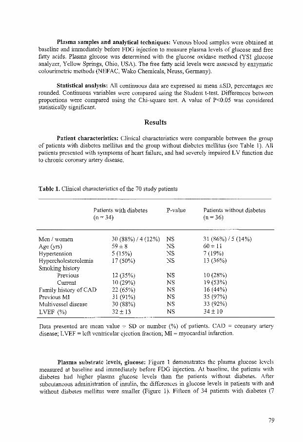
Plasma samples and analytical techniques: Venous blood samples were obtained at baseline and immediately before FDG injection to measure plasma levels of glucose and free fatty acids. Plasma glucose was determined with the glucose oxidase method (YSI glucose analyzer, Yellow Springs, Ohio, USA). The free fatty acid levels were assessed by enzymatic colourimetric methods (NEFAC, Wako Chemicals, Neuss, Germany).
Statistical analysis: All continuous data are expressed as mean ±SD, percentages are rounded. Continuous variables were compared using the Student t-test. Differences between proportions were compared using the Chi-square test. A value of P<0.05 was considered statistically significant.
Results
Patient characteristics: Clinical characteristics were comparable between the group of patients with diabetes mellitus and the group without diabetes mellitus (see Table 1). All patients presented with symptoms of heart failure, and had severely impaired LV function due to chronic coronary artery disease.
Table 1. Clinical characteristics of the 70 study patients
Men /women Age (yrs) Hypertension Hypercholesterolemia Smoking history
Previous Current
Patients with diabetes (n ~ 34)
30 (88%) /4 (12%) 59± 8 5 (15%) 17 (50%)
12 (35%) 10 (29%)
Family history of CAD 22 (65%) Previous MI 31 (91%) Multivessel disease 30 (88%) LVEF (%) 32 ± 13
P-value
NS NS NS NS
NS NS NS NS NS NS
Patients without diabetes (n ~ 36)
31 (86%) /5 (14%) 60 ± 11 7 (19%) 13 (36%)
10 (28%) 19 (53%) 16 (44%) 35 (97%) 33 (92%) 34 ± 10
Data presented are mean value ± SD or number (%) of patients. CAD = coronary artery disease; L VEF =left ventricular ejection fraction; MI =myocardial infarction.
Plasma substrate levels, glucose: Figure 1 demonstrates the plasma glucose levels measured at baseline and immediately before FDG injection. At baseline, the patients with diabetes had higher plasma glucose levels than the patients without diabetes. After subcutaneous administration of insulin, the differences in glucose levels in patients with and without diabetes mellitus were smaller (Figure 1 ). Fifteen of 34 patients with diabetes (7
79

IDDM, 8 NIDDM) had a plasma glucose level >9 mmol/L, which was lowered successfully in all patients with additional insulin.
Free fatty acids: At baseline, the patients witb diabetes had similar plasma levels of free fatty acids as the nondiabetic patients, at the time of FDG injection plasma levels of free fatty acids had declined significantly in both groups (Figure 2).
10
_J ::::0 8 0 E 6.1 ±1.3 E 6
<D (f)
0 4
()
2 2
" 0
Diabetes No diabetes
Figure 1. Bar graft demonstrating the plasma glucose levels measured at baseline (white bars) and immediately before FDG injection (black bars). * P<O.OOOJ versus no diabetes;** P<0.005 versus no diabetes; t P<O.OOJ versus immediately before FDG injection. Values are expressed as mean ± SD.
500
405 ± 247' ....J 400
350 ± 283' E -0 300 E c 200 <( L!.. L!.. 100 82 ± 123
0
Diabetes No diabetes
Figure 2. Plasma concentration of free fatty acids (FF A) in patients with diabetes mellitus and those without diabetes, at baseline (white bars) and after oral administration of Acipimox (black bars).* P<O.OOOJ versus after Acipimox.
80

Safety of FDG SPECT using Acipimox: No serious side effects (evaluated by direct questioning/physical examination) occurred during FDG SPECT imaging. Nineteen patients had a mild and transient skin flush after Acipimox administration. Mild skin flushing occurred in 5 of 34 (15%) patients with diabetes and in 14 of 36 (39%) patients without diabetes (P<0.05).
FDG SPECT image quality: Visual evaluation of the FDG images showed a good, moderate, and poor image quality in 27, 5, and 2 patients with diabetes mellitus respectively, and in 32, 4, and 0 patients without diabetes respectively (NS). The M/B ratio ofFDG SPECT images was comparable in patients with and without diabetes mellitus (see Figure 3).
5
4 3.5 ± 0.9 .2 - 3 "' ~ o:J 2 -:;;;
1
0
Diabetes No diabetes
Figure 3. The image quality (MIB ratio) of FDG SPECT images was comparable in patients with and without diabetes mellitus.
5
4 0 3.3 ± 1.1
:;::; 3 "' ~
o:J 2 -:;;; 1
0
IDDM NIDDM
Figure 4. Region-of-interest analysis demonstrated that the type of diabetes, insulin dependent diabetes mellitus (IDDM) versus non-insulin dependent diabetes mellitus (NIDDM), had no influence on FDG image quality.
81
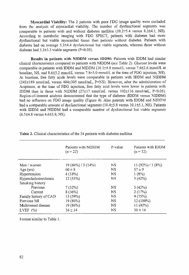
Myocardial Viability: The 2 patients with poor FDG image quality were excluded from the analysis of myocardial viability. The number of dysfunctional segments was comparable in patients with and without diabetes mellitus (10.2±5.4 versus 8.3±4.1, NS). According to metabolic imaging with FDG SPECT, patients with diabetes had more dysfunctional but viable myocardial tissue than patients without diabetes. Patients with diabetes had on average 5.3±4.4 dysfunctional but viable segments, whereas those without diabetes had 3.3±3.3 viable segments (P<0.05).
Results in patients with NIDDM versus IDDM: Patients with IDDM had similar clinical characteristics compared to patients with NIDDM (see Table 2). Glucose levels were comparable in patients with IDDM and NIDDM (10.1±4.8 mmo!JL versus 7.6±2.6 mmol/L at baseline, NS, and 8.6±3.2 mmol/L versus 7.8±3.0 mmol/L at the time ofFDG injection, NS). At baseline, free fatty acids levels were comparable in patients with IDDM and NIDDM (242±189 nmollmL versus 404±305 nmollmL, P~NS). However, after the administration of Acipimox, at the time of FDG injection, free fatty acid levels were lower in patients with IDDM than in those with NIDDM (27±17 nmollmL versus 102±116 nmollmL, P<0.05). Region-of-interest analysis demonstrated that the type of diabetes (IDDM versus NIDDM) had no influence on FDG image quality (Figure 4). Also patients with IDDM and NIDDM had a comparable amount of dysfunctional segments (10.4±5.8 versus 10.1±5.1, NS). Patients with IDDM and NIDDM had a comparable number of dysfunctional but viable segments (6.5±4.8 versus 4.6±3.9, NS).
Table 2. Clinical characteristics ofthe 34 patients with diabetes mellitus
Men /women Age (yrs) Hypertension Hypercholesterolemia Smoking history
Previous Current
Family history of CAD Previous MI Multivessel disease LVEF (%)
Format similar to Table 1.
82
Patients with NIDDM (n ~ 22)
19 (86%) /3 (14%) 60 ± 8 4 (18%) 12 (55%)
7 (32%) 8 (36%) 13 (59%) 19 (86%) 19 (86%) 34± 14
P-value
NS NS NS NS
NS NS NS NS NS NS
Patients with IDDM (n ~ 12)
11 (92%) /I (8%) 57± 9 I (8%) 5 (42%)
5 (42%) 2 (17%) 9 (75%) 12 (100%) II (92%) 30 ± 14

Discussion
Noninvasive assessment of myocardial viability in patients with ischemic cardiomyopathy has major implications for clinical decision making. \Vhen a substantial amount of dysfunctional but viable myocardium is present, coronary revascularization may improve LV function and long-term survival. Currently, both the number of patients with heart failure due to coronary artery disease and the prevalence of diabetes mellitus are increasing rapidly (16,17). Diabetes mellitus is a well-knovm risk factor for coronary artery disease. Patients with diabetes have a four-fold increased risk for developing heart failure after myocardial infarction (18). The assessment of myocardial viability is particularly relevant in these patients, since coronary revascularization in patients with diabetes mellitus is associated with a high morbidity and mortality (3). Currently, FDG PET is considered the gold standard technique for the assessment of myocardial viability (4-7). Recently, FDG metabolic imaging has become widely available with the introduction of FDG SPECT using dedicated collimators (19-24). The image quality of FDG imaging is influenced by the metabolic conditions (8,9). Several protocols were introduced to lower free fatty acid levels, which appears the main determinant of image quality during FDG imaging (8). FDG imaging following oral glucose loading is practical, but the image quality varies substantially, with 20-30% of the images being uninterpretable (in particular in patients with diabetes). The hyperinsulinemic euglycemic clamp ensures excellent image quality, but is labor-intensive and time consuming. Recently, FDG SPECT following oral administration of nicotinic acid derivatives (Acipimox) has been suggested as a clinically useful protocol that increases the patient throughput in busy nuclear cardiology laboratories (8,9). Acipimox is a nicotinic acid derivative, which inhibits lipolysis and decreases plasma concentrations of free fatty acids (11-13). Two preliminary studies showed good image quality of FDG imaging using Acipimox (8,9). However, the image quality with this protocol in patients with diabetes is unclear, while this is the most challenging patient subset to obtain a good image quality.
The present study assessed the feasibility and image quality of FDG SPECT following oral administration of Acipimox for the assessment of myocardial viability in patients with diabetes mellitus. Metabolic conditions (plasma substrate levels) were monitored. As anticipated baseline glucose levels were higher in patients with diabetes. In patients with plasma glucose levels >9 mmol/L additional insulin was added. An oral dose of Acipimox effectively lowered the free fatty acids concentrations in both patient groups. No serious side effects of Acipimox were observed. The image quality was comparable in patients with and without diabetes mellitus and the type of diabetes mellitus (IDDM, NIDDM) did not influence FDG image quality. These findings demonstrate that FDG SPECT following oral administration of Acipimox is a safe and clinically useful method for the assessment of myocardial viability that offers a good image quality, even in patients with diabetes mellitus.
It should be stated that Acipimox is currently not available in the United States. Recently, Vitale et aL (25) studied 10 patients with ischemic LV dysfunction and NIDDM using FDG PET. In that study, FDG imaging was performed following administration of Niacin (a nicotinic acid), yielding suboptimal results. However, nicotinic acid derivatives (e.g. Acipimox), are known to be 20 times as potent as nicotinic acids, which may explain the lesser results in the study by Vitale et al. using Niacin.
Of interest, patients with diabetes had more dysfunctional but viable myocardial tissue than patients without diabetes. This observation is in line with our previous work (26). The reason for this finding is currently unclear and awaits further research.
In conclusion, FDG SPECT metabolic imaging following Acipimox is safe and
83

practical for routine assessment of viability in patients with ischemic cardiomyopathy. Image quality is good, even in patients with diabetes, although additional insulin is sometimes needed.
References
1. Shindler DM, Kostis JB, Yusuf S, et aL Diabetes mellitus, a predictor of morbidity and mortality in the Studies of Left Ventricular Dysfunction (SOLVD) Trials and Registry. Am J CardieL 1996;77:1017-1020.
2. Dries DL, Sweitzer NK, Drazner MH, Stevenson L W, Gersh Bl Prognostic impact of diabetes mellitus in patients with heart failure according to the etiology of left ventricular systolic dysfunction. JAm Col! CardieL 2001;38:421-428.
3. Herlitz J, Wognsen GB, Emanuelsson H, et al. Mortality and morbidity in diabetic and nondiabetic patients during a 2-year period after coronary artery bypass grafting. Diabetes Care. 1996;19:698-703.
4. Tamaki N, Ohtani H, Yamashita K, et al. Metabolic activity in the areas of new fill-in after thalliurn-201 reinjection: comparison with positron emission tomography using fluorine-18-deoxyglucose. J Nucl Med. 1991 ;32:673-678.
5. Tillisch J, Brunken R, Marshall R, et aL Reversibility of cardiac wall motion abnormalities predicted by positron tomography. N Eng! J Med. 1986;314:884-888.
6. Lucignani G, Paolini G, Landoni C, et al. Presurgical identification of hibernating myocardium by combined use of technetium-99m hexakis 2-methoxyisobutylisonitrile single photon emission tomography and fluorine-18 fluoro-2-deoxy-D-glucose positron emission tomography in patients with coronary artery disease. Eur J Nucl Med. 1992; 19:874-881.
7. Eitzman D, Al-Aouar Z, Kanter H, et al. Clinical outcome of patients with advanced coronary artery disease after viability studies with positron emission tomography. J Am Col! CardieL 1992;20:559-565.
8. Knuuti MJ, Ykii-Jarvinen H, Voipio-Pulkki LM, et aL Enhancement of myocardial (fluorine-18)fluorodeoxyglucose uptake by a nicotinic acid derivative. J Nucl Med. 1994;35:989-998.
9. Bax JJ, Veening :MA, Visser FC, et al. Optimal metabolic conditions during fluorine-18 fluorodeoxyglucose imaging; a comparative study using different protocols. Eur J Nucl Med. 1997;24:35-41.
10. Bourdillon PD, Broderik TM, Sawada SG, et aL Regional wall motion index for infarct and non-infarct region after reperfusion in acute myocardial infarction: comparison with global wall motion index. JAm Soc Echo. 1989;2:398-407.
11. Fuccella LM, Goldaniga G, Lovisolo P, et aL Inhibition oflipolysis by nicotinic acid and by acipimox. Clin Pharmacal Ther. 1980;28:790-795.
12. Musatti L, Maggi E, Moro E, Valzelli G, Tamassia V. Bioavailability and pharmacokinetics in man of acipimox, a new antilipolytic and hypolipemic agent. J Int Med Res. 1981;9:381-386.
13. Nuutila P, Knuuti MJ, Raitakari M, et aL Effect of antilipolysis on heart and skeletal muscle glucose uptake in overnight fasted humans. Am J PhysioL 1994;267:E941-E946.
14. van Lingen A, Huijgens PC, Visser FC, et al. Performance characteristics of a 511-ke V collimator for imaging positron emitters with a standard gamma-camera. Eur J Nucl Med. 1992; 19:315-321.
15. Schinkel AFL, Bax JJ, Elhendy A, et aL Assessment of viable tissue in Q-wave regions by metabolic imaging using single-photon emission computed tomography in ischemic cardiomyopathy. Am J CardioL 2002;89: 1171-1175.
84

16. McCullough PA, Philbin EF, Spertus JA, et al. Confirmation of a heart failure epidemic: findings from the Resource Utilization Among Congestive Heart Failure (REACH) study. JAm Coil Cardia!. 2002;39:60-69.
17. Burke JP, Williams K, Gaskill SP, Hazuda HP, Haffner SM, Stem MP: Rapid rise in the incidence of type 2 diabetes from 1987 to 1996: results from the San Antonio Heart Study. Arch Intern Med. 1999;159:1450-1456.
18. Abbott RD, Donahue RP, Kannel WB, Wilson PW. The impact of diabetes on survival following myocardial infarction in men vs women. The Framingham Study. JAMA. 1988;260:3456-3460.
19. Sandler MP, Videlefsky S, Delbeke D, et al. Evaluation of myocardial ischemia using a rest metabolism/stress perfusion protocol with fluorine- 1 8 deoxyglucose/technetium-99m MIBI and dual-isotope simultaneous-acquisition single-photon emission computed tomography. JAm Coil Cardiol. 1995;26:870-878.
20. Martin WH, Delbeke D, Patton JA, et al. FDG-SPECT: correlation with FDG-PET. J Nucl Med. 1995; 36:988-995.
21. Chen EQ, Macintyre WJ, Go RT, et al. Myocardial viability studies using fluorine-18-FDG SPECT: a comparison with fluorine-18-FDG PET. J Nucl Med. 1997;38:582-586.
22. Bax JJ, Cornel JH, Visser FC, et al. Prediction of improvement of contractile function in patients with ischemic ventricular dysfunction after revascularization by fluorine-IS fluorodeoxyglucose single-photon emission computed tomography. J Am Coli CardieL 1997;30:377-383.
23. Sandler MP, Bax JJ, Patton JA, et al. Fluorine-18-fluorodeoxyglucose cardiac imaging using a modified scintillation camera. J Nucl Med. 1998;39:2035-2043.
24. Srinivasan G, Kitsiou AN, Bacharach SL, Bartlett ML, Miller-Davis C, Dilsizian V. (18F)fluorodeoxyglucose single photon emission computed tomography: can it replace PET and thallium SPECT for the assessment of myocardial viability? Circulation. 1998;97:843-850.
25. Vitale GD, deKemp RA, Ruddy TD, Williams K, Beanlands RS. Myocardial glucose utilization and optimization of (18)F-FDG PET imaging in patients with non-insulindependent diabetes mellitus, coronary artery disease, and left ventricular dysfunction. J Nucl Med. 2001;42:1730-1736.
26. Bax JJ, Visser FC, Poldermans D, et al. Feasibility, safety and image quality of cardiac FDG studies during hyperinsulinaemic-euglycaemic clamping. Eur J Nucl Med. 2002;29:452-457.
85


Chapter 8
Perfusion and contractile reserve in chronic
dysfunctional myocardium:
Relation to functional outcome after
surgical revascularization
Bax JJ, Poldermans D, Schinkel AFL,
Boersma E, Elhendy A, Maat A,
Valkema R, Krenning EP, Roelandt JRTC
Circulation 2002; 1 06:Il4-Il8
87


Perfusion and Contractile Reserve in Chronic Dysfunctional Myocardium: Relation to Functional Outcome After
Surgical Revascularization Jeroen J. Bax, MD; Don Poldermans, MD; Arend F.L. Schinkel, :MD; Eric Boersma, MSc;
Abdou Elhendy, MD; Alexander Maat, MD; Roelf Valkema, MD; Eric P. Krenning, :MD; Jos R.T.C. Roelandt, MD
Background--Chrorric dysfunctional but viable myocardium may exhibit contractile reserve and/or intact perfusion. Segments with intact perfusion without contractile reserve are frequently observed inpatients with ischemic cardiomyopathy. The clinical relevance of this observation is unclear; in particular, the functional outcome after revascularization is unknown. Thus, contractile reserve (using low-dose dobutamine echocardiography) and perfusion (using resting ~'lmTc tetrofosmin) were evaluated in 114 patients with ischemic cardiomyopathy and the findings were related to functional outcome (9 to 12 months after revascularization).
Methods and Results-Patients (n=114) with ischemic cardiomyopathy undergoing surgical revascularization were evaluated for perfusion (using 99mTc tetrofosmin) and contractile reserve (using low-dose dobutamine echocardiography). Contractile function (two-dimensional echocardiography) was assessed before and 9 to 12 months after revascularization. In the 1 336 dysfunctional segments, perfusion was preserved in 51% of the segments and contractile reserve in 31% (P<.05); 47% of the segments v.rith perfusion did not exhibit contractile reserve. The majority (66%) of segments v.rith recovery of function postrevascularization had intact perfusion and contractile reserve; the majority (58%) of segments v.rithout functional recovery lacked both perfusion and contractile reserve. Interestingly, 22% of segments v.rith functional recovery and 25% of segments v.rithout functional recovery showed intact perfusion without contractile reserve.
Conclusion-Segments v.rith intact perfusion/contractile reserve have a high likelihood of recovery of function postrevascularization; segments v.rithout contractile reserve/perfusion have a low likelihood of recovery and segments v.rith intact perfusion v.rithout contractile reserve have an intermediate likelihood of recovery. (Circulation. 2002; 106fsuppllj:I-14-I-18.)
Key Words: myocardial viability 11 hibernating myocardium a heart failure 11 perfusion 11 contractile reserve
I n patients with chronic ischemic left ventricular (LV) dysfunction, improvement of contractile function and fa
vorable long-term prognosis after surgical revascularization have been demonstrated in those patients with dysfunctional but viable myocardium.'- 6 Different techniques are available for the assessment of viable myocardium, based on the detection of different characteristics of viable myocardium.'-6 These characteristics include preserved glucose metabolism, intact cell membrane and mitochondria, preserved perfusion, and the presence of contractile reserve.'- 6 The first 4 characteristics can be evaluated by nuclear imaging techniques: Glucose metabolism can be evaluated by Fl8-fluorodeoxyglucose (FOG) imaging,7•8 intact cell membranes can be evaluated by 201Tl imaging,9 intact mitochondria can be assessed by 99mTc sestamibi imaging, 10 and preserved
perfusion can be evaluated by either imaging with thallium-201 or 99mTc-labeled tracersY·10 The presence of contractile reserve can be assessed during the infusion of low-dose dobutamine using two-dimensional (20) echocardiography or magnetic resonance imaging.'-6 However, dysfunctional but viable myocardium does not always eiliibit all characteristics. In particular, in a substantial percentage (approximately 25%) of the patients contractile reserve appeared absent, although other markers of viability (glucose metabolism, intact cell membrane, perfusion) were still present.'l.l 2 The precise clinical relevance of this observation is thus far unclear and remains a matter of debate;n in particular, the functional outcome after revascularization of these patients is uncertain. To further evaluate this phenomenon, we assessed contractile reserve (using low-dose dobutamine echocardiog-
From the Department of Cardiology, Leiden University Medical Center, Leiden, The Netherlands (J.J.B.), and the Departments of Cardiology (D.P .. A.F.L.S .• A. E., J.R.T.C.R.), Epidemiology and Statistics (E.B.), Thoracic Surgery (A.M.), and Nuclear Medicine (R.V., E.P.K.), Thorax Center Rotterdam, Rotterdam, The Netherlands.
Correspondence to Jeroen J Bax, Department of Cardiology, Lei den University Medical Center, Albinusdreef 2, 2333 ZA Leiden, The Netherlands. E-mail [email protected]
<D 2002 Amerir;on H""rr A"-""ci"tirm. Tnr
89

TABLE 1. Patient Characteristics (n=114)
Age {years) 61±7
Sex (M/F) 106/fl
Previous Ml 109 (96%)
MVD 104(91%}
Number stenosed coronary 2.7±.6 arteries
LVEF (%} 34±10
Previous CABG i4 (12%)
Previous PTCA 16(14%)
Diabetes 19(17%)
Renal failure 25 (22%)
COPD 19(17%}
Peripheral vascular diseese 18(16%)
CABG: coronary artery bypass grafting; COPD: chronic obstructive pulmOflary diSease; LVEF: left venlril:ular ejection fraction; Ml: myocanlial infarction; MVD: multi-vessel disease; PTCA: percutaneous transluminat coronary angioplasty.
raphy) and perfusion (using resting \>9mTc tetrofosmin) in a large group of patients with ischemic cardiomyopathy; the findings were subsequently related to functional outcome, assessed 9 to 12 months after revascularization.
Methods
Patients The study population existed of 114 patients with ischemic cardiomyopathy who were already scheduled for surgical revascularization. All patients presented with symptoms of heart failure whereas 38% of the patients had accompanying angina pectoris. Indications for surgery were heart failure in all (New York Heart Association [NYHA) score 3.2±0.8), together with angina in 38% (Canadian Cardiovascular Society [CCS] score 2.7±0.4). The decision for revascularization was based on clinical grounds (symptoms, presencefabsence of ischemia, angiographic findings).
Additional characteristics are presented in Table I. All patients were stable during the study. Patients with severe mitral regurgitation were not included. Each patient gave informed consent to the study protocol that was approved by the local Ethics Committee.
Study Protocol At the entrance of the study, the patients underwent radionuclide ventriculography (RNV) to assess LV ejection fraction (L VEF), resting 2D echocardiography to identifY regional contractile dysfunction, low-dose dobutaminc echocardiography to assess contractile reserve in dysfunctional myocardium, and resting -Tc tetrofosmin single-photon emission computed tomography (SPECT) to assess perfusion in dysfunctional myocardium. Late (9 to 12 months) after revascularization, resting segmental contractile function was reassessed using resting 2D echocardiography and global LV function (LVEF) by RNV.
Assessment of LVEF LVEF was assessed before and 9 to 12 months after surgical revascularization by using equilibrium RNV. 14 RNV was performed at rest with the patient in the supine position after the administration of 740 MBq of ""mTc. Images were acquired with a small field-ofview gamma camera (Orbiter, Siemens Corp, Iselin, NJ), oriented in the 45-degree left anterior oblique position with a 5- to 10-degree caudal tilt. The L VEF was calculated from the 45-degree left anterior oblique view by an automated technique. An improvement in L VEF by 5% or more was considered sit,>nificant. 15
90
Assessment of Resting Contractile Function Before/After Revascularization Resting contractile function was assessed by 2D echocardiography before and 9 to 12 months after revascularization. Four standard views of the LV were recorded (videotape and digitized in cine-loop format): parasternal long- and short-axis views and apical two- and four-chamber views. The prerevascularization and the postrevascularization images were reviewed (random order) off-line and consensus was achieved by 2 observers unaware of the other tests (low-dose dobutamine echocardiography, perfusion imaging). For analysis, a 16-segment model was used as suggested by American Society of Echocardiography. '" Both inward wall motion and wall thickening were analyzed. Each segment was assigned a wall motion score of 1 to 4: normal or mildly hypokinetic=l, severely hypokinetic=2 (decreased endocardial excursion and systolic wall thickening), akinetic=3 (absence of endocardial excursion and systolic wall thickening), and dyskinetic=4 (paradolliC outward movement in systole). Segments were considered dysfunctional when the wall motion score was 2 or more. Improvement of segmental wall motion score after revascularization by I grade or more was considered significant with the exception of improvement from dyskinesia to akinesia postrevascularization.
Assessment of Contractile Reserve Contractile reserve was assessed using low-dose dobutamine echocardiography. Dobutamine was infused at 5 and 10 J.tgfkg/min. 5 minutes per stage. Images were acquired at the end of both stages (parasternal long- and short·axis views and apical two- and fourchamber views) and analyzed by 2 observers (blinded to the postrevascularization echocardiograms and unaware of the perfusion data) using the 16-segment model and the 4-point scoring system as described above. Dysfunctional segments (resting wall motion score 2 or more) were evaluated for the presence of contractile reserve, defined as improvement of wall motion score by I grade or more, with the exception of improvement from dyskinesia to akinesia.
Assessment of Perfusion Perfusion was assessed at rest using ""'Tc tetrofosmin (600 MBq) SPECT. Data acquisition was performed with a triple-head gamma camera system (Picker Prism 3000 XP, Cleveland, OH); the energy was centered on the 140 keY photon peak of ""'Tc with a 15% window. Imaging was performed over 360 degrees (120 sectors of3 degrees) with a total imaging time of 32 minutes. Data were stored in 64X64, 16-bit matrix. The raw scintigraphic data were reconstructed by filtered back projection using a Butterworth filter (cut-off frequency at 0.17 cycle/pixel, of order 3.5). No attenuation correction was used. Further reconstruction yielded standard long- and short-axis projections perpendicular to the heart axis. Reconstructed slices were 8 rnm in all projections. The short-axis slices were displayed in polar map format, adjusted for peak myocardial activity (100%). The myocardium was divided into 16 segments, matching the echocardiographic segments (4 apical segments, 6 distal segments [anterior, anterolateral, inferolateral, inferior, inferoseptal, and anteroseptal], and 6 basal segments, comparable with the distal segments). Segments were divided into 4 categories: I =normal tracer uptake(> 75%), 2=moderatcly reduced tracer uptake (50% to 75%), 3=severely reduced tracer uptake (30% to 50%), and 4=absent tracer uptake ( <30%). In dysfunctional segments, perfusion was considered preserved when activity was 50% or more (score 1 or 2).
Statistical Analysis Descriptive results are expressed as mean±SD. Patient data were compared using the Student's t test for paired and unpaired data when appropriate. Comparison of proportions was performed using chi-square analysis. McNemar testing was used to evaluate differences in positivity for contractile reserve (as assessed by low-dose dobutamine echocardio~o'Taphy) and perfusion (assessed by """'Tc tetrofosmin SPECT). P<.05 was considered significant.

%segments
,.,r
"f ••
Hypokinesia Akinesia Dyskinesia
Figure 1. Relation between severity of contractife dysfunction at rest and the presence of contractile reserve; of the 412 segments wflh =ntractile reserve, 68% were hypokinetic, 31% akinetic, and 1% dyskinetic.
Results
Contractile Function, Preand Postrevascularization Six patients died during the study period (2 within 30 days after surgery (heart failure), 4 during the follow-up period (3 heart failure, I noncardiac death); 5 of these patients had absent perfusion and absent contractile reserve in all dysfunctional segments; 1 patient had 2 dysfunctional segments without perfusion/contractile reserve and 3 dysfunctional segments with preserved perfusion but without contractile reserve. In the remaining 108 patients, the symptoms improved significantly (mean CCS score reduced to 1.2::!::0.3. mean NYHA score reduced to 1.9:!:0.7, both P<.05 versus scores before revascularization).
Regional contractile function, as assessed by resting 20 echocardiography, demonstrated normal contraction in 392 (23%) segments and abnormal contraction in 1336 (77%) segments. Of the I 336 dysfunctional segments, 646 were severely hypokinetic, 629 akinetic, and 61 dyskinetic. Improvement of function postrevascularization occurred in 357 (27%) segments (63% severely hypokinetic, 37% akinetic and 0% dyskinetic segments), whereas 979 (73%) segments did not improve (or even deteriorate in contractile function).
Recovery of function was not observed more frequently in the 41 patients with angina as compared with the 67 patients without angina (153 versus 204 dysfunctional segments).
LVEF was 34:::':::10% before revascularization and did not significantly improve after revascularization (36::!:: II%). However, an improvement in L VEF of 5% or more was observed in 38 patients (from 28::!:8% before to 36::!::4% after revascularization).
Presence of Contractile Reserve in Dysfunctional Myocardium In the 1 336 dysfunctional segments, low-dose dobutamine echocardiography demonstrated improvement of function (contractile reserve) in 412 (31%) segments. Of these 412 segments with contractile reserve, approximately two-third were hypo kinetic 68% and one-third were akinetic (Figure l ). In the remaining 924 dysfunctional segments, no improve-
%segments
70
2
Hypokinesia Akinesia Dyskinesia
Figure 2. Relation between severity of contractile dysfunction at rest and the presence of intact perfusion; of the 683 segments with intact perfusion, 70% were hypokinetic, 28% akinetic. and 2% dyskinetic.
ment of contraction occurred during infusion of low-dose dobutamine.
Preserved Perfusion in Dysfunctional Myocardium In the I 336 dysfunctional segments, 09"'Tc tetrofosmin demonstrated preserved perfusion in 683 (51%) segments; 57% of the segments had perfusion score 1, and43% of the segments had perfusion score 2. Of the segments with preserved perfusion, the majority was hypokinetic (Figure 2). In the remaining 653 dysfunctional segments perfusion was not preserved (72% of the segments with perfusion score 3 and 28% of the segments with perfusion score 4).
Intact Perfusion Versus Preserved Contractile Reserve in Dysfunctional Myocardium In the 1 336 dysfunctional segments, perfusion was more frequently preserved as compared with contractile reserve (31% versus 51%, P<.05). The exact distribution of segments according to the presence/absence of petfusion/contractile reserve is shown in Table 2. In the 683 segments with preserved perfusion, contractile reserve was present in 360 (53%) and absent in 323 (47%) segments. Conversely, in the 653 segments without preserved perfusion, contractile reserve was present in 52 (8%) segments and absent in 601 (92%) segments (Figure 3). Disagreement between the 2 techniques was 28%, with 86% being related to segments with preserved perfusion without contractile reserve.
The distribution of segments according to the presence/ absence of perfusion/contractile reserve in relation to the absence/presence of angina is shown in Table 3.
TABLE 2. Characteristics of All1336 Dysfunctional Segments
category n (%)
P+ 683
CR+ 360 (53%)
CR- 323 (47%)
P- 653
CR+ 52 (8%)
CR- 601 (92%)
P: periusion; GR: contractile reserve; -.: present; : absent
91

OR• 8%
~1\ ((_} Perfusion intact Perfusion absent
Figure 3. Relation between contractile reserve and perfusion in chronic dysfunctional myocardium. Segments without intact perfusion virtually never have preserved contractile reserve (right); 47% of the segments with preserved perfusion do not exhibit contractile reserve.
Perfusion and Contractile Reserve Versus Functional Outcome The absence/presence of perfusion and absence/presence of contractile reserve in segments with and without improve~ ment of contractile function after revascularization is shown in Figure 4. Of the 357 segments with recovery of function, the majority (66%) had both preserved perfusion and contractile reserve, whereas only 13% of the 979 segments without improvement of contractile function exhibited preserved perfusion and contractile reserve (P<.05 versus seg~ ments with improvement of function postrevascularization). The majority (58%) of the segments without improvement of contractile function postrevascularization exhibited neither intact perfusion nor preserved contractile reserve. In contrast, only 9% of the segments with improvement of contractile function postrevascularization lacked both perfusion and contractile reserve (P<.OS versus segments without improvement of function postrevascularization). Of interest, 22% of the segments with improvement of contractile function and 25% of the segments without improvement of contractile function postrevascularization showed intact perfusion without contractile reserve. Finally, small percentages of segments with and without improvement of function (3% and 4%, respectively) showed preserved contractile reserve without intact perfusion.
Of the 360 segments with preserved perfusion/contractile reserve, 66% exhibited recovery of function. Of the 601 segments without preserved perfusion/contractile reserve, 95% did not improve in function. Of the 323 segments with preserved perfusion without contractile reserve, 24% improved in function. Of the 52 segments with contractile reserve without perfusion, 79% did not improve in function.
Finally, the segments with higher levels of preserved perfusion demonstrated a higher likelihood of recovery; 225 of 389 segments with tracer uptake >75% improved in
TABLE 3, Dysfunctional Segments in Patients With and Without Angina
P+ICR+ P+ICR P-/CR+
Angina+ (n-41 pts) 114 105 28
Angina - (n=67 pts) 246 218 " P: periusion; CR: contractile reserve; +: present; : allsent
92
ICR
229
372
Recovery .. Rscovary-
Figure 4. Distribution (according to presence/absence of contractile reserve/periusion) of segments with and without recovery postrevascularization. CR: contractile reserve; P: perfusion; +: present; - : absent.
function postrevascularization as compared with 132 of 294 segments with tracer uptake 50% to 75%.
Discussion In patients with ischemic cardiomyopathy, the assessment of residual viability in areas with chronic contractile dysfunction is important for prediction of improvement of function postrevascularization and prediction of long-term progno· sis.'-" Different techniques are available that rely on different characteristics of viable myocardium. 1- 6 Perfusion imaging (using 99"'Tc-labeled tracers or 201Tl) and assessment of contractile reserve (using low-dose dobutamine echocardiography) are widely used in the clinical setting for the assessment of viable myocardium. However, frequently chronic dysfunctional myocardium has preserved perfusion whereas contractile reserve is lacking. 11 ,12 Data on the functional outcome of these segments are scarce.l1 In the present study, perfusion imaging was compared with assessment of contractile reserve in a large number of patients with ischemic cardiomyopathy who were already scheduled for surgical revascularization. Both intact perfusion and the presence of contractile reserve were related to the severity of contractile dysfunction. Both perfusion and contractile reserve were more frequently observed in segments with severe hypokinesia as compared with akinetic segments, whereas dyskinetic virtually never exhibited intact perfusion of contractile reserve. Thus, the presence of these characteristics appears related to the severity of damage.
Moreover, perfusion was significantly more often preserved as compared with contractile reserve: 51% of the chronic dysfunctional segments showed intact perfusion and 31% showed contractile reserve. Head-to-head comparison of the individual segments revealed that the disagreement between the 2 imaging modalities was 28%, with 86% of these segments showing preserved perfusion without contractile reserve. Similar results have been reported by Panza et a!, 12
who compared 20 'Tl imaging with dobutamine echocardiography and demonstrated that a large number of segments demonstrated 20'Tl uptake but lacked contractile reserve. Studies comparing metabolic imaging with FDG to assessment of contractile reserve by dobutamine echocardiography have shown comparable results: A substantial percentage of the segments with FDG uptake did not exhibit contractile reserve. 18•19 Pagano and colleagues20 demonstrated that segments with FDG uptake without contractile reserve had more ultrastructural damage and a higher percentage of fibrosis as

compared with segments with preserved FDG uptake and intact contractile reserve. Nagueh and coworkers17 studied 20 patients with 201TI imaging and dobutamine echocardiography before surgical revascularization, and transmural biopsies were taken during surgery. The authors demonstrated a gradual increase of percentage fibrosis in different segments: 1% fibrosis in segments with both preserved 201Tl uptake and contractile reserve, 9% fibrosis in segments with either preserved 2n1Tl uptake or contractile reserve, and 28% fibrosis in segments without 20 'Tl uptake and without contractile reserve. These findings further substantiate the suggestion the severity of ultrastructural damage and fibrosis is related to the presence/absence of perfusion and contractile reserve. Segments with both characteristics have the least damage and fibrosis, segments with perfusion without contractile reserve have more damage/fibrosis and segments without perfusion/ contractile reserve have the most severe damage!fibrosis.
An important issue is how these findings affect improvement of function postrevascularization. In the present smdy, the majority of the segments with improvement of contractile function postrevascularization showed both preserved perfusion and contractile reserve. Segments with both characteristics presents may thus have a high likelihood of recovery of function post-revascularization (66% of the segments with both characteristics improved in function). In contrast, the majority of segments without recovery of function postrevascularization lacked both perfusion and contractile reserve. Segments without intact perfusion and contractile reserve may thus be considered to have a low likelihood of recovery of function postrevascularization (95% of the segments without perfusion/contractile reserve did not improve in function).
The segments with intact perfusion without contractile reserve pose a problem; 22% of the segments with recovery of function exhibited this pattern, but 25% of the segments without recovery of function also exhibited this pattern. Thus, segments with preserved perfusion without contractile reserve have an intermediate likelihood of recovery postrevascularization. However, from a clinical standpoint, it is important to emphasize that patients with preserved perfusion with contractile reserve may recover in function postrevascularization and should not routinely be denied surgical revascularization. Future studies are needed to identify which patients with this pattern are likely to improve and which patients will not improve in function postrevascularization.
Limitations Functional follow-up was performed at 9 to 12 months. whereas longer follow-up may be needed for complete recovery of function. This may be of particular importance in patients with severely damaged myocardium. In addition, long-term survival may be a superior end-point than improvement of function.
Graft patency was not assessed; thus, graft occlusion may have prevented some viable segments from recovery of function.
Finally, although a similar 16-segment model was used for echo and SPECT data, misalignment may have influenced the results.
Conclusions In chronic dysfunctional myocardium, perfusion is preserved more frequently than contractile reserve. Segments with both preserved perfusion and contractile reserve have a high likelihood of recovery of function postrevascularization; segments without preserved perfusion/contractile reserve have a low likelihood of recovery. Segments with preserved perfusion without contractile reserve have an intermediate likelihood of recovery after revascularization. Additional testing needs to be developed to predict which ofthese segments will improve and which segments will not improve in function.
References 1. Wijns W, Vainer SF, Camici PG. Hibernating myocardium. N Eng/
J Med. 1998;339:173-181. 2. Marwick TH. The viable myocardium: epidemiology, detection, and
clinical implications. Lancer. 1998;351:815-819. 3. Beller GA. Selecting patients with ischemic cardiomyopathy for medical
treaonent, revascularization, or heart transplantation. J Nucl Cardia/. 1997;4:Sl52-Sl57.
4. Bonow RO. Identification ofviabk myocardium. Circulmian. 1996;94: 2674-2678.
5. Udelson JE. Testing our tests: surrogate end points versus driving patient management and outcomes. JAm Call Cardia/. 2001 ;37:89-92.
6. Bax JJ, Poldennans D, Elhendy A, et al. Sensitivity, specificity, and predictive accuracies of various noninasive techniques for detecting hibernating myocardium. Curr Probl Cordia/_ 2001:26:141-186. Schclbcrt HR. Measurements of myocardial metabolism in patients with iochemic heart disease. Am J C@diol. l998;82(5A):61K-67K. Schwaiger M. Melin I_ Cardiological applications of nuclear medicine. Lancet. 1999;354:661-666. Bell .or GA, Ragosta ?\.f_ Extent of myocardial viability in regions of left ventricular dysfunction by rest·redistribution thallium·201 imaging: a p<>werful predictor of outcome. J Nucl Cardiol. 1998;5:445-448.
10. Gibbons RJ. Myocardial perfusion imaging. Hearl. 2001;83:555-560 11. Dilsizian V. Myocardial viability: contrnctile reserve or cell membrane
integrity? JAm Coli Cardia/. 1996;28:443-446. 12. Panza JA, Di!sizian V, Laurienzo JM, et al. Relation between thallium
uptake and contractile response to dobutamine: implication> regarding myocardial viability in patients with chronic coronary artery disease and left ventricular dysfunction. Circulation. 1995:91:990-998
13 Vanovcrschelde JLJ, Melin JA. Metabolic imaging and contrnctilc re'!<:rve for assessment of myocardial viability: friends or foes? J N~c/ CardioL 1999;6:458-461.
14 Wackers FJT, Berger HJ, Johnstone DE, et al. Multiple gated cardiac blood pool imaging for left ventricular ejection ffaction: validation of the technique and assessment of variability. Am J Cardia/. 1979;43·
1159-1165. 15. Bm: JJ, Visser FC, Poldermans D, et al. Relationship between preop
erative viability and postoperative improvement in LVEF and heart failure symptoms. J Nuc/ Med_ 2001 ;42:79-86.
16. Schill~r NB, Shah PM, Cra;vford M, ct al. Recommendation for quantification of the left ventricle by two·dirnensional echocardiob>raphy. JAm Sco Echocardiogr. 1989;2:358-367.
17. Naguch SF, Mikati 1, Weilbaecher D, et al. Relation of contrnetilc reserve of hibernating myocardium in relation to myocardial structure in humans Circulation. 1999;100:490-496.
18. Sawada S, Elsner G, Segar DS, et al. Evaluation of patterns of perfusion and metabolism in dobutamine·rcsponsive myocardium_ J Am Coli
CardioL 1997:29:55-61. 19 Sun KT, Czemin J, Krivokapich J, et al. Effects of dobutamine stimu
lation on myocardial blood flow, glucose metabolism, and wall motion in normal and dysfunctional myocardium. Circu/aiion. 1996:94:3146-3154
20 Pagano D, Tom1end JN, Parums DV, et al_ Hibernating myocardium morphological correlates of inotropic stimulation and glucose uptake. Hear/_ 2000;83:456-461
93


Chapter 9
Long-term prognostic value of dobutamine
stress technetium-99m-sestamibi SPECT:
A single-center experience
with 8-year follow-up
Schinkel AFL, Elhendy A, van Domburg RT,
Bax JJ, Valkema R, Roelandt JRTC, Poldermans D
Radiology, in press
95
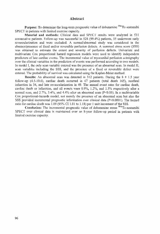
Abstract
Purpose: To determine the long-tenn prognostic value of dobutamine 99mTc-sestamibi SPECT in patients with limited exercise capacity.
Material and methods: Clinical data and SPECT results were analyzed in 531 consecutive patients. Follow-up was successful in 528 (99.4%) patients, 55 underwent early revascularization and were excluded. A normal/abnormal study was considered in the absence/presence of fixed and/or reversible perfusion defects. A summed stress score (SSS) was obtained to estimate the extent and severity of perfusion defects. Univariate and multivariate Cox proportional hazard regression models were used to identify independent predictors of late cardiac events. The incremental value of myocardial perfusion scintigraphy over the clinical variables in the prediction of events was performed according to two models. In model I, the only scan variable entered was the presence of an abnormal scan. In model II, scan variables including the SSS, and the presence of a fixed or reversible defect were entered. The probability of survival was calculated using the Kaplan-Meier method.
Results: An abnormal scan was detected in 312 patients. During the 8 ± 1.5 year follow-up (4.5-10.6), cardiac death occurred in 67 patients (total death 165), nonfatal infarction in 34, and late revascularization in 49. The annual event rates for cardiac death, cardiac death or infarction, and all events were 0.9%, 1.2%, and 1.5% respectively after a normal scan, and 2.7%, 3.4%, and 4.4% after an abnormal scan (P<0.05). In a multivariable Cox proportional-hazards model, not merely the presence of an abnormal scan but also the SSS provided incremental prognostic information over clinical data (P<O.OOOI). The hazard ratio for cardiac death was 1.09 (95% CI 1.01 to 1.18) per 1 unit increment of the SSS.
Conclusion: The incremental prognostic value of dobutamine stress 99mTc-sestamibi SPECT over clinical data is maintained over an 8-year follow-up period in patients with limited exercise capacity.
96

Introduction
The primary goal of risk stratification in patients with suspected or known coronary artery disease is to distinguish high-risk patients who may benefit from further invasive strategies, from low-risk patients who do not require further invasive evaluation. Stress 99mT csestamibi myocardial perfusion imaging provides useful information for risk stratification and determination of optimal clinical management (1-3). Multiple studies have reported the prognostic value of 99mTc-sestamibi myocardial perfusion imaging in various patient-subsets at short- to intermediate-term follow-up (4-7). To overcome a reduced exercise capacity and therefore an insufficient test, various types of non-exercise dependent pharmacological stressors, such as adenosine or dobutamine, were developed in conjunction with myocardial perfusion imaging.
However, it is important to note that currently no long-term prognostic studies using 99mTc-sestamibi myocardial perfusion imaging are available (1-7). The purpose of our study was to determine the long-term prognostic value of dobutamine 99mTc-sestamibi SPECT in patients with limited exercise capacity.
Material and methods
Study population: The study population comprised 531 consecutive patients with limited exercise capacity referred for dobutamine stress 99mTc-sestamibi SPECT imaging for the evaluation of suspected or known coronary artery disease, between 1990 and 1995. Follow-up was successful in 528 of 531 patients (99.4%). Fifty-five patients underwent early coronary revascularization (35 CABG, and 20 PTCA) within 60 days of the scintigraphy and were excluded from analysis. In these patients the decision to revascularize was already made and the test was performed as part of a research protocoL As a result, the prognostic data reported are based on 473 patients. All patients gave informed consent before testing. The local Medical Ethics Committee approved the study protocoL
In all patients a structured interview and clinical history were obtained, including assessment of cardiac risk factors, prior to the dobutamine stress test. Hypertension was defined as a blood pressure :?140/90 mm Hg, or treatment with antihypertensive medication. Diabetes mellitus was defined as a fasting glucose level :?7.8 mmol/L or the need for insulin or oral hypoglycemic agents. Hypercholesterolemia was defined as a total cholesterol :?6.4 mmol!L, or treatment with lipid-lowering medication.
Dobutamine stress protocol: Dobutamine stress testing was performed as described previously (8). Dobutamine was injected intravenously, first at a dose of 10 ~-tg/kg/min for 3 minutes, increasing by 10 ~-tg/kg/min every 3 minutes up to a maximum dose of 40 ~-tg/kg/min. If the test end-point was not reached at a dobutamine dose of 40 flg/kg/min, atropine (up to 1 mg) was given intravenously. Blood pressure, heart rate, and electrocardiography were continuously monitored. Test end-points were: achievement of target heart rate (85% of maximum age and gender predicted heart rate), horizontal or downs loping ST -segment depression >2 mm at an interval of 80 ms after the J-point compared with baseline, STsegment elevation > 1 mm in patients without previous MI, severe angina, systolic blood pressure fall >40 mm Hg compared to baseline, blood pressure >240/120 mm Hg, or significant cardiac arrhythmias. Metoprolol was available to reverse the (side) effects of dobutamine/atropine if these did not revert spontaneously after termination of dobutamine infusion. Side effects were registered, and the relation between the occurrence of cardiac arrhythmias during the test and subsequent cardiac events was evaluated.
97

SPECT acquisition and interpretation: An intravenous dose of370 MBq of99mTcsestamibi was administered approximately 1 minute prior to the termination of the stress test. For resting studies 370 MBq of sestamibi were injected at least 24 hours after the stress study. Image acquisition was achieved with a Siemens Gammasonics single-head Rota camera (Orbiter, Siemens, Iselin, NJ) without attenuation or scatter correction, using a low-energy allpurpose collimator. Thirty-two projections were obtained over a 180° arc, from left posterior oblique to right anterior oblique, with an acquisition time of 45 seconds per projection. Data were collected in a 64x64 matrix (word mode), and images were reconstructed using a filtered back projection algorithm and a ramp reconstruction filter. Transaxial tomograms were reconstructed using the SPETS software package (Nuclear Diagnostics AB, Ha.gersten, Sweden). From the three-dimensional data, oblique (short-axis) and sagittal (vertical longaxis), perpendicular and parallel to the long-axis, respectively, were reconstructed. For each study 6 oblique (short axis) slices from the apex to the base, and 3 sagittal (vertical long axis) slices were defined. Each of the 6 short axis slices was divided into 8 equal segments. The septal part of the 2 basal slices was excluded from analysis because this region corresponds to the fibrous portion of the interventricular septum and normally exhibits reduced uptake. Therefore, a total of 47 segments were identified (3 long axis and 44 short axis). The interpretation of the scan was semiquantitatively performed by visual analysis assisted by the circumferential profiles analysis. Profile curves 2.5 SDs below normal perfusion were considered abnormal. Stress and rest tomographic views were reviewed side-by-side on a computer display in consensus reading by two experienced observers who were unaware of the patients1 clinical data. In case of disagreement a majority decision was achieved by a third observer. In this study the original interpretations of the images were used. A reversible perfusion defect was defined as a perfusion defect on stress images that partially or completely resolved at rest in ;:::2 contiguous segments or slices in the 47-segment modeL A fixed perfusion defect was defined as a perfusion defect on stress images in 2 or more contiguous segments or slices, which persists on rest images in the 47-segment model. An abnormal study was considered in the presence of a fixed and/or reversible perfusion defect. To assess the severity of perfusion abnormalities, the left ventricular myocardium was divided into 6 segments: anterior, inferior, septal anterior, septal posterior, posterolateral and apical. Each of the 6 major left ventricular segments was scored using a 4-point scoring system; 0 = normal (100%;:, tracer activity> 75%); l ~slightly reduced (75%;:, tracer activity> 50%); 2 = moderately reduced (50% '2': tracer activity> 25%); 3 = severely reduced or absent uptake (25%;::: tracer activity;::: 0%). A summed stress score (SSS) was obtained by adding the scores of the 6 myocardial segments at stress. The SSS incorporates the extent and severity of a perfusion defect.
Follow-up: The follow-up data were collected in the year 2001. One author (A.S.) contacted the patient's general practitioner and/or reviewed hospital records. This author was blinded to the scan results. The date of the last examination or consultation was used to determine follow-up time. The mean follow-up period was 8 ± 1.4 years (range 4.5 to 10.6). End points were overall death, cardiac death, nonfatal Ml, and late (>60 days) coronary revascularization. Cardiac death was defined as a death caused by acute MI, significant cardiac arrhythmias, or refractory congestive heart failure, together with ECG and autopsy results when available. Sudden death occurring without another explanation was included as cardiac death. Cardiac enzyme levels and ECG-changes defined nonfatal MI. Patients were censored at the time of late coronary revascularization.
Statistical analysis: Values were expressed as mean value ±SD or number, and compared using the Student t test or Chi-square test. Univariate and multivariate Cox
98

proportional hazard regression models (BMDP statistical software) were used to identify independent predictors of late cardiac events (9). Variables were selected in a stepwise forward selection manner with entry and retention set at a significance level of 0.05. The risk of a variable was expressed as a hazard ratio with a corresponding 95% confidence interval. The incremental value of myocardial perfusion scintigraphy over the clinical variables in the prediction of events was performed according to two models. In model I, the only scan variable entered was the presence of an abnormal scan. In model II, scan variables including the SSS, and the presence of a fixed or reversible defect were entered. The probability of survival was calculated using the Kaplan-Meier method, and survival curves were compared using the log-rank test. P value <0.05 was considered statistically significant.
Results
Demographics and stress test results: The characteristics of the 473 patients (273 men, age 61 ± 12 years) are presented in Table 1.
Table 1. Study population (n=173)
Age (years) Male gender Hypertension Diabetes mellitus Smoking Hypercholesterolemia Congestive heart failure Beta-blocker use Prior myocardial infarction Prior surgical revascularization Prior percutaneous revascularization
61 ± 12 273 (58) 214 (45) 69 (15) 122 (26) 116 (25) 84(18) 185 (39) 210 (44) 92 (19) 75 (16)
There was a significant increase of heart rate (70 ± 14 to 136 ± 17 bpm, P<O.OOOl), and systolic blood pressure (140 ± 23 to 146 ± 31 mm Hg, P<O.OOl) observed during the dobutamine stress test. The highest dobutamine dose was 10 !J-g · kg-1 min-1 in 3 (1%) patients, 20 ~g · kg·1
· min-1 in 15 (3%), 30 ~g · kg·1 • min·1 in 66 (14%), and 40 ~g · kg·1
•
min- 1 in 389 (82%). In 196 (41%) patients atropine was added. Patients who were using betablockers (120 of 185, 65%) more frequently received atropine than those without beta-blocker therapy (76 of 288, 26%, P<O.OOl). The test was inconclusive (failure to achieve target heart rate in the absence of perfusion abnormalities) in 43 (9%) patients. The prognostic data reported were based on 473 patients, including the 43 patients who did not achieve target heart rate. No patient experienced a MI or ventricular fibrillation. Side effects were atrial fibrillation in 5 patients (I%), short ventricular tachycardia (<10 complexes) in 19 patients (4%), severe hypotension (decrease in systolic blood pressure >40 mm Hg) in 3 (0.6%), and severe hypertension (blood pressure >2401130 mm Hg) in 3 (0.6%). Minor side effects included nausea in 18 (4%), chills in 22 (5%), and headache in 29 (6%). No relation was
99
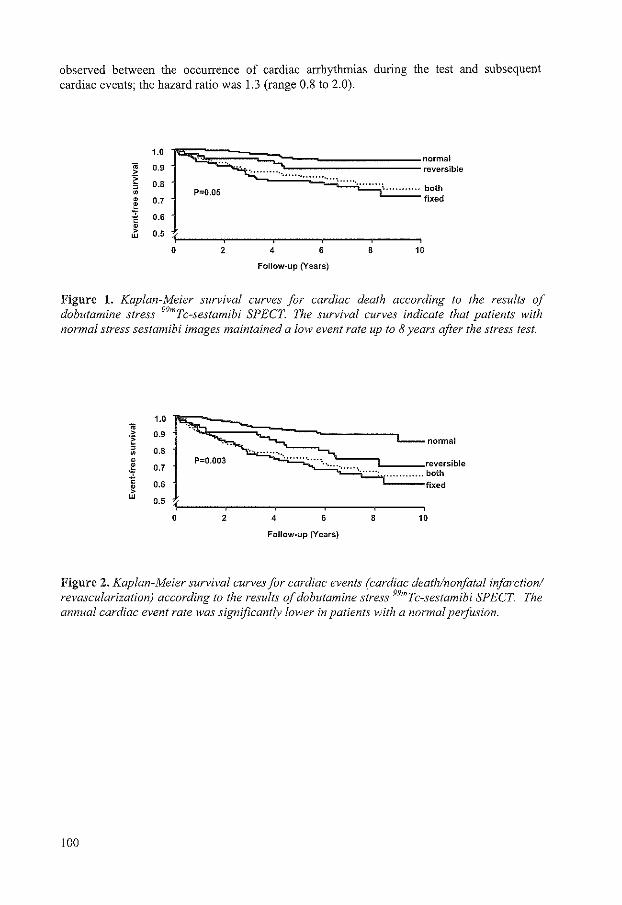
observed between the occurrence of cardiac arrhythmias during the test and subsequent cardiac events; the hazard ratio was 1.3 (range 0.8 to 2.0).
1.0 .. 0.9 > -~
0.8 , • P=0.05 • 0.7
~ 0.6
r~~==;~~--: .. ~ .. ~ .. : .. ~ ... ~ .. = .. ~ .. ; .. : .. : .. : .. ~ .. ; .. ~ .. ~ .. ~---~ .. ~ .. ~ .. ~-,~ .. ~"~"~"~"~'' ~~~::blo • > 0.5 w
0 2 4 6 8 10
Follow-up (Years)
Figure 1. Kaplan-Meier survival curves for cardiac death according to the results of dobutamine stress 99mTc-sestamibi SPECT. The survival curves indicate that patients with normal stress sestamibi images maintained a low event rate up to 8 years after the stress test.
1.0 .. ·~ 0.9 ,
0.8 • • • 0.7 ! " 0.6 • > w 0.5
0 2 4 6 8 10
Follow-up (Years)
Figure 2. Kaplan-Meier survival curves for cardiac events (cardiac death/nonfatal infarction! revascularization) according to the results of dobutamine stress 99mTc-sestamibi SPECT. The annual cardiac event rate was significantly lower in patients with a normal perfitsion.
100

Table 2. Predictors of cardiac death.
Multivariate analysis
Univariate Clinical data Modell Model II Clinical characteristics
Age* 1.03 (l.Ol-1.06) 1.03 (1.01-1.05) 1.02( 1.01-1.04) 1.03 (1.01-1.05) Male gender 2.4 (1.4-4.1) 2.5 (1.6-3.9) 2.1 (1.3-3.3) 2.3 (1.4-3.6) Prior MI 2.0 (1.2-3.3) NS NS NS Diabetes mellitus 2.3 (1.3-4.0) 1.8 (1.1-2.8) 1.7 (1.1-2.7) 1.7 (1.1-2.8) Hypertension 0.7 (0.4-1.2) NS NS NS Hypercholesterolemia 0.9 (0.5-1.5) NS NS NS Typical angina 1.1 (0.6-1.9) NS NS NS Smoking 1.6 (0.9-2.6) NS NS NS Congestive heart failure 4.7 (2.9-7.6) 2.1 (1.4-3.2) 1.9 (1.2-2.8) 1.7 (1.1-2.7)
Stress test results Typical angina 1.0 (0.6-1.7) NS NS ST -segment changes 1.5 (0.9-2.5) NS NS
Scan parameters Abnormal scan 3.4 (1.7-6.8) 2.1 (1.2-3.7) SSS* 1.22 (1.13-1.32) 1.09 (1.01-1.18) Fixed defect 1.8 (1.1-2.9) NS Reversible defect 1.2 (0.8-2.0) NS
*Per 1 unit increment.- indicates variable excluded. Values are expressed as Cox proportional hazard ratio and 95% confidence interval. In model I, the only scan variable entered was the presence of an abnormal scan. In model II, the SSS, and presence of a fixed defect or reversible defect were included. MI = myocardial infarction; NS = not statistically significant.
Table 3. Predictors of cardiac events (cardiac death, myocardial infarction, late coronary revascularization).
Clinical characteristics Age* Male gender Prior MI Diabetes mellitus Hypertension Hypercholesterolemia Typical angina Smoking Congestive heart failure
Stress test results Typical angina ST -segment changes
Scan parameters Abnormal scan SSS* Fixed defect Reversible defect
Format similar to Table 2.
Univariate
1.02 (1.00-1.04) 2.5 (1.6-3.8) 2.2 (1.5-3.2) 1.9 (1.2-3.0) 0.7 (0.5-1.1) 1.1 (0.7-1.7) 1.1 (0.7-1.7) 1.2 (0.8-1.9) 2.7 (1.8-4.1)
1.1 (0.7-1.6) 1.6 (0.8-1.8)
3.2 (1.9-5.4) 1.17 (1.10-1.25) 1.4 (1.0-2.1) 1.4 (1.0-2.1)
Clinical data
1.03 (1.01-1.05) 2.5 (1.6-3.9) NS 1.8 (1.1-2.8) NS NS NS NS 2.1 (1.4-3.2)
Multivariate analysis
Model I
1.02 (1.01-1.04) 2.1 (1.3-3.3) NS 1.7 (1.1-2.7) NS NS NS NS 1.9 (1.2-2.8)
NS NS
2.1 (1.2-3.7)
Model II
1.03 (1.01-1.05) 2.3 (1.4-3.6) NS 1.7 (1.1-2.8) NS NS NS NS 1.7 (1.1-2.7)
NS NS
1.09 (1.01-1.18) NS NS
101
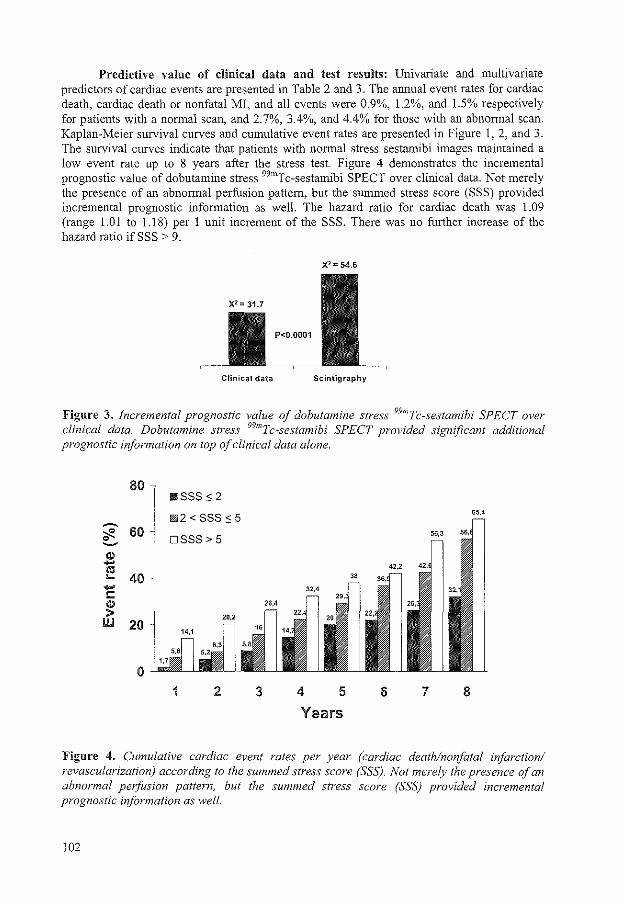
Predictive value of clinical data and test results: Univariate and multivariate predictors of cardiac events are presented in Table 2 and 3. The annual event rates for cardiac death, cardiac death or nonfatal MI, and all events were 0.9%, 1.2%, and 1.5% respectively for patients with a normal scan, and 2. 7%, 3.4%, and 4.4% for those with an abnormal scan. Kaplan-Meier survival curves and cumulative event rates are presented in Figure 1, 2, and 3. The survival curves indicate that patients with normal stress sestamibi images maintained a low event rate up to 8 years after the stress test. Figure 4 demonstrates the incremental prognostic value of dobutamine stress 99mTc-sestamibi SPECT over clinical data. Not merely the presence of an abnormal perfusion pattern, but the summed stress score (SSS) provided incremental prognostic information as well. The hazard ratio for cardiac death was 1.09 (range 1.01 to 1.18) per 1 unit increment of the SSS. There was no further increase of the hazard ratio ifSSS > 9.
X2 = 54.6
Clinical data Scintigraphy
Figure 3. Incremental prognostic value of dobutamine stress 99mTc-sestamibi SPECT over clinical data. Dobutamine stress 99mTc-sestamibi SPECT provided significant additional prognostic information on top of clinical data alone.
1 2 3 4 5 6 7 8
Years
Figure 4. Cumulative cardiac event rates per year (cardiac death/nonfatal infarction/ revascularization) according to the summed stress score (SSS). Not merely the presence of an abnormal perfusion pattern, but the summed stress score (SSS) provided incremental prognostic iriformation as well.
102

Discussion
Risk stratification in patients with suspected or known coronary artery disease is essential for optimal clinical decision making (10). The prognostic value of stress technetium imaging bas been reported in various patient-subsets at short- to intermediate-term follow-up (2-7). The mean follow up of this study is 8 ± 1.4 years as compared to an average follow up of 0.8-3.6 years after exercise and 0.8-2.3 years after pharmacological stress sestamibi myocardial perfusion imaging among previous studies (2-7). The main finding from this study is that the prognostic value of dobutamine stress 99mTc-sestamibi SPECT was maintained at long-term fo1low-u~. Univariate and multivariate Cox proportional regression analysis demonstrated that 9 mTc-sestamibi SPECT provided incremental prognostic information in addition to clinical and stress test parameters all through a nearly complete 8-year follow-up period. Patients with a normal scan had a good long-term prognosis, and in contrast, patients with an abnonnal test had an elevated risk of future cardiac events. Not merely the presence of an abnormal perfusion pattern, but the extent and severity of the perfusion defect provided incremental prognostic information as well
Long-term prognostic significance: Although long-term prognostic studies are currently not available, the medium-term prognostic value of dobutamine stress 99mTcsestamibi SPECT has been reported in several studies (4-7). Geleijnse eta!. (4) studied 392 patients with a median follow-up period of 22 ± 13 months. There were 27 (7%) cardiac deaths during follow up, nonfatal Ml occurred in 17 (4%) patients, and 34 (9%) patients underwent late coronary revascularization (10%). The hard cardiac event rate (cardiac death or nonfatal MI) was 0.8% for patients with a normal scan, and 9.2% per year in subjects an abnormal dobutamine stress 99mTc-sestamibi SPECT scintigraphy. The medium-term prognosis of women with chest pain and normal dobutamine stress 99mTc-sestamibi SPECT images is excellent (5). None of the study population of 80 women died or had a nonfatal MI during the follow-up period of 23 ± 13 months. Senior et a!. (6) followed up 61 patients undergoing coronary angiography and dobutamine stress 99mTc-sestamibi SPECT for 19 ± 11 months. There were 2 (3%) deaths, 2 (3%) nonfatal Ml, 13 (21 %) patients with unstable angina, and 3 (5%) subjects with congestive heart failure during the follow-up period. The event rate was 3% in patients with normal and 44% in patients with abnormal results. Recently, Calnon et a!. (7) studied 308 high-risk patients who underwent dobutamine stress 99mTc-sestamibi SPECT. During an average follow-up of 1.9 ± 1.1 years, there were 15 cardiac deaths and 18 Mis. Event rates were 10.0% in patients with an abnormal SPECT images and 2.3% in those with normal results.
The findings from the present study extend the conclusions drawn from these four medium-term prognostic studies (4-7). In the current study population of 473 patients, the prognostic value of dobutamine sestamibi SPECT imaging was maintained over an 8-year period. The event rates for cardiac death, cardiac death or nonfatal MI, and all events were respectively, 0.9%, 1.2%, and 1.5% per year for patients with a normal scan, and 2.7%, 3.4%, and 4.4% per year for those with an abnormal scan. Moreover, Kaplan-Meier survival curves continued to diverge over time, whereas the curve of patients with a normal SPECT study showed a very favorable event-free survival, indicating that the prognostic value was maintained during the entire follow-up period. This is particularly relevant since recently some investigators have suggested that the "warranty" period for normal stress sestamibi scans appeared to be 2 years (2). In contrast, the findings from the present study indicate that patients with normal stress sestamibi images have a durable good prognosis up to 8 years after the index stress test. The annual event rate for cardiac death or nonfatal MI in patients with normal scintigraphy results was comparable to the event rate in the general population.
103
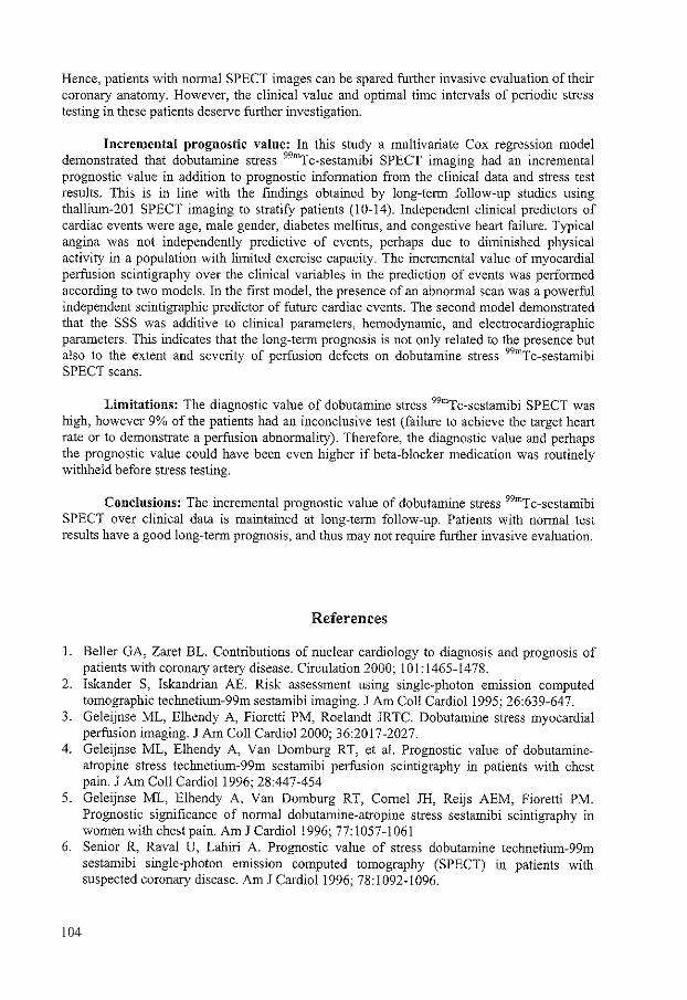
Hence, patients with normal SPECT images can be spared further invasive evaluation of their coronary anatomy. However, the clinical value and optimal time intervals of periodic stress testing in these patients deserve further investigation.
Incremental prognostic value: In this study a multivariate Cox regression model demonstrated that dobutarnine stress 99rncrc-sestamibi SPECT imaging had an incremental prognostic value in addition to prognostic information from the clinical data and stress test results. This is in line with the findings obtained by long-term follow-up studies using thallium-201 SPECT imaging to stratify patients (10-14). Independent clinical predictors of cardiac events were age, male gender, diabetes mellitus, and congestive heart failure. Typical angina was not independently predictive of events, perhaps due to diminished physical activity in a population with limited exercise capacity. The incremental value of myocardial perfusion scintigraphy over the clinical variables in the prediction of events was performed according to two models. In the first model, the presence of an abnormal scan was a powerful independent scintigraphic predictor of future cardiac events. The second model demonstrated that the SSS was additive to clinical parameters, hemodynamic, and electrocardiographic parameters. This indicates that the long-term prognosis is not only related to the presence but also to the extent and severity of perfusion defects on dobutamine stress 99mTc-sestamibi SPECT scans.
Limitations: The diagnostic value of dobutamine stress 99mTc-sestamibi SPECT was high, however 9% of the patients had an inconclusive test (failure to achieve the target heart rate or to demonstrate a perfusion abnormality). Therefore, the diagnostic value and perhaps the prognostic value could have been even higher if beta-blocker medication was routinely withheld before stress testing.
Conclusions: The incremental prognostic value of dobutamine stress 99mTc-sestamibi SPECT over clinical data is maintained at long-term follow-up. Patients with normal test results have a good long-term prognosis, and thus may not require further invasive evaluation.
References
1. Beller GA, Zaret BL. Contributions of nuclear cardiology to diagnosis and prognosis of patients with coronary artery disease. Circulation 2000; 101:1465-1478.
2. Iskander S, Iskandrian AE. Risk assessment using single-photon emission computed tomographic technetium-99m sestamibi imaging. JAm Coli Cardiol!995; 26:639-647.
3. Geleijnse ML, Elhendy A, Fioretti PM, Roeland! JRTC. Dobutamine stress myocardial perfusion imaging. JAm Col! Cardio12000; 36:2017-2027.
4. Geleijnse ML, Elhendy A, Van Domburg RT, et al. Prognostic value of dobutamineatropine stress technetium-99m sestamibi perfusion scintigraphy in patients with chest pain. JAm Coli Cardiol1996; 28:447-454
5. Geleijnse ML, Elhendy A, Van Domburg RT, Cornel JH, Reijs AEM, Fioretti PM. Prognostic significance of normal dobutamine-atropine stress sestarnibi scintigraphy in women with chest pain. Am J Cardiol1996; 77:1057-1061
6. Senior R, Raval U, Lahiri A. Prognostic value of stress dobutamine technetium-99m sestarnibi single-photon emission computed tomography (SPECT) in patients with suspected coronary disease. Am J Cardiol!996; 78: I 092-1096.
104

7. Calnan DA, McGrath PD, Doss AL, Harrell FE, Watson DD, Beller GA. Prognostic value of dobutamine stress technetium-99m-sestamibi single-photon emission computed tomography myocardial perfusion imaging: stratification of a high-risk population. J Am Call Cardiol2001; 38;1511-1517.
8. Elhendy A, Valkema R, van Domburg RT, et al. Safety of dobutamine-atropine stress myocardial perfusion scintigraphy. J Nuc Med 1998; 39: 1662-1666.
9. Cox DR. Regression models and life-tables. J R Stat Soc (B). 1972; 34:187-202. 10. Brovro KA. Prognostic value ofthallium-201 myocardial perfusion imaging: a diagnostic
tool comes of age. Circulation 1991; 83:363-381. 11. Vanzetto G, Ormezzano 0, Fagret D, Comet M, Denis B, Machecourt J. Long-term
additive prognostic value ofthallium-201 myocardial perfusion imaging over clinical and exercise stress test in low to intermediate risk patients: study in 1137 patients with 6-year follow-up. Circulation 1999; 100:1521-1527.
12. Pavin D, Delonca J, Siegenthaler M, Doat M, Rutishauser W, Righetti A. Long-term (10 years) prognostic value of a normal thallium-201 myocardial exercise scintigraphy in patients with coronary artery disease documented by angiography. Eur Heart J 1997; 18:69-77.
13. Steinberg EH, Koss JH, Lee M, Grunwald AM, Bodenheimer MM. Prognostic significance from 1 0-year follow-up of a qualitatively normal planar exercise thallium test in suspected coronary artery disease. Am J Cardia! 1993; 71:1270-1273.
14. Marie PY, Danchin N, Durand JF, et al. Long-term prediction of major ischemic events by exercise thallium-201 single-photon emission computed tomography: incremental prognostic value compared with clinical, exercise testing, catheterization and radionuclide angiograpbic data. JAm Call Cardioll995;26:879-886.
105


Chapter 10
Prognostic value of dobutamine-atropine
stress 99mTc-tetrofosmin myocardial
perfusion SPECT in patients with known
or suspected coronary artery disease
Schinkel AFL, Elhendy A, van Domburg RT,
Bax JJ, Roelandt JRTC, Poldermans D
J Nucl Med 2002;43:767-772
107

Prognostic Value of Dobutamine-Atropine Stress 99mTc-Tetrofosmin Myocardial Perfusion SPECT in Patients with Known or Suspected Coronary Artery Disease Arend F.L. Schinkel, :MD; Abdou Elhendy, MD, PhD; Ron T. van Domburg, PhD; Jeroen J. Bax, MD, PhD; Jos R.T.C. Roelandt, MD, PhD; and Don Poldennans, :MD, PhD
Department of Cardiology, Thoraxcenter. Erasmus Medical Center Rotterdam, Rotterdam, The Netherlands
~smrc-Tetrofosmin is a recently introduced radioactive isotope for the assessment of myocardial perfusion. Data regarding the prognostic value of stress imaging using this isotope are scarce. The aim of this study was to assess the prognostic value of dobutamine-atropine ~~"'Tc-tetrofosmin SPECT for the prediction of late cardiac events in patients with known or suspected coronary artery disease. Methods: A total of 72i consecutive patients with limited exercise capacity underwent dobutamineatropine stress 99mTc-tetrofosmin SPECT. Follow-up was successful in 719 of 721 patients (99.7%). Twenty-eight patients who underwent early revascularization were excluded. Results: Myocardial perfusion abnormalities were detected in 381 patients (55%) and included fixed defects in 190 patients (27%) and reversible defects 191 patients (28%). During a mean follow-up period of 37 :':. 17 mo, there were 150 deaths (22%), of which 62 (41%) were attributed to cardiac causes. Nonfatal myocardial infarction occurred in 23 patients {3%), and late (>3 mo) coronary revascularization was performed on 21 patients (3%). The cardiac death rate was 1 %/yin patients with a normal scan and 5.1 %/y in patients with an abnormal scan (P < 0.0001). In a multivariable Cox proportional-hazards model, the presence of abnormal perfusion was independently associated with an increased risk of cardiac death, after adjusting for clinical and stress test data (hazard ratio, 8.2; 95% confidence interval, 3.2-21). Conclusion: Dobutamine-atropine stress g9"'Tc-tetrofosmin SPECT is a useful imaging method for distinguishing patients at high and low risk of future cardiac events. The presence of perfusion abnormalities provides incremental prognostic information to clinical, stress electrocardiographic, and hemodynamic data. Key Words: tetrofosmin: coronary artery disease; SPECT
J Nucl Med 2002; 43:767-772
Received Aug. 29. 2001: revision accepted Jan. 31. 2002. For correspondence and reprints contact: Don Poldermans. MD. PhD,
Department of Cardiology. Thoraxcenter. Room Ba 300. Erasmus Medical Center Rotterdam. Dr. Molewaterplein 40, 3015 GD Rotterdam. The Netherlands.
E-mail: [email protected]
Rannacologic stress myocardial perfusion imaging is a useful alternative to exercise stress in patients with limited exercise capacity (1-3). For many years, 201Tl was the most widely used radioactive isotope in conjunction with pharmacologic stress testing for the diagnosis and risk stratification of coronary artery disease (CAD) (I). The use of 99"'Tc-labeled agents provides the advantages of improved image quality, increased consistency of image analysis, and a larger injectable dose because of a shorter half-life compared with 201 Tl (2-4). 99mTc-Labeled agents (sestamibi and tetrofosmin) share similar phannacokinetic properties. These radiopharmaceuticals are distributed "Within the myocardium in proportion to the regional cardiac blood flow. After intravenous injection, a relatively rapid clearance of the tracer from the blood and extracardiac structures occurs with minimal redistribution from the myocardium. Although several studies have revealed the diagnostic value of 99"'Tc-tetrofosmin SPECT (5-JJ), the data to define the prognostic value of stress imaging with 99mTc-tetrofosmin are limited. In a recent study ( 12), the extent of reversible perfusion abnormalities during dipyridamole infusion was lower with tetrofosmin than with sestamibi. It was concluded that the data relating to the prognostic value of 99mTc-sestamibi in the literature should not be empirically extrapolated to 99mTc-tetrofosmin imaging because of these differences. In addition, few studies have shown a reduced sensitivity oftetrofosmin imaging in patients with intermediate stenotic lesions due to a low extraction fraction and early plateau phase (13). These findings may have important prognostic implications in patients with normal tetrofosmin perfusion studies. In these cases CAD may be missed. Recent stuclies have shown that hyperemia induced by a dobutamineatropine stress test is of a magnitude at least equal to hyperemia induced by vasodilator stress agents such as dipyridamole (14). The aim of this study was to assess the prognostic value of dobutamine-atropine 99"'Tc-tetrofosmin SPECT in patients v.':ith known or suspected CAD.
109

MATERIALS AND METHODS
Patient Selection Between 1994 and 2000, 721 consecutive patients with limited
exercise capacity were referred for dobutamine-atropine stress 9'hnTc-tetrofosmin SPECT for evaluation of suspected or known CAD. Follow-up was successful in 719 of 721 patients (99.7%). Twenty-eight patients who underwent coronary revascularization within 3 mo of scintigraphy were excluded from analysis. In these patients, the decision to revascularize may be influenced by test results. Consequently. the prognostic data reported are based on 693 patients. All patients gave infonned consent before testing. The Hospital Ethics Committee approved the protocol.
Clinical Data Before the dobutamine stress test. a structured interview and
clinical history, including assessment of cardiac risk factors, were obtained. Hypertension was defined as a blood pressure of 2::!40/90 mm Hg or treatment with antihypertensive medication. Diabetes mellitus was defined as a fasting glucose level of 2::7.8 mmol'L or the need for insulin or oral hypoglycemic agents. Hypercholesterolemia was defined as a total cholesterol of 2::6.4 mmol/L or treatment with lipid-lowering medication.
Dobutamine Stress Test The dobutamine-atropine stress test was performed as described
(7). Dobutamine was administered intravenously, starting at a dose of 5 !J,g/kglmin for 3 min, then 10 fig/kg/min for 3 min, increasing by 10 j.!.g/kg/min every 3 min up to a maximum dose of 40 j.!.g/kg/min. If the test endpoint was not reached at a dobutamine dose of 40 fLgfkglmin, atropine (up to 2 mg) was given intravenously. Blood pressure, heart rate, and electrocardiography were monitored continuously. Test endpoints were achievement of target heart rate (85% of maximum age-predicted heart rate), horizontal or dO\.vnsloping ST-segment depression of >2 mm, STsegment elevation of > 1 mm in patients without previous myocardial infarction, severe angina, systolic blood pressure fall of >40 mm Hg, blood pressure of >240/120 mm Hg, or significant arrhythmia. Metoprolol was available to reverse the (side) effects of dobutamine or atropine if these did not revert spontaneously after tennination of dobutamine infusion.
SPECT Approximately 1 min before the termination of the stress test, an
intravenous dose of370 MBq 99mTc-tetrofosmin was administered. For resting studies, 370 MBq tetrofosmin were injected at least 24 h after the stress study. Image acquisition was performed with a triple-head gamma-camera system (Prism 3000 XP; Picker International, Cleveland, OH). For each study, 6 oblique (short axis) slices from the apex to the base and 3 sagittal (vertical long axis) slices were defined. Each of the 6 short-axis slices was divided into 8 equal segments. The septal part of the 2 basal slices was excluded from analysis because this region corresponds to the fibrous portion of the interventricular septum and normally exhibits reduced uptake. Therefore, 47 segments were identified (3 long axis and 44 short axis). The interpretation of the scan was performed semiquantitatively by visual analysis assisted by analysis of the circumferential profiles. Stress and rest tomographic views were reviewed side by side by an experienced observer who was unaware of each patient's clinical data. A reversible perfusion defect was defined as a perfusion defect on stress images that partially or completely resolved at rest in ~2 contiguous segments
110
or slices in the 47-segment model. A fixed perfusion defect was defined as a perfusion defect on stress images in ~2 contiguous segments or slices, which persists on rest images in the 47-segment model. An abnormal study was considered in the presence of a fixed or reversible perfusion defect (or both). To assess the severity of perfusion abnormalities, the left ventricular myocardium was divided into 6 segments: anterior, inferior, septal anterior, septal posterior, posterolateral, and apical. Each of the 6 major left ventricular segments was scored using a 4-grade scoring method (0 = normal, I "' slightly reduced, 2 = moderately reduced, 3 = severely reduced or absent uptake). The perfusion defect score was derived by the summation of the score of the 6-myocardial segments at stress (SSS) and at rest (SRS). The difference was expressed as the summed difference score (SDS).
Patient Follow-Up Follow-up data were obtained in 2000. The mean follow-up
period was 37 ± 17 mo. The present status was determined by contacting the patient's general practitioner or by review of hospital records. The date of the last review or consultation was used to calculate the follow-up time. Outcome events were overall death, cardiac death, nonfatal myocardial infarction, and late (> 3 mo) coronary revascularization. Cardiac death was defined as death caused by acute myocardial infarction, significant cardiac arrhythmias, or refractory congestive heart failure. Sudden death occurring without another explanation was included as cardiac death. Nonfatal myocardial infarction was defined by cardiac enzyme levels and electrocardiographic changes.
Statistical Analysis Data were expressed as mean value ± SD or number and
compared using the Student t test or x2 test. Univariate and multivariate Cox proportional-hazards regression models were used to identify independent predictors of late cardiac events. Variables were selected in a stepwise forward selection marmer with entry and retention set at a significance level of0.05. The risk of a variable was expressed as a hazard ratio with a corresponding 95% confidence interval. The incremental value of myocardial perfusion scintigraphy over the clinical variables in the prediction of events was performed according to 2 models. In model 1, the only scan variable entered was the presence of an abnormal scan. In model 2, the presence of a fixed defect or reversible defect was included separately. The probability of survival was calculated using the Kaplan-Meier method, and survival curves were compared using the log·rank test. P < 0.05 was considered statistically significant.
RESULTS
Demographics and Stress Test Results Clinical characteristics are presented in Table 1. During
the dobutamine-atropine stress test, there was a significant increase of heart rate (from 77 ± 17 to 133 ± 17 beats per minute; P < 0.0001) and systolic blood pressure (from 144 ± 23 to 155 ± 32 mm Hg; P < 0.0001). The highest dobutamine dose was I 0 p.g.'kg/min in 1% of the patients, 20 f.Lg/kg!min in 16%, 30 f.Lgfkg/min in 17%, and 40 f.Lg/ kg/min in 66%. Atropine was added in testing 258 patients (37%); it was administered more frequently in testing patients who received (3-blocker therapy (150/253 [59%]) than in patients who did not (108/440 [25%]; P < 0.001). The

TABLE 1 Baseline Characteristics
Characteristic
Patients (n) Age (y) Moo Women Hypertension Diabetes mellitus Smoking Hypercholesterolemia Congestive heart failure 13-Biocker use History
Myocardial infarction Coronary angioplasty Coronary artery bypass surgery
Value(%)
693 60 ;!: 10 419 (60) 274 (40} 327 (47} 125 (18} 169 (24) 248 (36) 121 (17} 253 (37)
194 (28} 111 (16) 100 (14)
test was inconclusive (failure to achieve the target heart rate in the absence of perfusion abnormalities) in 7% of the patients. The cardiac event rate in patients who did not achieve the target heart rate did not differ from the event rate in patients with a complete test (P = 0.48). No relationship was found between the achieved final stage of dobutamine infusion and subsequent cardiac death or infarction.
Side effects during dobutamine-atropine stress were short ventricular tachycardia (<10 complexes) in 23 patients (3.3%), atrial fibrillation in 7 patients (1.0%), severe hypotension (decrease in systolic blood pressure of >40 mm Hg) in 7 patients (1.0%), and severe hypertension (blood pressure of >240/130 mm Hg) in 5 patients (0.7%). Minor side effects were chills in 52 patients (7.5%), headache in 46 patients (6.6%), and nausea in 38 patients (5.5%). No patient experienced a myocardial infarction or ventricular fibrillation.
SPECT and Follow-Up Results Myocardial perfusion abnormalities were present in 381
of the 693 patients (55%) and included fixed defects alone in 190 patients (27%) and reversible defects in 191 patients (28%). These defects were completely reversible in 61 patients (9%) and partially reversible in 130 patients (19%). There were 150 deaths (22%) during follow-up, of which 62 were attributed to cardiac causes. Nonfatal myocardial infarction occurred in 23 patients (3%), and late coronary revascularization (>3 mo) was performed on 21 patients (3%).
Univariate Analysis The cardiac death rate (adjusted for number of patients at
risk) was I %/yin patients with normal scans versus 5.1%/y in patients with abnormal scans during the 5-y follow-up period (P < 0.0001) (Fig. 1). Patients with a normal 99mTctetrofosmin SPECT scan had a hard~event rate (cardiac death or myocardial infarction) of 1.5%/y compared with 5.5%/y in those with abnormal scans (P < 0.0001). Univariate predictors of endpoints of interest are shown in Table 2. The most powerful predictor for cardiac death. cardiac death or (re)infarction, coronary revascularization, and all events was abnormal perfusion. Moreover, the extent and severity of the defect (expressed by the SSS, SRS, and SDS) were related to cardiac death or nonfatal myocardial infarction.
Multivariate Analysis A stepwise logistic regression model showed that an
abnormal scan was the strongest independent predictor of cardiac death. Multivariate predictors of cardiac death are presented in Table 3. The addition of the perfusion parameters to the clinical variables was performed according to 2 models. In the first model (Table 3, model!) the presence of an abnormal scan was added to the clinical characteristics,
Normal scan 1.0
" 0.8
> -~ , "' 0.6
0.4 0
P<0.0001 Abnormal scan
2 3 4
Follow-up (y)
5 FIGURE 1. Kaplan-Meier curves for cardiac death during follow-up in patients with normal and abnormal 99mTc-tetrofosmin SPECT scans.
Ill

TABLE 2 Univariate Predictors of Late Cardiac Events
Hazard ratio (95% Cl)
Cardiac death All cardiac
Predictor Cardiac death orMI Revascularization events•
Clinical characteristics Age (per year) 1.03 (1.01-1.05) 1.01 (1.01-1.05) 0.99 (0.98-1.00) 1.01 (1.00-1.03)
Age >70 y 1.4(0.8-2.7) 1.4 (0.8-2.5) 0.8 (0.4-1.7) 1.2(0.7-1.9)
Male gender 1.5 (0.9-2.7) 2.0 (1.6-3.4) 2.7 (1.4-5.4) 2.3 (1.5-3.6) Previous Ml 1.6 (0.9-2.7) 1.8 (1.1-2.8) 2.4 (1.4-4.2} 2.1 (1.4-3.2) Diabetes mellitus 2.2 (1.2-3.9) 2.0 (1.2-3.3) 1.2 (0.6-2.4} 1.7(1.1-2.7)
Hypertension 1.3 (0.8-2.2) 1.3 (0.8-2.1) 0.8 (0.4-1.3) u (0.8-1.7) Hypercholesterolemia 0.7 (0.4-1.2) 0.6 (0.4-1.0) 1.4 (0.8-2.4) 1.0(0.6-1.4) Smoking 2.0 (1.1-3.4} 1.8 (1.1-3.0) 2.1 (1.2-3.8) 2.2 (1.4-3.3) Congestive heart failure 4.4 (2.5-7.9) 3.7 (2.2-6.3) 0.6 (0.3-1.5) 2.4 (1 .5-3.8)
Stress test results Angina pectoris 0.7(0.4-1.5) 1.0 (0.5-1.7) 2.1 (1.1-3.8) 1.2 (0.8-2.0) ST-segment changes 1.0 (0.6-1.8) 0.9 (0.6-1 .6) 0.8 (0.4-1.6) 0.8 (0.5-1.3)
Scan parameters AbnollT!al scan 8.2 (3.5-19.4) 5.4 (2.8-10.1) 4.0 (1 .9-8.4) 5.6 (3.3-9.5) Rxed defect 2.7 (1.7-4.4) 2.1 (1.4-3.1) 1.2 (0.7-2.1) 1.4(1.1-1.8) Reversible defect 2.0 (1.3-3.3) 1.8 (1.2-2.7) 3.0 (1.8-5.1) 1.5 (1.2-1.8)
sss 1.18 (1.09-1.27) i .19 (1.11-1.27) 1.13 (1.04-1.23} 1.17(1.11-1.24)
SRS 1.16 (1.08-1.26) i .17 (1.09-1.25) 1.05 (0.95-1.16) 1.13 (1.06-1.19} SDS 1.13 (0.95-1.33) 1.14 (0.99-1.32) 1.34 (1.17-1.57) 1.26 (1.13-1.41}
'Cardiac death, Ml, revascularization. Cl =confidence interval; Ml = myocardial infarction.
stress electrocardiographic data, and hemodynamic data. In model 2 the presence of a fixed or reversible perfusion defect was added separately. The presence of an abnormal scan (model!) provided incremental prognostic value over clinical, stress electrocardiographic, and hemodynamic data (log-likelihood, -324 to -305; P < 0.0001). Model2 also offered incremental prognostic information compared with
the clinical, stress electrocardiographic, and hemodynamic parameters (log-likelihood, -324 to -313; P < 0.0001).
Predictor
Clinical characteristics Age (per year) Diabetes mellitus Smoking Congestive heart failure
Stress test results Typical angina ST-segment changes
Scan parameters Abnormal scan
DISCUSSION
99mT c-Tetrofosmin is currently used in many nuclear laboratories in the United States and in Europe. Because of
TABLE 3 Multivariate Predictors of Cardiac Death
Clinical data
1.05 (1.02-1.08) 2.0 (1.1-3.4) 2.1 (1.2-3.6) 4.2 (2.5-7.0)
Hazard ratio (95% Cl)
Model1
1.05 (1.02-1.06) 1.9 (1.1-3.2) 1.9 (1.1-3.2) 3.9 (2.3-{':;.6)
NS NS
8.2 (3.2-21)
Model2
1.04 (1.01-1.07) NS
1.6 (1.0-3.0) 3.7 (2.2-6.2)
NS NS
Reversible defect 2.1 (1.2-3.5) Fixed defect 2.2 (1.2-4.0)
Cl =confidence interval; NS = not significant;-= variable excluded. In model 1, presence of abnormal scan was only scan variable entered. In model 2, presence of fixed defect or reversible defect was
included separately.
112

considerations of sensitivity in moderately stenotic lesions and the possibility of underestimating perfusion abnormalities, studies of the prognostic information obtained by imaging with this tracer are important to clarify-whether the physician can rely on the results of the test in planning further management of patients-particularly in deferring further diagnostic evaluation or interventions in patients with a negative study.
To our lmowledge, this is the first study to assess the prognostic value of dobutamine 99mTc-tetrofosmin imaging. This study showed that dobutamine-atropine 99mTc-tetrofosmin SPECT provided incremental prognostic value over clinical data, for distinguishing patients with high and low risk of future cardiac events. A normal perfusion scan was related to a good prognosis and identified the patient subset at low risk for cardiac death (cardiac death rate, 1%/y). This event rate is comparable with that in the general population (15). Conversely, the cardiac death rate was 5.1 %/y in patients with abnormal scan results (P < 0.0001).
The most important predictor of late cardiac events was an abnormal perfusion scan, which increased the risk for cardiac death by 8-fold. Fixed and reversible perfusion abnormalities provided incremental independent prognostic information. Survival curves continued to diverge over time, indicating that the prognostic value of 99mTc-tetrofosmin SPECT was maintained during the entire follow-up period.
In contrast to the limited experience with prognostic applications of 99mT c-tetrofosmin myocardial perfusion imaging, multiple studies (16-21) have reported the prognostic value of imaging with 99mTc-sestamibi, the perfusion agent that was introduced earlier. Most of these studies, with patient populations that were comparable with our study, showed that normal perfusion scintigraphy was related to a very low probability of future cardiac events. Nevertheless, most of these studies showed that the event rate was relatively higher in patients with normal pharmacologic stress compared with normal exercise stress perfusion imaging, probably attributed to the referral of patients with a higher risk status to pharmacologic stress testing (22). Stratmann et al. (10 reported the prognostic value of exercise 99mTc-sestamibi SPECT in 521 patients with a median follow-up period of 13 mo. Patients with normal sestam.ibi images had a hard-event rate (cardiac death or myocardial infarction) of 0.5%/y compared with 7%/y for those with abnormal sestamibi scans. Heller et a!. (17) reported hard-event rates of 1.4% and 7.4% during a 12.8-mo follow-up period for subjects with normal and abnormal dipyridamole 99mTc-sestamibi scintigraphy, respectively. Berman eta!. (18) reported a hard-event rate of 0.2% in patients with normal or equivocal exercise 99mTcsestam.ibi studies and 7.5% in patients with abnormal results over a follow-up period of 20 mo. Geleijnse et al. (19) reported that, in 392 patients, hard cardiac-event rates were 0.8%/y and 6.8%/y in subjects with normal and abnormal
dobutamine 99mTc-sestamibi SPECT scintigraphy, respectively. A recent study ofHachamovitch eta!. (20) applied a dual-isotope protocol (rest thallium and exercise sestarnibi) in 2,113 patients. The event rates were 0.3%, 4.7%, and 10% over the 19-mo follow-up period for subjects with a normal study, mildly abnormal study, and severely abnormal study, respectively.
Our study implies that dobutamine tetrofosmin scintigraphy can differentiate patients into low-risk and high-risk categories, with event rates after a normal study comparable with that reported in patients undergoing sestamibi imaging. The observed limitations of tetrofosmin with regard to the low extraction fraction and estimation of the extent of hypoperfusion were reported with vasodilator stress and, therefore, similar findings may not apply with exercise or exercise simulating stress modalities such as dobutamine. In a recent study by Galassi et al. (23), 459 patients underwent exercise tetrofosmin SPECT. During follow-up perfusion, 15 cardiac deaths occurred, compared with 62 in our study, probably reflecting the higher risk status in patients who were unable to perform exercise stress test.
Several studies have shown a relationship between the extent and severity of99mTc-sestamibi perfusion defects and the prognosis. In our study, this issue was addressed, and the extent and severity of 99mTc-tetrofosmin perfusion defects were expressed by the SSS and SRS and evaluated as a continuous variable. The univariate analysis showed that SSS and SRS were predictors of cardiac death, cardiac death or (re)infarction, and all cardiac events. Furthermore, the SSS was a predictor for late coronary revascularization. A recent study by Hachamovitch et a!. (20) reported that the extent and severity of reversible defects present on dualisotope SPECT scans (as measured by the SDS) provided important prognostic information. In line with this, our study shows that the SDS, which represents the amount of stress-induced ischemia, is a strong predictor of the need for coronary revascularization and all future cardiac events. Nevertheless, in univariate and in multivariate analyses, the most powerful predictor of cardiac events was an abnormal scan (Tables 2 and 3).
Previous studies have revealed the safety and feasibility of high-dose dobutamine-atropine stress testing (24-28). Consistently, dobutamine-atropine stress testing was accomplished in this study without serious side effects: No sustained ventricular tachycardia, ventricular fibrillation, myocardial infarction, or death occurred. Side effects were generally well tolerated and rarely required termination of the study (24-28).
Although the feasibility of the test was high, 7% of patients had an inconclusive test (failure to achieve the target heart rate or to show a perfusion abnormality). The feasibility and perhaps the prognostic value could have been even higher if !)-blocker therapy was routinely discontinued before the stress test.
113
CONCLUSION
Dobutamine-atropine stress 99mTc-tetrofosmin SPECT is a useful imaging method for distinguishing patients at high and low risk of future cardiac events. The presence of perfusion abnormalities provides incremental prognostic information in addition to clinical, stress electrocardiographic, and hemodynamic data. Patients with normal perfusion studies have a low rate of cardiac death (1 %). The presence of abnormal perfusion was associated with an 8-fold increase in the risk of cardiac events.
ACKNOWLEDGMENT
The authors thank the general practitioners for their assistance in the patient follow-up.
REFERENCES
l Brown KA. Prognostic value ofthallium-201 myocardial perfusion imaging: a diagnostic tool comes of uge. Circulation. 1991;83:363-380.
2 lskander S, lskandrian AE. Risk OSO<,tmi<Ont using single-photon emi,;ion computed tomogr:<phic t<ehnclium·99m sestamibi imaging. JAm Col/ Cardwl. 1998; 32:57-62
3. Beller GA Zaret BL. Contributions of nuclear cardiology to diagnosis and prognosis of patients with coronary artery disease. Cin-rdalion. 2000; I 01: 1465-1478
4. Hcndd RC, Parker MA, Wacker> FJT, Rigo P, Lohiri A Zar<t BL Reduced variability of interpretation and improved imoge quality with a technetium 99m
myocordi~l perfu,ion agent: comparison of thallium 201 and technetium 99mlabeled tetrofosmin. J Nud Cardiol. 1994;1:509-514
5. Zarot BL Rigo P, Wacker.< FJ. et al. The Tetrofosmin lntcmationol Trial Study Group: myocardial perfusion imagmg with ""'Tc"letrofosmin-comparison to 101 TI imaging and coronary angiogr:<phy in a phu.<e Ill multicenter tnal. Cirtulmion. 1995:91:313-319.
6 Matsunari t Fujino S, Tak1 J, et al. Comparison of defect size between thai hum· 201 and technetium-99m tetrofosmin myocardial single-photon emis.ion com· puled tomography in patients wiD1 single-vessel coronary arte<Y di;eosc. Am J
Cardia/_ 1996:77:350-354. 7. Elhendy A. Soz:<i FB, van Domburg RT, Volkema R, Bax JJ, Roeland! JRTC.
Accumcy of dobutamine 99m technetium tetrofosmin SPECT imaging for the diagnosis of coronary arte<Y disease. J Nuc/ Cardiol. 2000:7:432-439
8. Tamaki N, Tokahoshi N, Kawamoto -"1, et al. Myocardial tomogr:<phy using technetium-99m-tetrofosmin to evaluate coronary arte<Y disease. J Nucl Mod. 1994;35:594-600.
9. Takohasi N, Tannk\ N, Tadamurn E., et al. Combined assessment of regional perfusion a:nd wall motion in patients with coronary arrery disease with techne· tium 99m tetrufosmin. J ,Vue/ Cardiol. 1994;\:29-38.
10. Rigo P, Leclercq B, ltti R, Lohiri A, Brnat S. Technetium-99m-tetrofosmin myooardial imaglng: a comparison with thallium-201 and angiography. J /Vue/ Med. 1994:35:587-593
II Sridhara BS, Brnat S. Rigo P, Itti R, Cload P, Lohiri A. Comparison of myocardial perfusion imaging with tcchnetium-99m tetrofosmin versus thalliurn-201 in coronary artery disease. Am J Cardiol. 1993:72:1015-1019.
12. Soman P, Taillefer R. DePucy EG, Udelson JE, Lohiri A. Enhanced detection of reversible perfusion defecos by Tc-99m sestamibi compared to Tc-99m tetrofoo·
114
min during vasodilator stros.s SPECT imaging in mild-to-moderato corona<Y artery disoaw. JAm Col/ Cardio/. 2001:37:458-462.
13. Glover DK, Ruiz M, Yong JY, Smith WH, Watson DD, Beller GA. Myocardial "mTc·tetrofosmin uptake during adenosine-induced vasodilation wtth either a critical or mild coronary stenosis: comparison with "'TI and regional myocardi•l blood flow. Circulation_ 1997:96:2332-2338.
14 Tadrtmura E, lidrt l-1, Matswnoto K, ct al. Comparison of myocardial blood flow during dobutominc-atropine infusion with that after dipyridamole administration in nonna] men. JAm Call Cardiol. 200\;37:130-136.
15. Hoyer! DL, Kochanek KD, Murphy SL Dearhs: Final Darajor 1997-National Vital Statistics Report. Vol. 47, no. 19. Hyo(tsv\lk, MD: National Center for Health Statistics: 1999
16 Stratmann HG, William.< G.-\. Wittry MD. Chaitmon BR. Miller DD. Exercise tcchnetium-99m scst.lmibi tomography for cnrdioc risk stratification of patients with stable chest pain. Circulation. )994;89:615-622
17. Heller GV, Herman SD, Trnvin Ml, B.,.on Jt Santos-Ocornpo C, McClellan JR Independent prognostic value ofintra>enous d1pyridornole wirh technetium-99m scstamibi tomogrophic imaging in prcdictmg cardiac events "nd cardiac-related ho>pital admissions. JAm Col/ Cardiol. 1995;26:1202-1208.
18. Berman DS, Hachamovitch R, Kiat H, et al. !ncremenllll value of prognostic testing in patients with known or suspected ischemic heart disease: a b..,;, for optimal utilization of exercise tcchnctium-99m scot.lmtbi myocnrdial pcrfi..u;ion SPECT. JAm Col/ Cardio/. 1995:26:639-647.
19 Geleijnse ML. Elhcndy A, Van Domburg RT, el al. Prognostic value of clobutarnine-atropinc stre;, tcohnctium-99m SC";tamibi perfusion scmtigraphy in pa· ticnts with chest pnin. JAm Coli Cardio/. 1996:28:447-454
20 Hachamovitch R, Berman DS. Kiat R et aL Exercise myocardial perfusion SPECT in patients without known CAD: incremental prognostic value and usc in risk stratification. Circulation. 1996;93:905-914.
21 Zhang X, Liu XJ, Wu Q, "'a], Clinkal outcome of patients with previous myocard1al infarction and left ventricular dysfunction assess<d with myocordial "'"'Tc-\1181 SPECT and "F-FDG PET. J N•<Ci Med. 2001;42:1166 --1173
22 Calnon DA, McGrath PD, Doso AL, Horrell FE Jr, Watson DD, Beller GA. Prognostic \'alue of dobuUimine stress technetium.99m·oestamibi single-photon emission computed tomo~'rnphY myocardial perfi..u;ion imaging: stratification of a high-risk population. JAm Coli Cardiol. 2001:38:1511~1517
23. Galassi AR, Azzarelli S, Tomaselli A, ot ol. Incremental prognostic value of teohn<tium-99m-tetrofosmin exercise rnyocardju] perfusion imaging for pn:dicting outcomes in patients with su;pcctcd or known coronary artery discos¢. Am J
Cardiol. 2001;88:10\-106. 24 Marwick T. Wilkmart B, D'Hondt AM, <tal. Selection of the optimal nonex
ercise stress for <he evalu:ltion of ischemio regional myocardial dysfunction and malpcrfusion: comparison of dobutaminc and adenosine using cchocardiogmphy and ~!"'Tc-MlBI single photon emission computed tomogrnphy. Circulation. 1993:87:345-354
25. Picano E, Mathias W, Pin~~tore A, Bigi R, Previt.>Ji M. Safety and tolerability of dobutaminc-atropine stress cchocardwgmphy: a prospcctjve, multicentrc otudyEcho Dobutomine International Cooperotive Study Group. Lance/. 1994;344· 1190-1192.
26. Gclcijnsc ML, Elhendy A, van Domburg RT, ct al. C.,.diac imaging for ri'k stratification with dobutamine-atropinc stress testing in patients with chest pain echocardiogr:<phy, perfusion scintigr:<phy, or both? Circulation. 1997;96:137-
'" 27. Poldenrums D. Fioretti PM, BoerSma E, c( ol. Long-term prognostic value of dobutamine-atrupine stress echoc.,.diogr:<phy in 1737 patients with known or suspected coronary arte<Y diseuse: a single-center experience. Circoi/arwn. 1999; 99:757-762.
28. Elhendy A. Valkema R, vun Domburg RT, et oL Snfety of dobutomine-atropinc stress myocardial perfusion scintigraphy. J Nud Med. lg98;39:1662-1666

Chapter 11
Prognostic value of dobutamine-atropine
stress myocardial perfusion imaging in
patients with diabetes mellitus
Schinkel AFL, Elhendy A, van Domburg RT,
Bax JJ, Vourvouri EC, Sozzi FB,
Valkema R, Roelandt JRTC, Poldermans D
Diabetes Care 2002;25:1637-1643
115

Prognostic Value of Dobutamine· Atro,ine Stress Myocardial Perfusion Imaging in Patients With Diabetes ARE:-lD F.L. Scm:>~KEL-, ~o ABDOll ELHENDY, MD
RoN T. YAK 00MBURG, PHD
jEROENj. B,v;:, MD
£LEN! C. VOURVOURl, MD
fA!llOLA B. SOUl, MD
RoELF V ALKDI!A, MD
]OS R.T.C. ROELANDT, MD
Dol' POLDERMANS. MD
OBJECTIVE- Exercise tolerance in pmients 'Wlth d1abetes IS frequently impmrcd due to
noncardiac dis~asc such as claudication and polyneuropathy_ This study assesses th~ prognostic value of dobutamine stress myocardial perfusion imaging in paliem.s with diabetes
RESEARCH DESIGN AND METHODS- A total of 107 consecuuve d1abetic patients who were unable w undergo exc1nsc stress testing underwent dobutaminc-atropine stress myocardial perfuswn 1maging. Follow-up was successful in 206 of2.07 (99_5%) pauents. A total of 12 patients underwent enr!y (<60 days) revasculanzatwn and were excluded from the onolysis End points during follow-up were hard cardiac events, defined as cardiac demh and nonfatal myocardial infarctwn.
RESULTS- Abnormal myocardial perfuston was detected ill 125 (64%) paucnts. During 4.1 ± 2_4 years of follow-up, 73 (_)8%) deaths occurred. 36 ( '1-9%) of which were due to cardiac causes. Nonfatal myocardial infarction occurred in 7 (4%) patients, and '1-5 (23%) patients underwcm late coronMy rcvascularization. Cardiat death occurred in 2 o[ 69 (3%) patients with normal myocardml perfusion and in 34 of 125 (27%) patients with perfuswn abnormalities (P < 0_0001)_ A muhivariable Cox proportional hazard made! demonstrated that. m addition \0
clinical and stress test data, arr abnormal scm had an incremental prognostic vallle for predicnon of cardiac death (hazard rat10 7.2, 95% Cl l.7-30)_ The summed "tre"s score ;vas an imponant predictor of cardiac death: 1 he hazard rauo was 1.2 (95% Cl l 07-l .34) p~r one-unit mcrement
CONCLUSIONS- Dobuwmme-atropine stress myocardial perfusion imaging provides additional prognosuc mformation incremental to clinical daw in pauents with diobnes who are unable to undngo exercise stress testing
D iabetes is considered a major risk factor for cardiovascular disease (l-4). Coronary artery d1sease 1s a ma
jor cause of morbidity and mortality in patients with diabetes_ When clinical coronary hean disease develops in patients with diabetes, the clinical outcome is worse than in patients with coronary
Diabetes Care 25:1637-1643, 2002
heart disease without diabetes (5,6)_ Diabetic pauents have higher cardiac event rates. more silem ischemia, and a higher morbidity after acute myocardial infarction than nondiabetic patients (7-10) Myocardial revasculari::ation procedures in diabeuc patients are associated with higher morbidity and mortality rates than
eeeeooeeeeeee oo oe ee eooeoeoeo ee eooooeeeoeoooeee eo •
From 1he Dc'portmmt uf Cardic>logy, Thor:Jxccma, Eraomus l\·lcd•c:Jl Center, Rotterdam, the Netherlands Address correspcmdence :Jncl rcpTir.t requests to Don Poldermans. MD. PC. D. Thc>raxcemcr Room Bil 300,
Dcpa:tmcnt o[ Cardiology, [rosmus :..kdtud C~ntcc. Dr. ~-lolcwJLCrpldn 40, 3015 GD Rot<crdam. the Nctherl.mcls. E-ma,]: poldcnmns®hlkd ozr.nl
Rccei\'Cd [c,, public~1.on 12 \brcil 2002 o~d accepted m rc,•i:;cd form lB ).by 2002 Abbrevi~tions: '1'''"Tc. lcchnwum-99m, SPECT, sir>gle-rhoton cmLSS1on cnm?utccltamc>graphy: SSS.
summed strcso score A t;Jbk cbc.,.h~rc in th<> tssuc ,;hows con,;en;ional nnd Systcme lm~rnnuonol (SI) umts JnJ cconvcr>wn
bcwrs for m~ny subsumc·cs
nondiabetic patients However, coronary artery bypass grafting may substantially improve long"term outcome m selected diabetic patients (ll.l2.). Therefore, tlts clinically important to determine the risk of cardiac events in patients wtth diabetes in order to select the appropriate management strategy
Several techniques have been pro~ posed for the prognostic stratification of diabetic patients, such as exercise stress testing, in conjunction with thallium scintigraphy or echocardiography (13-17). However. many dtabetic patients are unable to undergo an exercise stress test due to the higher prevalence of stroke, peripheral vascular disease, and neuropathy (18-20). Such patients generally represent a higher risk population than patients who are able to undergo exercise stress testing, and therefore, pharmacologic stress testing may predict a greater number of cardiac events in these pa" tients_ Dobutamine-atropine stress myocardial perfusion tmaging may be a useful alternative in these htgh-risk patients (21,22). However, the value of dobutamine-atropine stress myocardtal perfusion imaging in the prognostic strallftcation of diabetic patients has not been prevwusly studied_ The aim of this study was to assess the incremental prognostic value of dobutamine"atropine stress myocardial perfusion imaging relative to clinical data in diabetic pallents who are unable to undergo an exercise test.
RESEARCH DESIGN AND METHODS
Patient population The study population comisted o[ 207 consec:uuve patients with diabetes who were unable to undergo an exercise test and underwent dobutamine-atropine stress myocardial perfuston imaging These patients were included in an electronic registry that accumulated in the course of daily clinical care. The Lest was requested for evaluation o[ myocardial ischemia in all patients; 91 pauents had
117

Table 1-Ba.seline chamcteristics
N Age (years) Male sex Hypenenslorr Smokirrg Hypercholesterolemia Heart failure History of myocardial
mfarction History of myocardial
revasculati2ation f)-blockers Calcium channel blockers Type I diabetes Duration of diabetes (years) Complications of diabetes
Nephropathy -:\europathy Retinopathy Peripheral atherosclerosis Stroke
D~ta are means ± SE or n (%).
194 61 ± 10
116 (60) 110 (57) 48 (25) 64 (33) 51 (26) 82 (42)
52 (27)
66 (34) 102 (53) 58 (30)
9 ± 10
37 (19) 31 (16) 37 (19) 34 {18) 17 (9)
knovm or suspected coronary artery disease, 63 had atypical angina, and 53 had typical angina_ Diabetes was defined as a fasting blood glucose level > 140 mgfdl or the need for insulin or oral hypoglycemic agents. Follow-up was successful in 206 o£207 patients (99.5%)_ A total of 12 patients who unden.vent coronary revascularization within 60 days of the test were excluded from the analysis. This exclusion was based on previously published data indicating that referral to coronary revascularization in the first 60 days after nuclear testing tends to be based on the results of the scan and that referral to revascularization >60 days after nuclear testing tends to be based on worsening of the patient's clinical status (23). Data from the remaining 194 patients are reported_ All patients gave informed consent before testing. The medical ethics committee of the hospital approved the study protocoL
Clinical data Before the test, a structured interview was performed and a clinical history -was obtained, including assessment of cardiac nsk factors. Hypertension was defined as blood pressure ~ 140/90 mmHg or need for antihypertensive medication. Hypercholesterolemia -was defined as total cholesterol level ~6.4 mmolll or need for lipid-lowering medication
118
The type and duration of diabetes. as well as the presence of diabetic complications at baseline, including nephropathy, neuropathy, stroke, retinopathy, and peripheral atherosclerosis, were assessed by review of hospital records or contacting the patient's treating physician. Nephropathy was defined as a serum creatinine concentration >250 jJIDOlll, urinary albumin excretion rate in an overnight specimen > 200 fi-g/min on more than one consecutive occasion, or the need for dialysis or renal transplantation. Stroke was defined as a sudden neurologic deficit that persisted for >24 hand was evidenced by a leswn in the expected site of injury by computed tomography or magnetic resonance imaging when available Diabetic retinopathy was assessed by experienced ophthalmologists via ophthalmoscopy with pupil dilatation and/or retinal photography. Peripheral atherosclerosis was defined as the need for peripheral by"Pass surgery or amputation of at least one digit.
Dobutamine stress protocol Dobutamine-atropine stress testing was performed according to a standard protocol as previously reported (24)_ Dobutamine was administered intravenously, starting at a dose of lO fi-g· kg-1
• min-1 for 3 min, increasing by 10 fi-g· kg-1 • min-1
every 3 min, up tO a maximum dose of 40 f'.g · kg-1
• min-1. If the test end point was
not reached at a dobutamine dose of 40 f'.g · kg-1 • min-1, atropine (:s:;l mg) was administered intravenously. Blood pressure and heart rate were monitored and electrocardiography was performed constantly_ Test end points included the following: achievement of target heart rate (85% of maximum age- and sex-predicted heart rate), horizontal or dovmsloping ST-segment depression >2 mm at an interval of 80 ms after thej-point compared with baseline, ST -segment elevation > 1 mm in patients without previous myocardial infarction, severe angina, decrease in systolic blood pressure >40 mmHg, blood pressure > 2401120 mmHg, or significant cardiac arrhythmia. Metoprolol was available for administration to reverse the adverse effects of dobutamine/ atropine.
Myocardial perfusion imaging Approximately l min before the termination of the dobutamine-atropine stress test, an intravenous dose of 370 MBq of
technetium-99m (99mTc)-sestamibi (in 69 patients) or 99mTc-tetrofosmin (in 125 patients) was injected. For resting studies, 370 MBq of the same tracer was injected at least 24 h after the stress test. Image acquisition was performed with a commercially available single-photon emission computed tomography (SPECT) camera system (Orbiter camera; Siemens, Iselin, NJ; or Picker Prism 3000XP camera; Picker, Cleveland, OH). For each study, six oblique (short axJs) slices from the apex to the base and three sagittal (venical long axis) slices were defined Each of the six short-axis slices was divided into eight equal segments. The septal part of the two basal slices was excluded from analysis because this region corresponds to the fibrous ponion of the interventricular septum and normally exhibits reduced uptake. Therefore, a total of 4 7 segments were identified (3 long-axis and 44 short-axis). The interpretation of the scan was semiquantitatively performed by visual analysis assisted by the clrcumferential profiles analysis_ Stress and rest tomographic views were reviewed s!deby-side by two experienced observers who were unaware of the patients' clinical data. In case of disagreement, a majority decision was achieved by a third observer. A reversible perfusion defect was defined as a perfusion defect on stress images that partially or completely resolved at rest in ~2 contiguous segments or slices in the 47 -segment modeL A fixed perfusion defect -was defined as a perfusion defect on stress images in two or more contiguous segments or slices that persisted on rest images in the 4 7 -segment model. An abnormal study was considered in the presence of a fixed and! or reversible perfusion defect. To assess the severity of perfusion abnormalities, the left ventricular myocardium -was divided into six segments: anterior, inferior, septal anterior, septal posterior, posterolateral, and apicaL Each of the six major left vemricular segments was scored using a four-point scoringsystem (0 = normal, I = slightly reduced, 2 = moderately reduced, and 3 = severely reduced or absent uptake). The perfusion defect score was derived by the summation of the score of the six myocardial segments at stress (summed stress score [SSS]).
Data collection and end points Follow-up data were obtained by reviewing hospital records and/or by contacting

Table 2-Pre:dictors of cardiac de:ath
Multivariate analysis
Univariate Clinical data Modell Modell\
Clinical characteristics Age* 1.07 (1.03-1.12) 1.08 (l.03-Ll2) 1.08 (1.03-1.13) 1.08 (1.04-1.13) Male sex 0.8 (0.4-1.6) NS NS NS Previous myocardial infarction 1.8 (0.9-3.5) NS NS NS Hypenension 0.6 (0.3-1.2) NS NS NS Hypercholesterolemia 1.1 (0.6-2.2) :\!S NS NS Smoking 1.2 (0.6-2.5) NS NS NS Heart failure 3.8 (2.0-7.-t) 3.1 (1.6-6.0) 2.5 (1.3-4.9) 2.2 (l.l-4.4) Type l diabetes 1.8 (0.8-3.9) NS NS NS Durauon of diabetes (years)" 1.05 (1.01-1.08) NS NS NS Diabetic complications
Nephropathy 1.9 (0.8-4.3) :--:!5 NS NS Neuropathy 0.8 (0.3-2.3) NS NS NS Retinopathy l.3 (0.5-3.1) NS NS N$ Peripheral atherosclerosis 3.7 (1.6--8.4) 3.2 (1.6-6.5) 3.1 (1.5-6.2) 3.4(1.7--6.9} Stroke 2.7 (0.9-7.8) NS " NS
Stress test results Typical angina 0.8 (0.3-1.7) NS NS ST-segment changes l.l (0.5-2.2) NS NS
Scan parameters Abnormal scan 9.7(2.3-40) 7.2 (1.7-30) sss• 1.21 (l.l0-1.34) 1.20 (1.07-1.34)
Data are Cox propomonal hazard ratio (95% CI). ln model l. the varlabk entered was the presence of an abnormal scan; m modelll, the SSS was includec 'Per one"unit increment;-, varil!ble excluded: NS, not statlstically significant.
the patient's general practitioner. The date of the last review or consultation was used to determine follow-up time. The mean follow-up period was 4.1 :.t 2.4 years (range 6 months to 10 years). End points comprised overall death, cardiac death, nonfatal myocardial infarction, and late (>60 days) coronary revascularization. Cardiac death was defined as a death caused by acute myocardial infarction, sigmficant cardiac arrhythmias, or refractory congestive heart failure. Sudden death occurring without another explanation was included as cardiac death Nonfatal myocardial infarction was defined by elevated cardiac enzyme levels and typical changes on electrocardiography.
Statistical analysis Values were expressed as mean value :.t SD or number and were compared using the Student's t test or x2 test. Univariate and multivariate Cox proportional hazard regression models (BMDP statistical software, Los Angeles, Cll,.) were used to identify independent predictors of late cardiac events (25). Variables were selected in a stepwise forw11rd-selection manner with
entry and retention set at a significance level of 0.05. The risk of a variable was expressed as a hazard ratio with corresponding 95% CI. The incremental value of myocardial perfusion scintigraphy over the clinical variables in the prediction of events was determined according to two models. In model I, the variable entered was the presence of an abnormal scan; in model II, the SSS was entered. The probability of survival was calculated using the Kaplan-Meier method, and survival curves were compared using the log-rank test. P < 0.05 was considered statistically significant.
RESULTS
Patient characteristics and dobutamine stress test Baseline data from the 194 patients (116 men and 78 women aged 61 :.t 10 years) are summarized in Table l. During the dobutamine-atropine stress test, heart rate increased from 77 :.t 17 to 136 :.t 17 bpm (P < 0_0001), and systolic blood pressure increased from 144 ::'::: 26 to
158 :.t 33 mmHg (P < 0.001). The peak dobutamine dose was 10 ~J-g· kg-t ·min-J
in 5 patients (3%), 20 ~J-g · kg-1 ·min-' in 2+ patients (12%), 30 ~J-g · kg-1 ·min-1 m 35 patients (18%), and 40 ~J-g · kg-1 •
min-1 in 130 patients (67%). In 78 pa~ tients (40%), atropine was added. Atropine was more frequently administered in patients using 13-blocker therapy ( 41 of 66 patients, 62 %) than in those not taking 13-blockers (37 of 128,29%. P < 0.0001) Adverse effects during the test were ventricular tachycardia (> 10 beats) in 1 patient (0.5%), short ventricular tachycardia (<10 beats) in 7 patients (3.6%), atrial fibrillation in 2 patients (1.0%), severe hypotension (decrease in systolic blood pressure >40 mmHg) in 3 patients (1.5%), nausea in ll patients (5.7%), and headache in 12 patients (6.2%). No patient experienced a myocardial infarction or fatal complication_ Typical angina occurred in 53 patients (27%), whereas 54 patients (28%) exhibited ST-segment changes.
Myocardial perfusion images and follow-up Abnormal myocardial perfusion was detected in 125 patients (64%). A total of 17 patients (9%) had reversible perfusion de-
119
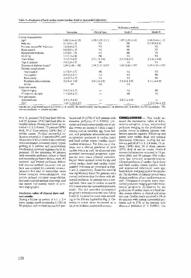
Table 3-Predictors of hard cardiac events (cardiac death or myocardial infarction)
Multivariate analysis
Univariate Clinical data Modell Model II
Chn'tcal characteristics Age~ 1.06 (1.03--1.12) 1.06 (1.03--l.ll) 1.07 (L03~Lll) 1.06 (1.03-l.lO)
Male sex 0_7 (0.4---1.3) NS NS 0.5 (0.3-0.9) Previous myocardial infarction 1.6 (0.9-2.9) NS NS NS Hypertension 0.9 (0_5~1.6) N$ NS NS Hypercholesterolemia 1.0 (0.5-1.9) NS NS NS Smoking 1.4 (0.7~2.6) NS NS NS Heart failure 3.2 (1.7-5.9) 2.7 (1.5-5.0) 2.5 (1.4-4.7) 2.1 (1.1~4.0) Type l diabetes 2.0 (0.9-4.2) NS NS NS Duration of diabetes (years)* 1.04 (LOI-1.08) 1.04 (l.Ol-l.07) 1.04 (l.Oi-1.07) 1.04 (l .01-1_07) Diabetic complications
Nephropathy 1.7 (0.7-3.7) NS NS NS Neuropathy 0.8 (0.3~2.1) NS NS NS Retmopathy 1.2 (0_7~1.4) NS NS NS Peripheral atherosclerosis 3.2 (1.4-7.0) 3.0 (1.6~5.8) 3.3 0.6--5.9) 3.1 (1.6--6.0) Stroke NS NS NS
Stress test results Typical angina 0.8 (0.4---1.7) NS NS ST-segment changes Ll (0.6--2.0) NS NS
Scan parameters Abnormal scan 3.4 (1_4--S.l) 2.8 (1.2~6.8) sss~ U6 (1.06--U?) Ll3 (1.02-1.25)
Data are Cox proponional hazard ratio (95% (j)_ In model!, the variable entered was the presence of an abnormal scan: in rnodellL the SSS was include( •Per one-unit increment;-, variable ""eluded
fects, 61 patients (31 %) had fixed defects, and 47 patients (24%) had fixed plus reversible defects. During the follow-up period of 4.1 ::: 2.4years, 73 patients {38%) died; 36 of these patients {19%) died of cardiac causes. Nonfatal myocardial infarction occurred in 15 patients (8%), and 18 patients (9%) unden.vent late coronary revascularization (coronary artery bypass grafting in 6 patients and percutaneous transluminal coronary angiography in l2 patients)_ Of the remaining 46 patients with reversible perfusion defects or fixed and reversible perfusion defects, most { 43 patients) had limited perfusion defects and received medical treatment; one patient was accepted for coronary revascularization but died of noncardiac cause before coronary revascularization, one patient refused coronary revascularization and received medical treatment, and one patient had normal results of coronary angiography.
Predictive value of clinical data and test results During a follow-up period of 4.1 :!: 2.4 years, cardiac death occurred in2 (3%) of 69 patients with normal myocardial per-
120
fusion and 34 (27%) of 125 patients with abnormal perfusion {P < 0.0001). Univariate and multivariate predictors of cardiac events are shown in Tables 2 and 3. Among clinical variables, age, heart failure, and peripheral atherosclerosis were independent predictors of cardiac death and hard cardiac events (cardiac death! nonfatal infarction). The duration of diabetes was a clinical predictor of hard cardiac events as well. An abnormal scan provided incremental prognostic information over these clinical variables. Kaplan-Meier survival curves for the end points cardiac death and cardiac death! nonfatal infarction are presented in Figs l and 2, respectively. Event-free survival was significantly better for patients with normal perfusion than for those with abnormal perfusion. In patients with a normal scan, there was no cardiac mortality $2.5 years after the myocardial perfusion study. The SSS provided incremental prognostic information as well (see Tables 2 and 3). Cumulative event rates according to the SSS are depicted in Fig. 3. Cumulative event rates increased as a function of defect extent and severity.
CONCLUSIONS- This study assessed the incremental value of dobutamine-atropine stress myocardial perfusion imaging in the prediction of cardiac events in diabetic patients vvith limited exercise capacity. Follow-up end points were cardiac death and nonfatal myocardial infarction_ During the follow-up period of 4_1:!: 2.4 years, 73 patients (38%) died; 36 of these patients (27%) died of cardiac causes. Nonfatal myocardial infarction occurred in 15 patients (8%), and 18 patients (9%) underwent late coronary revascularization Clinical predictors of cardiac death alone and hard cardiac events (cardiac death and myocardial infarction) were age, heart failure, and peripheral atherosclerosis. The duration of diabetes (years) was a clinical predictor of hard cardiac events as well. Dobutamine-atropine stress myocardial perlusion imaging provided incremental prognostic information for the prediction of cardiac death and hard cardiac events relative to clinical and stress Lest data. Cardiac death occurred in 3% of the patiems with normal myocardial perfusion and in 27% of the patients with abnormal perfusion (P < 0.0001)_ The
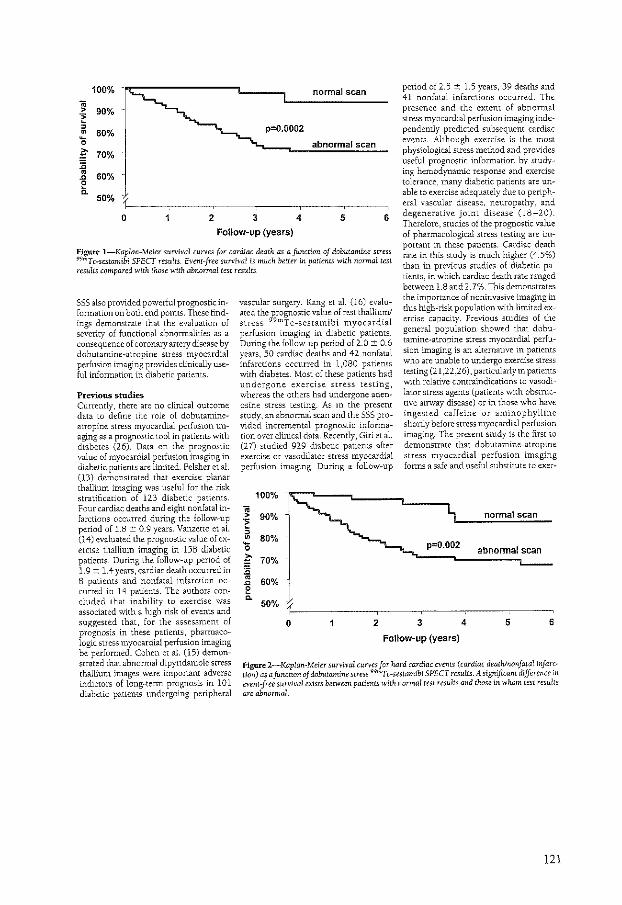
100% normal scan .. > 90% -~ , • 80% 0
p=0.0002
abnormal scan
~ 70% :;; • 60% .c
~ 50%
0 2 3 4 5 6
Follow-up (years)
period of 2.5 ± 1.5 years, 39 deaths and 41 nonfatal infarctions occurred. The presence and the extent of abnormal stress myocardial perfusion imaging independently predicted subsequent cardiac events. Although exercise is the most physiological stress method and provides useful prognostic information by studying hemodynamic response and exercise tolerance, many diabetic patients are unable to exercise adequately due to peripheral vascular disease, neuropathy, and degenerative joint disease (18-20). Therefore, studies of the prognostic value of pharmacological stress testing are important in these patients. Cardiac death rate in this study is much higher (4.5%) than in previous studies of diabetic patients, in which cardiac death rate ranged between 1.8 and 2. 7%. This demonstrates the importance of noninvasive imaging in this high-risk population with limited exercise capacity. Previous studies of the general population showed that dobutamine-atropine stress myocardial perfusion imaging is an alternative in patients who are unable to undergo exercise stress testing (21 ,22,26), particularly in patients with relative contraindications to vasodilator stress agents (patients with obstructive airway disease) or in those who have ingested caffeine or aminophylline shortly before stress myocardial perfusion imaging. The present study is the first to
demonstrate that dobutamine-atropine stress myocardial perfusion imaging forms a safe and useful substitute to exer-
Figure !-Kaplan-Meier survival curves for cardiac death as a function of dobutamine stress 99mTc-sestamibi SPECT results. Event-free survival is much better in patients with normal test results compared with those with abnormal test results
SSS also provided powerful prognostic information on both end points. These findings demonstrate that the evaluation of severity of functional abnormalities as a consequence of coronary artery disease by dobutamine-atropine stress myocardial perfusion imaging provides clinically useful information in diabetic patients.
Pre\ious studies Currently, there are no clinical outcome data to define the role o[ dobutamineatropine stress myocardial perfusion imaging as a prognostic tool in patients with diabetes (26). Data on the prognostic value of myocardial perfusion imaging in diabetic patients are limited. Felsher et al. (13) demonstrated that exercise planar thallium imaging was useful for the risk stratification of 123 diabetic patients. Four cardiac deaths and eight nonfatal infarctions occurred during the follow-up period of 1.8 ± 0.9 years. Vanzetto et al. (14) evaluated the prognostic value of exercise thallium imaging in 158 diabetic patients. During the follow-up period of 1. 9 ± l. 4 years, cardiac death occurred in 8 patients and nonfatal infarction occurred in 14 patients. The authors concluded that inability to exercise was associated with a high risk of events and suggested that, for the assessment of prognosis in these patients, pharmacologic stress myocardial perfusion imaging be performed. Cohen et al. (15) demonstrated that abnormal dipyridamole stress thallium images were important adverse indictors of long-term prognosis in 101 diabetic patients undergoing peripheral
vascular surgery. Kang et al. (16) evaluated the ~rognostic value of rest thallium/ stress 9 rnTc-sestamibi myocardial perfusion imaging in diabetic patients. During the follow-up period of 2.0 ± 0.6 years, 50 cardiac deaths and 42 nonfatal infarctions occurred in 1,080 patients with diabetes. Most o[ these patients had undergone exercise stress testing, whereas the others had undergone adenosine stress testing. As in the present study, an abnormal scan and the SSS provided incremental prognostic information over clinical data. Recently, Giri et al. (27) studied 929 diabetic patients after exercise or vasodilator stress myocardial perfusion imaging. During a follow-up
100% .. > 90% i: , • 0
80%
~ 70% :;; • 60% .c e 0.
50%
0 2
p=0.002
3
Follow-up (years)
normal scan
abnormal scan
4 5 6
Figure l-Kaplan-Meier survival curves for hard cardiac events (cardiac death/nonfatal infarction) as a function of dobutamine stress 99"'T c-sestamibi SPECT results. A significant difference in event -:free survival exists between patients with normal test results and those in whom test results are abnormal.
121
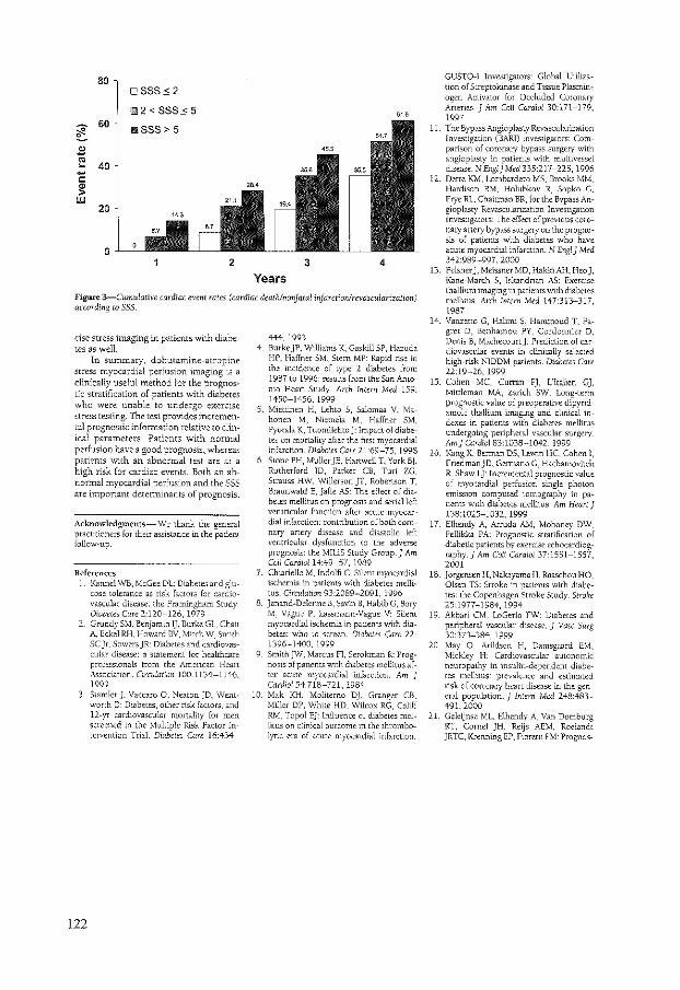
4
Years Figure 3-Cumutativc cardiac event rates (cardiac deaihlnonjatal infarctionlnvaswlarizatlon) according to 555.
cise stress imaging in patients vvith diabetes as welL
In summary, dobutamine-atropine stress myocardial perfusion imaging is a clinically useful method for the prognostic stratification of patients with diabetes who were unable to undergo exercise stress testing. The lest provides incremental prognostic information relative to clinical parameters. Patients with normal perfusion have a good prognosis, whereas patients with an abnormal test are at a high risk for cardiac events. Both an abnormal myocardial perfusion and the SSS are imponant determinants of prognosis.
Acknowledgments- We thank the general practitioners for thetr assistance in the patient follow-up.
References l Kanne! WB, McGee DL: Diabetes and glu
cose tolerartce as risk factors for cardiovascular disease: the Framingham Study Dwbeteo Care 2:120-126, 1979
122
Grundy S:vl, Benjamin I], Burke GL, Chait A, Eckel RH, Howard BV, ~itch W, Smith SC jr, Sowers jR: Diabetes and cardiovascular disease: a statement for healthcare professionals from the Amencan Hean Association. Circulation 100:1134-1146, 1999 Stamler), Vaccaro 0, ~eatonjD, Went· worth 0: Dmbetes, other risk factors, and 12-yr cardwvascular monality for men screened in the Multiple Risk Factor Intervention TriaL Diabetes Care 16:434-
444, 1993 4 BurkejP, Williams K, Gaskill SP, Hazuda
HP, Haffner SM, Stem MP: Rapid rise in the incidence of type 2 diabetes from 1987 to 1996: results from the San Amonio Heart Study_ Arch Imem Med 159: 1450-1456, 1999
5. Miettinen H, Lehto 5, Salomaa V, Mahonen M, Niemela M, Haffner SM, Pyorala K, Tuomilehto j: Impact of diabetes on mortality after the first myocardial infarction. Diabetes Care 21:69-75, 1998
6 Stone PH, Muller jE, Hartwell T, York Bj, Rutherford JD, Parker CB, Turi ZG, Strauss HW, Willerson jT, Roberuon T, Braunwald E, Jaffe AS: The effect of diabetes mellitus on prognosis and serial left ventricular function after acute myocardial infarction: contribution of both coronary artery disease and diastolic left ventricular dysfunction to the adverse prognosis: the MILlS Study Group.] Am Call Cardioll4:49-57, 1989 Chiariello M, lndolfi C: Silent myocardial ischemia in patients >with diabetes mellitus. Circulation 93:2089-2091, 1996
8 ]anand-Delenne B, Savin B, Habib G, Bory M, Vague P. Lassmann-Vague V: Silent myocardial ischemia in patients with diabetes: who to screen. Diabetes Care 22· 1396-1400, 1999 SmithjW, Marcus Fl, Serokman R: Prognosts of panents with diabetes mellitus after acute myocardial infarction. Am J Cardia! 54:718-721, 1984
10 Mak KR :vloliterno Dj, Granger CB, Miller DP, White HD, Wilcox RG, Califf RM, Topol EJ: lniluence of diabetes mellitus on clinical outcome in the thrombolytic era of acute myocardial infarction
GUSTO-! Investigators: Global Utilization of Streptokmase and Tissue Plasminogen Activator for Occluded Coronary Arteries. 1 Am Call Cardia! 30:171-179, 1997
11. The Bypass Angioplasty Revascularization Investigation (BARJ) investigators: Comparison of coronary bypass surgery with angioplasty in patients with multivessel disease. N Eng!) Mcd 335:217-225, 1996
12 Detre KM, Lombardero MS, Brooks MM, Hard1son RC\1, Holubkov R, Sopko G, Frye RL, Chaitman BR, for the Bypass Angioplasty Revascularizatwn Investigation Investigators: The effect of previous coronary artery bypass surgery on the prognosis of patients with diabetes who have acute myocardial infarction. N Eng I 1 Mcd 342:989-997, 2000
13. Felsherj, Meissner MD, HakkiAH, Heo ), Kane-March S, lskandrian AS: Exercise thallium imaging in patients with diabetes mellitus_ Arch Intern Med 147:313-317, 1987
14. Vanzetto G, Halimi S, 1-lammoud T, Fagret D, Benhamou PY, Cordonnier D, Denis B, Machecourtj: Prediction of cardiovascular events in clinically selected high-risk NIODM patients. Dwbetcs Care 22:19-26, 1999
15. Cohen MC. Curr:a.n Pj, L'ltalien G], Mittleman l\1A, Zarich SW: Long-term prognostic value of preoperative dipyridamole thallium imaging and clinical indexes in patients with diabetes mellitus undergoing peripheral vascular surgery Am 1 Cardiol 83:1038-1042, 1999
16. Kang X, Berman DS, Lewin HC, Cohen l, FriedmanJD, Germano G, Hachamovitch R, ShawL]: Incremental prognostic value of myocardial perfusion single photon emisswn computed tomography in patients with diabetes mellitus. Am Heart J 138:1025-1032, 1999
17. Elhendy A, Arruda AM, Mahoney OW, Pellikka PA: Prognostic stratification of diabetic patients by exercise echocardiography.1 Am Coli Cardia! 37:1551-1557, 2001
18. jorgensen H, Nakayama H, RE.aschou HO, Olsen TS: Stroke in patients with diabetes: the Copenhagen Stroke Study. Stroke 25:1977-1984, 1994
19 Akbari CM. LoGerfo FW: Diabetes and peripheral vascular disease. 1 Vase Surg 30:373-38+, 1999
20 May 0, Arildsen H. Damsgaard EM, Mickley H: Cardiovascular autonomic neuropathy in insulin-dependent diabetes mellitus: prevalence and estimated risk of coronary heart disease in the general population_ J Intern Med 248:483--491, 2000
21 Geleljnse ML, Elhendy A Van Domburg RT, Cornel jH, Reijs AEM, Roelandt JRTC, Krenning EP, Fioretti PM: Prognos-

tic value of dobutamine·atropine stress technetium-99m sestamibi perfusion scintigraphy in patients with chest pain. ] Am Col! Cardiol28:447-454, 1996
22 Senior R, Raval U, lahiri A: Prognostic value of stress dobutamine technetium-99m sestamibi single-photon emisston computed tomography (SPECT) in patients with suspected coronary disease. Am] Cardiol78:1092-l096, 1996
23. Hachamovitch R, Bennan DS, Kiat H, Cohen l, Cabico JA, Friedman J, Dtamond GA: E:~:ercise myocardiill perfusion
SPECT m patients without kno;vn coronary artery disease: incremental prognostic value and use in risk stratification Cirrulation 93:905-914, 1996
24 Elhendy A, Valkema R, van DomburgRT, Bax ], N\erop PR, Come! JH, Geleijnse 'ML, Reijs AE, Krenning EP, Roelandt JRTC: Safety of dobutamine-atropine stress myocardial perfusion scmtigraphy. J Nud Med 39:1662-1666, 1998
25. Cox DR: Regresswn models and life-tables.] R Stat Soc (B) 34:187-202, 1972
26 Geleijnse ML, Elhendy A, Fioretti PM, Ro-
elandtJRTC: Dobutamine stress myocardial perfusion imaging.] Am Coli Cardia! 36:2017-2027,2000
27. Gin 5, Shaw LJ, Murthy DR, Travin Ml, Miller DO, Hachamovuch R, BorgesNeto S, Berman OS, Waters DO, Heller GV: Impact of diabetes on the risk stratification usmg stress single-photon emission computed tomography myocardial perfusion imagmg in patients v..ith symptoms suggestive of coronary artery disease CirculatiOn 105:32.-40, 2002
123


Chapter 12
Incremental value of exercise 99mTc
tetrofosmin myocardial perfusion
single-photon emission computed
tomography for the prediction
of cardiac events
Schinkel AFL, Elhendy A, van Domburg RT,
Bax JJ, Vourvouri EC, Bountioukos M, Rizzello V, Agricola E,
Valkema R, Roelandt JRTC, Poldermans D
Am J Cardiol, in press
125
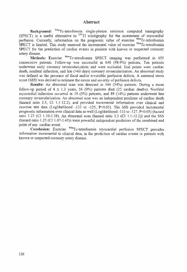
Abstract
Background: 99mTc-tetrofosmin single-photon emission computed tomography (SPECT) is a useful alternative to 201Tl scintigraphy for the assessment of myocardial perfusion. Currently, information on the prognostic value of exercise 99mTc-tetrofosmin SPECT is limited. This study assessed the incremental value of exercise 99mTc-tetrofosmin SPECT for the prediction of cardiac events in patients with known or suspected coronary artery disease.
Methods: Exercise 99mTc-tetrofosmin SPECT imaging was performed in 655 consecutive patients. Follow-up was successful in 648 (98.9%) patients. Ten patients undenvent early coronary revascularization and were excluded. End points were cardiac death, nonfatal infarction, and late (>60 days) coronary revascularization. An abnormal study was defined as the presence of fixed and/or reversible perfusion defects. A summed stress score (SSS) was derived to estimate the extent and severity of perfusion defects.
Results: An abnormal scan was detected in 344 (54%) patients. During a mean follow-up period of 4 ± 1.3 years, 56 (9%) patients died (22 cardiac deaths). Nonfatal myocardial infarction occurred in 19 (3%) patients, and 89 (14%) patients underwent late coronary revascularization. An abnormal scan was an independent predictor of cardiac death (hazard ratio 3.5, CI 1.1-12.2), and provided incremental information over clinical and exercise test data (Loglikelihood -133 to -125, P<0.05). The SSS provided incremental prognostic information over clinical data as well (Loglikelihood -133 to -127, P<O.OS) (hazard ratio 1.23 (CJ l.I0-1.38). An abnormal scan (hazard ratio 3.3 (CJ 1.1-12.2)) and the SSS (hazard ratio 1.25 (CI 1.07-1.45)) were powerful independent predictors of the combined end point of any cardiac event.
Conclusion: Exercise 99mTc-tetrofosmin myocardial perfusion SPECT provides information incremental to clinical data, in the prediction of cardiac events in patients with known or suspected coronary artery disease.
126

Introduction
Exercise thallium-201 e01 Tl) myocardial perfusion imaging has been used for the grediction of cardiac events over the last two decades (1-3). Previous studies have shown that
01 Tl myocardial perfusion variables have incremental value for the prediction of cardiac events over clinical and exercise test information alone (1-3). The new technetium-99m C9mTc) labeled perfusion tracers provide an improved image quality, and have a much shorter half-life compared to 201 Tl (4-6). Several studies have confirmed the diagnostic accuracy of exercise 99mTc-tetrofosmin SPECT imaging (7-10). However, information on the prognostic value of exercise 99mTc-tetrofosmin myocardial perfusion imaging is scarce (11,12). Additionally, the value of exercise 99mTc-tetrofosmin SPECT for the prediction of cardiac death as an individual end-point has not yet been demonstrated. From a clinical point of view this is important to know, since the prevention of cardiac death differs from the prevention of other cardiac events (13-16). The aim of this study was to assess the value of exercise 99mTctetrofosmin SPECT for the prediction of cardiac events in patients with known or suspected coronary artery disease.
Material and methods
Patient Selection: The study population consisted of 655 consecutive patients referred for exercise 99mTc-tetrofosmin SPECT imaging for the evaluation of suspected or known coronary artery disease. Follow-up was successful in 648 patients (98.9%). Ten patients undef'Nent coronary revascularization in the first 60 days after the nuclear testing. These patients were excluded from the analysis, because referral to coronary revascularization in the first 60 days after nuclear testing tends to be based on the results of the scan, and referral to revascularization >60 days after testing tends to be based on worsening of the patient's clinical status (17). The data from the remaining 638 patients are reported. All patients gave informed consent before the test The protocol was approved by the Hospital Ethics Committee.
Clinical data: Before nuclear testing, a stmctured interview and clinical history were acquired and cardiac risk factors were assessed. Hypertension was defined as a blood pressure 2':140/90 mmHg, or treatment with antihypertensive medication. Diabetes mellitus was defined as a fasting glucose level 2':7.8 mmol/L or the need for insulin or oral hypoglycemic agents. Hypercholesterolemia was defined as a total cholesterol 2':6.4 mmoVL, or treatment with lipid-lowering medication.
Exercise protocol: All patients performed a symptom-limited upright bicycle ergometry test with stepwise increment of 20 W every minute. Three electrocardiographic leads were continuously monitored. Cuff blood pressure measurements and tvvelve-lead electrocardiography were recorded at rest and every minute during exercise and recovery. Computer averaging of the electrocardiographic complexes was perfonned by the Schiller system Cardiovit CSG/12. Significant ST-segment depression was defined as a >1 mm horizontal or downsloping St-segment depression occurring at 80 ms after the J point.
99mTc-Tetrofosmin SPECT imaging: An intravenous dose of 370 MBq of 99mTctetrofosmin (Myoview, Amersham, Buckinghamshire, United Kingdom) was administered approximately 1 minute before the termination of the exercise test For resting studies 370 MBq of tetrofosmin were injected at least 24 hours after the exercise study. Image acquisition was performed with a triple head gamma camera system (Picker Prism 3000 XP, Cleveland,
127

Ohio, USA). For each study six oblique (short axis) slices from the apex to the base, three sagittal (vertical long axis) slices were defined. Each of the 6 short axis slices was divided into 8 equal segments. The septal part of the 2 basal slices was excluded from analysis because this region corresponds to the fibrous portion of the interventricular septum and normally exhibits reduced uptake. Consequently, a total of 47 segments were identified (3 long axis and 44 short axis). The interpretation of the scan was semiquantitatively performed by visual analysis assisted by the circumferential profiles analysis. Exercise and rest tomographic views were reviewed side by side by an experienced observer who was unaware of the patients' clinical data. A reversible perfusion defect was defined as a perfusion defect on the exercise images that partially or completely resolved at rest in ~ contiguous segments or slices in the 47-segment model. A fixed perfusion defect was defined as a perfusion defect on exercise images in 2 or more contiguous segments or slices, which persists on rest images in the 47-segment modeL An abnormal study was considered in the presence of fixed/and or reversible perfusion defect. To assess the severity of perfusion abnormalities, the left ventricular myocardium was divided into 6 segments: anterior, inferior, septal anterior, septal posterior, posterolateral and apical. Each of the 6 major left ventricular segments was scored using a 4-point scoring method (0 ~ normal, 1 ~ slightly reduced, 2 ~moderately reduced, 3 ~ severely reduced or absent uptake). The summed stress score was calculated to estimate the extent and severity of perfusion defects.
Follow-up: Follow-up data collection was performed by contacting the patient's general practitioner and by review of hospital records. The date of the last review or consultation was used to calculate follow-up time. Outcome events were: overall death, cardiac death, nonfatal myocardial infarction, and late (>60 days) coronary revascularization. Cardiac death was defined as a death caused by acute myocardial infarction, significant cardiac arrhythmias, or refractory congestive heart failure. Sudden death occurring without another explanation was included as cardiac death. Nonfatal myocardial infarction was defined by cardiac enzyme levels and ECG-changes.
Statistical analysis: Continuous data were expressed as mean value ± SD. The Student's t test was used to analyze continuous data. Differences behveen proportions were compared using the Chi-square test. Univariate and multivariate Cox proportional hazard regression models (BMDP Statistical Software Inc., Los Angeles, California, USA) were used to identify independent predictors of late cardiac events (18). Variables were selected in a stepwise forward selection manner with entry and retention set at a significance level of 0.05. The risk of a variable was expressed as a hazard ratio with a corresponding 95% confidence interval. The incremental value of myocardial perfusion scintigraphy over the clinical variables in the prediction of events was performed according to hvo models. In model I, the presence of an abnormal scan was included, and in model II, the SSS was included. The probability of survival was calculated using the Kaplan-Meier method, and survival curves were compared using the log-rank test. A p value <0.05 was considered statistically significant.
Results
Patient characteristics and exercise test results: The clinical characteristics of the 638 patients are presented in Table l.There was a significant increase of heart rate (76 ± 18 to 140 ± 24 beats/min, P<O.OOOl), and systolic blood pressure (140 ± 21 to 180 ± 30 mm Hg, P<O.OOOl) from rest to peak exercise. The mean work load was 152 ± 39 W. During the exercise test, 106 patients had typical angina, and 124 had significant ST-segment depression.
128

Side effects were short ventricular tachycardia (<10 complexes) in 7 patients (1%), and atrial fibrillation in 8 patients (1 %). Minor side effects were dizziness in 40 patients (6%), headache in 18 (3%), and nausea in 12 (2%). No patient experienced a myocardial infarction or ventricular fibrillation.
Table 1. Baseline Characteristics
Age (y) Man Women Congestive heart failure Diabetes mellitus Hypercholesterolemia Hypertension Previous myocardial infarction Smoking Beta blockers Calcium channel blockers
Number(%)
56±11 428 (67) 210 (33) 70 (11) 58 (9) 268 (42) 275 (43) 171 (27) 153 (24) 261 (41) 269 (42)
SPECT and follow-up results: An abnormal scan was detected in 344 (54%) patients. Myocardial perfusion abnormalities included fixed defects alone in 186 (29% ), and reversible defects in 158 (25%). These defects were completely reversible in 56 (9%) patients and partially reversible in 102 (16%) patients. During a mean follow-up period of 4 ± 1.3 years, 56 (9%) patients died, of these patients 22 (39%) died due to cardiac causes. Nonfatal myocardial infarction occurred in 19 (3%) patients, and 89 (14%) patients underwent late coronary revascularization.
100% Normal scan
iii > -~ 90% P<0.001
"' Abnormal scan
(J)
80%
0 2 3 4
Follow-up (y)
Figure 1. Kaplan-Meier survival curves(end point of cardiac death) in patients with normal and abnormal exercise 99mTc-tetrofosmin SPECT.
129
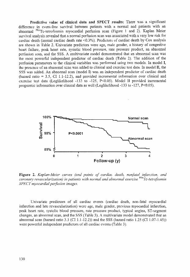
Predictive value of clinical data and SPECT results: There was a significant difference in event-free survival between patients with a normal and patients with an abnormal 99mTc-tetrofosmin myocardial perfusion scan (Figure 1 and 2). Kaplan Meier survival analysis revealed that a normal perfusion scan was associated with a very low risk for cardiac death (annual cardiac death rate <0.3%). Predictors of cardiac death by Cox analysis are shown in Table 2. Univariate predictors were age, male gender, a history of congestive heart failure, peak heart rate, systolic blood pressure, rate pressure product, an abnormal perfusion scan, and the SSS. A multivariate model demonstrated that an abnormal scan was the most powerful independent predictor of cardiac death (Table 2). The addition of the perfusion parameters to the clinical variables was performed using two models. In model I, the presence of an abnormal scan was added to clinical and exercise test data. In model II, the SSS was added. An abnormal scan (model I) was an independent predictor of cardiac death (hazard ratio ~ 3.5, Cl 1.1-12.2), and provided incremental information over clinical and exercise test data (Loglikelihood -133 to -125, P<0.05). Model ll provided incremental prognostic information over clinical data as well (Loglikelihood -133 to -127, P<0.05).
P<0.0001
80%
0 2 3 4
Follow-up (y)
Figure 2. Kaplan-Meier curves (end points of cardiac death, nonfatal infarction, and coronary revascularization) in patients with normal and abnormal exercise 99mTc-tetrofosmin SPECT myocardial perfusion images.
Univariate predictors of all cardiac events (cardiac death, non-fatal myocardial infarction and late revascularization) were age, male gender, previous myocardial infarction, peak heart rate, systolic blood pressure, rate pressure product, typical angina, ST -segment changes, an abnormal scan, and the SSS (Table 3). A multivariate model demonstrated that an abnormal scan (hazard ratio 3.3 (Cl 1.1-12.2)) and the SSS (hazard ratio 1.25 (Cll.07-1.45)) were powerful independent predictors of all cardiac events (Table 3).
130

Table 2. Predictors of cardiac death
Univariate Multivariate
(HR. CI) HR(CI) Model I Model II Age* 1.04 (1.00-1.09) 1.04 (1.00-1.09) NS NS Male gender 3.4 (1.0-11.6) 3.4(1.0-11.5) NS NS Congestive heart
2.7 (1.0-7.3) NS NS NS failure
Diabetes mellitus 0.5 (0.1-3.8) NS NS NS History of angina 0.5 (0.2-1.6) NS NS NS Hypercholesterolemia 0.5 (0.2-1.4) NS NS NS Hypertension 1.0 (0.4-2.4) NS NS NS Previous MI 1.9 (0.8-4.4) NS NS NS Smoking 0.9 (0.3-2.5) NS NS NS
Exercise test results Peak heart rate 0.97 (0.96-0.99) NS NS Peak systolic BP 0.98 (0.97-0.99) NS NS Peak RPP 0.88 (0.82-0.95) 0.90 (0.83--D.97) NS Typical angina 1.1 (0.4-3.3) NS NS ST -segment changes 0.8 (0.3-2.1) NS NS
Scan parameters Abnormal scan 3.4 (1.3-11.7) 3.5 (1.1-12.2) SSS* 1.23 (1.10-1.39) 1.23 (1.1 0-1.38)
Values are expressed as Cox proportional hazard ratio and 95% confidence intervaL In model!, the presence of an abnormal scan was entered. In model II, the SSS was included. MI = myocardial infarction; BP= blood pressure; RPP =rate pressure product; NS =not statistically significant;*Per 1 unit increment;- indicates
Table 3. Predictors of cardiac events (cardiac death, myocardial infarction, late coronary revascularization)
Univariate Multivariate
HR (CI) HR(CI) Modell Model II Age* 1.02 (1.01-1.04) 1.02 (1.01-1.04) NS 1.02 (1.00-1.04) Male gender 1.9 (1.3-2.9) 1.9 (1.2-2.8) NS NS Congestive heart failure 1.5 (0.9-2.4) NS NS NS Diabetes mellitus 1.5 (0.9-2.5) NS NS NS History of angina 1.3 (0.9-1.9) NS NS NS H ypercholes tero lemi a 0.9 (0.7-1.3) NS NS NS Hypertension 0.9 (0.6-1.3) NS NS NS Previous MI 1.6 (1.1-2.3) NS NS NS Smoking 1.3 (0.9-1.8) NS NS NS
Exercise test results Peak heart rate 0.98 (0.97-0.99) 0.98 (0.97-0.99) 0.98 (0.97-0.99) Peak systolic BP 0.98 (0.97-0.99) NS NS Peak RPP 0.91 (0.86-0.96) NS NS Typical angina 2.0 (1.3-2.9) 1.6 (1.1-2.4) NS ST -segment changes 1.4 (1.0-2.0) NS NS
Scan parameters Abnormal scan 3.1 (2.1-4.6) 3.3 (1.1-12.2) SSS* 1.16 (1.10-1.22) 1.25 (1.07-1.45)
Fonnat similar to Table 2.
131

Discussion
The main finding of this study is that exercise 99mTc-tetrofosmin SPECT myocardial perfusion imaging provides prognostic information incremental to clinical data and exercise test results, in the prediction of cardiac events. A normal perfusion scan was associated with a very low risk for cardiac death (annual cardiac death rate <0.3%). The cardiac death rate was significantly increased in patients with an abnormal 99mTc-tetrofosmin SPECT study. The survival curves continued to diverge during the follow-up period, which indicates a sustained prognostic value of 99mTc-tetrofosmin SPECT over the 4 years of follow up. Multivariate Cox analysis showed that after adjusting for clinical and exercise stress data, the SPECT results had significant incremental prognostic value. An abnormal scan and a larger SSS were related to a significant increase of the risk of cardiac death and all cardiac events.
Comparison to previous studies: To date, information on the prognostic value of exercise 99mTc-tetrofosmin SPECT myocardial perfusion imaging is limited (11,12). Groutars et al. (11) reported a very favorable outcome in 246 patients with a normal exercise dualisotope 201 TV99mTc-tetrofosmin test during the follow-up period of 2.1 ± 0.3 years. Recently, Galassi et al. (12) studied 459 patients with exercise 99mTc-tetrofosmin SPECT. During follow-up 25 hard cardiac events (cardiac death and nonfatal infarction) occurred. Multivariate analysis showed that exercise 99mTc-tetrofosmin SPECT provided incremental information over clinical and exercise data for the prediction of hard cardiac events. However, nuclear data had no incremental value over clinical and exercise information when the combined endpoint of all cardiac events (cardiac death, infarction, and coronary revascularization).
Hence, the currently available studies on the prognostic value of exercise 99mTctetrofosmin SPECT have only evaluated patients with a normal scan, or used a combined study endpoint (11,12). Compared to the previous studies, our study included a large number of patients, with a nearly complete follow-up, and there was a larger number of cardiac events. In this setting, exercise 99mTc-tetrofosmin SPECT information predicted cardiac death as a single endpoint, as well as all cardiac events.
Clinical implications: 99mTc-tetrofosmin provides an imrrroved image quality, and has a much shorter half-life compared to 201Tl (4-6). Nevertheless, 9 mTc-tetrofosmin has a lower extraction fraction and a less parallel relationship between myocardial blood flow and myocardial tracer uptake during vasodilator stress compared to 99mTc-sestamibi (19,20). It is not known whether these findings are reproducible with other forms of stress. Furthermore, the prognostic implications of these findings are uncertain.
The present study demonstrated that exercise 99mTc-tetrofosmin imaging provides important ~rognostic information in patients with known or suspected coronary artery disease (1-3,21). 9 mTc-tetrofosmin effectively identified patients at high and low risk for cardiac death. Patients with a normal scan have very favorable prognosis, and a cardiac death rate comparable to that in the general population (22). Therefore, these patients can be excluded from further invasive procedures. Conversely, high-risk patients with an abnormal myocardial perfusion scan, particularly those with a large perfusion abnormality, may benefit from coronary revascularization. The clinical management of these patients should be individualized and further studies are needed to demonstrate the value of revascularization in that subset of patients.
Limitations: No attenuation correction or scatter correction was used during 99mTctetrofosmin SPECT myocardial perfusion imaging. Application of attenuation and scatter
132

correction may have further improved the accuracy of the 99mTc-tetrofosmin myocardial perfusion images.
Conclusion: Exercise 99mTc-tetrofosmin SPECT myocardial perfusion imaging effectively identifies patients at low and high risk of cardiac death. Perfusion variables are strong predictors of cardiac death and all cardiac events. 99mTc-tetrofosmin SPECT data are incremental to both clinical and exercise stress parameters in the prediction of cardiac events.
References
1. Beller GA, Zaret BL. Contributions of nuclear cardiology to diagnosis and prognosis of patients with coronary artery disease. Circulation. 2000; I 01:1465-1478.
2. Brown KA. Prognostic value ofthallium-201 myocardial perfusion imaging: a diagnostic tool comes of age. Circulation. 1991 ;83:363-380.
3. Brown KA. Prognostic value of myocardial perfusion imaging: state of the art and new developments. J Nucl Cardia!. 1996;3:516-37.
4. Kelly JD, Forster AM, Higley B, et a!. Technetium-99m-tetrofosmin as a new radiophannaceutical for myocardial perfusion imaging. J Nucl Med. 1993;34:222-227.
5. Jain D, Wackers FJ, Mattera J, McMahon M, Sinusas AJ, Zaret BL. Biokinetics of technetium-99m-tetrofosmin: myocardial perfusion imaging agent: implications for a oneday imaging protocoL J Nucl Med. 1993;34: 1254-1259.
6. Hendel RC, Parker MA, Wackers FJ, Riga P, Lahiri A, Zaret BL. Reduced variability of interpretation and improved image quality with a technetium 99m myocardial perfusion agent: comparison of thallium 201 and technetium 99m-labeled tetrofosmin. J Nucl Cardia!. 1994; 1 :509-14.
7. Zaret BL, Riga P, Wackers FJ, et a!. The tetrofosmin international trial study group. Myocardial perfusion imaging with 99mTc-tetrofosmin: comparison to 201Tl imaging and coronary angiography in a phase III multicenter triaL Circulation. 1995;91 :313-319.
8. Tamaki N, Takahashi N, Kawamoto M, et a!. Myocardial tomography using technetium-99m-tetrofosmin to evaluate coronary artery disease. J Nucl Med. 1994;35:594-600.
9. Rigo P, Leclercq B, Itti R, Lahiri A, Braat S. Technetium-99m-tetrofosmin myocardial imaging: a comparison with thallium-201 and angiography. J Nucl Med. 1994;35:587-593.
10. Sridhara BS, Braat S, Riga P, ltti R, Cload P, Lahiri A Comparison of myocardial perfusion imaging with technetium-99m tetrofosmin versus thallium-201 in coronary artery disease. Am J Cardia!. 1993;72: 1015-1019.
II. Groutars RG, Verzijlbergen JF, Muller AJ, eta!. Prognostic value and quality of life in patients with normal rest thallium-201/stress technetium 99m-tetrofosmin dual-isotope myocardial SPECT. J Nucl Cardia!. 2000;7:333-341.
12. Galassi AR, Azzarelli S, Tomaselli A, et al. Incremental prognostic value of technetium-99m-tetrofosmin exercise myocardial perfusion imaging for predicting outcomes in patients with suspected or known coronary artery disease. Am J Cardia!. 2001;88:101-106.
13. Hachamovitch R, Berman DS, Shaw LJ, et al. Incremental prognostic value of myocardial perfusion single photon emission computed tomography for the prediction of cardiac death: differential stratification for risk of cardiac death and myocardial infarction. Circulation. 1998;97:535-43.
14. Aldennan EL, Bourassa MG, Cohen LS, et a!. Ten-year follow-up of survival and myocardial infarction in the randomized Coronary Artery Surgery Study. Circulation. 1990;82: 1629-1646.
133
15. Shepherd J, Cob be SM, Ford I, eta!. Prevention of coronary heart disease with pravastatin in men with hypercholesterolemia. West of Scotland Coronary Prevention Study Group. N Engl J Med. 1995;333: 1301-1307.
16. Gaspoz JM, Coxson PG, Goldman PA, eta!. Cost effectiveness of aspirin, clopidogrel, or both for secondary prevention of coronary heart disease. N Eng] J Med. 2002;346:1800-1806.
17. Hachamovitch R, Berman DS, Kiat H, et al. Exercise myocardial perfusion SPECT in patients without known coronary artery disease: incremental prognostic value and use in risk stratification. Circulation. 1996;93 :905-914.
18. Cox DR. Regression models and life-tables. J R Stat Soc (B). 1972; 34:187-202. 19. Soman P, Taillefer R, DePuey EG, Udelson JE, Lahiri A. Enhanced detection of
reversible perfusion defects by Tc-99m sestamibi compared to Tc-99m tetrofosmin during vasodilator stress SPECT imaging in mild-to-moderate coronary artery disease. J Am Coil Cardiol. 2001;37:458-462.
20. Glover DK, Ruiz M, Yang JY, Smith WH, Watson DD, Beller GA. Myocardial 99mTctetrofosmin uptake during adenosine-induced vasodilation with either a critical or mild coronary stenosis: comparison with 201 Tl and regional myocardial blood flow. Circulation. 1997;96:2332-2338.
21. Iskander S, Iskandrian AE. Risk assessment using single-photon emission computed tomographic technetium-99m sestamibi imaging. JAm Coil Cardiol. 1995;26:639-647.
22. Hoyert DL, Arias E, Smith BL, Murphy SL, Kochanek KD. Deaths: Final Data for 1999.
134
National vital statistics reports; vol 49 no. 8. Hyattsville, Maryland: National Center for Health Statistics. 2001.

Chapter 13
Long-term prognosis after a normal
exercise stress technetium-99m-sestamibi
SPECT study
Elhendy A, Schinkel AFL, van Domburg RT,
Bax JJ, Valkema R, Roelandt JRTC, Poldennans D
Submitted for publication
135
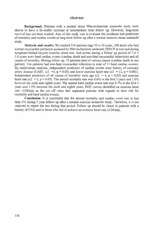
Abstract
Background: Patients with a normal stress 99m-technetiurn sestamibi study were shown to have a favorable outcome at intermediate term follow up. However, long-term survival has not been studied. Aim of this study was to evaluate the incidence and predictors of mortality and cardiac events at long term follow up after a normal exercise stress sestamibi study.
Methods and results: We studied 218 patients (age 53± 10 years, 108 men) who had normal myocardial perfusion assessed by 99m-technetium sestamibi SPECT at rest and during symptom-limited bicycle exercise stress test. End points during a follow up period of 7.4 ± 1.8 years were hard cardiac events (cardiac death and non-fatal myocardial infarction) and all causes of mortality. During follow up, 13 patients died of various causes (cardiac death in one patient). Ten patients had non-fatal myocardial infarction (a total of 11 hard cardiac events). By multivariate analysis, independent predictors of cardiac events were history of coronary artery disease (CAD) (c2 ~s, p ~ 0.03) and lower exercise heart rate (c2 ~ 12, p ~ 0.001). Independent predictors of all causes of mortality were age (c2 = 4, p = 0.05) and exercise heart rate (c2 ~ 5, p ~ 0.03). The annual mortality rate was 0.6% in the first 5 years and 1.8% betvveen the sixth and eighth years. The annual hard cardiac event rate was 0.7% in the first 5 years and 1.5% bet\veen the sixth and eighth years. ROC curves identified an exercise heart rate <130/min as the cut off value that separated patients with regards to their risk for mortality and hard cardiac events.
Conclusion: It is concluded that the annual mortality and cardiac event rate is less than 1% during 5 year follow up after a normal exercise sestamibi study. Therefore, it is not required to repeat the test during that period. Follow up should be closer in patients with a history of CAD and in those who fail to achieve an exercise heart rate 2::130/min.
136
Introduction
The management of patients with suspected coronary artery disease (CAD) should be based on proper evaluation of the risk of mortality and cardiac events in an individual patient, based on clinical data and the results of non-invasive stress testing techniques whenever these are indicated. Identification of patients at low risk of cardiac events has an important impact on patients' management by avoiding the risk and the cost related to further diagnostic and therapeutic approaches, which are unlikely to improve the outcome in low risk patients (1-3). Stress 99m-technetium sestamibi SPECT imaging is a useful technique for the diagnosis and prognostic stratification of patients with known or suspected coronary artery disease (4-23). Patients with a normal stress sestamibi myocardial perfusion study were reported to have a low event rate at short and intermediate term follow up (9, 19). However, the long-term outcome after a normal study is unknown. The mean duration of follow in previous reports ranged between 13 and 28 months. Therefore, it is not known whether the low risk warrantee of a normal study can be maintained at longer-term follow up. Due to the lack of long tenn follow up data, it is not known whether patients with a normal study may require repeated testing, and if so what would be the time interval at which the low risk status is no longer guaranteed in a patient with a previously normal study. Additionally, data regarding overall survival after a normal stress sestamibi study are scarce.
The aim of this study was to evaluate the incidence and predictors of mortality and cardiac events at long term follow up after a normal exercise stress 99m-technetium sestamibi SPECT imaging.
Methods
Patient selection: Study population comprised consecutive patients referred for exercise stress testing in conjunction with 99m technetium sestamibi SPECT imaging for evaluation of suspected CAD between 1988-1995, who had normal myocardial perfusion at rest and during exercise stress. Exclusion criteria were the presence of significant valvular heart disease and unstable chest pain. Mean age was 53 ± 10 years. Inclusion criteria were fulfilled in 218 patients (108 men and 110 women). Follow-up data was collected in the year 2000 and could be completed in all patients. Mean follow up duration in was 7.4 ± 1.8 (maximal= 11.7 years). The minimum follow up duration (in patients without events) was 5.2 years.
Exercise stress test: All patients undenvent a symptom limited upright bicycle ergometry test with stepwise increment of 20 Watts each minute. 3 electrocardiographic leads were continuously monitored. 12-lead electrocardiogram was recorded at rest and every minute until the end of recovery phase. The level of ST -segment was calculated by averaging the signals using a computerised system (Cardiovet, CSG/12, Schiller, Baar, Switzerland). An ischemic response was defined as 2:1-mm horizontal or downsloping ST-segment depression persisting 80 ms after the J point.
SPECT imaging: Approximately 1 minute before the termination of the exercise stress test, an intravenous dose of 3 70 MBq of 99m-technetium sestamibi was administered. Stress images were acquired 1 hour after termination of the exercise test. For resting studies, 370 MBq of sestamibi were injected 24 hours after the stress study. Image acquisition was performed with a Siemens Gammasonics single-head Rota Camera (Orbiter; Siemens Corp., Iselin, NJ). Thirty-two projections were obtained, from the left posterior oblique to the right anterior oblique over 180 degrees. For each study six oblique (short axis) slices from the apex
137

to the base and three sagittal (vertical long axis) slices from the septum to the lateral wall were defined. Each of the 6 short axis slices was divided into 8 equal segments. The interpretation of the scan was semiquantitatively performed by visual analysis assisted by the circumferential profiles analysis. Images were reviewed side by side by an experienced observer who was unaware of the patients' clinical or electrocardiographic data. A reversible perfusion defect was defined as a perfusion defect on stress images that partially or completely resolved at rest in 2:2 contiguous segments or slices. A fixed perfusion defect was defined as a perfusion defect on stress images in 2 or more contiguous segments or slices, which persists on rest image. A normal study was defined as absence of perfusion abnormalities.
Follow-up: Follow-up was obtained by mailed questionnaires and scripted telephone interviews. Events were verified by contacting the patients' primary physician and reviewing medical records and death certificates. The end points considered were all causes of mortality and hard cardiac events defined as nonfatal myocardial infarction and cardiac death. Sudden death occurring without another explanation was included as cardiac death. Myocardial infarction was defined according to usual clinical, electrocardiographic and enzymatic criteria. Patients who had coronary revascularization (angioplasty or coronary artery bypass surgery) prior to other events were censored at the time ofrevascularization.
Statistical analysis: Unless specified, data are presented as mean values ± SD. The Chi square test was used to compare differences between proportions. The Student t test was used for analysis of continuous data. P value <0.05 was considered statistically significant. Logistic regression models were used to identify independent predictors of follow up events. P value <0.05 was considered statistically significant. Parameters included in the multivariate analysis model were those found to be significant (or of borderline significance, p=O.l) in the univariate analysis.
Results
Clinical features: Mean age was 53 ± 10 years. There were 108 men and 110 women. Forty-seven patients (22%) had a history of coronary artery disease (previous coronary angioplasty in 33 patients and previous non-Q myocardial infarction in 14 patients). In the remaining patients, the pretest probability of CAD based on Diamond and Forrester (24) classification was low in 57 patients and intermediate or high in 114 patients. Medications at the day of the test included beta-blockers in 68 (31 %) patients, calcium channel blockers in 59 (27%) patients and angiotensin converting enzyme inhibitors in 27 (12%) patients. Chest pain was the main complaint in 158 (72%) patients. This was classified as atypical in 114 patients and typical of angina in 44 patients. Risk factors for CAD were diabetes mellitus in 14 (6%), cigarette smoking in 58 (27%), hypercholesterolemia in 53 (24%) and hypertension in 71 (33%) patients.
Hemodynamic response: There was a significant increase of the heart rate (78 ± 15 vs 148 ± 24 beats/minute, p<O.OOOl); systolic blood pressure (137 ± 21 vs 188 ± 25 mm Hg, p<O.OOOl) and rate pressure product (10691 ± 2584 vs 28703 ± 9964, p<O.OOOOl) from rest to peak exercise respectively. The target heart rate (85% of the maximal predicted heart rate) was reached in 161 patients (74%). The mean working capacity was 144 ± 42 Watts. Nine (4%) patients had ST segment depression and 25 (11 %) patients had angina induced by exerctse.
138
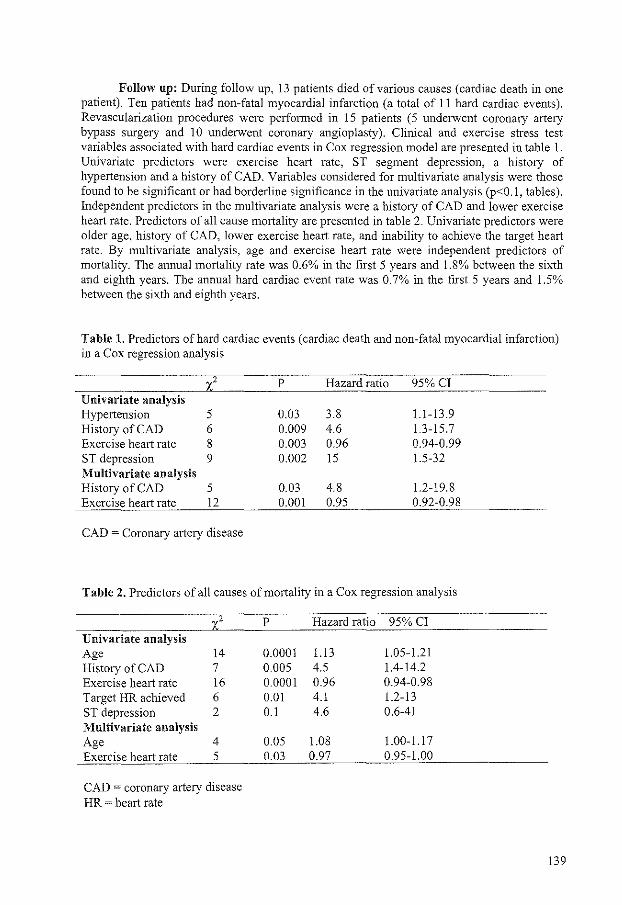
Follow up: During follow up, 13 patients died of various causes (cardiac death in one patient). Ten patients had non-fatal myocardial infarction (a total of 11 hard cardiac events). Revascularization procedures were performed in 15 patients (5 underwent coronary artery bypass surgery and 10 underwent coronary angioplasty). Clinical and exercise stress test variables associated with hard cardiac events in Cox regression model are presented in table 1. Univariate predictors were exercise heart rate, ST segment depression, a history of hypertension and a history of CAD. Variables considered for multivariate analysis were those found to be significant or had borderline significance in the univariate analysis (p<0.1, tables). Independent predictors in the multivariate analysis were a history of CAD and lower exercise heart rate. Predictors of all cause mortality are presented in table 2. Univariate predictors were older age, history of CAD, lower exercise heart rate, and inability to achieve the target heart rate. By multivariate analysis, age and exercise heart rate were independent predictors of mortality. The annual mortality rate was 0.6% in the first 5 years and 1.8% between the sixth and eighth years. The annual hard cardiac event rate was 0.7% in the first 5 years and 1.5% between the sixth and eighth years.
Table 1. Predictors of hard cardiac events (cardiac death and non-fatal myocardial infarction) in a Cox regression analysis
2 p Hazard ratio 95%CI Univariate analysis Hypertension 5 0.03 3.8 1.1-13.9 History of CAD 6 0.009 4.6 1.3-15.7 Exercise heart rate 8 0.003 0.96 0.94-0.99 ST depression 9 0.002 15 1.5-32 Multivariate analysis History of CAD 5 0.03 4.8 1.2-19.8 Exercise heart rate 12 0.001 0.95 0.92-0.98
CAD ~ Coronary artery disease
Table 2. Predictors of all causes of mortality in a Cox regression analysis
2 p Hazard ratio 95% Cl Univariate analysis Age 14 0.0001 1.13 1.05-1.21 History of CAD 7 0.005 4.5 1.4-14.2 Exercise heart rate 16 0.0001 0.96 0.94-0.98 Target HR achieved 6 0.01 4.1 1.2-13 ST depression 2 0.1 4.6 0.6-41 Multivariate analysis Age 4 0.05 1.08 1.00-1.17 Exercise heart rate 5 0.03 0.97 0.95-1.00
CAD = coronary artery disease HR ~ heart rate
139
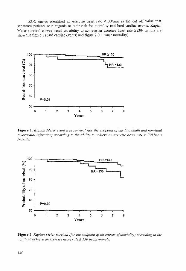
ROC curves identified an exercise heart rate <130/min as the cut off value that separated patients with regards to their risk for mortality and hard cardiac events. Kaplan Meier survival curves based on ability to achieve an exercise heart rate ;:::130/ minute are shown in figure 1 (hard cardiac events) and figure 2 (all cause mortality).
100 c::=J HR ~130
~ \ HR <130 e... 90
"iii > -~ 80 " "' "' "' 70 ~ ... -!: "' 60 > w P=0.02
50 0 2 3 4 5 6 7 8
Years
Figure 1. Kaplan Meier event free survival (for the endpoint of cardiac death and non-fatal myocardial infarction) according to the ability to achieve an exercise heart rate 2 130 beats /minute.
100 ~ e... "iii 90 > -~
"' 80 "' -0 ,.,
70 ~ :c "' .Q 60 0 P=0.01 ~
c.. 50
0 2 3 4 5 6 7 8
Years
Figure 2. Kaplan Meier survival (for the endpoint of all causes of mortality) according to the ability to achieve an exercise heart rate 2 130 beats /minute.
140
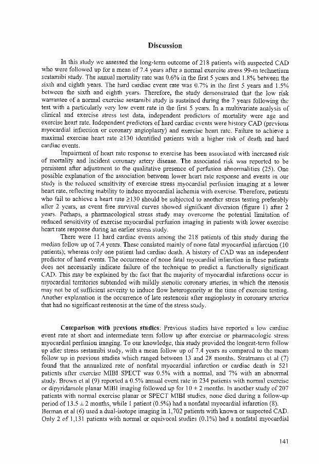
Discussion
In this study we assessed the long-term outcome of 218 patients with suspected CAD who were followed up for a mean of 7.4 years after a normal exercise stress 99-rn technetium sestamibi study. The annual mortality rate was 0.6% in the first 5 years and 1.8% between the sixth and eighth years. The hard cardiac event rate was 0. 7% in the first 5 years and 1.5% between the sixth and eighth years. Therefore, the study demonstrated that the low risk warrantee of a normal exercise sestamibi study is sustained during the 7 years following the test with a particularly very low event rate in the first 5 years. In a multivariate analysis of clinical and exercise stress test data, independent predictors of mortality were age and exercise heart rate. Independent predictors of hard cardiac events were history CAD (previous myocardial infarction or coronary angioplasty) and exercise heart rate. Failure to achieve a maximal exercise heart rate ;:::130 identified patients with a higher risk of death and hard cardiac events.
Impairment of heart rate response to exercise has been associated with increased risk of mortality and incident coronary artery disease. The associated risk was reported to be persistent after adjustment to the qualitative presence of perfusion abnormalities (25). One possible explanation of the association between lower heart rate response and events in our study is the reduced sensitivity of exercise stress myocardial perfusion imaging at a lower heart rate, reflecting inability to induce myocardial ischemia with exercise. Therefore, patients who fail to achieve a heart rate ;:::130 should be subjected to another stress testing preferably after 2 years, as event free survival curves showed significant diversion (figure 1) after 2 years. Perhaps, a pharmacological stress study may overcome the potential limitation of reduced sensitivity of exercise myocardial perfusion imaging in patients with lower exercise heart rate response during an earlier stress study.
There were 11 hard cardiac events among the 218 patients of this study during the median follow up of 7.4 years. These consisted mainly of none fatal myocardial infarction (1 0 patients); whereas only one patient had cardiac death. A history of CAD was an independent predictor of hard events. The occurrence of none fatal myocardial infarction in these patients does not necessarily indicate failure of the technique to predict a functionally significant CAD. This may be explained by the fact that the majority of myocardial infarctions occur in myocardial territories subtended with mildly stenotic coronary arteries, in which the stenosis may not be of sufficient severity to induce flow heterogeneity at the time of exercise testing. Another explanation is the occurrence of late restenosis after angioplasty in coronary arteries that had no significant restenosis at the time of the stress study.
Comparison with previous studies: Previous studies have reported a low cardiac event rate at short and intermediate term follow up after exercise or pharmacologic stress myocardial perfusion imaging. To our knowledge, this study provided the longest-term follow up after stress sestamibi study, with a mean follow up of 7.4 years as compared to the mean follow up in previous studies which ranged between 13 and 28 months. Stratmann et al (7) found that the annualized rate of nonfatal myocardial infarction or cardiac death in 521 patients after exercise MIBI SPECT was 0.5% with a normal, and 7% with an abnormal study. Brown et al (9) reported a 0.5% annual event rate in 234 patients with normal exercise or dipyridamole planar MIBI imaging followed up for 10 ± 2 months. In another study of 207 patients with normal exercise planar or SPECT MIBI studies, none died during a follow-up period of 13.5 ± 2 months, while 1 patient (0.5%) had a nonfatal myocardial infarction (8). Berman et al (6) used a dual-isotope imaging in 1,702 patients with known or suspected CAD. Only 2 of 1,131 patients with normal or equivocal studies (0.1%) had a nonfatal myocardial
141
infarction or cardiac death during a follow-up of 20 ± 5 months, compared with 43 of 571 patients with an abnonnal study (8%). In 2,200 patients without documented CAD followed up after exercise testing with dual-isotope SPECT (5), Hachamovitch et al reported an event rate of0.3% in 1,623 patients with normal SPECT studies.
In 412 patients with intermediate pretest probability of CAD followed up for 17 ± 13 months, Nallamouthu et al (13) reported only one event in 295 patients with normal studies (0.3%; p < 0.0001). Travin et al (21) studied 1,226 men and 1,151 women who underwent MIBI SPECT with either exercise or dipyridamole testing, a normal rest-stress MIBI study was associated with an annual rate of cardiac death or nonfatal myocardial infarction of 1. 7% in men and 0.8% in women.
Studies that evaluated the prognostic value of pharmacological stress sestarnibi SPECT have reported a low risk of cardiac events in patients with a normal study. However, event rate tended to be higher after a normal pharmacologic as compared to exercise stress studies, reflecting the higher risk status of a population unable to perform an exercise stress test (26).
Evaluation of all cause of mortality. Recently, the evaluation of all causes of mortality as a separate follow up endpoint in patients with known or suspected CAD has gained an increasing interest. This is due to the difficulties encountered in determining the actual cause of death in some patients (27) and the fact that CAD is associated with other conditions which are independently associated with increased risk of death such as diabetes mellitus, hypertension, smoking and peripheral vascular disease. This is the first study that evaluates long-term mortality rate after stress sestamibi studies. We found that patients with normal sestamibi studies have a very low annual mortality rate during a mean of 7.4 years of follow up.
Limitations: Although this study reports the longest term follow up after a sestamibi study, the number of patients was relatively low as well as the number of events. Further studies from other centers are needed to confirm these findings.
Summary and clinical implications: The optimal use of stress imaging techniques in the risk stratification of patients with known or suspected CAD requires adequate knowledge of the outcome of patients relative to the results obtained by these techniques. Although it is well established that a normal stress sestamibi study identifies patients at low risk of cardiac events at intermediate term follow up, it is not known whether the physician can rely upon these results in predicting longer term outcome. Performing the test at regular intervals in individuals with a normal study may not be cost effective. On the other hand deferring these individuals from future non-invasive testing may result in failure to predict events at longer term follow up. This study showed that the annual mortality and cardiac event rate is less than 1% during 5 year follow up after a normal exercise sestamibi study and therefore, it is not required to repeat the test during that period, particularly in those patients who have no history of CAD and who were able to achieve an exercise heart rate ;:::130/min. Follow up should be closer in patients with a history of CAD and in those who fail to achieve an exercise heart rate ;:::130/min.
References
I. Gibbons RJ, Balady GJ, Beasley JW, Bricker JT, Duvemoy WF, Froelicher VF, Mark DB, Marwick TH, McCallister BD, Thompson PD, Winters WL Jr, Yanowitz FG, Ritchie JL, Cheitlin MD, Eagle KA, Gardner TJ, Garson A Jr, Lewis RP, O'Rourke RA, Ryan TJ. A CCI AHA guidelines for exercise testing: executive summary-a report of the American
142
College of Cardiology/American Heart Association Task Force on Practice Guidelines (Committee on Exercise Testing). Circulation 1997;96:345-354.
2. Brown KA. Prognostic value of myocardial perfusion imaging: state of the art and new developments. J Nucl Cardiol 1996;3:516-537.
3. Iskander S, Iskandrian AE. Risk assessment using singleCphoton emission computed tomographic technetium-99m sestamibi imaging. JAm Coli Cardiol 1998;32:57-62.
4. Boyne TS, Koplan BA, Parsons WJ, Smith WH, Watson DD, Beller GA. Predicting adverse outcome with exercise SPECT technetium=99m sestamibi imaging in patients with suspected or known coronary artery disease. Am J Cardiol1997;79:270-274.
5. Hachamovitch R, Berman DS, Kiat H, Cohen 1, Cabico JA, Friedman J, Diamond GA. Exercise myocardial perfusion SPECT in patients without known coronary artery disease: incremental prognostic value and use in risk stratification. Circulation 1996;93:905-914.
6. Berman DS, Hachamovitch R, Kiat H, Cohen I, Cabico JA, Wang FP, Friedman JD, Germano G, Van Train K, Diamond GA. Incremental value of prognostic testing in patients with known or suspected ischemic heart disease: a basis for optimal utilization of exercise technetium-99m sestamibi myocardial perfusion single-photon emission computed tomography. JAm Coli Cardiol1995;26:639-647.
7. Stratmann HG, Williams GA, Wittry MD, Chaitrnan BR, Miller DD. Exercise technetium-99m sestamibi tomography for cardiac risk stratification of patients with stable chest pain. Circulation 1994;89:615-622.
8. Soman P, Parsons A, Lahiri N, Lahiri A. The prognostic value of a normal Tc-99m sestamibi SPECT study in suspected coronary artery disease. J Nucl Cardiol 1999;6:252-256.
9. Brown KA, Altland E, Rowen M. Prognostic value of normal technetium-99m-sestamibi cardiac imaging. JNucl Med 1994;35:554-557.
10. Raiker K, Sinusas AJ, Wackers FJ, Zaret BL. One-year prognosis of patients with normal planar or single-photon emission computed tomographic technetium 99m-labeled sestamibi exercise imaging. J Nucl Cardiol 1994; 1 :449-456.
11. Zanco P, Zampiero A, Favero A, Borsato N, Chierichetti F, Rubello D, Saitta B, Ferlin G. Myocardial technetium-99m sestamibi single-photon emission tomography as a prognostic tool in coronary artery disease: multivariate analysis in a long-term prospective study. Eur J Nucl Med 1995;22: 1023-1028.
12. Desideri A, Candelpergher G, Zanco P, Suzzi G, Terlizzi R, Borsato N, Colangeli G, Daskalakis C, Chierichetti F, Celegon L. Exercise technetium 99m sestamibi singlephoton emission computed tomography late after coronary artery bypass surgery: longterm follow-up. Clin Cardiol 1997:20:779-784.
13. Nallamouthu N, Araujo L, Russell J, Heo J, Iskandrian AE. Prognostic value of simultaneous perfusion and function assessment using technetium-99m sestamibi. Am J Cardio1 1996;78:562-564.
14. Hachamovitch R, Berman DS, Kiat H, Bairey CN, Cohen I, Cabico A, Friedman J, Germano G, Van Train KF, Diamond GA. Effective risk stratification using exercise myocardial perfusion SPECT in women: gender-related differences in prognostic nuclear testing. JAm Col! Cardiol 1996;28:34-44.
15. Heller GV, Herman SD, Travin MI, Baron JI, Santos-Ocampo C, McClellan JR .. Independent prognostic value of intravenous dipyridamole with technetium-99m sestamibi tomographic imaging in predicting cardiac events and cardiac-related hospital admissions. JAm Col! Cardio11995;26:1202-1208.
16. Haehamovitch, R, Berman, DS, Kiat, H, Cohen I, Lewin H, Amanullah A, Kang X, Friedman J, Diamond GA. Incremental prognostic value of adenosine stress myocardial perfusion single-photon emission computed tomography and impact on subsequent
143

management in patients with or suspected of having myocardial ischemia. Am 1 Cardiol 1997;80:426-433.
17. Senior R, Raval U, Lahiri A. Prognostic value of stress dobutamine technetium-99m sestamibi single-photon emission computed tomography (SPECT) in patients with suspected coronary artery disease. Am J Cardiel 1996;78: I 092-1096.
18. Geleijnse ML, Elhendy A, van Domburg RT, Cornel JH, Reijs AE, Roeland\ JR, Krenning EP, Fioretti PM. Prognostic value of dobutamine-atropine stress technetium-99m sestamibi perfusion scintigraphy in patients with chest pain. J Am Col! Cardiel 1996;28:447-454.
19. Geleijnse ML, Elhendy A, van Domburg RT, Cornel JH, Reijs AE, Fioretti PM. Prognostic significance of normal dobutamine-atropine stress sestamibi scintigraphy in women witb chest pain. Am J Cardioll996;77:1057-!16!.
20. Stratmann HG, Younis LT, Wittry MD, Amato M, Miller DD. Exercise technetium-99m myocardial tomography for the risk stratification of men with medically treated unstable angina pectoris. Am J Cardio\1995;76:236-240.
21. Travin MI, Duca MD, Kline GM, Herman SD, Demus DD, Heller GV. Relation of gender to physician use of test results and to the prognostic value of stress technetium 99m sestamibi myocardial single-photon emission computed tomography scintigraphy. Am Heart J 1997;134:73-82.
22. Stratmann HG, Tamesis BR, Younis LT, Wittry MD, Miller DD. Prognostic value of dipyridamole technetium-99m sestamibi myocardial tomography in patients with stable chest pain who are unable to exercise. Am J Cardioll994;73:647-652.
23. Calnon DA, McGrath PD, Doss AL, Harrell FE, Watson DD, Beller GA. Prognostic value of dobutamine stress technetium-99m-sestamibi single-photon emission computed tomography myocardial perfusion imaging: stratification of a high-risk population. J Am Col! Cardiol2001;38:1511-1517.
24. Diamond GA, Forrester JS. Analysis of probability as an aid in the clinical diagnosis of coronary-artery disease. N Eng! J Med 1979;300:1350-1358.
25. Lauer MS, Francis GS, Okin PM, Pashkow FJ, Snader CE, Marwick TH. Impaired chronotropic response to exercise stress testing as a predictor of mortality. JAlviA 1999;281:524-529.
26. Mandalapu BP, Amato M, Stratmann HG. Technetium Tc 99m sestamibi myocardial perfusion imaging: current role for evaluation of prognosis. Chest 1999;115:1684-94. Review.
27. Lauer MS, Blackstone EH, Young JB, Topol EJ. Cause of death in clinical research: time for a reassessment? JAm Col! Cardio\1999;34:618-620.
144

Chapter 14
Prognostic significance of silent ischemia
assessed by dobutamine stress
99-m technetium sestamibi SPECT imaging
Elhendy A, Schinkel AFL, van Domburg RT,
Bax JJ, Valkema R, Roelandt JRTC, Poldennans D
Am J Cardiol, in press
145
Abstract
Rationale: The aim of this study was to assess the prognostic significance of reversible perfusion abnormalities in patients without angina during dobutamine stress 99mtechnetium sestamibi SPECT.
Method: The study comprised 224 patients (age~ 60 ± 11 year, 144 men) with completely or partially reversible perfusion abnormality during dobutamine stress sestamibi SPECT. Follow up end points were hard cardiac events (cardiac death and non-fatal myocardial infarction).
Results: Angina occurred in 93 (42%) patients during dobutamine stress test (symptomatic ischemia group). The 131 patients without angina represented the silent ischemia group. There was no significant difference between patients with and without angina with regards to summed stress perfusion score (5.3 ± 2.5 vs 5.2 ± 2.2, p ~ 0.9), or summed ischemic score (3.1 ± 1.7 vs 3.2 ± 1.4, p ~ 0.7). During a median follow up of 7.2 years, cardiac death occurred in 14 (15%) patients with and in 21 (16%) patients without angina. Non-fatal myocardial infarction occurred in 8 (9%) patients with and in 13 (10%) patients without angina. In a multivariate analysis model of clinical and perfusion data, independent predictors of cardiac events were age (hazard ratio= 1.02, CI 1.01-1.05 per year increment), diabetes mellitus (HR ~ 1.9, Cll.2-3.4) and ischemic perfusion score (HR 2.1, Cll.3-3.8).
Conclusion: Patients with silent ischemia defined as reversible perfi.rsion abnormalities without associated angina during dobutamine stress sestamibi SPECT imaging have similar extent of ischemia and similar cardiac event rate compared to patients with symptomatic ischemia. Therefore, the absence of angina in association with reversible perfusion abnormalities should not be interpreted as a sign of more benign prognosis.
146

Introduction
Dobutamine stress 99m technetium sestamibi SPECT myocardial perfusion imaging is a sensitive technique for eliciting myocardial ischemia in patients with coronary artery disease (CAD) (!). The technique was additionally shown to provide incremental data for risk stratification in patients with known or suspected CAD (2,3). Reversible perfusion abnormalities are the wholemark of myocardial ischemia in patients undergoing stress myocardial perfusion imaging (4). In many patients with CAD, reversible perfusion abnormalities may occur during stress without symptoms (5-17). It is not known whether the absence of angina in patients with reversible perfusion abnormalities during dobutamine stress test implies a better outcome as compared to patients with symptomatic ischemia. There is a controversy in the literature regarding the extent of ischemic burden in silent versus symptomatic ischemia during exercise or dipyridamole stress myocardial perfusion imaging with some reports indicating similar extent (8,12) and others indicating less severe ischemia in patients without angina (5,6,17). However, data regarding the prognosis of silent versus symptomatic ischemia during stress myocardial perfusion imaging are scarce. Furthermore, the prognostic significance of silent ischemia on dobutamine perfusion imaging has not been previously investigated. The aim of this study is to compare the outcome of patients with reversible perfusion abnormalities on dobutamine stress 99m-technetium sestamibi SPECT in the presence and in absence of angina during the test.
Methods
Study population: The study included 224 consecutive patients with limited exercise capacity referred for dobutamine-atropine stress 99mTc-sestamibi SPECT imaging for the evaluation of suspected or known CAD, between 1990 and 1995 in our institute, who demonstrated completely or partially reversible perfusion abnonnalities. All patients gave informed consent before testing. The medical ethics commission approved the protocol of the study. A structured interview and clinical history were obtained, including assessment of cardiac risk factors, and the symptoms prior to the dobutamine stress test.
Dobutamine stress protocol: Dobutamine-atropine stress test was performed as described previously (16). Dobutamine was injected intravenously, first at a dose of 10 mg/kg/min for 3 minutes, increasing by 10 mg/kg/min every 3 minutes up to a maximum dose of 40 mg/kg/min. Test end-points were: achievement of target heart rate (85% of maximum age and gender predicted heart rate), horizontal or downsloping ST -segment depression >2 mrn at an interval of 80 ms after the J-point compared with baseline, ST -segment elevation > 1 mrn in patients without previous myocardial infarction, severe angina, systolic blood pressure fall >40 mm Hg, blood pressure >2401120 mm Hg, or significant cardiac arrhythmia. If the test end-point was not reached at a dobutamine dose of 40 mgikg/min, atropine (up to I mg) was given intravenously. Blood pressure, heart rate, and electrocardiography were continuously monitored. Metoprolol was available to reverse the side effects of dobutamine/atropine if these did not revert spontaneously after termination of dobutamine infusion.
SPECT acquisition and interpretation: An intravenous dose of370 MBq of99mTcsestamibi was administered approximately 1 minute prior to the termination of the stress test. For resting studies 370 MBq of sestamibi were injected at least 24 hours after the stress study. Image acquisition was performed with a Siemens Gammasonics single-head Roter camera (Orbiter, Siemens, Iselin, New Jersey). For each study 6 oblique (short axis) slices from the
147

apex to the base, and 3 sagittal (vertical long axis) slices were defined. Each of the 6 short axis slices was divided into 8 equal segments. The septal part of the 2 basal slices was excluded from analysis because this region corresponds to the fibrous portion of the interventricular septum and normally exhibits reduced uptake. Therefore, a total of 4 7 segments were identified (3 long axis and 44 short axis). The interpretation of the scan was semiquantitatively performed by visual analysis assisted by the circumferential profile analysis. Stress and rest tomographic views were reviewed side-by-side by two experienced observers who were unaware of the patients' clinical data. In case of disagreement a majority decision was achieved by a third observer. A reversible perfusion defect was defined as a perfusion defect on stress images that partially or completely resolved at rest in 2::::2 contiguous segments or slices in the 4 7 -segment model. A fixed perfusion defect was defined as a perfusion defect on stress images in 2 or more contiguous segments or slices, which persists on rest images in the 47-segment modeL By inclusion criteria, only patients with partially or completely reversible perfusion abnormalities were enrolled. To assess the severity of perfusion abnormalities, the left ventricular myocardium was divided into 6 segments: anterior, inferior, septal anterior, septal posterior, posterolateral and apical. Each of the 6 major left ventricular segments was scored using a 4-point scoring system (0 = normal, 1 = slightly reduced, 2 ~ moderately reduced, 3 ~ severely reduced or absent uptake). Perfusion defect score was derived by the summation of the score of the 6-myocardial segments at stress (SSS) and at rest (SRS). The difference between stress and rest scores was considered representative of the severity of myocardial ischemia (SDS).
Follow-up: The follow-up data were collected by contacting the patient's general practitioner and/or by review of hospital records. The date of the last examination or consultation was used to determine follow-up time. The mean follow-up period was 7.4 ± 3 years. End points were all cause mortality, cardiac death, and nonfatal myocardial infarction. Myocardial revascularization procedures were classified as early (<60 days), and late (>60 days) after the stress study. Patients who underwent early revascularization were censored at the time of revascularization. Cardiac death was defined as a death caused by acute myocardial infarction, significant cardiac arrhythmias, or congestive heart failure. Sudden death occurring without another explanation was included as cardiac death. Nonfatal myocardial infarction was defined by standard criteria of chest pain, cardiac enzyme levels and ECG-changes. Hard cardiac events were defined as cardiac death and non-fatal myocardial infarction.
Statistical analysis: Values were expressed as mean value ±SD or number, and compared using the Student t test or Chi-square test. Univariate and multivariate Cox proportional hazard regression models (BMDP statistical software) were used to identify independent predictors of hard cardiac events among clinical stress test and perfusion data. Variables were selected in a stepwise forward selection manner with entry and retention set at a significance level of 0.05. The risk of a variable was expressed as a hazard ratio with a corresponding 95% confidence interval. The probability of survival was calculated using the Kaplan-Meier method, and survival curves were compared in patients with and without angina during dobutamine stress test using the log-rank test. P value <0.05 was considered statistically significant.
148

Results
Clinical characteristics and hemodynamic data: There were 144 men and 80 women. Mean age was 60 ± 12 years. A history of previous myocardial infarction was obtained in 113 (50%) patients. Ninety-two (41 %) patients had typical anginal complaints before the test. There was a significant increase of heart rate (68 ± 13 to 133 ± 17 bpm, P<O.OOO!), and systolic blood pressure (139 ± 22 to 148 ± 24 mm Hg, P<O.Ol) from rest to peak dobutamine-atropine stress test respectively. No patient experienced a myocardial infarction or ventricular fibrillation during the test. Ninety-three (42%) patients had angina during dobutamine stress test. The remaining 131 patients were considered to have silent ischemia. Side effects were atrial fibrillation in 2 patients (1 %), short ventricular tachycardia (<10 complexes) in 9 patients (5%), and symptomatic hypertension in 2 (1 %) patients. Minor side effects were nausea in 13 (6%), chills in 13 (6%), and headache in 15 (7%) patients. The test was terminated due to achievement of the target heart rate in 166 patients (74%), angina in 30 (13%), maximal dobutamine and atropine dose in 17 (8%), ST segment changes in 4 (2%), arrhythmias in 5 (2%) and hypotension in 2 (1%) patients.
Table 1. Clinical characteristics in patients with and without angina during dobutamine stress myocardial perfusion imaging
Angina during dobutamine stress Yes No P value N~93 N~ 131
Age 60 ±10 60 ±12 0.9 Men 66 (71 %) 78 (60%) 0.5 History of myocardial infarction 53 (57%) 60 (46%) 0.6 Diabetes Mellitus 15 (16%) 20 (15%) 0.9 Hypertension 48 (52%) 51 (39%) 0.8 Hypercholesterolemia 25 (27%) 36 (27%) 0.9 Smoking 20 (22%) 39 (30%) 0.7 History of anglna 47 (51%) 35 (27%) 0.01 Atypical chest pain 38 (41 %) 51 (45%) ACE inhibitors 27 (29%) 33 (25%) 0.8 Beta blockers 40 (43%) 48 (37%) 0.7 Calcium channel blockers 42 (45%) 47 (36%) 0.5 ACE - angiotensin converting enzyme
Clinical characteristics of patients with and those without angina during dobutamine stress test are presented in table 1. Dobutamine stress data are presented in table 2. No significant difference was detected between the 2 groups with regards to age, gender, prevalence of previous myocardial infarction, risk factors for CAD, dobutamine systolic blood pressure, dobutamine dose and frequency of administration of atropine. Patients with angina during dobutamine stress more often had a history of angina prior to the test and had significantly lower stress heart rate compared to patients without angina.
149

SPECT results: Table 2 demonstrates perfusion parameters in patients with and without angina during dobutamine stress. No significant difference was found between the 2 groups regarding SSS, SRS, and SDS,
Table 2. Dobutamine stress test and perfusion data in patients with and without angina during the test
Angina during dobutamine stress Yes No P value N~93 N~ 131
Stress heart rate 127 ± 18 137 ± 17 0,0001 Stress systolic blood pressure 141 ± 22 138 ± 17 0.3 Dobutamine dose 37.6 ± 6 38.5 ± 4 0.9 Atropine use 39 (42%) 63 (48%) 0.8 ST -segment depression 32 (34%) 35 (27%) 0.7 Abnormal resting perfusion 60 (65%) 73 (56%) 0.5 Summed rest score 2.2 ± 1.5 2 ± 1.5 0.4 Summed stress score 5.3 ± 2.5 5.2 ± 2.2 0.9 Summed difference score 3.1+1.7 3.2 + 1.4 0.5
Follow up data: During follow-up, 78 (35%) deaths occurred of which 35 (45%) were due to cardiac causes. Nonfatal myocardial infarction occurred in 21 (9%) patients. A total of 53 patients (24%) underwent revascularization, which was early (<60 days) in 11 patents and late (>60 days) in 42 patients. Patients with angina during dobutamine stress had a higher incidence of unstable angina during follow up. Survival curves in patients with and without angina during dobutamine stress test are presented in figure 1. No significant difference was found regarding incidence of mortality and cardiac events during the follow up in the 2 groups (table 3).
Table 3. Follow up events in patients with and without angina during dobutamine stress myocardial perfusion imaging
Death Cardiac death Non-fatal Ml Unstable angina Early revascularization Late revascularization
PC! CABG
Angina during dobutamine stress Yes No N~93 N~ 131
35 (38%) 14 (15%) 8 (9%) 23 (25%) 6 (6%) 18 (19%) 8 (9%) 10(11%)
43 (33%) 21 (16%) 13 (10%) 14(11%) 5 (4%) 24 (18%) ll (8%) 13 (10%)
PCI =percutaneous intervention; CABG = coronary artery bypass grafting
150
P value
0.7 0.9 0.9 0.01 0.5 0.9
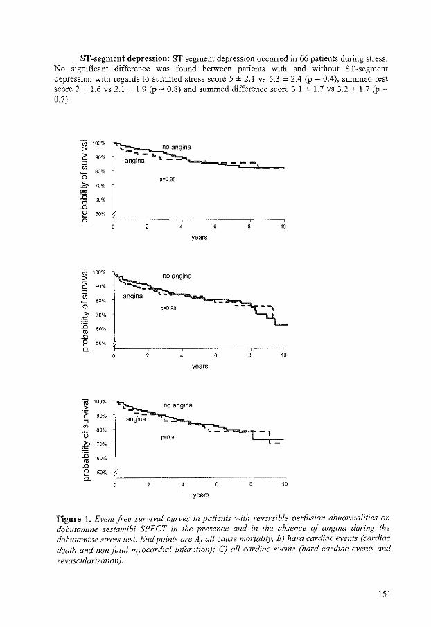
ST-segment depression: ST segment depression occurred in 66 patients during stress. No significant difference was found between patients with and without ST-segment depression with regards to sunnned stress score 5 ± 2.1 vs 5.3 ± 2.4 (p ~ 0.4), summed rest score 2 ± !.6 vs 2.1 ± 1.9 (p ~ 0.8) and summed difference score 3.1 ± !.7 vs 3.2 ± 1.7 (p ~ 0.7).
~ 100%
·:;:;
" "' 90%
..... 80% 0 g 70%
]5
J1l 2 0.
60%
50%
~ 100%
2' " "'
90%
0 80%
~ 70%
15 60%
'" .0
2 50% 0.
~ 100%
2: " "'
90%
0 80%
~ 15
'" .0
2 0.
70%
60%
50%
'- no angina ~- ~
angina "
p=0.98
2 6 8 10
years
p=0.98
4 6 10
years
p=0.9
2 8 10
years
Figure 1. Event free sun;ival cun;es in patients with reversible perfusion abnormalities on dobutamine sestamibi SPECT in the presence and in the absence of angina during the dobutamine stress test. End points are A) all cause mortality, B) hard cardiac events (cardiac death and nonfatal myocardial infarction); C) all cardiac events (hard cardiac events and revascularization).
151

Predictors of cardiac events: In a multivariate analysis model of clinical and perfusion data, independent predictors of cardiac events were age (hazard ratio = 1.02, CI 1.01-1.05 per year increment), diabetes mellitus (HR ~ 1.9, CI L2-3A) and ischemic perfusion score (HR 2.1, CI 1.3-3.8).
Discussion
Identification of stress test variables associated with adverse cardiovascular outcome is important in clinical decision-making (4). In this study, we assessed the outcome of patients with reversible perfusion abnormalities in the absence of angina during dobutamine stress 99m sestamibi SPECT, as compared to patients who experienced angina during the test. Among 224 patients with reversible abnormalities, 93 (42%) patients had angina. There was no significant difference between patients with and without angina with regards to extent and severity of reversible or fixed perfusion abnormalities. Patients with angina during dobutamine stress were more likely to have a history of angina prior to the stress test and had a lower peak stress heart rate, which may be explained by earlier termination of the test due to angina. During a median follow up period of 7.4 years, there was no difference between patients with and without angina during dobutamine stress with regards to cumulative total mortality (38% vs 33%), cardiac death and non-fatal myocardial infarction (24% vs 26%) rates, and the composite endpoint of all cardiac events (cardiac death, myocardial infarction and late revascularization) (43% vs 44%). The incidence of unstable angina during follow up was higher in patients with angina during dobutamine stress test (25% vs 11 %). In a multivariate analysis model, independent predictors of cardiac events were age, diabetes mellitus and ischemic perfusion defect score, while angina was not predictive. The incidence of ST segment depression was similar in patients with and without angina during stress. The extent of perfusion abnormalities was not different in patients with and without ST -segment depression.
Comparison with previous studies: Although many previous studies have assessed the extent of ischemia in presence and in absence of angina during stress myocardial perfusion imaging, prognostic studies are scarce, particularly with 99m technetium labeled agents. Additionally, our study provided the longest term follow up as well as the largest number of hard cardiac events during follow up, when compared to previous reports (11 ,13 ). Some investigators have reported a greater extent and severity of ischemia in symptomatic patients than in those with silent ischemia. Travin et al (6) reported that patients with exercise-induced thallium redistribution without angina had less ischemia than patients with angina. Klein et al (5) reported that the induction of chest pain is associated with more extensive thallium perfusion abnormalities when analyzed in a broad-spectrum population with CAD. The analysis of chest pain tended to lose its value when it was restricted to a CAD population with a greater prior likelihood of inducible ischemia. Marcassa et al (17) studied 300 patients with ischemic heart disease and reversible hypoperfusion on exercise sestamibi tomography. Patients with painful ischemia had more reversible hypoperfusion, and higher incidence of ST segment depression than did patients with silent ischemia. In contrast, numerous studies have failed to find a difference in the amount of ischemic myocardium between patients with silent and symptomatic ischemia during exercise or pharmacologic stress testing. Mahmarian et al (12) studied 356 low risk patients with treadmill exercise thallium-201 SPECT. There were no differences in the extent, severity of reversible perfusion abnormalities, or distribution of coronary stenoses in patients with silent or symptomatic ischemia. Gasperetti et al (8) studied 103 consecutive patients undergoing symptom-limited exercise thallium-201 scintigraphy. Fifty-nine patients (57%) had no angina on exercise
152

testing. There was no difference between the two groups with respect to extent of reversible perfusion abnormalities or extent of angiographic CAD. We have recently shown that in 85 patients with CAD and reversible perfusion abnormalities on dobutamine sestamibi SPECT, the extent of reversible abnormalities was similar in patients with and without angina.
Some previous studies suggested a common pathway for both the electrical and painful response to ischemic stimulus (7). In contrast with previous studies on exercise stress test which reported a higher prevalence of ST -segment depression with symptomatic ischemia ( 6-8), the prevalence of ST segment depression was not different in patients with and without angina in this study. This may be explained by the lower sensitivity of the electrocardiogram with dobutamine as compared with exercise stress. In this study, only 29% of patients with ischemia on SPECT demonstrated ST segment depression. This low sensitivity of dobutamine ECG was attributed to lower rate pressure product achieved by dobutamine as compared to exercise stress test ( 1).
Prognostic studies: The presence of silent ischemia on exercise thallium imaging was shown to be associated with adverse prognosis in asymptomatic population (14,15). Travin et a! (6) studied 134 patients with redistribution on a thallium-201 exercise test who did not experience angina and 134 patients also having redistribution who had angina during the test. Patients without angina had less ischemic thallium-201 images and were less likely to undergo early revascularization, but the occurrence of adverse cardiac events was similar (21% vs 29%). Heller eta!. (13) reported follow up events in 146 patients with reversible perfusion defects on 201- thallium scans (82 patients with and 74 patients without angina). There was no difference between both groups with regards to mortality and cardiac event rate during a mean follow up period of mean 5.2 years. Pancholy eta!. (II) studied 521 patients with coronary artery disease. Symptomatic ischemia (ST depression or reversible defects) was detected in 210 patients, whereas 311 patients had silent ischemia. During a mean follow-up of24 months, there were 30 cardiac events. There was no significant difference in the eventfree survival in patients with symptomatic or silent ischemia.
Study limitations: Most patients were receiving anti-anginal medication, which may have decreased the prevalence of angina or ST -segment depression and influenced the extent and severity of ischemia. Nevertheless, the percentage of patients with or without angina who received medication was not different, and the effect of medications in both groups can be expected to be similar.
Clinical implications: The outcome of patients after a positive dobutamine stress sestamibi study is predicted by the extent of reversible hypoperfusion, but not by symptoms of angina during the test. Patients with silent ischemia defined as reversible perfusion abnormalities without associated angina during dobutamine stress sestamibi SPECT imaging have similar mortality and hard cardiac event rate compared to patients with symptomatic ischemia, but are less likely to have unstable angina during follow up. Therefore, the absence of angina in association with perfusion abnonnalities should not be interpreted as a sign of more benign prognosis.
References
I. Geleijnse ML, Elhendy A, Fioretti PM, Roeland! JRTC. Dobutamine stress myocardial perfusion imaging. JAm Coli Cardiol2000;36:2017-27.
2. Geleijnse ML, E!hendy A, Van Domburg RT, et a!. Prognostic value of dobutamineatropine stress technetium-99m sestamibi perfusion scintigraphy in patients with chest pain. JAm Coli Cardia! 1996;28:447-54.
153
3. Calnan DA, McGrath PD, Doss AL, Harrell FE, Watson DD, Beller GA. Prognostic value of dobutamine stress technetium-99m-sestamibi single-photon emission computed tomography myocardial perfusion imaging: stratification of a high-risk population. J Am Col! Cardiol2001;38:1511-7.
4. Beller GA, Zaret BL. Contributions of nuclear cardiology to diagnosis and prognosis of patients with coronary artery disease. Circulation 2000; 101: 1465-78.
5. Klein J, Chao SY, Berman DS, Rozanski A. Is silent myocardial ischemia really as severe as symptomatic ischemia? The analytic effect of patient selection biases. Circulation 1994;89: 1958-1966.
6. Travin MI, Flores AR, Boucher CA, Newell JB, LaRaia PJ. Silent versus symptomatic ischemia during a thallium-201 exercise test. Am J Cardioll991;68: 1600-1608.
7. Hecht HS, Shaw RE, Bruce T, Mylder RK. Silent ischemia: evaluation by exercise and redistribution tomographic thallium 20 I myocardial imaging. J Am Col! Cardiol 1989; 14:895-900.
8. Gasperetti CM, Burwell LR, Beller GA. Prevalence and variables associated with silent myocardial ischemia on exercise thallium-201 stress testing. J Am Coil Cardiel 1990; 16: 115-123.
9. Amanullah AM, Lindvall K. Prevalence and significance of transient--predominantly asymptomatic--myocardial ischemia on Holter monitoring in unstable angina pectoris, and correlation with exercise test and thallium-201 myocardial perfusion imaging. Am J Cardioll993;72:144-148.
!0. Travin MI, Boucher CA, Newell JB, LaRaia PJ, Flores AR, Eagle KA. Variables associated with a poor prognosis in patients with an ischemic thallium-201 exercise test. Am Heart J 1993;125:335-344.
II. Pancholy SB, Schalet B, Kuhlmeier V, Cave V, Heo J, Iskandrian AS. Prognostic significance of silent ischemia. J Nucl Cardiel 1994; I :434-40.
12. Mahmarian JJ, Pratt CM, Cocanougher MK, Verani MS. Altered myocardial perfusion in patients with angina pectoris or silent ischemia during exercise as assessed by quantitative thalliurn-201 single-photon emission computed tomography. Circulation 1990 ;82:1305-15.
13. Heller LI, Tresgallo M, Sciacca RR, Blood DK, Seldin DW, Johnson LL. Prognostic significance of silent myocardial ischemia on a thallium stress test. Am J Cardiel 1990;65 :718-21.
14. Fleg JL, Gerstenblith G, Zonderman AB, Becker LC, Weisfeld! ML, Costa PT Jr, Lakatta EG. Prevalence and prognostic significance of exercise-induced silent myocardial ischemia detected by thallium scintigraphy and electrocardiography in asymptomatic volunteers. Circulation 1990;81:428-36.
15. Younis LT, Byers S, Shaw L, Barth G, Goodgold H, Chaitman BR. Prognostic importance of silent myocardial ischemia detected by intravenous dipyridamole thallium myocardial imaging in asymptomatic patients with coronary artery disease. J Am Coll Cardiol. 1989;14:1635-41.
16. Elhendy A, Geleijnse ML, Roeland! JR, Cornel JH, van Domburg RT, Reijs AE, Nierop PR, Fioretti PM. Altered myocardial perfusion during dobutamine stress testing in silent versus symptomatic myocardial ischaemia assessed by quantitative MIBI SPET imaging. Eur JNucl Med 1996;23:1354-60.
17. Marcassa C, Galli M, Baroffio C, Campini R, Giannuzzi P. Ischemic burden in silent and painful myocardial ischemia: a quantitative exercise sestamibi tomographic study. J Am Col! Cardiel 1997;29:948-54.
154

Chapter 15
The influence of left ventricular myocardial
contractile reserve on atrial natriuretic
peptide and brain natriuretic peptide
Schinkel AFL, Vourvouri EC, Bax JJ,
Boomsma F, Bountionkos M, Rizzello V,
Roelandt JRTC, Poldermans D
Submitted for publication
155
Abstract
Background: Left ventricular (LV) dysfunction is correlated with elevated natriuretic peptides, however the presence of myocardial contractile reserve may inversely influence natriuretic peptide levels in patients with reduced LV function.
Methods: Plasma atrial natriuretic peptide (ANP) and brain natriuretic peptide (BNP) were determined in 66 consecutive patients referred to dobutamine stress echocardiography for the evaluation of myocardial viability. ANP and BNP were measured using immunoradiometric assays. Left ventricular ejection fraction (L VEF) was assessed by echocardiography at rest. Echocardiograms were analyzed using a 16-segment 5-point model. Contractile reserve was defined as an improvement of segmental wall motion score by ::::1 grade following infusion of low-dose dobutamine (10 !lg/kg/min) in 2:2 severely dyssynergic segments.
Results: ANP and BNP plasma concentrations were higher in patients with a LVEF,;35% compared to patients with a LVEF>35% (ANP: 11.1 ± 9.7 versus 34.4 ± 37.0, p<0.0005, BNP: 62.4 ± 79.0 versus 11.6 ± 14.0 pmoVL, p<0.0005, respectively). The presence of contractile reserve influenced the ANP and BNP levels in patients with wall motion abnormalities. Patients with a preserved myocardial contractile reserve had lower ANP and BNP levels than patients without contractile reserve (ANP: 15.7 ± 8.0 versus 44.8 ± 41.9, p<0.05, BNP: 17.9 ± 12.0 versus 78.3 ± 89.4 pmoi/L, p<0.05, respectively). Cardiovascular medical therapy, including beta-blockers and ACE-inhibitors, was comparable between the patient groups.
Conclusions: Plasma natriuretic peptide levels are elevated in patients with LV dysfunction. However, in the presence of preserved myocardial contractile reserve, relatively low ANP and BNP levels are present.
!56

Introduction
Ischemic left ventricular (LV) dysfunction is the principal cause of congestive heart failure, which is associated with a poor prognosis (1-3). The management of these patients is challenging, whereas the prevalence of congestive heart failure is increasing over the last decade (2). Substantial reductions of morbidity and mortality can be achieved with medical therapy, additionally coronary revascularization may improve outcome in patients with severe ischemic LV dysfunction with a preserved myocardial contractile reserve ( 4-6).
Recently, atrial natriuretic peptide (ANP) and brain natriuretic peptide (BNP) have been proposed for the detection and management of patients with LV dysfunction (7 -11 ). ANP is a cardiac hormone that is synthesized and secreted primarily in atrium, whereas BNP is produced in the ventricles in response to changes in wall stretch. Plasma natriuretic peptide concentrations may be elevated in patients with LV dysfunction, and elevated ANP and BNP levels are related to an adverse outcome (7 -11 ). Previous studies demonstrated that a preserved myocardial contractile reserve is related to a favorable outcome in patients with LV dysfunction. Currently, the relation between the presence of myocardial contractile reserve and plasma natriuretic peptides is not clear. In this study, we investigated the effect of myocardial contractile reserve on plasma ANP and BNP levels in patients referred to dobutamine stress echocardiography with a varying degree of heart failure.
Methods
Patient population, study protocol: The study population consisted of 66 consecutive patients referred to dobutamine stress echocardiography for the evaluation of known or suspected coronary artery disease. Patients with primary cardiomyopathy, concomitant significant valvular disease, or left ventricular hypertrophy were not included. Plasma ANP and BNP concentrations were determined using immunoradiometric assays. All patients underwent resting echocardiography to assess the LV ejection fraction (LVEF) and to identify dysfunctional myocardial tissue. Left ventricular myocardial contractile reserve was assessed during low-dose dobutamine stress echocardiography. The local medical ethics committee approved the study protocol and all patients gave informed consent.
Cardiac peptide measurements: Before stress echocardiography a blood sample was drawn from a peripheral vein, after the patient had rested for at least 30 minutes in a supine position. The blood sample was dra\¥!1 into a pre-chilled tube containing edetic acid (EDTA, 1.9 mg/ml) and the protease inhibitor aprotonin (Trasylol, 100 kiU/ml) to prevent breakdown of the cardiac peptides. The sample was placed on ice and promptly centrifuged at 3000 rpm (4°C) for 10 minutes. The plasma was separated and stored at -80°C. Plasma concentrations of ANP and BNP were determined using standard commercially available immunoradiometric assay kits (Shionoria ANP and BNP kits, Shionogi, Osaka, Japan).
Echocardiography: A commercially available imaging system (Hewlett Packard Sonos 5500, Andover, Mass.) and a 1.8 MHz transducer using second harmonic imaging to optimize endocardial border visualization were used. Two-dimensional imaging was performed with the patient in the left lateral position; standard views were recorded on optical disk (cine loops).
After the venous blood samples were drawn, dobutamine stress echocardiography was performed to assess the contractile reserve in dysfunctional myocardium. Following the resting echocardiographic study, dobutamine was administered intravenously, starting at a dose of 5 ~g/kg body weight per minute for 5 minutes, followed by a 10 ~g/kg/min dose for 5
157

minutes (low-dose). Incremental doses of 10 !J-g/kg/min dobutamine were given at 3-minute intervals up to a dose of 40 !J-glkg/min, and atropine was added if target heart rate was not achieved.
Global LV function, assessment of LVEF: The LVEF was determined off-line by the 2-dimensional biplane disk method. using the modified Simpson's rule (13). The endocardial borders of the 2- and 4-chamber apical views were digitally traced at end-diastole and end-systole. Subsequently, the LV end-diastolic and end-systolic volumes ejection fraction were measured and the LVEF was calculated. A L VEF $35% was considered abnormal.
Regional LV function, segmental analysis: Two experienced observers, unaware of the clinical data, scored the digitized echocardiograms offline. In case of disagreement, a majority decision was achieved by a third observer. The left ventricle was divided into 16 segments according to the American Society ofEchocardiography (12). Regional wall motion and systolic wall thickening were scored using a 5-point grading scale: 1 =normal, 2=mildly hypokinetic, 3=severely hypokinetic, 4=akinetic, 5=dyskinetic. Segments with severe hypokinesia, akinesia or dyskinesia were considered abnormaL The wall motion score index (WMSI) was calculated as the sum of the segmental scores divided by the number of analyzed segments. Contractile reserve was defined as an improvement of segmental wall motion score by 21 grade following infusion of low-dose dobutamine (10 )lg/kg/min) in 22 severely dyssynergic segments. Ischemia was defined as new or worsened wall motion abnormalities during high-dose dobutamine stress indicated by a deterioration of segmental wall motion score by ~1 grade.
Statistical analysis: Values are expressed as mean ± SD, when appropriate, percentages are rounded. Continuous variables were compared using the Student t-test for unpaired samples. Differences bet\.veen proportions were compared using the Chi-square test. A value ofp<0.05 was considered statistically significant.
Results
Patient characteristics: The clinical characteristics of the 66 patients are presented in Table 1. A total of 44 patients were in New York Heart Association (NYHA) functional class Jill, 22 in class III/IV.
Global function, LVEF: The L VEF at rest was on average 47±15%, 21 patients had a L VEF$35%. Dobutamine-stress echocardiography was performed in all patients without side effects. The hemodynamic changes in response to low-dose dobutamine infusion are presented in Table 2.
Wall motion analysis: Segmental wall motion abnormalities were present in 27 patients. Of these patients, 13 patients had a preserved myocardial contractile reserve, the remaining 14 patients had no contractile reserve. The clinical characteristics were comparable in patients with and in patients without contractile reserve (see Table 3). Patients with a preserved contractile reserve less often had a history of myocardial infarction compared with patients without contractile reserve.
158

Table 1. Baseline characteristics
Men/Women Age (years) NYHA functional class LVEF (%) Ischemia Diabetes mellitus Hypercholesterolemia Smoking History
Myocardial infarction Coronary angioplasty Coronary bypass surgery
Medical therapy Beta-blockers Calcium channel blockers Nitrates ACE-inhibitors Diuretics Digoxin Aspirin Cholesterol lowering drugs
Data are presented as number(%).
50
40 p < 0.0005
~ 0 30 E "-a_ 20 z 11.1 <1:
10
0
LVEF>35%
41 (62) /25 (38) 63 ± 13 1.8 ± 1.0 47 ± 15 18 (27) 2 (3) 20 (30) 8 (12)
19 (29) 3 (5) 10(15)
20 (30) I 0 (15) 8 (12) 13 (20) 8 (12) 5 (8) 26 (39) 20 (30)
34.4
LVEF_:::35%
Figure 1. Plasma ANP concentrations in patients with a LVEF > 35% and in patients with a
LVEF-535%.
!59
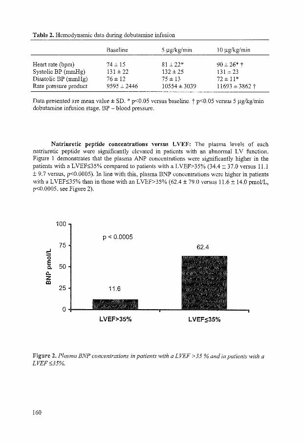
Table 2. Hemodynamic data during dobutamine infusion
Baseline 5 11g/kg/min 1 0 !lglkg/min
Heart rate (bpm) 74 ± 15 81 ± 22* 90±26* t Systolic BP (mmHg) 131 ± 22 132 ± 25 131±23 Diastolic BP (mmHg) 76± 12 75 ± 13 72±11* Rate pressure product 9595 ± 2446 10554 ± 3039 11693 ± 3862 t
Data presented are mean value± SD. * p<0.05 versus baseline. t p<0.05 versus 5 ~g/kg/min dobutamine infusion stage. BP = blood pressure.
Natriuretic peptide concentrations versus LVEF: The plasma levels of each natriuretic peptide were significantly elevated in patients with an abnormal LV function. Figure 1 demonstrates that the plasma ANP concentrations were significantly higher in the patients with a LVEE35% compared to patients with a LVEF>35% (34.4 ± 37.0 versus 11.1 ± 9.7 versus, p<0.0005). In line with this, plasma BNP concentrations were higher in patients with a LVEF:>35% than in those with an LVEF>35% (62.4 ± 79.0 versus 11.6 ± 14.0 pmoVL, p<0.0005, see Figure 2).
100
p < 0.0005 75 62.4
..J -0 E 50 c.
a.. z Ill
25 11.6
0
LVEF>35% LVEF$35%
Figure 2. Plasma BNP concentrations in patients with a LVEF > 35 %and in patients with a LVEF0:35%.
160

Natriuretic peptide concentrations versus contractile reserve: The presence of contractile reserve influenced the natriuretic peptide concentrations in patients with wall motion abnormalities. The patients with a preserved myocardial contractile reserve had a lower ANP concentration than the patients without contractile reserve (15.7 ± 8.0 versus 44.8 ± 41.9, p<O.OS, see Figure 3). Also, plasma BNP levels were lower in patients with a preserved myocardial contractile reserve than in patients without contractile reserve (17.9 ± 12.0 versus 78.3 ± 89.4 pmol/L, p<O.OS, respectively, Figure 4).
50
40
<:! 0 30 E c. "- 20 z <(
10
0+---
44.8
p < 0.05
15.7
CR+ CR-
Figure 3. Plasma ANP concentrations in patients with wall motion abnormalities with and without a preserved left ventricular myocardial contractile reserve.
100 78.3
p < 0.05
75 <:! 0 E 50 c. "-z !!l
25 17.9
0
CR+ CR-
Figure 4. Plasma BNP concentrations in patients with wall motion abnonnalities with and without a preserved left ventricular myocardial contractile reserve.
161

Discussion
Previous studies have demonstrated that plasma natriuretic peptide concentrations are elevated in patients congestive heart failure (7 -II). Although the number of patient with congestive heart failure due to ischemic heart disease is increasing rapidly (2), there are no data available on the relation between myocardial contractile reserve and plasma natriuretic peptide concentrations. In this study, ANP and BNP levels were determined in a patient cohort with known or suspected coronary artery disease and a varying degree of heart failure. The main finding from the present study is that the presence of myocardial contractile reserve during low-dose dobutamine stress echocardiography influences plasma levels of both natriuretic peptides in patients with an impaired LV function. Plasma ANP and BNP levels were markedly elevated in patients without contractile reserve, compared to patients with a preserved contractile reserve. Cardiovascular medical therapy, including beta-blockers and ACE inhibitors, was comparable between the patient groups.
Pathophysiological role of ANP and BNP: The natriuretic peptides have a major role in the protection of the heart from volume overload (14). The cardiac hormones ANP and BNP are produced in the atria and ventricles, respectively, in response to an increase in wall stretch, or pressure. Elevated plasma ANP and BNP concentrations have a natriuretic and diuretic effect. In addition, high plasma BNP levels cause a fluid-shift from the capillary bed to the interstitium, decreasing preload and blood pressure. Hence, ANP and BNP are functional counterparts of the renin-angiotensin aldosterone system. Therefore there may be a future for these natriuretic peptides in screening and guiding management in patients with LV dysfunction. In the present study, the ANP and BNP plasma concentrations were significantly elevated in the patients with LV dysfunction, whereas the patients with a normal function had normal natriuretic peptide levels. These findings indicate that an increased wall tension or stretch in abnonnally contracting myocardial tissue may lead to elevated plasma natriuretic peptide levels, and confirm the "volume overload" hypothesis for the production and secretion of these peptides. This is in line with the study of Sumida et al. (15) showing that the secretion of natriuretic peptides increases in proportion to the severity of LV dysfunction, and is elevated in infarct regions.
Contractile reserve in patients with LV dysfunction: Since the introduction of the concepts of myocardial viability, hibernation, and stunning, it has become clear that ischemic LV dysfunction is not an irreversible process (16,17). In more than 50% of the patients with ischemic cardiomyopathy and heart failure, a clinically significant amount of viable myocardium is present and coronary revascularization may be considered (18). The evaluation of myocardial contractile reserve may have important clinical implications in patients with ischemic LV dysfunction. Recently, Chaudry et al. (19) evaluated contractile reserve during low-dose dobutamine stress echocardiography in 80 patients with ischemic LV dysfunction. Contractile reserve was a significant predictor of survival in these patients. Moreover, the presence of contractile reserve is related to the extent of interstitial fibrosis and predicts the recovery of systolic function after coronary revascularization (20). Several studies have reported that plasma natriuretic peptides are predictive of long-term survival after myocardial infarction, although the mechanism for this is not clear (21 ,22). In the current study myocardial contractile reserve influenced plasma natriuretic peptide levels. Plasma ANP and BNP levels were high in patients without contractile reserve, and relatively low in patients with a preserved contractile reserve. Hence, natriuretic peptides are correlated with myocardial viability in dysfunctional myocardium. This may partially explain why natriuretic
162
peptides predict survival in patients who had a myocardial infarction. Further research is needed to fully elucidate this issue.
Conclusions: The presence of myocardial contractile reserve influences the plasma natriuretic peptide concentrations in patients with LV dysfunction. Plasma ANP and BNP levels were markedly elevated in patients with LV dysfunction without contractile reserve, compared with patients with a preserved contractile reserve. These findings may give further insights into the role of these natriuretic peptides in patients with LV dysfunction.
References
l. Ho KK, Pinsky JL, Kannel WB, Levy D. The epidemiology of heart failure: the Framingham Study. JAm Coli Cardioll993;22:6A-l3A.
2. McCullough PA, Philbin EF, Spertus JA, Kaatz S, Sandberg KR, Weaver WD. Confirmation of a heart failure epidemic: findings from the Resource Utilization Among Congestive Heart Failure (REACH) study. JAm Coli Cardiol2002;39:60-69.
3. Gheorghiade M, Borrow RO. Chronic heart failure in the United States - a manifestation of coronary artery disease. Circulation 1998;97:282-289.
4. Pfeffer MA, Braunwald E, Moye LA, Basta L, Brown EJ Jr, Cuddy TE, Davis BR, Geltman EM, Goldman S, Flaker GC, on behalf of the SAVE investigators. Effect of captopril on mortality and morbidity in patients with left ventricular dysfunction after myocardial infarction: results of the survival and ventricular enlargement triaL N Engl J Med 1992;327:669-677.
5. Pitt B, Zannad F, Remme WJ, Cody R, Castaigne A, Perez A, Palensky J, Wittes J, for the randomized aldactone evaluation study investigators. The effect of spironolactone on morbidity and mortality in patients with severe heart failure. N Eng! J Med 1999;341 :709-717.
6. The cardiac insufficiency bisoprolol study II (CIBIS-11): a randomised trial. Lancet I999;353:9-l3.
7. Davidson NC, Naas AA, Hanson JK, Kennedy NS, Coutie WJ, Struthers AD. Comparison of atrial natriuretic peptide B-type natriuretic peptide, and N-terminal proatrial natriuretic peptide as indicators of left ventricular systolic dysfunction. Am J Cardiol 1996;77:828-831.
8. Cowie MR, Struthers AD, Wood DA, Coats AJ, Thompson SG, Poole-Wilson PA, Sutton GC. Value of natriuretic peptides in assessment of patients with possible new heart failure in primary care. Lancet 1997;350:1349-1353.
9. Koglin J, Pehlivanli S, Schwaiblmair M, Vogeser M, Cremer P, vonScheidt W. Role of brain natriuretic peptide in risk stratification of patients with congestive heart failure. J Am Coli Cardiol200l ;38: 1934-!941.
10. McDonagh TA, Robb SD, Murdoch DR, Morton JJ, Ford I, Morrison CE, Tunstall-Pedoe H, McMurray JJ, Dargie Hl Biochemical detection of left-ventricular systolic dysfunction. Lancet 1998;351 :9-13.
11. Ishii J, Nomura M, Nakamura Y, Naruse H, Mori Y, Ishikawa T, Ando T, Kurokawa H, Kondo T, Nagamura Y, Ezaki K, Hishida H. Risk stratification using a combination of cardiac troponin T and brain natriuretic peptide in patients hospitalized for worsening chronic heart failure. Am J Cardiol2002;89:69l-695.
12. Bourdillon PD, Broderick TM, Sawada SG, Armstrong WF, Ryan T, Dillon JC, Fineberg NS, Feigenbaum H. Regional wall motion index for infarct and non-infarct region after reperfusion in acute myocardial infarction: comparison with global wall motion index. J Am Soc Echocardiogr 1989;2:398-407.
163
13. Schiller NB, Shah PM, Crawford M, DeMaria A, Devereux R, Feigenbaum H, Gutgesell H, Reichek N, Sabn D, Scbnittger I. Recommendations for quantitation of the left ventricle by two-dimensional echocardiography. JAm Soc Echocardiogr 1989;2:358-367.
14. Schrier RW, Abraham WT. Hormones and hemodynamics in heart failure. N Eng! J Med 1999;341 :577-585.
15. Sumida H, Yasue H, Yoshimura M, Okumura K, Ogawa H, Kugiyama K, Matsuyama K, Kikuta K, Morita E, Nakao K. Comparison of secretion pattern between A-type and Btype natriuretic peptides in patients with old myocardial infarction. J Am Coll Cardiel. 1995;25: 1105-1110.
16. Rahimtoola SH. The hibernating myocardium. Am Heart J 1989;117:211-221. 17. Diamond GA. Hibernating myocardium. Am Heart J 1989;118:1361. 18. Schinkel AFL, Bax JJ, Boersma E, Elhendy A, Roeland! JRTC, Poldermans D. How
many patients with ischemic cardiomyopathy exhibit viable myocardium? Am J Cardiel 2001 ;88:561-564.
19. Chaudhry FA, Tauke JT, Alessandrini RS, Vardi G, Parker MA, Borrow RO. Prognostic implications of myocardial contractile reserve in patients with coronary artery disease and left ventricular dysfunction. JAm Coli Cardiel 1999;34:730-738.
20. Nagueh SF, Mikati I, Weilbaecher D, Reardon MJ, Al-Zaghrini GJ, Cacela D, He ZX, Letsou G, Noon G, Howell JF, Espada R, Verani MS, Zoghbi WA. Relation of the contractile reserve of hibernating myocardium to myocardial structure in humans. Circulation 1999; 100:490-496.
21. Omland T, Aakvaag A, Bonarjee VV, Caidahl K, Lie RT, Nilsen DW, Sundsfjord JA, Dickstein K. Plasma brain natriuretic peptide as an indicator of left ventricular systolic function and long-term survival after acute myocardial infarction. Comparison with plasma atrial natriuretic peptide and N-terminal proatrial natriuretic peptide. Circulation 1996;93: 1963-1969.
22. Arakawa N, Nakamura M, Aoki H, Hiramori K. Plasma brain natriuretic peptide concentrations predict survival after acute myocardial infarction. J Am Coll Cardiel 1996;27: 1656-1661.
164
Chapter 16
Left ventricular hypertrophy screening
using a hand-held ultrasound device
Vourvouri EC, Poldennans D, Schinkel AFL,
Koroleva L Y, Sozzi FB, Parharidis GE,
Bax JJ, Roelandt JRTC
Eur Heart J 2002;23: 1516-1521
165


Left ventricular hypertrophy screening using a hand-held ultrasound device
E. C. Vourvouri, D. Poldermans, A. F. L. Schinkel, L. Y. Koroleva, F. B. Sozzi, G. E. Parharidis, J. J. Bax and J. R. T. C. Roeland!
Department of Cardiology, Thoraxcentre, Erasmus Medical Centre Rotterdam, Rotterdam, The Netherlands
Aims To test the diagnostic potential of a hand-held ultrasound device for screening for left ventricular hypertrophy in a hypertensive population using a standard echocardiographic system as a reference.
Methods One hundred consecutive hypertensive patients were enrolled. An experienced investigator performed measurements of the thickness of the anterior septum and posterior wall using the parasternal 2D-long axis view and the end-diastolic dimension of the left ventricle with both imaging devices. Left ventricular hypertrophy was defined as an increase in left ventricular mass ~ 134 g. m - 2 for men and ~ 110 g . m- z for women, when indexed for body surface area and ~ 143 g. m- 1 for men and ~ 102 g. m- 1
for women, when indexed for height.
Results Sixty+five men and 35 women were studied (age 60 ± 11 years); mean duration of hypertension: 13 ± 11 years; mean blood pressures: systolic !50± 20 mmHg and diastolic 89 ± 11 mrnHg. The anterior septum and posterior wall were visualized in all patients ;vith both imaging devices. The standard echocardiographic system identified
Introduction
Left ventricle hypertrophy which expresses end-organ damage from hypertension, is an independent potent marker of cardiovascular risk in arterial hypertension[l-31. lt is considered as an asymptomatic pre-clinical stage of the cardiovascular disease, that may lead to cardiac eventsf4l. Also, reversal of left ventricular hypertrophy can improve the patient's outcome[51. Early identification of left ventricular hypertrophy and treatment
Revision submitted 31 December 2001, and accepted 2 January 2002.
Correspondence: Don Poldermans, MD, PhD, Thoraxcentre, Room Ba 300. Erasmus Medical Centre Rotterdam, Dr. Molewaterplein 40, 3015 GD Rotterdam, The Netherlands.
left ventricular hypertrophy by body surface area in 18 (18%) patients and by height in 26 (26%) patients. The agreement between the standard echocardiographic system and the hand-held device for the assessment of left ventricular hypertrophy was 93%, kappa: 0·77 (left ventricular mass/body surface area) and 90%, kappa: 0·76 (left ventricular mass/height).
Conclusions We conclude that hand-held devices can be effectively applied for screening for left ventricular hypertrophy in hypertensive patients. (Eur Heart J, 2002; 23: 1516-1521, doi:10.1053/euhj.2002. 3162) © 2002 Published by Elsevier Science Ltd on behalf of The European Society of Cardiology.
Key Words: Left ventricular hypertrophy, left ventricular mass, hand-held ultrasound device.
See doi: 10.1053/euhj.2002.3292 for the Editorial comment on this article
is therefore the cornerstone of appropriate management. The electrocardiogram (ECG), although commonly available and inexpensive has proven insensitive in detecting the presence of left ventricular hypertrophy[6-SJ_ Echocardiography is a sensitive means for measurement of left ventricular thickness and has comparable accuracy to the magnetic resonance imaging (MRI) especially in patients with normal left ventricular geometry[~-loJ_ New and small echocardiographic devices are now becoming available which could be used as screening tools for various pathomorphologies of the heart.
The aim of the present study was to evaluate the potential and diagnostic accuracy of a recently developed portable hand-held ultrasound system for screening for left ventricular hypertrophy in hypertensive patients using a standard two-dimensional echocardiographic system as a reference.
167
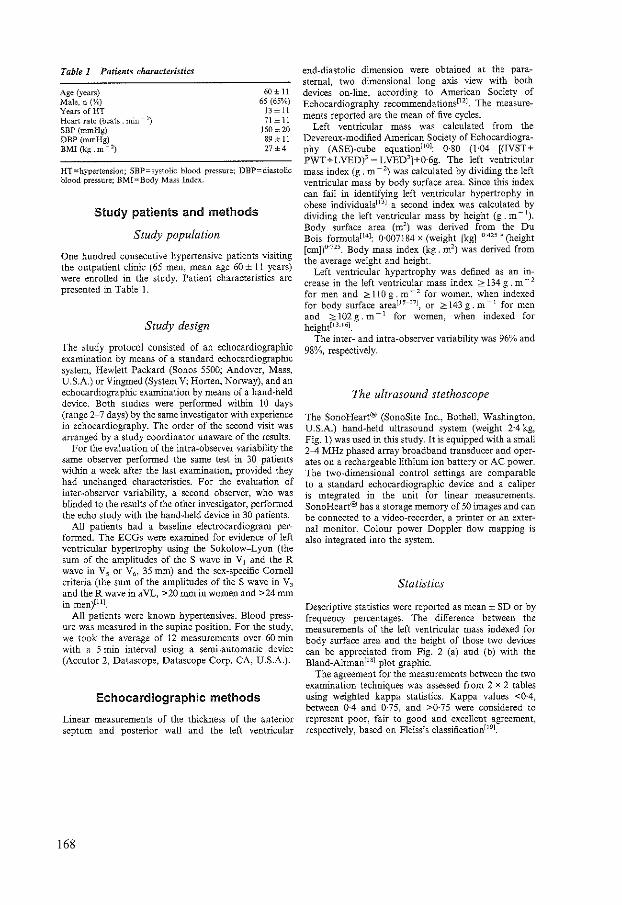
Table 1 Patients characteristics
Age (years) Male, n (%) Years ofHT Heart rate (beats , min~ 1)
SBP (rnrnHg) DBP(=Hg) BMI(kg.m~ 2)
60 ± 11 65 (65%)
13 ± 11 71 ± 11
150 ± 20 89 ± ll 27±4
HT=hypertension: SBP=systolic blood pressure; DBP-diastolic blood pressure; BMI =Body Mass Index.
Study patients and methods
Study population
One hundred consecutive hypertensive patients visiting the outpatient clinic (65 men, mean age 60 ± 11 years) were enrolled in the study. Patient characteristics are presented in Table I.
Study design
The study protocol consisted of an echocardiographic examination by means of a standard echocardiographic system, Hewlett Packard (Sones 5500; Andover, Mass, U.S.A) or Vingmed (System V; Harten, Norway), and an echocardiographic examination by means of a hand-held device. Both studies were performed within 10 days (range 2-7 days) by the same investigator with experience in echocardiography. The order of the second visit was arranged by a study coordinator unaware of the results.
For the evaluation of the intra-observer variability the same observer performed the same test in 30 patients within a week after the last examination, provided they had unchanged characteristics. For the evaluation of inter-observer variability, a second observer, who was blinded to the results of the other investigator, performed the echo study with the hand-held device in 30 patients.
All patients had a baseline electrocardiogram performed. The ECGs were examined for evidence of left ventricular hypertrophy using the Sokolow-Lyon (the sum of the amplitudes of the S wave in V1 and the R wave in V5 or V6, 35 mm) and the sex-specific Cornell criteria (the sum of the amplitudes of the S wave in V3
and the R wave in aVL, >20 mm in women and >24 mm in men)llll_
All patients were known hypertensives. Blood pressure was measured in the supine position. For the study, we took the average of 12 measurements over 60 min with a 5 min interval using a semi-automatic device (Accutor 2, Datascope, Datascope Corp. CA, U.S.A.).
Echocardiographic methods
Linear measurements of the thickness of the anterior septum and posterior wall and the left ventricular
168
end-diastolic dimension were obtained at the parasternal, two dimensional long axis view with both devices on-line, according to American Society of Echocardiography recommendations[l 2l. The measurements reported are the mean of five cycles.
Left ventricular mass was calculated from the Devereux-modi:fied American Society ofEchocardiography (ASE)-cube equationP01: 0·80 (1·04 [(IVST+ PWT+ L VED)3 - L VED3]+0·6g. The left ventricular mass index (g. m - 2) was calculated by dividing the left ventricular mass by body surface area. Since this index can fail in identifying left ventricular hypertrophy in obese individuaJsll 3l a second index was calculated by dividing the left ventricular mass by height (g. m- 1
).
Body surface area (m2) was derived from the Du
Bois formulaP 4l: 0·007184 x (weight [kg] o-425" (height
[cm])0 ' 725 . Body mass index (kg. m2) was derived from
the average weight and height. Left ventricular hypertrophy was defined as an in
crease in the left ventricular mass index ;::>: 134 g. m - 2
for men and ;::.: 110 g. m- 2 for women, when indexed for body surface areaU 5 -- 17l, or ;::.: 143 g. m -t for men and ;::.: 102 g . m- 1 for women, when indexed for height[l 3"161.
The inter- and intra-observer variability was 96% and 98%, respectively.
The ultrasound stethoscope
The SonoHeart® (SonoSite Inc., Bothell, Washington, U.S.A.) hand-held ultrasound system (weight 2·4 kg, Fig. 1) was used in this study.lt is equipped with a small 2-4 MHz phased array broadband transducer and operates on a rechargeable lithium ion battery or AC power. The two-dimensional control settings are comparable to a standard echocardiographic device and a caliper is integrated in the unit for linear measurements. SonoHeart® has a storage memory of 50 images and can be connected to a video-recorder, a printer or an external monitor. Colour power Doppler flow mapping is also integrated into the system.
Statistics
Descriptive statistics were reported as mean± SD or by frequency percentages. The difference between the measurements of the left ventricular mass indexed for body surface area and the height of those two devices can be appreciated from Fig. 2 (a) and (b) with the Bland-Altman[18l plot graphic.
The agreement for the measurements between the two examination techniques was assessed from 2 x 2 tables using weighted kappa statistics. Kappa values <0·4, between 0·4 and 0·75, and >0·75 were considered to represent poor, fair to good and excellent agreement, respectively, based on Fleiss's classification[191.

Figure 1 Photograph of the SonoHeartS device, a handheld ultrasound imager, used in this study.
Results
Clinical characteristics
The mean systolic blood pressure was 150 ± 20 mmHg and the diastolic blood pressure 89 ± 11 mmHg. The mean heart rate was 71 ± 11 beats . min- 1•
Electrocardiography
Four patients were found to have left ventricular hypertrophy according to the Sokolov~Lyon criteria and 13 according to the Cornell criteria. The sensitivity of the ECG for the detection of left ventricular hypertrophy was, respectively, 5% and 16% and the specificity was, respectively, 96% and 87%.
Measurements and agreement
Visualization was feasible in all patients with both imaging devices. The results of the measurements of the thickness of the anterior septum and the posterior
wall and the dimension of the left ventricle with both examination techniques are summarized in Table 2.
The mean left ventricular mass indexed by body surface area was 96·2 ± 36 g. m - 2 with the standard echocardiographic system and 103 ± 33 g. m- 2 with the hand-held device. Using the threshold of ;;::: 134 g. m- 2
for men and ;;::: 110 g. m- 2 for women the standard echocardiographic system identified left ventricular hypertrophy in 18 (18%) patients (nine women and nine men). The agreement between the two methods was 93%, kappa 0·77 (Fig. 3(a)).
The mean left ventricular mass indexed by height was Ill· 5 ± 43 g . m- 1 with the standard echocardiographic system and 120 ± 40 g. m -I with the hand-held device. Using the threshold of ;;:: 143 g. m- 1 for men and ;;::: 102 g. m- 1 for women the standard echocardiographic system identified left ventricular hypertrophy in 26 patients (13 women and 13 men). The agreement between the two methods was 90%, kappa=0·76 (Fig. 3(b)).
Discussion
The presence of left ventricular hypertrophy, calculated as an absolute left ventricular mass has an independent prognostic value on top of age and blood pressure[3·20·21l. Recent studies have reported good reliability for echocardiographic measurements of left ventricular massl22.2JJ_
Our study showed that this new, hand-held device could be effectively used for screening for left ventricular hypertrophy in office practice. Recently, we demonstrated in a previous study the efficacy and high accuracy of this small imaging device in assessing the pathomorphology and function of the heart enhancing and extending the physical examination to allow goaloriented examination, such as screeningl24l.
Although echocardiography can assess left ventricular hypertrophy accurately compared to the 'gold standard' MRI, the World Health Organisation~Intemational Society of Hypertension (WHO~ISH)i25l and the Joint National Committee on prevention, detection, evaluation and treatment of high blood pressurel26l do not recommend routine echocardiography in all hypertensive patients. Thus, in patients categorized as high risk patients (having cardiovascular risk factors or an endorgan damage), treatment is already indicated and echocardiography results will not change their managementl27J. However, echocardiography is recommended in patients with concomitant heart diseasel27·28l and in patients with 'stage one' hypertension (patients with high~normal blood pressure who do not have clinical cardiovascular disease, target organ damage or other risk factors). This is recommended in order to avoid misclassi:fication as 'mild' hypertension in patients that have an end-organ damage as left ventricular hypertrophyl25·26·28·29l. Both, Black and Shepsl30·31l support this view introducing limited echocardiographic protocols.
169

(a)
Sensitivity"' 83% (95% CI: 0·76 to 0·91) Specificity = 95% (95% CI: 0·91 to 0·99) PPV =79% (95% CI: 0·71 to 0·87}
75 NPV = 96% (95% CI: 0·93 to 1·00) Accuracy ::: 93% (95% Cl: 0·88 to 0·98)
50 •
25 ---------------.;-.--·------------------
0Fc_ _____ ~ .... r.s:·~·ij~~~·,,;r~·~·--~ .. ~~--·--A-ol • fli .. •• • •
~25 ... :--;-.. ,. • • • ________ 1.__.:--------.------------
• -50
-75
~100 L_ __ ,L-__ -,Lc--~cc--~ 0 50 100 150 200
Average mass/BSA index
(b)
Sensitivity= 96% (95% CI: 0·92 to 1·00) Specificity= 88% (95% CI: 0·81 to 0·94) PPV :74% (95% CI: 0·65 to 0·82)
75 NPV = 98% (95% CI: 0·96 to 1·00) Accuracy = 90% (95% CI: 0·84 to 0·96)
w • ----------- -- -------•----•--------------25 •• • # • . ., .. .
o L .J.~· .: • • • • _,,r------. • .-'-j .. ~n~•1TI_i;lc.~....,:"'i.eC:,'o,:-'-eo--j • - •• ¥ .. • '
• • • -50 ----- -- -- --------------- -- --- .... ---- ---
-75f 0
l -1000 L ------~5~0 -------:1~0~0-------;1750::------2:::00
Average mass/height index
Figure 2 Bland-Altman plot, demonstrating the magnitude of the difference between the measurements with the two techniques (differences plotted against their mean average) of (a) left ventricular mass indexed for body surface area (BSA) and (b) left ventricular mass indexed for height. SD=2 standard deviations of the mean difference in the measurements of the two devices. Using standard echocardiographic system as the gold standard, the sensitivity and specificity of the band-held device for screening of the presence of left l'entricular hypertrophy were calculated as was the positive predicthe value (PPV), negative predictive value (NPV) and accuracy.
Table 2 Measurements (in em) of the anterior septum (AS), the posterior wall (PW) and the left ventricular end-diastolic dimension (LVED) with both imaging devices
Type of examination
Standard echo Hand-held device SE-hand-held
AS
1·18 1·21
-0·03
so
PW LVEO AS PW LVED
4·85 ... 0·05
0·29 0·20 0·26 0·16 0·03 0·04
SD=standard deviation; Number of patients: 100.
However, the indication of echocardiography in hypertensive patients may be broadened, as this new and inexpensive (...., 1/lOth of the price of a standard echocardiographic system) hand-held ultrasound device becomes widely available. In our view, such small handheld imaging devices, reducing the cost and being ultraportable and easy to use, will allow routinely echocardiographic examination in all hypertensive patients. Performing as an extension to physical examination they will provide the clinician with immediate, valuable information about prognosis and risk classification, assisting him in his decision of therapy. Of course, the initiation of aggressive therapy is dependent on not only the presence of left ventricular hypertrophy but also on other parameters such as cardiovascular risk factors and end-organ damage. Furthermore, it is becoming increasingly clear that we should aim for aggressive treatment in most hypertensive patients.
The efficacy of the selected therapy could be followed with the hand-held device by serial estimation of left ventricular mass with every visit at the outpatient clinic.
170
AS
Range of measurements
PW LVED
0·5-2·2 0·4--1·3 3-4--6·8 0·7-2·3 0·6-1·4 3·5--6·6
However, the reliability ofleft ventricular mass measurements depends on many factors, such as the experience of the operator, the age of the patient, the body habitus or the presence of an abnormal left ventricular geometry or emphysema. Furthermore the amount of regression with therapy also plays a significant role in the likelihood of true changes[23l.
By analysing the left ventricular geometric pattern, risk stratification can be carried out: patients "With normal left ventricular architecture have the best prognosis, those with concentric remodelling or eccentric hypertrophy have intermediate, and those with concentric left ventricular hypertrophy have the worst prognosis[3·25·32l. Furthermore, echocardiography provides us not only with left ventricular mass determination, but with additional valuable information such as left ventricular systolic function or valvular abnormalities.
The method used most frequently for the diagnosis of left ventricular hypertrophy is still standard electrocardiography. Although the ECG has low sensitivity and specificity in recognising left ventricular hypertrophy, it
(a)
-~ 0
Abnormal ~
~ :;; ' Normal
"'
LVMJBSA (g.m - 2)
Standard echo
Abnormal Normal
15 4
3 78
Agreement= 93%, k = 0·77
Number of patients: 100.
(b)
j Abnormal
~ 'i ~
' Normal • "'
LVM!height (g.m -l)
Standard echo
Abnormal Normal
25 9
1 65
Agreement= 90%, k = 0·76
The numbers inside the table express the absolute number of patients Abnormal "' left ventricular hypertrophy
Figure 3 Agreement of the left ventricular mass (LVM) indexed by body surface area (BSA) (a) and by height {b), measured by the band-held device and the standard echocardiographic system.
should not be abandoned in patients with known or suspected coronary artery disease as it provides additional information on ischaemia, previous myocardial infarction and rhythm abnormalities.
Left ventricular mass determination, especially with theM-mode based methodology, can be unreliable in an asymmetric heart. In the presence of such an anatomy, the 3D echocardiogram and the ECG-gated magnetic resonance imaging have a higher accuracy and reliability. However, albeit they are superior compared to conventional echocardiographic methods, they have a higher cost and a varied availabilityt33l_
The study was perfonned by a cardiologist with experience in echocardiography. We believe that physicians can be trained to use this hand-held device and to recognize and distinguish nonnal from abnonnal findings. In case of an abnormal finding or in case of doubt an echocardiographic study with a standard echocardiographic system perfonned by an experienced investigator should follow. However, training and licensing for use of these devices by non-cardiologists will become an important issue in the future.
Recently, Goodkin et af.134l studied the use of the hand-held device at the point-of-care and compared it to the physical examination. They reported that the use of this device by cardiologists improved the detection of important cardiovascular findings. However, they pointed out that such a hand-held device cannot be a substitute for the final diagnosis, in case of abnormal findings. This is in concordance with the study performed by Spencer et af.13 5J in critically ill patients. Moreover, Schiller[36l comments that further evaluation of these devices will improve their practical use.
Limitations
In this study, we calculated the left ventricular mass by the Devereux modified (ASE)-cube equation. Due to the
absence of the M-mode feature of the hand-held device the measurements were performed with the use of callipers on the two-dimensional parasternal long axis view according to the American Society ofEchocardiography recommendations[11 l. The same measuring technique was used for both devices for perfonnance comparison.
The hand-held device used in this study had colour power Doppler flow mapping instead of the traditional colour Doppler. Furthermore, it had no Doppler modalities with which to obtain haemodynamic data. By now, spectral Doppler and colour Doppler are integrated in the new generation of personal ultrasound imagers.
Conclusion
The hand-held ultrasound device, being ultra-portable, and inexpensive could become part of the clinical examination in high-risk patient groups, performing like an excellent screening tool.
We are grateful to Eric Boersma, PhD, for expert statistical advise.
References
[1] Kannel WB, Gordon T. Castelli WP, Margolis JR. Electrocardiographic left ventricular hypertrophy and risk of coronary heart disease: the Framingham Study. Ann Intern Med 1970; 72: 813-22.
[2] Kannel WB, Schatzkin A. Sudden death: lessons from subsets in population studies. J Am Coll Cardiol 1985; 5 (Suppl): 141B-149B.
[3] Koren MJ, Devereux RB, Casale PN, Savage DD, Laragh JH. Relation of left ventricular mass and geometry to morbidity and mortality in uncomplicated essential hypertension. Ann Intern Med 1991; 114: 345-52.
[4] Devereux RB, Alderman MH. Role of preclinical cardiovascular disease in the evolution from risk factor exposure to development of morbid events. Circulation 1993; 88: 1444-55.
171

[5] Perlini S, Muiesan ML, Cuspidi Ce/ a!. Midwall mechanics are improved after regression of hypertensive left ventricular hypertrophy and normalization of chamber geometry. Circulation 2001; 6: 678-83.
[6] Norman JE, Levy D, Campbell G, Bailey JJ. Improved detection of echocardiographic left ventricular hypertrophy using a new electrocardiographic algorithm. J Am Coil Cardioll993; 21: 1680-6.
[7] Molloy T, Okin PM, Devereux RB. K.ligfield P. Electrocardiographic detection of left ventricular hypertrophy by the simple QRS voltage..duration product. J Am Coil Cardiel 1992; 20: 1180-6.
[8] Woythaler JN, Singer SL, Kwan OLer a!. Accuracy of echocardiography versus electrocardiography in detecting left ventricular hypertrophy: comparison with postmortem mass measurements. JAm Col! Cardiol1983; 2: 305--11.
[9] Devereux RB, Reichek N. Echocardiographic determination of left ventricular mass in man: anatomic validation of the method. Circulation 1977: 55: 613-8.
[10] Devereux RB, Alonso DR, Lutas EMet a!. Echocardiographic assessment of left ventricular hypertrophy: comparison to necropsy findings. Am J Cardiel 1986; 57: 450---8.
[11] Schillaci G, Verdecchia P, Borgioni C, Ciucci A, Guerrieri M, Zampi I. Improved electrocardiographic diagnosis of left ventricular hypertrophy. Am J Cardiel 1994; 74: 714-9.
[12] Schiller NB, Shah PM, Crawford Met a!. Reco=endations for quantitation of the left ventricle by two-dimensional echocardiography. 1989; 2: 358-67.
[13] Liebson PR, Grandits G, Prineas Ret a/. Echocardiographic correlates of left ventricular structure among 844 mildly hypertensive men and women in the treatment of mild hypertension study (TOMHS). Circulation 1993; 87: 476--86.
[14] DuBois D, DuBois E. A fonnula to estimate the approximate surface area if height and weight be known. 1916. Nutrition 1989; 5: 303-11.
[15] Lauer MS, Anderson KM, Levy D. Separate and joint influences of obesity and mild hypertension on left ventricular mass and geometry: the Framingham Heart Study. JAm Coll Cardiel 1992; 19: 130-4.
[16] Devereux RB, Lutas EM, Casale PN et a!. Standardization of ::vi-Mode echocardiographic left ventricular anatomic measurements. JAm Coll Cardiel 1984; 4: 1222-30.
[17] Devereux RB, Reichek N. Repolarization abnormalities of left ventricular hypertrophy. Clinical, echocardiographic and hemodynamic correlates. J Electrocardiol 1982; 15:47-54.
[18] Bland JM, Altman DG. Statistical methods for asses.sing agreement between two methods of clinical measurement. Lancet 1986; 8: 307-10.
[19] Fleiss JL. Statistical methods for rates and proportions. 2nd edn. New York, NY: Wiley, 1981.
[20] Levy D, Garrison RJ. Savage DD, Kannel WB, Castelli WP. Prognostic implications of echocardiographically detennined left ventricular mass in the Framingham Heart Study. N Eng! J Med 1990; 322: 1561--6.
[21] Casale PN, Devereux RB, Milner Met a!. Value of echocardiographic measurement of left ventricular mass in predicting cardiovascular morbid events in hypertensive men. Ann Intern Med 1986; 105: 173-8.
172
[n] De Simone G, Muiesan ML, Ganau Aet a/. Reliability and limitations of echocardiographic measurements of left ventricular mass for risk stratification and follow-up in single patients: The RES trial. Working Group on Heart and Hypertension of the Italian Society of Hypertension. Reliability of M-Mode echocardiographic studies. 1 Hypertens 1999; 17 (12 Pt2): 1955--63.
[23] Palmieri V, Dahlof B, DeQuattro Vet al. Reliability of echocardiographic assessment of left ventricular structure and function; The PRESERVE Study. 1 Am Coli Cardiel 1999; 34: 1625 .. 32.
[24] Vourvouri EC, Poldermans 0, De Sutter 1, So:ui FB, I:uo P, Roelandt JR TC. Experience with an ultrasound stethoscope. JAm Soc Echocardiogr 2002; 15: 80-5.
[25] Guidelines Subcommittee of the World Health Organisation International Society of Hypertension (WHO-ISH) Mild Hypertension Committee. 1999 World Health Organization International Society of Hypertension guidelines for management of hypertension. J Hypcrtens 1999; 17: 151--83.
[26] The sixth report of the Joint National Committee on prevention, detection, evaluation and treatment of high blood pressure. Arch Intern Med 1997; 157:2413-46.
[27] De Simone G, Ganau A, Verdecchia P, Devereux RB. Echocardiography in arterial hypertension: when, why and how? J Hypertens 1994; 12: 1129-36.
[28] De Simone G, Schillaci G, Palmieri V, Devereux RB. Should all patients with hypertension have echocardiography? J Hum Hypertens 2000; 14:417-21.
[29] Abergel E, Chatellier G, Battaglia C, Menard J. Can echocardiography identify mildly hypertensive patients at high risk, left untreated based on current guidelines? J Hypertens 1999; 17: 817-24.
[30] Black HR, WeltinG, Jalfe CC. The limited echocardiogram: a modification of standard echocardiography for use in the routine evaluation of patients with systemic hypertension. Am 1 Cardiel 1991: 67: 1027-30.
[31] Sheps SG, Frohlich ED. Limited echocardiography for hypertensive left ventricular hypertrophy. Hypertension 1997; 29: 560--3.
[32] Ganau A, Devereux RB, Roman MJet al. Patterns of left ventricular hypertrophy and geometric remodelling in essential hypertension. J Am Coli Cardiel \992; 19: 1550-8.
[33] Gopal AS, Keller AM, Shen Zet a!. Three-dimensional echocardiography: in vitro and in vivo validation of left ventricular mass and comparison with conventional echocardiographic methods. J Am Coli Cardiel 1994; 24: 504-13.
[34] Godkin GM, Spevack DM, Tunick PA, Kronzon I. How useful is hand-carried bedside echocardiography in critically ill patients? 1 Am Coil Cardiel 2001; 37: 2019-22.
[35] Spencer KT, Anderson AS, Bhargava A et a!. Physicianperformed point-of-care echocardiography using a laptop platfonn compared with physical examination in the cardiovascular patient. JAm Coli Cardiel 2001; 37: 2013-8.
[36] Schiller NB. Hand-held echocardiography: a revolution or hassle? JAm Coli Cardiol2001; 37: 2023-4.

Chapter 17
Screening for left ventricular dysfunction
using a hand-carried cardiac
ultrasound device
V ourvouri EC, Schinkel AFL, Roelandt JR TC,
Boomsma F, Sianos G, Bountioukos M, Sozzi FB,
Rizzello V, Bax JJ, Poldermans D
Submitted for publication
173

Abstract
Background: Background: A hand-carried cardiac ultrasound (HCU) device is a recently introduced imaging device which may be potential useful in the primary care setting.
Aim: To test the screening potential of a HCU for the detection of LV dysfunction by evaluating LV ejection fraction (LVEF) and inferior vena cava (IVC) collapse. A standard echocardiographic system (SE) and plasma brain natriuretic peptide (BNP) measurements were used as a reference.
Methods: Eighty-eight (88) consecutive patients (56 male, age 59±12 years) with suspected LV dysfunction were emolled in the study. The HCU-LVEF was visually estimated and the SE-LVEF derived by the Simpson's biplane method. A LVEF ,;35% represented LV dysfunction. An IVC collapse of <50% and BNP levels :2:15 pmol!L were considered abnormaL The correlation of the HCU-LVEF, the HCU-IVC and BNP to the SE-LVEF and SE-IVC were analysed independently using two-by-two tables.
Results: Six patients were excluded because of poor echo images. 17/82 patients had LV dysfunction. Both, the HCU and BNP could identifY 16 out of these 17 patients. The agreement for L VEF and IVC collapse between SE and HCU was 98% and 96% respectively. The sensitivity of IVC collapse, HCU-LVEF and BNP in identifYing patients with LV dysfunction were respectively 30%, 94% and 94%.
Conclusion: A HCU device can reliably be used as a screening tool for LV dysfunction.
174
Introduction
Congestive heart failure is a disease associated with high morbidity, mortality and cost (1-4). One of the main precursor forms of heart failure is left ventricular (LV) dysfunction, which at early stages is asymptomatic. Appropriate and early treatment can delay if not prevent the development of chronic heart failure (5-7) which makes screening for this disorder worthwhile (8). However, clinical diagnosis of LV dysfunction with the existing conventional criteria is often difficult and inaccurate (9-11).
Brain natriuretic peptide (BNP) is a cardiac neurohormone secreted in the ventricles as a response to volume and pressure overload (12,13) and may be elevated in patients with LV dysfunction (14, 15). Studies have suggested measurements of plasma BNP levels as a potential new screening method for the diagnosis of patients with impaired LV dysfunction (14-17). Echocardiography on the other hand, is known to be the screening method of choice for LV dysfunction assessment (3,18-19) but is considered to be unpractical and costly (15,16,20). Furthermore, the inspiratory changes in diameter of the IVC collapse as an indicator of right-sided filling pressure can be measured by echocardiography (21-22). However, the utility of this parameter as a screening parameter for LV dysfunction has not been studied yet. Hand-carried cardiac ultrasound (HCU) devices aim to bring echocardiography into the community setting allowing screening programmes for various cardiac pathologies (23-25).
The purpose of the current study was to test the diagnostic potential of a HCU device (SonoHeartTM, SonoSite Inc and OptiGoTM, Philips Medical Systems) in screening for LV dysfunction by evaluating LV ejection fraction (LVEF) and the IVC collapse. A standard echocardiographic system (SE) evaluating LV function and IVC collapse and BNP concentration measurements were used as a reference.
Methods
Study patients: The study was approved by the Institutional Medical Ethical Committee and informed consent for the study was obtained from all patients. Eighty-eight (88) consecutive patients referred from the outpatient cardiology clinic to the echo lab with suspected LV dysfunction were included in the study. All patients were clinically stable and cardiac medication was unchanged during the study period. Patients characteristics are listed in table I.
The HCU device: Two HCU devices were used: the OptiGoTM (Philips Medical Systems) and the SonoHeartTM Plus (SonoSite Inc) (Figure 1 ). Both devices operate on a rechargeable battery or AC current and allow quantitative assessment of the heart with inclusive linear callipers. SonoHeartTM has a storage memory of 50 images which can be downloaded into a PC and has also connection to a VCR, a printer or an external monitor. The OptiGo TM uses a Compactflash card to archive images which also can be downloaded into a PC. Colour flow Power Doppler and Colour flow Doppler is integrated to the SonoHeartTM and OptiGoTM respectively. SonoHeartTM Plus has in addition M-Mode and pulsed Doppler.
175

Figure 1. Photograph of the two ultrasound stethoscopes used in this study. (A) The OptiGoTM and (B) the SonoHeartTMplus.
Echocardiographic data: The study protocol consisted of t\vo consecutive echocardiographic examinations: one examination by means of a standard echocardiographic system (Sonos 5500, Andover, Mass) and the other by means of a HCU device (SonoHeartTM, SonoSite Inc or OptiGoTM, Philips Medical Systems). All images were stored in the memory of the portable devices and as digital loops onto optical discs for the SE. Both studies were performed on the same day by tvvo independent cardiologists blinded to each other's results and to medical history or clinical status of the patient.
L VEF evaluation using SE: Images were acquired with the SE at standard cardiac views. We used LVEF, derived with the previously validated modified Simpson's biplane discs method (26), as our gold standard for classification of LV function. The analysis was performed on a computerised off-line station by an independent third observer blinded to the HCU device and BNP results. The cinematic frames corresponding to end-diastole and endsystole were selected from 2-chamber and 4-chamber views.
LVEF evaluation using a HCU device: Global LV systolic function was estimated visually from the same with the SE cardiac views in all patients. Normal LV systolic function was defined by normal LV end-diastolic ($5.5 em) and end-systolic ($3.5 em) dimensions and no major wall motion abnormalities (27) whereas EF ::::.;35% was considered to represent a severely reduced LV function.
IVC measurements: The expiratory and inspiratory IVC diameter and percent collapse were measured with SE and HCU devices from the subcostal view with the patient in supine position. The diameter was measured within 2 em of the right atrium origin of IV C. In case of quiet respiration and minimal IVC variation the patient was asked to suddenly inhale ("sniff') and the subsequent IVC was measured (Figure 2). The collapse index (IVC-CI) was calculated by taking the difference of the two dimensions and dividing it by end-expiratory IVC dimensions. An IV C-CI < 50% represented an elevated right atrium (RA) pressure (>I 0 mmHg) (22).
176
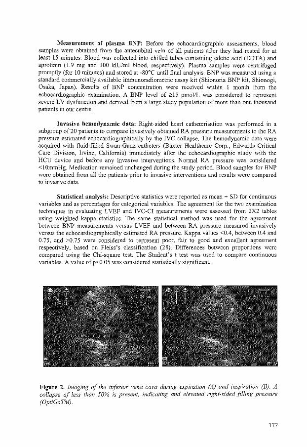
Measurement of plasma BNP: Before the echocardiographic assessments, blood samples were obtained from the antecubital vein of all patients after they had rested for at least 15 minutes. Blood was collected into chilled tubes containing edetic acid (EDTA) and aprotinin (!.9 mg and 100 klU/ml blood, respectively). Plasma samples were centrifuged promptly (for 10 minutes) and stored at -SO"C until final analysis. BNP was measured using a standard commercially available imrnunoradiometric assay kit (Shionoria BNP kit, Shionogi, Osaka, Japan). Results of BNP concentration were received within 1 month from the echocardiographic examination. A BNP level of ?15 pmol!L was considered to represent severe LV dysfunction and derived from a large study population of more than one thousand patients in our centre.
Invasive hemodynamic data: Right-sided heart catheterisation was performed in a subgroup of 20 patients to compare invasively obtained RA pressure measurements to the RA pressure estimated echocardiographically by the IVC collapse. The hemodynamic data were acquired with fluid-filled Swan-Ganz catheters (Baxter Healthcare Corp., Edwards Critical Care Division, Irvine, California) immediately after the echocardiographic study with the HCU device and before any invasive interventions. Normal RA pressure was considered <10mmHg. Medication remained unchanged during the study period. Blood samples for BNP were obtained from all the patients prior to invasive interventions and results were compared to invasive data.
Statistical analysis: Descriptive statistics were reported as mean ± SD for continuous variables and as percentages for categorical variables. The agreement for the two examination techniques in evaluating L VEF and IV C-CI measurements were assessed from 2X2 tables using weighted kappa statistics. The same statistical method was used for the agreement between BNP measurements versus L VEF and between RA pressure measured invasively versus the echocardiographically estimated RA pressure. Kappa values <0.4, between 0.4 and 0.75, and >0.75 were considered to represent poor, fair to good and excellent agreement respectively, based on Fleiss's classification (28). Differences between propmiions were compared using the Chi-square test. The Student's t test was used to compare continuous variables. A value ofp<0.05 was considered statistically significant.
Figure 2. Imaging of the inferior vena cava during expiration (A) and inspiration (B). A collapse of less than 50% is present, indicating and elevated right-sided filling pressure (OptiGoTM).
177

Results
Patients characteristics: Eighty-eight (88) patients (52 male, age 59±12) referred for echocardiography with suspected LV dysfunction were included in the study. Out of the initial 88 patients, 6 were excluded from the study due to poor visualisation of the LV (2 patients), and IVC (4 patients) leaving 82 patients for analysis. Sixty-four out of 82 patients were classified according to the New York Heart Association functional class I or II, 14 were class III and 4 patients were class IV. The classification was performed by an investigator blinded to the results of the echocardiographic examinations.
Table 1. Baseline characteristics of the 88 patients with suspected LV dysfunction
Age (years) Male, n History of myocardial infarction Hypertension, knuwn Diabetes mellitus, treated No history of cardiovascular disease Beta-blockers Nitrates Calcium-antagonists Diuretics Aspirin/anticoagulants Lipid-lowering agents
52±12 57 (59%) 34 (39%) 15 (17%) 9 (10%)
32 (36%) 36 (41%) 15 (17%) 15 (17%) 12 (14%) 40 (45%) 37 (42%)
Echocardiographic data: Out of the 82 patients to analyse, 17 had an EF 535% as assessed by theSE Simpson's biplane discs method. The HCU examination detected 16/17 patients showing a sensitivity of 94% and specificity of 100% in the diagnosis of LV dysfunction (table 2A). The agreement between the two imaging techniques for the IVC-CI was very good (96%, kappa~0,87) (table 3A). However, there was no correlation between IVC-CI and LVEF assessed by SE (agreement:76%,kappa~0.19) (table 3B).
BNP data: Results of BNP were available for all 82 patients. BNP levels were elevated in 16 out of the 17 patients with LV dysfunction. However, in 8 patients with normal L VEF the BNP levels were also elevated resulting in a sensitivity of BNP in diagnosing patients with LV dysfunction of94% and a specificity of 88% (table 2B).
Subgroup analysis: 20 patients underwent a right-sided heart catheterisation as part of diagnostic procedure. The characteristic data of the patients are listed in table 4. The mean RA pressure measured invasively was 8±9 mmHg. The agreement for the measurement of the RA pressure between the two techniques was 95%, kappa 0.9 (table 5). The correlation between BNP and invasively measured RA pressure was poor (60%, kappa~0.29).
178
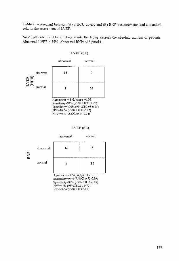
Table 2. Agreement between (A) a HCU device and (B) BNP measurements and a standard echo in the assessment ofLVEF.
No of patients: 82. The numbers inside the tables express the absolute number of patients. Abnormal LVEF: o;35%. Abnormal BNP: <15 pmol!L
abnormal
normal
abnormal
normal
LVEF (SE)
abnormal normal
16 0
1 65
" Agreement -99 Yo, kappa 0.96. Sensitivity=94% (95%CI:0.77-0.77) Specificity""lOO% (95%CI:0.95-0.95) PPV=IOO% (95%CI:0.82-0.82) NPV=98% (95%CI:0.94-0.94)
LVEF (SE)
abnormal normal
16 8
57
Agreement =89%, kappa -0.71. Sensitivity=94% (95%CI:0.73-0.99) Specificity=87% (95%CI:0.82-0.89) PPV=67% (95%CI:0.51-0.70) NPV=98% (95%CI:0.92-l.O)
179
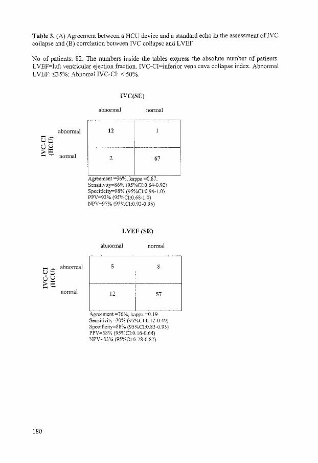
Table 3. (A) Agreement between a HCU device and a standard echo in the assessment ofJVC collapse and (B) correlation between IVC collapse and LVEF
No of patients: 82. The numbers inside the tables express the absolute number of patients. LVEF=left ventricular ejection fraction. IVC-CI=inferior vena cava collapse index. Abnormal LVEF: S35%; Abnomal IV C-CI: <50%.
abnormal
normal
abnormal
normal
180
IVC(SE)
abnormal normal
12 1
2 67
- 0 -Agreement 96 Yo, kappa -0.87. Sensitivity=86% (95%CI:0.64-0.92) Specificity=98% (95%CI:0.94-l.O) PPV=92% (95%Cl:0.68-I.O) NPV=97% (95%CI:0.93-0.98)
LVEF (SE)
abnormal normal
5 8
12 57
0 Agreement -76 Yo, kappa -0.19. Sensitivity=30% (95%CI:0.12-0.49) Specificity=88% (95%Cl:0.83-0.93) PPV=38% (95%CI:0.16-0.64) NPV=83% (95%CI:0.78-0.87)

Discussion
Heart failure and LV systolic dysfunction occur frequently, especially in the elderly population and are related to poor prognosis and considerable health-care cost (3). Early recognition and initiation of appropriate treatment can improve survival (6). However, diagnosis of LV dysfunction, especially in asymptomatic patients, may be difficult to assess by physical examination only, even when routine lab values, electrocardiograms and chest Xrays are added (29).
BNP is a 32 amino acid polypeptide containing a 17 amino acid ring structure common to all natriuretic peptides. The source of BNP is the cardiac ventricles and its release is directly proportional to ventricular volume expansion and pressure overload (12,13). BNP levels are elevated in patients with heart failure and LV dysfunction. Also the effect of treatment in these patients can be monitored with repeated BNP measurements, as suggested by Troughton et al (30). Furthermore BNP levels seem to be an independent predictor of longtenn survival after myocardial infarction (31) and all cause mortality for patients with LV dysfunction (32,33). BNP measurements have therefore been proposed as a new simple and inexpensive screening tool for LV dysfunction (14-17, 34,35). Furthermore the European Society of Cardiology has recently incorporated the BNP measurements into the diagnosis of heart failure (18). BNP levels are useful in "ruling out" this disorder due to very high negative predictive values, especially in untreated patients. In accordance with previous studies we demonstrated that BNP measurements show high sensitivity in detecting patients with LV dysfunction. However, we have to take into account that BNP, as an indicator of raised intracardiac pressure, can be elevated in various forms of heart disease besides LV systolic dysfunction including atrial fibrillation, LV diastolic dysfunction, LV hypertrophy and significant valve disease (36-37). This may explain our results regarding BNP measurements.
Echocardiogaphy as a screening tool for LV dysfunction: According to the guidelines of the European Society of Cardiology, objective evidence of LV dysfunction must be added to clinical symptoms to establish the diagnosis of heart failure. Echocardiography has been proposed to be the screening method of choice to demonstrate cardiac dysfunction (18,19). However, echocardiography is considered not cost-effective as a screening tool, especially for patients with low probability of cardiac dysfunction, and its availability is often limited in the different clinical settings. Newly developed HCU devices offer high image quality, ultra-portability and significantly lower capital cost (!/lOth of the cost of a SE). The value of such devices for screening for various cardiac pathologies has been shown in previous studies (23-25, 27, 38-39). The main finding of the current study was that a HCU device, estimating LVEF, is a sensitive tool for screening for LV dysfunction as assessed by SE.
We tested furthermore the hypothesis of diagnosing LV dysfunction by assessing the percentage collapse of IV C. However, this parameter appeared to be oflow sensitivity (30%) and positive predictive value (38%) for the detection of LV dysfunction with both devices, SE and HCU echocardiography. To our knowledge, this is the first study that evaluated the parameter of the percentage collapse of IVC as a potential screening parameter for LV dysfunction. In the contrary and as previously shown (22), the correlation of the echocardiogaphically estimated RA pressure, assessed by the JVC collapse, compared to the invasively assessed RA pressure (agreement 95%).
Echocardiography, can provide non-invasively, additional valuable information about other significant abnormalities beyond LV function. Thus, LV hypertrophy, valvular abnormalities or mass lesions can be diagnosed instantly with echocardiography but could be missed by physical examination or a blood test. Furthermore, the addition of the Doppler
181
feature in some of these devices enables the differentiation between systolic and diastolic dysfunction.
Use of ultrasound stethoscopes: The American Society of Cardiology recommends Level I of training as an absolute minimal level required for the use of HCU devices ( 40). Studies have shown that minimal echo training may enable physicians to use HCU for interpreting simple abnormalities with high efficacy and accuracy (41-42).
Limitations: The HCU devices used in this study were SonoHeart plusTM and OptiGoTM. Although the former has two additional modalities (pulsed Doppler and the MMode ), we used only the 2D feature for the L VEF and the IVC collapse evaluation to avoid bias between the two devices. There are therefore no data about the diastolic LV function of the heart or flow data. Furthermore no additional data about other cardiac abnormalities were reported since the purpose of the study was to test the potential of a HCU device as a screening tool for LV dysfunction.
Conclusion: Echocardiography is a most practical tool to demonstrate cardiac dysfunction. The HCU devices lead to incorporation of echocardiography into the physical examination and can broaden the availability of echocardiography allowing screening programmes for the identification of patients with LV dysfunction.
References
l. McKee PA, Castelli WP, McNamara PM, Kannel! WB. The natural history of heart failure: the Framingham Study. N Engl J Med 1971;285: 1441-46.
2. Cowie MR, Mosterd A, Wood DA, Deckers JW, Poole-Wilson PA, Sutton GC, Grobbee DE. The epidemiology of heart failure. Eur Heart J 1997;18:208-25.
3. Mosterd A, Hoes AW, de Bruyne MC, Deckers JW, Linker DT, Hofman A, Grobbee DE. Prevalence of heart failure and left ventricular dysfunction in the general population. The Rotterdam Study. Eur Heart J 1999;20:447-55.
4. Macintyre K, Capewell S, Stewart S, Chalmers JWT, Boyd J, Finlayson A, Redpath A, Pell JP, McMmray JN. Evidence of improving prognosis in heart failure. Trends in case fatality in 66 547 patients hospitalized between 1986 and 1995. Circulation 2000; 102:1126-31.
5. The CONSENSUS Trial Study Group. Effects of Enalapril on mortality in severe congestive heart failure. N Eng! J Med 1987;316: 1429-35.
6. The SOL VD investigators. Effect of Enalapril on mortality and the development of heart failure in asymptomatic patients with reduced left ventricular ejection fractions. N Eng J Med 1992;327:685-91.
7. The MERIT-HF Study Group. Effect of metoprolol CRIXL in chronic heart failure: Metoprolol CRIXL Randomised Intervention Trial in Congestive Heart Failure (MERITHF). Lancet 1999;353:2001-07.
8. Grimes DA, Shulz KF. Uses and abuses of screening tests. Lancet 2002;359:881-84. 9. Remes J, Miettinen H, Reunanen A, Pyorala K. Validity of clinical diagnosis of heart
failure in primary health care. Eur Hear J 1991; 12:315-21. 10. Stevenson LW. The limited availability of physical signs for estimating hemodynamics in
chronic heart failure. JAMA 1989;261;884-8. 11. Hobbs FDR, Jones MI, Allan TF, WilsonS, Tobias R. European survey of primary care
physician perceptions on heart failure diagnosis and management (EURO-HF). Eur Heart J 2000;2!:1877-87.
182

12. Yandle TG. Biochemistry of natriuretic peptides. J Intern Med 1994;235:561-76. 13. Daggubati S, Parks JR, Overton RM, Cintron G, Schocken DD, Vesely DL.
Adrenomedullin, endothelin, neuropeptide Y, atrial, brain, and C-natriuretic prohonnone peptides compared as early heart failure indicators. Cardiovasc Res 1997;36:246-55.
14. Cowie MR, Struthers AD, Wood DA, Coats AJS, Thompson SG, Poole-Wilson PA. Value of natriuretic peptide in assessment of patients with possible new heart failure in primary care. Lancet 1997;350: 1349-53.
15. McDonagh TA, Robb SD, Murdoch DR, Morton JJ, Ford I, Morrison CE, Tunstali-Pedoe H, McMurray JN, Dargie HJ. Biochemical detection of left-ventricular systolic dysfunction. Lancet 1998;351 :9-13.
16. Maisel AS, Koon J, Krishnaswamy P, Kazenegra R, Clopton P, Gardetto N, Morrisey R, Garcia A, Chiu A, De Maria A. Utility of B-natriuretic peptide as a rapid, point-of-care test for screening patients undergoing echocardiography to determine left ventricular dysfunction. Am Heart J 2001 ;141 :367-74.
17. Morrison KL, Harrison A, Krishnaswamy P, Kazanegra R, Clopton P, Maisel A. Utility of a rapid B-natriuretic peptide assay in differentiating congestive heart failure from lung disease in patients presenting with dyspnea. JAm Coli Cardiol2002;39:202-9.
18. Remme WJ, Swedberg K, (co-chairmen). Guidelines for the diagnosis and treatment of chronic heart failure. Task Force for the diagnosis and treatment of chronic heart failure, European Society of Cardiology. Eur Heart J 2001 ;22: 1527-60.
19. Guidelines for the evaluation and management of heart failure. Report of the ACC/AHA task force on practice guidelines. JAm Coli Cardioll995;26:1376-98.
20. Talreja D, Gruver C, Sklenar J, Dent J, Kaul S. Efficient utilisation of echocardiography for the assessment ofleft ventricular systolic function. Am Heart J 2000;139:394-8.
21. Sorrell VL, Reeves WC. Noninvasive right and left heart catheterization: taking the echo lab beyond an image-only laboratory. Echocardiography 2001; 18:31-41.
22. Stein JH, Neumann A, Marcus RH. Comparison of estimates of right atrial pressure by physical examination and echocardiography in patients with congestive heart failure and reasons for discrepancies. Am J Cardioll997;80:1615-18.
23. Vourvouri EC, Poldermans D, Schinkel AFL, Koroleva L, Sozzi FB, Bax JJ, Roeland! JRTC. Left ventricular hypertrophy screening using a hand-held ultrasound device. Eur Heart J 2002,26:1516-21.
24. Vourvouri EC, Poldermans D, Schinkel AFL, Sozzi FB, Bax JJ, van Urk H, Roeland! JRTC. Abdominal aortic aneurysm screening using a hand-held ultrasound device. A pilot study. Eur J Vase Endovasc Surg 2001;22:352-4.
25. Bruce CJ, Spitteli PC, Montgomery SC, Bailey KR, Tajik AJ, Seward JB: Personal Ultrasound Imager: Abdominal aortic aneurysm screening. J Am Soc Echoc. 2000;13:674-79.
26. Schiller NB, Shah PM, Crawford M, DeMaria A, Devereux R, Feigenbaum H, Gutgesell H, Reichek N, Sahn D, Schnittger I, et al. Recommendations for quantitation of the left ventricle by two dimensional echocardiography: American Society of Echocardiography Committee on Standards Subcommittee. JAm Soc Echocardiogr 1989;2:358-367.
27. Vourvouri EC, Poldermans D, de Sutter J, Sozzi FB, Izzo P, Roeland! JRTC. Experience with an ultrasound stethoscope. JAm Soc Echocardiogr 2002;15:80-5.
28. Fleiss JL. Statistical methods for rates and proportions. 2nd ed. New York, NY:Wiley,l981.
29. Davie AP, Francis CM, Love MP, Caruana L, Starkey IR, Shaw TR. Value of the electrocardiogram in identifYing heart failure due to left ventricular systolic dysfunction. BMJ 1996;312:222.
183
30. Troughton RW, Frampton CM, Yandle TG, Espiner EA, Nicholls MG, Richards AM. Treatment of heart failure guided by plasma aminoterminal brain natriuretic peptide (NBNP) concentrations. Lancet. 2000;355:1126-30.
31. Arakawa N, Nakmura M, Aoki H, Hiramori K. Plasma brain natriuretic peptide concentrations predict survival after acute myocardial infarction. J Am Call Cardiel 1996;27: 1656-61.
32. McDonagh TA, Cunningham AD, Morrison CE, McMurray JN, Ford E, Morton JJ, Dargie HJ. Left ventricular dysfunction, natriuretic peptides and mortality in an urban population. Heart 2001;86:21-26.
33. Berger R, Huelsman M, Strecker K, Bojic A, Moser P, Stanek B, Pacher R. B-type natriuretic peptide predicts sudden death in patients with chronic heart failure. Circulation 2002;105:2392-97.
34. McCullough PA, Nowak RM, McCord J, Hollander JE, Herrmann HC, Steg PG, Due P, Westheim A, Omland T, Knudsen CW, Storrow AB, Abraham WT, Lamba S, Wu AH, Perez A, Clopton P, Krishnaswamy P, Kazanegra R, Maisel AS. B-type natriuretic peptide and clinical judgment in emergency diagnosis of heart failure: analysis from Breathing Not Properly (BNP) Multinational Study. Circulation 2002;106:416-22
35. Maisel AS, Krishnaswamy P, Nowak RM, McCord J, Hollander JE, Due P, Omland T, Storrow AB, Abraham WT, Wu AH, Clopton P, Steg PG, Westheim A, Knudsen CW, Perez A, Kazanegra R, Herrmann HC, McCullough PA. Rapid measurement of B-type natriuretic peptide in the emergency diagnosis of heart failure. N Eng J Med 2002;347:161-7.
36. Nakamura M, Endo H, Nasu M, Arakawa N, Segawa T, Hiramori K. Value of plasma B type natriuretic peptide measurement for heart disease screening in a Japanese population. Heart 2002;87: 131-135.
37. Struthers AD. Introducing a new role for BNP: as a general indicator of cardiac structural disease rather than a specific indicator of systolic dysfunction only. Heart 2002;87:97-98.
38. Roelandt JRTC. A personal ultrasound imager (ultrasound stethoscope). A revolution in the physical cardiac diagnosis' Eur Heart J 2002;23: 523-27.
39. Spencer KT, Anderson AS, Bhargava A, Bales AC, Sorrentino M, Furlong K, Lang RM. Physician-performed point-of-care echocardiography using a laptop platform compared with physical examination in the cardiovascular patient. JAm Col! Cardiol2001;37:2013-8.0.
40. Seward JB, Douglas PS, Erbel R, Kerber RE, Kronzon I, Rakowski H, Sahn DJ, Sisk EF, Tajik AJ, Wann S. Hand-carried cardiac ultrasound (HCU) device: recommendations regarding new technology. A report from the echocardiography task force on new technology of the nomenclature and standards committee of the American Society of Echocardiography. JAm Soc Echocardiogr 2002;15:369-73.
41. Alexander JH, Peterson ED, Chen AY, Harding TM, Adams DB, Kisslo J. Training and accuracy of non-cardiologists in simple use of point-of-care echo: A preliminary report from the Duke Limited Echo Assessment Project (LEAP). Thoraxcenter Journal 2001;13:105-110.
42. Kimura BJ, Bocchicchio M, Willis CL, DeMaria AN. Screening cardiac ultrasonographic examination in patients with suspected cardiac disease in the emergency department. Am Heart J. 2001;142:324-30.
184
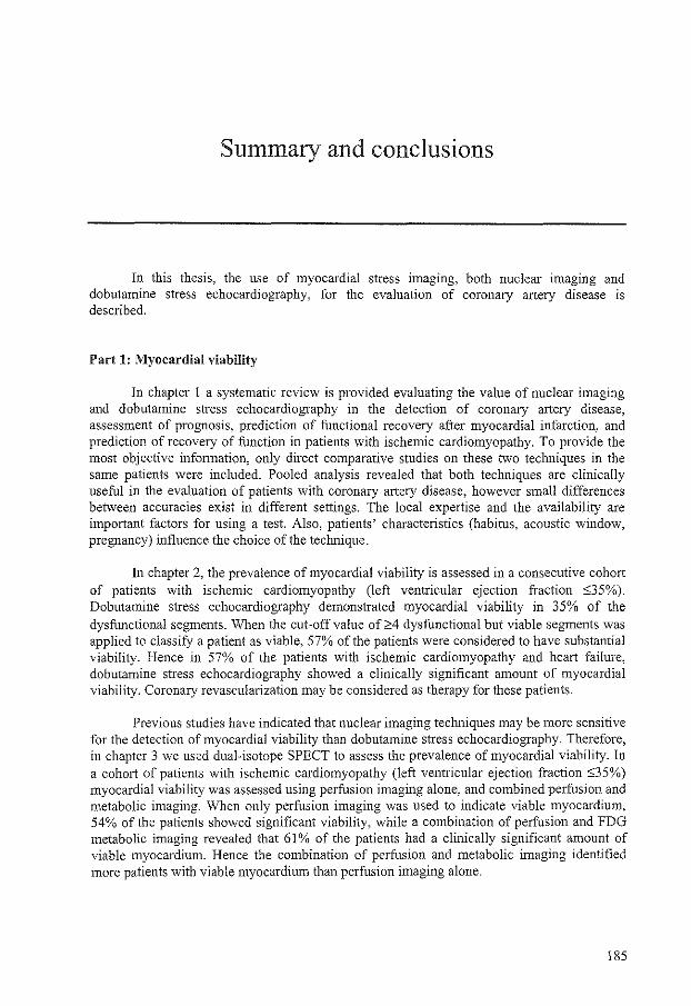
Summary and conclusions
In this thesis, the use of myocardial stress imaging, both nuclear imaging and dobutamine stress echocardiography, for the evaluation of coronary artery disease is described.
Part 1: Myocardial viability
In chapter 1 a systematic review is provided evaluating the value of nuclear imaging and dobutamine stress echocardiography in the detection of coronary artery disease, assessment of prognosis, prediction of functional recovery after myocardial infarction, and prediction of recovery of function in patients with ischemic cardiomyopathy. To provide the most objective information, only direct comparative studies on these two techniques in the same patients were included. Pooled analysis revealed that both techniques are clinically useful in the evaluation of patients with coronary artery disease, however small differences between accuracies exist in different settings. The local expertise and the availability are important factors for using a test. Also, patients' characteristics (habitus, acoustic window, pregnancy) influence the choice of the technique.
In chapter 2, the prevalence of myocardial viability is assessed in a consecutive cohort of patients with ischemic cardiomyopathy (left ventricular ejection fraction :::;;35%). Dobutamine stress echocardiography demonstrated myocardial viability in 35% of the dysfunctional segments. \Vhen the cut-off value of ;:::4 dysfunctional but viable segments was applied to classify a patient as viable, 57% of the patients were considered to have substantial viability. Hence in 57% of the patients with ischemic cardiomyopathy and heart failure, dobutamine stress echocardiography showed a clinically significant amount of myocardial viability. Coronary revascularization may be considered as therapy for these patients.
Previous studies have indicated that nuclear imaging teclmiques may be more sensitive for the detection of myocardial viability than dobutamine stress echocardiography. Therefore, in chapter 3 we used dual-isotope SPECT to assess the prevalence of myocardial viability. In a cohort of patients with ischemic cardiomyopathy (left ventricular ejection fraction :::;;35%) myocardial viability was assessed using perfusion imaging alone, and combined perfusion and metabolic imaging. \Vhen only perfusion imaging was used to indicate viable myocardium, 54% of the patients showed significant viability, while a combination of perfusion and FDG metabolic imaging revealed that 61% of the patients had a clinically significant amount of viable myocardium. Hence the combination of perfusion and metabolic imaging identified more patients with viable myocardium than perfusion imaging alone.
185

Chronic electrocardiographic Q waves are often believed to reflect scarred tissue. In chapter 4, the relation between chronic electrocardiographic Q waves, end-diastolic wall thickness, and myocardial viability is addressed. A consecutive series of patients with chronic electrocardiographic Q wave infarction, heart failure symptoms, and chronic coronary artery disease underwent dobutamine stress echocardiography. In 58% of the Q wave regions viable tissue was present. Hence, chronic Q waves on electrocardiography do not exclude the presence of viable myocardium. Initial evaluation by resting echocardiography may already exclude the presence of viable tissue when the end-diastolic wall thickness is 6 mm or less. However, in regions with Q waves and an end-diastolic wall thickness >6 nun, additional testing is needed since 38% of this regions did not show viability during dobutamine stress echocardiography.
Next, in chapter 5, myocardial viability in Q-wave regions in patients with ischemic cardiomyopathy, who had heart failure as the predominant symptom was assessed with dualisotope SPECT. The patients underwent echocardiography at rest to identify dysfunctional myocardial tissue and dual-isotope nuclear imaging to assess myocardial perfusion and metabolism. According to FDG SPECT metabolic imaging viable tissue persisted in a high proportion ( 61 %) of dysfunctional myocardial regions with chronic Q waves. Hence, chronic Q waves on electrocardiography do not necessarily imply irreversibly scarred myocardium.
In chapter 6, the presence of contractile reserve in response to dobutamine infusion was studied in patients with stunned and hibernating myocardium. Dysfunctional myocardium was assessed by resting echocardiography, the dysfunctional segments were subsequently evaluated for the presence of stunning or hibernation by dual-isotope SPECT. The presence of contractile reserve was assessed by dobutamine stress echocardiography. Contractile reserve was present in 61% of the stunned segments as compared to 51% of the hibernating myocardium. Patients with more severe heart failure (NYHA functional class III-IV) less frequently showed contractile reserve as compared to patients with mild heart failure. Contractile reserve was absent in the majority of scar segments; only 14% of the scar segments exhibited contractile reserve. In addition, residual contractile reserve was related to the extent of scar tissue, nontransmural scar tissue more often exhibited contractile reserve than transmural scars. The progressive reduction of contractile reserve in stunned, hibernating, nontransmural and transmural scar tissue, supports the hypothesis that stunning, hibernation, and scar are not circumscript pathofysiological entities but represent gradual ultrastructural damage on the myocyte level.
In chapter 7, the feasibility and image quality of dual-isotope SPECT imaging using acipimox for the evaluation of myocardial viability in patients with diabetes mellitus are assessed. The study population consisted of 70 patients, subsets of patients with insulindependent diabetes mellitus and with non-insulin dependent diabetes mellitus were studied. Metabolic conditions (plasma substrate levels) were monitored. As anticipated baseline glucose levels were higher in patients with diabetes. In patients with plasma glucose levels >9 mmol/L additional insulin was administered. An oral dose of acipimox effectively lowered the free fatty acids concentrations in patients with and without diabetes. No serious side effects of Acipimox were observed. The image quality (assessed by the myocardium to background ratio) was comparable in both patient groups. The type of diabetes (insulin dependent or noninsulin dependent diabetes) did not influence FDG image quality. In conclusion, FDG SPECT is a safe and clinically useful method to assess myocardial viability that offers a good image quality, even in patients with diabetes mellitus.
186
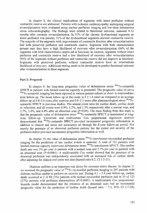
In chapter 8, the clinical implications of segments with intact perfusion without contractile reserve are addressed. Patients with ischemic cardiomyopathy undergoing surgical revascularization were evaluated using nuclear perfusion imaging and low-dose dobutamine stress echocardiography. The findings were related to functional outcome, assessed 9-12 months after coronary revascularization. In 51% of the chronic dysfunctional segments an intact perfusion was present, 31% of the dysfunctional segments showed contractile reserve. The majority of the segments with improvement of contractile function after revascularization had both preserved perfusion and contractile reserve. Segments with both characteristics present may thus have a high likelihood of recovery after revascularization (66% of the segments with both characteristics improved in function). In contrast, segments without intact perfusion and contractile reserve had a low likelihood of recovery after revascularization (95% of the segments without perfusion and contractile reserve did not improve in function). Segments with preserved perfusion without contractile reserve have an intermediate likelihood of recovery. Additional testing needs to be developed to predict functional outcome after revascularization in these segments.
Part 2: Prognosis
In chapter 9, the long-term prognostic value of dobutamine stress 99mTc-sestamibi SPECT in patients with limited exercise capacity is presented. The prognostic value of stress 99mTc-sestamibi imaging has been reported in various patient-subsets at short- to intermediateterm follow-up. The mean follow up of this study is 8 ± l A years as compared to an average follow up of 0.8-3.6 years after exercise and 0.8-2.3 years after pharmacological stress 99mTcsestamibi SPECT in previous studies. The annual event rates for cardiac death, cardiac death or infarction, and all events were 0.9%, 1.2%, and 1.5% respectively after a normal scan, and 2.7%, 3.4%, and 4.4% after an abnormal scan (P<0.05). The main finding from this study is that the prognostic value of dobutamine stress 99mTc-sestamibi SPECT is maintained at longterm follow-up. Univariate and multivariate Cox proportional regression analysis demonstrated that 99mTc-sestamibi SPECT provided incremental prognostic information in addition to clinical and stress test parameters all through the 8-year follow-up period. Not merely the presence of an abnormal perfusion pattern, but the extent and severity of the perfusion defect provided incremental prognostic information as well.
In chapter 10, the value of dobutamine stress 99mTc-tetrofosmin myocardial perfusion imaging for the prediction of late cardiac events is assessed. A total of 721 patients with limited exercise capacity underwent dobutamine stress 99mTc-tetrofosmin SPECT. The cardiac death rate was 1% per year in patients with a normal scan and 5.1% per year in patients with an abnormal scan (P<O.OOOl). A multivariable Cox model showed that the presence of an abnormal perfusion was independently associated with an increased risk of cardiac death, after adjusting for clinical and stress test data (hazard ratio 8.2, CI 3.2-21).
Diabetes mellitus is an important risk factor for coronary artery disease. In chapter 11 we assessed the prognostic value of 99mTc myocardial perfusion imaging in 207 patients with diabetes mellitus unable to perform an exercise test. During 4.1 ± 2.4 year follow-up, cardiac death occurred in 2 of 69 (3%) patients with normal myocardial perfusion and in 34 of 125 (27%) patients with perfusion abnormalities (P<O.OOOI). A multivariable Cox proportionalhazards model demonstrated that the presence of an abnormal scan had an incremental prognostic value for the prediction of cardiac death (hazard ratio ~ 7.2, 95% CI 1.7-30),
187
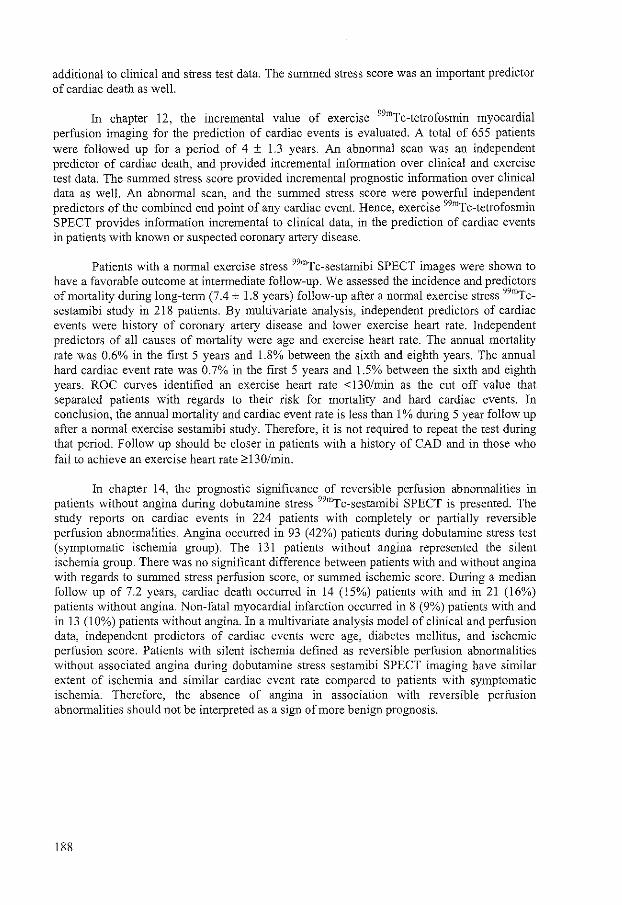
additional to clinical and stress test data. The summed stress score was an important predictor of cardiac death as well.
In chapter 12, the incremental value of exercise 99mTc-tetrofosmin myocardial perfusion imaging for the prediction of cardiac events is evaluated. A total of 655 patients were followed up for a period of 4 ± 1.3 years. An abnormal scan was an independent predictor of cardiac death, and provided incremental information over clinical and exercise test data. The summed stress score provided incremental prognostic information over clinical data as well. An abnormal scan, and the summed stress score were powerful independent predictors of the combined end point of any cardiac event. Hence, exercise 99mTc-tetrofosmin SPECT provides information incremental to clinical data, in the prediction of cardiac events in patients with known or suspected coronary artery disease.
Patients with a nonnal exercise stress 99mTc-sestamibi SPECT images were shown to have a favorable outcome at intermediate follow-up. We assessed the incidence and predictors of mortality during long-term (7.4 ± 1.8 years) follow-up after a normal exercise stress 99mTcsestarnibi study in 218 patients. By multivariate analysis, independent predictors of cardiac events were history of coronary artery disease and lower exercise heart rate. Independent predictors of all causes of mortality were age and exercise heart rate. The annual mortality rate was 0.6% in the first 5 years and 1.8% between the sixth and eighth years. The annual hard cardiac event rate was 0. 7% in the first 5 years and 1.5% between the sixth and eighth years. ROC curves identified an exercise heart rate <130/min as the cut off value that separated patients with regards to their risk for mortality and hard cardiac events. In conclusion, the annual mortality and cardiac event rate is less than 1% during 5 year follow up after a normal exercise sestamibi study. Therefore, it is not required to repeat the test during that period. Follow up should be closer in patients with a history of CAD and in those who fail to achieve an exercise heart rate :;?:130/min.
In chapter 14, the prognostic significance of reversible perfusion abnormalities in patients without angina during dobutamine stress 99mTc-sestamibi SPECT is presented. The study reports on cardiac events in 224 patients with completely or partially reversible perfusion abnormalities. Angina occurred in 93 (42%) patients during dobutamine stress test (symptomatic ischemia group). The 131 patients without angina represented the silent ischemia group. There was no significant difference between patients with and without angina with regards to summed stress perfusion score, or summed ischemic score. During a median follow up of 7.2 years, cardiac death occurred in !4 (!5%) patients with and in 2! (!6%) patients without angina. Non-fatal myocardial infarction occurred in 8 (9%) patients with and in 13 (10%) patients without angina. In a multivariate analysis model of clinical and perfusion data, independent predictors of cardiac events were age, diabetes mellitus, and ischemic perfusion score. Patients with silent ischemia defined as reversible perfusion abnormalities without associated angina during dobutamine stress sestamibi SPECT imaging have similar extent of ischemia and similar cardiac event rate compared to patients with symptomatic ischemia. Therefore, the absence of angina in association with reversible perfusion abnormalities should not be interpreted as a sign of more benign prognosis.
!88
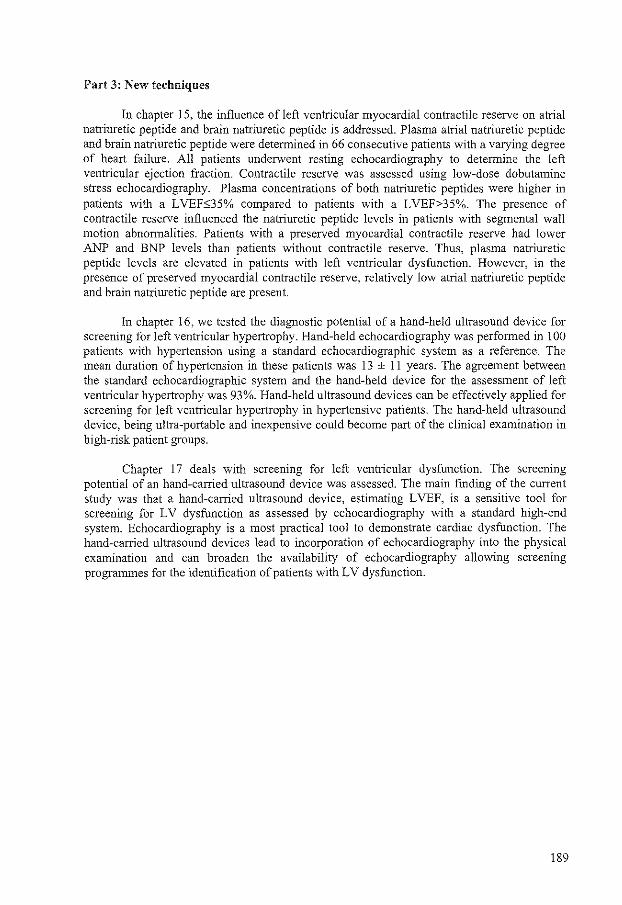
Part 3: New techniques
In chapter 15, the influence of left ventricular myocardial contractile reserve on atrial natriuretic peptide and brain natriuretic peptide is addressed. Plasma atrial natriuretic peptide and brain natriuretic peptide were determined in 66 consecutive patients with a varying degree of heart failure. All patients underwent resting echocardiography to determine the left ventricular ejection fraction. Contractile reserve was assessed using low-dose dobutamine stress echocardiography. Plasma concentrations of both natriuretic peptides were higher in patients with a LVEF:s;35% compared to patients with a LVEF>35%. The presence of contractile reserve influenced the natriuretic peptide levels in patients with segmental wall motion abnonnalities. Patients with a preserved myocardial contractile reserve had lower ANP and BNP levels than patients without contractile reserve. Thus, plasma natriuretic peptide levels are elevated in patients with left ventricular dysfunction. However, in the presence of preserved myocardial contractile reserve, relatively low atrial natriuretic peptide and brain natriuretic peptide are present.
In chapter 16, we tested the diagnostic potential of a hand-held ultrasound device for screening for left ventricular hypertrophy. Hand-held echocardiography was performed in 100 patients with hypertension using a standard echocardiographic system as a reference. The mean duration of hypertension in these patients was 13 ± 11 years. The agreement between the standard echocardiographic system and the hand-held device for the assessment of left ventricular hypertrophy was 93%. Hand-held ultrasound devices can be effectively applied for screening for left ventricular hypertrophy in hypertensive patients. The hand-held ultrasound device, being ultra-portable and inexpensive could become part of the clinical examination in high-risk patient groups.
Chapter 17 deals with screening for left ventricular dysfunction. The screening potential of an hand-carried ultrasound device was assessed. The main finding of the current study was that a hand-carried ultrasound device, estimating L VEF, is a sensitive tool for screening for LV dysfunction as assessed by echocardiography with a standard high-end system. Echocardiography is a most practical tool to demonstrate cardiac dysfunction. The hand-carried ultrasound devices lead to incorporation of echocardiography into the physical examination and can broaden the availability of echocardiography allowing screening programmes for the identification of patients with LV dysfunction.
189

Samenvatting en conclusies
In dit proefschrift wordt bet gebruik van beeldvormend cardiaal stress onderzoek, zowel nucleaire beeldvorrning als dobutamine stress echocardiografie, voor bet evalueren van coronaire hartziekten beschreven.
Deell: Myocardvitaliteit
In hoofdstuk 1 wordt een systematisch overzicht gegeven van de waarde van nucleaire beeldvorming en dobutamine stress echocardiografie voor bet diagnostiseren van coronaire hartziekten, bepaling van de prognose, voorspellen van functieherstel na een myocardinfarct en bet voorspellen van functieherstel bij patienten met ischemische cardiomyopathie. Om objectieve informatie te verschaffen, werden alleen onderzoeken geanalyseerd die deze twee technieken in dezelfde patienten hebben vergeleken. Gecombineerde analyse liet zien dat dit klinisch waardevolle technieken zijn voor bet evalueren van patienten met coronaire hartziekten, hoewel er in verschillende situaties kleine verschillen in nauwkeurigheid bestaan. De plaatselijke expertise en de beschikbaarheid zijn belangrijke factoren voor het gebruiken van een onderzoek Ook de eigenschappen van de patient (habitus, akoestisch raam, zwangerschap) beinvloeden de keuze voor een van de technieken.
In hoofdstuk 2 wordt the prevalentie van dysfunctioneel maar vitaal myocardweefsel bepaald in een groep van opeenvolgende patienten met ischemische cardiomyopathie (linker ventrikel ejectie fractie :535%). Dobutamine stress echocardiografie toonde myocardvitaliteit aan in 35% van de dysfunctionele segmenten. Wanneer de grenswaarde van ?4 dysfunctionele maar vi tale segmenten werd toegepast om een patient als vitaal the classificeren, had 57% van de patienten een substantiele hoeveelheid vitaal weefsel. Dus in 57% van de patienten met ischemische cardiomyopathie en hartfalen, liet dobutamine stress echocardiografie een substantiele hoeveelheid vitaal myocardweefsel zien. Coronaire revascularisatie kan als behandeling overwogen worden bij deze patienten.
Eerder onderzoek heeft laten zien dat nucleaire technieken gevoeliger kunnen zijn dan dobutamine stress echocardiografie voor bet vaststellen van myocardvitaliteit. Daarom gebruikten we in hoofdstuk 3 dual-isotope SPECT om de prevalentie van myocardvitaliteit vast te stellen. In een cohort patienten met ischemische cardiomyopathie (linker ventrikel ejectie fractie :S:35%) werd myocardvitaliteit bepaald met aileen perfusie beeldvorming, en met gecombineerde perfusie en metabolisme beeldvorming. Wanneer alleen de perfusie beelden werden gebruikt, had 54% van de patienten een significante hoeveelheid dysfunctioneel maar vitaal weefsel, de combinatie van perfusie en metabolisme liet zien dat 61% van de patienten een significante hoeveelheid vitaal myocardweefsel had. Dus met de combinatie van perfusie en metabolisme beeldvorming werden meer patienten met vitaal myocardweefsel ontdekt dan met aileen perfusie beeldvorming.
191

Er wordt vaak gedacht dat chronische Q golven op het electrocardiogram op littekenweefsel duiden. In hoofdstuk 4 werd de relatie tussen chronische Q golven, einddiastolische wanddikte en myocardvitaliteit onderzocht. Een opeenvolgende serie patienten met een chronisch Q golf infarct, hartfalen, en chronische coronaire hartziekte ondergingen dobutamine stress echocardiografie. In 58% van de Q golf gebieden was dysfunctioneel maar vitaal weefsel aanwezig. Derhalve sluiten chronische Q golven op bet electrocardiogram de aanwezigheid van vitaal myocard niet uit. Een eerste evaluatie met een rust echocardiogram kan de aanwezigheid van vitaal weefsel al uitsluiten wanneer de einddiastolische wanddikte 6 rnm of minder is. In gebieden met Q golven en een einddiastolische wanddikte grater dan 6 mm is echter een aanvullende test nodig, omdat 38% van deze gebieden geen vitaliteit toonde tijdens dobutamine stress echocardiografie.
Vervolgens werd in hoofdstuk 5 de myocardvitaliteit bepaald in gebieden met Q golven bij patienten met ischemische cardiomyopathie, die hartfalen als het belangrijkste symptoom hadden, door middel van dual-isotope SPECT. De patienten ondergingen een rust echocardiogram om dysfunctioneel myocardweefsel vast te stellen en dual-isotope nucleaire beeldvorming om de myocardiale perfusie en het glucose metabolisme te bepalen. Volgens FDG SPECT afueeldingen van het metabolisme was er nog vitaal weefsel aanwezig in een groat gedeelte (61 %) van de dysfimctionele gebieden met chronische Q golven. Chronische Q golven op het electrocardiogram wijzen dus niet eenvoudigweg op irreversibel verlittekend myocardweefsel.
In hoofdstuk 6 wordt de aanwezigheid van een contractiele reserve in respons op dobutamine infusie bestudeerd in patienten met stunned en hibernating myocardweefsel. Dysfunctioneel myocard werd vastgesteld met een echocardiogram in rust, de dysfimctionele segmenten werden vervolgens geevalueerd op de aanwezigheid van stunned of hibernating myocardweefsel met dual-isotope SPECT. De aanwezigheid van contractiele reserve werd vastgesteld met dobutamine stress echocardiografie. Contractiele reserve was aanwezig in 61% van de stunned segmenten en in 51% van de hibernating segmenten. Patienten met emstig hartfalen (NYHA functie klasse III-IV) hadden minder vaak een behouden contractiele reserve vergeleken met patienten met een geringere vorm van hartfalen. Contractiele reserve was ahvezig in de meerderheid van de verlittekende segmenten; slechts 14% van de verlittekende segmenten had een contractiele reserve. Bovendien was een behouden contractiele reserve gerelateerd aan de uitbreiding van het Iitteken, een niet-transmuraal litteken liet vaker contractiele reserve zien dan een transmuraal litteken. De progressieve vermindering van contractiele reserve in stunned, hibernating, niet-transmuraal en transmuraal littekenweefsel, steunt de hypothese dat stunning, hibernation en littekenweefsel geen scherp omschreven pathosfysiologische begrippen zijn, maar een geleidelijke hoeveelheid schade op het niveau van de myocyten vertegenwoordigen.
In hoofdstuk 7 wordt de toepasbaarheid en de beeldkwaliteit van dual-isotope SPECT na orale toediening van acipimox voor de evaluatie van myocardvitaliteit bij patienten met diabetes mellitus vastgesteld. De onderzoekspopulatie bestond uit 70 patienten, waarvan subgroepen van patienten met insulineafhankelijke en non-insulineafhankelijke diabetes mellitus werden bestudeerd. Zeals venvacht waren de uitgangsglucosewaarden hager bij patienten met diabetes. Bij patienten met een plasma glucosewaarde >9 mmol/L werd extra insuline toegediend. Een orale dosis acipimox verlaagde vrije vetzuur concentraties effectief bij patienten met en zonder diabetes. Er werden geen ernstige bijwerkingen van acipimox gezien. De beeldkwaliteit (bepaald met de myocard versus achtergrond ratio) was vergelijkbaar in beide groepen patienten. Ret type diabetes (insulineafhankelijke en non-
192
insulineafhankelijke diabetes) had geen inv1oed op de FDG bee1dkwa1iteit. FDG SPECT is een veilige en klinisch nuttige methode en biedt een goede beeldbvaliteit om de myocardvitaliteit vast te stellen, zelfs bij patienten met diabetes mellitus.
In hoofdstuk 8 worden de klinische implicaties van segmenten met een intacte perfusie zonder contractiele reserve vastgesteld. Patienten met ischemische cardiomyopathie ondergingen een coronaire bypass operatie en werden geevalueerd met nucleaire beeldvorming en lage-dosis dobutamine stress echocardiografie. De bevindingen werden gerelateerd aan de verbetering van functie, die 9-12 maanden na de coronaire bypass operatie werd bepaald. In 51% van de chronisch dysfunctionele segmenten was er een intacte perfusie en 31% van de dysfunctionele segmenten had een behouden contractiele reserve. De meerderheid van de segmenten met een verbeterde contractiele functie na de revascularisatie had een behouden perfusie en contractiele reserve. Segmenten met beide eigenschappen hadden een hoge kans op functieherstel na revascularisatie ( 66% van deze segmenten had een functie verbetering). Bij segmenten zonder intacte perfusie en contractiele reserve was de kans op functieherstel na revascularisatie laag (95% van de segmenten zonder perfusie en contractiele reserve hadden geen functieherstel). Segmenten met een behouden perfusie zonder contractiele reserve hadden ook een kans op functieherstel. Aanvullende tests moeten ontwikkeld worden om het functieherstel na revascularisatie in deze segmenten te voorspellen.
Deel2: Prognose
In hoofdstuk 9 wordt de lange-termijn prognostische waarde van dobutamine stress 99mTc-sestamibi SPECT bij patienten met een beperkte inspanningscapaciteit gepresenteerd. De prognostische waarde van stress 99mTc-sestamibi beeldvorming is reeds onderzocht in verschillende patienten groepen met een korte of middellange termijn. De gemiddelde vervolg termijn van dit onderzoek is 8 ± 1.4 jaar in vergelijking tot een gemiddelde termijn van 0.8-3.6 jaar na inspanning en 0.8-2.3 jaar na farmacologische stress 99mTc-sestamibi SPECT in eerdere onderzoeken. De jaarlijkse kans op cardiale dood, cardiale dood of een myocardinfarct, en cardiale dood of een myocardinfarct of een coronaire revascularisatie was 0.9%, 1.2%, en 1.5% respectievelijk na een normale scan, en 2.7%, 3.4%, en 4.4% na een afwijkende scan (P<0.05). De be1angrijkste bevinding van dit onderzoek is dat de prognostische waarde van dobutamine stress 99mTc-sestamibi SPECT behouden blijft tijdens lange-termijn vervolgonderzoek. Univariabel and multivariabel Cox proportionele regressie analyse toonde dat 99mTc-sestamibi SPECT meer informatie biedt dan klinische en stress test parameters gedurende de gehele follow-up periode van 8 jaar. Niet aileen de aanwezigheid van een afwijkend perfusie, maar ook de uitbreiding en de ernst van een perfusie defect verschaffen een toegevoegde prognostische waarde.
In hoofdstuk 10 wordt de waarde van dobutamine stress 99mTc-tetrofosmin nucleaire beeldvorming voor het voorspellen van late cardiale aandoeningen vastgesteld. Een groep van 721 patienten met een beperkte inspanningscapaciteit onderging dobutamine stress 99mTctetrofosmin SPECT. De kans op een cardiale dood was 1% per jaar bij patiE:nten met een normale scan en 5.1% per jaar bij patienten met een afwijkende scan (P<O.OOOl). Een multivariabel Cox model toonde dat de aanwezigheid van een afwijkende perfusie was gerelateerd aan een verhoogd risico op een cardiale dood, na correctie voor de klinische en stress test informatie (hazard ratio 8.2, CI 3.2-21).
193

Diabetes mellitus is een belangrijke risicofactor voor coronaire hartziekten. In hoofdstuk 11 bepaalden we de prognostische waarde van 99mTc nucleaire beeldvorming bij 207 patienten met diabetes mellitus die niet in staat waren om een inspanningstest te volbrengen. Gedurende een follow-up periode van 4.1 ± 2.4 jaar, trader cardiale dood op bij 2 van de 69 (3%) patienten met een normale myocard perfusie en bij 34 van de 125 (27%) patienten met perfusie afwijkingen (P<O.OOO!). Een multivariabel Cox model toonde dat de aanwezigheid van een afwijkende scan een toegevoegde prognostische waarde had voor bet voorspellen van een cardiale dood (hazard ratio~ 7.2 95% Cl 1.7-30), boven klinische en stress test gegevens. De stress score was ook een belangrijke voorspeller van cardiale dood.
In hoofdstuk 12 wordt de toegevoegde waarde van inspannings 99mTc-tetrofosmin SPECT voor bet voorspellen van cardiale aandoeningen geevalueerd. Een groep van 655 patienten werd gevolgd gedurende 4 ± 1.3 jaar. Een afwijkende scan was een onafhankelijke voospeller van cardiale dood en bood een toegevoegde waarde naast de klinische en inspannings test gegevens. The stress score bood ook toegevoegde prognostische informatie naast de klinische gegevens. Een abnormale scan en de stress score waren sterke onafhankelijke voorspellers voor bet gecombineerde eindpunt van cardiale dood, infarct, of coronaire revascularisatie. Inspannings 99mTc-tetrofosmin SPECT onderzoek, naast klinische gegevens, biedt een toegevoegde informatie voor bet voorspellen van cardiale aandoeningen bij patienten met bewezen of vermoedelijke coronaire hartziekten.
Patienten met norma1e inspannings 99mTc-sestamibi SPECT scans hebben een gunstige prognose op middellange termijn. We stelden de incidentie en voorspellers van ( cardiale) sterfte vast, ~edurende een 1ange-termijn follow-up (7 .4 ± 1.8 jaar) na een normale inspannings 9 mTc-sestamibi scan bij een groep van 218 patienten. Multivariabele analyse toonde dat een voorgeschiedenis met coronaire hartziekte en een lage hartfrequentie tijdens inspanning onafhankelijke voorpellers zijn van cardiale aandoeningen. Leeftijd en hartfrequentie tijdens inspanning zijn onafhankelijke voorspellers voor de totale mortaliteit. De jaarlijkse mortaliteitskans was 0.6% in de eerste 5 jaar en 1.8% van het zesde tot bet achtste jaar. De kans op cardiale dood of een myocardinfarct was 0. 7% in de eerste 5 jaar en 1.5% tussen het zesde en achtste jaar. Een hartfrequentie tijdens inspanning <130/min is de grenswaarde die patienten verdeelt wat betreft hun sterfterisico en de kans op een myocardinfarct. De jaarlijkse mortaliteit en kans op een myocardinfarct is minder dan 1% gedurende een vervolgperiode van 5 jaar na een nonnale inspannings sestamibi scan. Tijdens deze periode lijkt het daarom niet nodig om de test te herhalen bij patienten met een normale scan. Patienten met een voorgeschiedenis van coronaire hartziekten en zij die een hartfrequentie tijdens inspanning <130/min hebben dienen intensiever gevolgd te worden.
In hoofdstuk 14 wordt bet prognostische belang van reversibele perfusie afwijkingen bij patienten zonder angina pectoris tijdens de dobutamine stress 99mTc-sestamibi SPECT test gepresenteerd. Dit onderzoekt richt zich op de cardiale aandoeningen bij 224 patienten met een compleet of gedeeltelijk reversibel perfusie defect. Angina tract bij 93 (42%) patienten op tijdens de dobutamine stress test (groep met symptomatische ischemie). De 131 patienten zonder angina vertegenwoordigden de groep met stille ischemie. Er was geen significant verschil tussen patienten met en zonder angina wat betreft de stress perfusie score, of de ischemie score. Tijdens een vervolgperiode van 7.2 jaar, trad cardiale dood op bij 14 (15%) patienten met en bij 21 (16%) patienten zonder angina. Een niet-fataal myocardinfarct trad op bij 8 (9%) patienten met en 13 (10%) patienten zonder angina. Een multivariabele analyse van klinische en perfusie gegevens, toonde dat leeftijd, diabetes mellitus en de ischemie perfusie score onafhankelijke voorspellers voor cardiale aandoeningen waren. Patienten met stille
194
ischemie gedefinieerd als reversibele perfusie defecten zonder angina tijdens de dobutamine stress sestamibi SPECT test hebben een vergelijkbare hoeveelheid ischemie en een vergelijkbare kans op cardiale aandoeningen als patienten met symptomatische ischemie. De af\vezigheid van angina pectoris bij patienten met een reversibel perfusiedefect moet daarom niet gezien worden als een teken dat wijst op een gunstige prognose.
Deel3: Nieuwe technieken
In hoofdstuk 15 wordt de invloed van contractiele reserve van het linker ventrikel op atrium natriuretisch peptide (ANP) en brein natriuretisch peptide (BNP) bestudeerd. De plasma concentraties van ANP en BNP werden bepaald bij 66 opeenvolgende patienten met hartfalen. Bij aile patienten werd een rust echocardiogram gemaakt om de linker ventrikel ejectie ftactie (L VEF) te bepalen. Contractiele reserve werd vastgesteld met lage-dosis dobutamine stress echocardiografie. Plasma concentraties van beide natriuretische peptiden waren boger in patienten met een LVEF$;35% vergeleken met patienten met een LVEF>35%. De aanwezigheid van contractiele reserve beinvloedde de concentratie van de natriuretische peptiden bij patienten met wandbewegingsstoomissen. Patienten met een behouden myocardiale contractiele reserve hadden lagere ANP en BNP niveaus dan patienten zonder contractiele reserve. Plasma natriuretische peptide concentraties zijn dus verhoogd bij patienten met een dysfunctie van de linker ventrikel. Echter, in aanwezigheid van een behouden myocardiale contractiele reserve waren de ANP en BNP concentraties relatief laag.
In hoofdstuk 16 hebben we de diagnostische moge1ijkbeden van een draagbaar echoapparaat geevalueerd voor het screenen op linker ventrikel hypertrofie. Bij 100 patienten met hypertensie werd een echocardiogram gemaakt met een draagbaar echoapparaat. Een standaard echocardiografiesyteem werd gebruikt als referentiemethode. De patienten hadden gemiddeld 13 ± 11 jaar hypertensie. De overeenkomst tussen het standaard echocardiografie systeem en het draagbare apparaat voor bet vaststellen van linker ventrikelhypertrofie was 93%. Draagbare echoapparaten kunnen effectief ingezet worden om te screenen op linker ventrikel hypertrofie bij patienten met hypertensie. Deze apparaten zijn relatief goedkoop en kunnen deel uitmaken van het klinische onderzoek bij patienten met een hoog risico voor linker ventrikel hypertrofie.
Hoofdstuk 17 beschrijft de screeningsmogelijkheden van draagbare echoapparaten voor bet opsporen van linker ventrikel dysfunctie. De belangrijkste bevinding was dat het schatten van de L VEF met een draagbaar echoapparaat een gevoelig instrument is voor bet screenen op LV dysfunctie. Draagbare echoapparaten kunnen een onderdeel worden van bet lichamelijk onderzoek. Echocardiografie wordt zo voor meer patienten beschikbaar en een screeningsprogramma voor het opsporen van patienten met LV dysfunctie wordt mogelijk.
195


Dankwoord
Wetenschap is een teamsport. Alle cardiologen, fellows, statistici, arts-assistenten, echocardiografisten, verpleegkundigen en medewerkers van het Thoraxcentrum wil ik hartelijk danken voor het tot stand brengen van dit proefschrift. Ook de samenwerking met de afdeling Nucleaire Geneeskunde en de afdeling Interne Geneeskunde heb ik zeer gewaardeerd. Enkele mensen wil ik in het bijzonder bedanken:
Prof.dr. Jos RT.C. Roelandt, promotor, ben ik zeer erkentelijk, omdat ik van hem het vertrouwen kreeg om te werken in het Thoraxcentrum. Het is een voorrecht klinisch wetenschappelijk onderzoek te doen in deze inspirerende omgeving. Zijn kritische instelling en enthousiasmerende persoonlijkheid waren van groat belang voor dit onderzoek Daamaast heb ik de humor en gastvrijheid van m'n promotor en zijn vrouw Martine buitengewoon gewaardeerd.
Dr. Don Poldermans, copromoter, is een grote steun. Op hem heb ik kunnen bouwen. Zoals vele fellows, heb ik van hem de geheimen van dobutamine stress echocardiografie geleerd. Niet aileen zijn discipline-overstijgende expertise op klinisch gebied, maar vooral zijn heldere wetenschappelijke ideeen vormen een uniek voorbeeld. Zijn enthousiasme en humor zorgen voor een aangename en productieve werksfeer. Zeer veel dank voor de vriendschap en het vertrouwen.
Dr. Jeroen J. Bax is een drijvende kracht achter dit onderzoek. Zelden heb ik iemand zoals Jeroen ontmoet: zijn enthousiasme, kennis op het gebied van noninvasieve cardiale beeldvorming, en snelle marrier van werken waren belangrijke ingredienten voor dit proefschrift. Dank voor de vriendschap, een betere samenwerking had ik me niet kunnen wensen.
De beoordelingscommissie van dit proefschrift: prof.dr.ir. A.F.W. van der Steen, prof.dr. P.J. De Feijter en prof.dr. E.P. Krenning.
Het Thoraxcentrum is befaamd vanwege haar internationale karakter en het is mij een waar genoegen om met collega's uit verschillende Ianden te mogen werken. Mijn dank gaat uit naar Dr. Abdou Elhendy, USA, wiens waardevolle suggesties en kennis van de literatuur een belangrijke bijdrage aan dit proefschrift vormen. Mijn directe collega's en kamergenoten van kamer Ba 302 zijn vrienden geworden. Dr. Fabiola Sozzi uit Italie heeft haar dissertatie een jaar eerder volbracht, onze fijne samenwerking en gesprekken heb ik zeer gewaardeerd.
Aparte aandacht verdient Eleni Vourvouri. In vele studies hebben we intensief samengewerkt en we verdedigen ons proefschrift op dezelfde dag. Het is mooi om te zien hoe
197
je de aandacht voor draagbare echocardiografie hebt doen opleven. Bovendien wil ik jou en Georgios Sianos bedanken voor de kennismaking met de Griekse cultuur: euxaptcvr:co noA.u!
Dank aan Manolis Bountioukos uit Griekenland voor de prettige wetenschappelijke samenwerking en vriendschap, je hebt je de vreemde Nederlandse humor snel eigen gemaakt.
Meer recent zijn Vittoria Rizello, uit Italie, Boudewijn Krenning, en Miklos Kertai ons team komen versterken. Dank voor de vriendschap en prettige samenwerking, ik wens jullie aile succes bij het verwezenlijken van jullie ideal en. Van de vele fellows die in de afgelopen tijd vanuit aile windstreken naar het Thoraxcentrum kwamen wil ik vooral Riccardo Rambaldi, Lubov Koroleva, Galal Elkilany, Nestor Mercado, Eustachio Agricola, en Maria Daniel bedanken.
Virginie Polderrnans voor de gezelligheid en waardevolle adviezen. We hebben een speciale band opgebouwd en dat waardeer ik buitengewoon.
Van de cardiologen wil met name Dr. Folkert J. Ten Cate, bedanken voor de gastvrijheid en de vele mooie ( contrast)echobeelden die hij met me gedeeld heeft, en Dr. Jaap W. Deckers, Dr. Folkert J. Meijboom, Dr. Jolien Roos, Dr. Tjebbe Galema en Dr. Jan GeversLeuven, lipidoloog en Dr. Anton van de Meiracker, internist, voor hun betrokkenheid. Veel dank aan Dr. MarcelL. Geleijnse voor de wetenschappelijke suggesties.
Dr. Eric Boersma en Dr. Ron van Domburg voor de fijne samenwerking, en de vele statistische adviezen en berekeningen. De databases die jullie ont\vikkeld hebben en beheren vorrnen een belangrijke bron van informatie.
De technische expertise, en niet te vergeten de aanstekelijke humor van Wim Vletter en Rene Frowijn heb ik zeer gewaardeerd. Zonder de hulp van de echocardiografisten was dit proefschrift onmogelijk geweest. Hartelijk dank aan Jackie, Debby, Marianne, Veronica, Hans, Dieny en Elly. Dank aan Nico Bruining en Jan Tuin voor hun assistentie.
De sarnenwerking met de afdeling Nucleaire Geneeskunde was erg productief, met name Dr. Roelf Valkema en Boen Kam hebben een belangrijke bijdrage aan dit proefschrift geleverd. Dank aan alle rnedewerkers van de afdeling Nucleaire Geneeskunde.
Van het laboratoriurn Interne Geneeskunde ben ik Dr. Frans Boomsma en Rene de Bruin dankbaar voor de adviezen en analyse van bloedrnonsters.
Van de diverse secretariaten wil ik speciaal Willeke, Arita, Helena, en Bea noemen en natuurlijk Ad van Drunen, voor de snelle afwikkeling van financiele zaken. Van de polikliniek Cardiologic dank ik Miranda, Willeke, Corrie, Lenne, Anja, Kirsten, Miijam, Arja, Conny, Hanneke, Rina, Tine, Celeste en Lydia voor het plezierige contact. Uiteraard dank aan Ellen van de balie Interne Geneeskunde. De forrnele voorbereiding van deze prornotie kwam tot stand dankzij Dr. Jan Willem de Jong en Annet Louw (Bureau Onderwijs Thoraxcentrum).
Mijn familie en vrienden, vooral Erik, Rogier, Rendel, Henriette, Feddo, Raymonda, en Willem-Johan, voor de belangstelling.
Tenslotte dank ik rnijn ouders en zusjes Lizeke en Rianne. Hun advies en vertrouwen, zeker niet aileen bij het voltooien van dit werk, waren essentieel.
198
Curriculum Vitae
Arend Schinkel was born on December 15, 1975 in Leiderdorp, The Netherlands. He completed his preparatory academic education (Gymnasium~. latin-mathematics) in 1994 at the Bonaventura College in Leiden. In the same year he started his medical training at the Leiden University Medical Center. In 2000 he passed the final examinations in medicine and became a research fellow at the Department of Cardiology of the Thorax center in Rotterdam The research described in this thesis was performed between 1999 and 2002 (supervisors professor Jos R.T.C. Roelandt and Dr. Don Poldermans). Subsequently, the clinical training in cardiology will be started.
199

Publications
1. Schinkel AFL, Elhendy A, Van Domburg RT, Bax JJ, Vourvouri EC, Sozzi FB, Valkema R, Roelandt JRTC, Poldennans D. Prognostic value of dobutamine-atropine stress myocardial perfusion imaging in patients with diabetes. Diabetes Care. 2002;25:1637-1643.
2. Schinkel AFL, Bax JJ, Sozzi FB, Boersma E, Valkema R, Elhendy A, Roeland! JRTC, Poldennans D. Prevalence of myocardial viability assessed by single photon emission computed tomography in patients with chronic ischaemic left ventricular dysfunction. Heart. 2002;88;125-130.
3. Schinkel AFL, Elhendy A, van Domburg RT, Bax JJ, Roeland! JRTC, Poldermans D. Prognostic value of dobutamine-atropine stress (99m)Tc-tetrofosmin myocardial perfusion SPECT in patients with known or suspected coronary artery disease. J Nucl Med. 2002;43;767-772.
4. Schinkel AFL, Bax JJ, Elhendy A, Boersma E, Vourvouri EC, Sozzi FB, Valkema R, Roeland! JRTC, Poldermans D. Assessment of viable tissue in Q-wave regions by metabolic imaging using single-photon emission computed tomography in ischemic cardiomyopathy. Am J Cardia!. 2002;89: 1171-1175.
5. Vourvouri EC, Poldermans D, Schinkel AFL, Sozzi FB, Bax JJ, van Urk H, Roeland! JRTC. Abdominal aortic aneurysm screening using a hand-held ultrasound device. The Thoraxcenter Journa/2002; 14;40-42.
6. Elhendy A, van Domburg RT, Vantrimpont P, Poldermans D, Bax JJ, van Gelder T, Baan CC, Schinkel AFL, Roeland! JRTC, Balk AH. Prediction of mortality in heart transplant recipients by stress technetium-99m tetrofosmin myocardial perfusion imaging. Am J Cardia/. 2002;89;964-968.
7. Sozzi FB, Poldermans D, Bax JJ, Elhendy A, Vourvouri EC, Valkema R, De Sutter J, Schinkel AFL, Borghetti A, Roeland! JR TC. Improved identification of viable myocardium using second harmonic imaging during dobutamine stress echocardiography. Heart. 2001;86:672-678.
8. Schinkel AFL, Bax JJ, Valkema R, Poldermans D. Evaluation of myocardial viability in ischemic cardiomyopathy: nuclear imaging versus dobutamine stress echocardiography. New Advances in Nuclear Cardiology. 2001 ;2;11-13.
201
9. Vourvouri EC, Poldermans D, Bax JJ, Sianos G, Sozzi FB, Schinkel AFL, de Sutter J, Parcharidis G, Valkema R, Roeland! JRTC. Evaluation of left ventricular function and volumes in patients with ischaemic cardiomyopathy: gated single-photon emission computed tomography versus two-dimensional echocardiography. Eur J Nucl Med. 2001 ;28: 1610-1615.
10. Vourvouri EC, Poldermans D, Schinkel AFL, Sozzi FB, Bax JJ, van Urk H, Roeland! JRTC. Abdominal aortic aneurysm screening using a hand-held ultrasound device. 11A pilot study". Eur J Vase Endovasc Surg. 2001 ;22:352-354.
11. Schinkel AFL, Bax JJ, Boersma E, Elhendy A, Roeland! JRTC, Poldermans D. How many patients with ischemic cardiomyopathy exhibit viable myocardium? Am J Cardia!. 2001 ;88:561-564.
12. Schinkel AFL, Bax JJ, van der Wall EE, Jonkers GJ. Echocardiographic follow-up of Chlamydia psittaci myocarditis. Chest. 2000;117:1203-1205.
13. Bax JJ, Poldermans D, Schinkel AFL, Boersma E, Valkema R, Krenning E, Roeland! JRTC. Perfusion and contractile reserve in chronic dysfunctional myocardium: relation to functional outcome after surgical revascularization. Circulation 2002;106:114-118.
14. Schinkel AFL, Bax JJ, Boersma E, Elhendy A, Vourvouri EC, Roeland! JR TC, Poldermans D. Residual myocardial viability on dobutamine stress echocardiography in regions with chronic electrocardiographic Q wave infarction. Am Heart J, in press.
15. Vourvouri EC, Poldermans D, Schinkel AFL, Koroleva L, Sozzi FB, Bax JJ, Parharidis GE, Roeland! JRTC. Screening for left ventricular hypertrophy using a hand-held ultrasound device. Eur Heart 12002;23:1516-1521.
16. Bax JJ, Kim CY, Poldermans D, Elhendy A, Boersma E, Schinkel AFL, Sloof GW, Sandler MP. Cardiac applications ofFDG Imaging with PET and SPECT. Bookchapter in "Clinical PET", in press.
17. Schinkel AFL, Bax JJ, Elhendy A, Valkema R, van Domburg R, Vourvouri EC, Sozzi FB, Roelandt JRTC, Poldermans D. Dobutamine-induced contractile reserve in stunned, hibernating, and scarred myocardium in patients with ischemic cardiomyopathy. J Nucl Med, in press.
18. Schinkel AFL, Elhendy A, van Domburg RT, Bax JJ, Valkema R, Roeland! JRTC, Poldermans D. Long-term prognostic value of dobutamine-atropine stress technetium-99m-sestamibi SPECT: A single-center experience with 8-year follow-up. Radiology, in press.
19. Sozzi FB, Elhendy A, Poldermans D, van Domburg RT, Schinkel AFL, Vourvouri EC, Bax JJ, De Sutter J, Borghetti A, Roelandt JRTC. Prognostic value of dobutamine stress echocardiography in patients with diabetes mellitus. Diabetes Care, in press.
20. Schinkel AFL, Bax JJ, Geleijnse ML, Boersma E, Elhendy A, Roeland! JRTC, Poldermans D. The noninvasive evaluation of ischemic heart disease: nuclear perfusion imaging or stress echocardiography? Eur Heart J, in press.
202

21. Schinkel AFL, Elhendy A, van Domburg RT, Bax JJ, Vourvouri EC, Bountioukos M, Rizzello V, Agricola E, Valkema R, Roeland! JRTC, Poldermans D. Exercise stress 99mTc-tetrofosmin myocardial perfusion single-photon emission computed tomography for the prediction of late cardiac events. Am J Cardia/, in press.
22. Vourvouri EC, Deckers JW, ten Cate FJ, Poldermans D, Schinkel AFL, Roeland! JRTC. Diagnosis of endocarditis of tricuspid valve with a small personal ultrasound imager. Submitted.
23. Elhendy A, Schinkel AFL, Poldermans D, van Domburg RT, Bax JJ, Roeland! JRTC. Long-term prognostic value of a normal exercise sestamibi myocardial perfusion study. Submitted.
24. Bountioukos M, Doorduijn JK, Vourvouri EC, Bax JJ, Schinkel AFL, Kertai MD, Roelandt JRTC, Sonneveld P, Poldermans D. Repetitive dobutamine stress echocardiography testing for the prediction of anthracycline cardiotoxicity. Submitted.
25. Schinkel AFL, Vourvouri EC, Bax JJ, Boomsma F, Bountioukos M, Roeland! JRTC, Poldermans D. The influence of myocardial contractile reserve during dobutamine stress echocardiography on atrial natriuretic peptide and brain natriuretic peptide. Submitted.
26. Vourvouri EC, Schinkel AFL, Boomsma F, Sianos G, Bountioukos M, Sozzi FB, Rizzello V, Bax JJ, Roeland! JRTC, Poldermans D. Screening for heart failure: inferior vena cava collapse assessment with a small personal ultrasound imager or brain natriuretic peptide measurement? Submitted.
27. Schinkel AFL, Bax JJ, Valkema R, Elhendy A, van Domburg RT, Vourvouri EC, Bountioukos M, Roeland! JRTC, Poldermans D. Effect of diabetes mellitus on myocardial 18F-fluorodeoxyglucose single-photon emission computed tomography using Acipimox for the assessment of myocardial viability. Submitted.
28. Vourvouri EC 1 Koroleva LY1 ten Cate FJ, Poldermans D, Schinkel AFL, van Domburg RT, Vletter WB, Roeland! JRTC. Clinical utility and cost-effectiveness of a personal ultrasound imager for cardiac evaluation during consultation rounds in patients with suspected cardiac disease. Submitted.
29. Elhendy A, Schinkel AFL, van Domburg RT, Bax JJ, Valkema R, Poldermans D, Roelandt JRTC. Assessment of silent ischemia using stress myocardial perfusion imaging. Submitted.
30. Bountioukos M, Poldermans D, Vourvouri EC, Rizzello V, Schinkel AFL, Krenning B, Bax JJ, Elhendy A, Roeland! JRTC. Safety and diaguostic accuracy of dobutamine stress echocardiography in patients with aortic stenosis. Submitted.
31. Rizzello V, Poldermans D, Schinkel AFL, Roeland! JRTC. Ventricular tachycardia during dobutamine stress echocardiography in a patient with severe aortic valve stenosis. Submitted.
203
32. Elhendy A, Schinkel AFL, van Domburg RT, Bax JJ, Roeland! JRTC, Poldermans D. Prediction of cardiac events in patients with heart failure using stress myocardial perfusion scintigraphy. Submitted.
33. Bountioukos M, Poldermans D, Vourvouri EC, Schinkel AFL, Rizzello V, Bax JJ, Roelandt JR TC. Atorvastatin improves myocardial contractility assessed by tissue Doppler imaging in patients with hypercholesterolemia. Submitted.
34. Rizzello V, Poldermans D, Schinkel AFL, Bountioukos M, Vourvouri EC, Bax JJ, Roelandt JRTC. Role of scar and biphasic segments on the recovery of viable myocardium after revascularization. Submitted.
35. Sozzi FB, Elhendy A, Poldermans D, van Domburg RT, Vourvouri EC, Schinkel AFL, Bax JJ, Romano S, Roelandt JRTC. Prognostic value of dobutamine stress echocardiography in patients with hypertension. Submitted.
36. Elhendy A, Schinkel AFL, van Domburg RT, Bax JJ, Valkema R, Roeland! JRTC, Polderrnans D. Prediction of cardiac death in hypertensive patients by stress myocardial perfusion imaging. Submitted.
37. Elhendy A, Schinkel AFL, van Domburg RT, Bax JJ, Valkema R, Roeland! JRTC, Poldermans D. Prognostic value of stress myocardial perfusion SPECT in patients with prior coronary revascularization. Submitted.
38. Poldermans D, Bax JJ, Kertai MD, Krenning B, Westerhout CM, Schinkel AFL, Landsberg P, Fleisher LA, Klein J, van Urk H, Roelandt JRTC, Boersma E. Statins are associated with a reduced incidence of perioperative mortality in patients undergoing major noncardiac vascular surgery. Submitted.
39. Bax JJ, Maddahi J, Poldermans D, Elhendy A, Schinkel AFL, Boersma E, Valkema R, Krenning EP, Roelandt JRTC, van der Wall EE. Accuracy to predict improvement of LVEF after revascularization: Comparison of different non-invasive imaging strategies. Submitted.
40. Bountioukos M, Schinkel AFL, Bax JJ, Boersma E, Vourvouri EC, Roeland! JR TC, Poldermans D. Comparison of regional left ventricular function in Q-wave and non-Qwave infarcted regions by tissue Doppler imaging in patients with ischemic cardiomyopathy. Submitted.
41. Agricola E, Oppizzi M, Schinkel AFL, Galderisi M, Maisano F, Roeland! JR TC, Alfieri 0. Pulsed tissue Doppler imaging can detect early myocardial dysfunction in asympomatic patients with severe mitral regurgitation. Submitted.
204

Awards:
KNMG Dick Held Junior-Prize for the manuscript 'Prevalence of myocardial viability assessed by single-photon emission computed tomography in patients with chronic ischemic left ventricular dysfuuction', Leiden, The Netherlands, 2001.
Winner of the Travel Grant Competition of the Working Group on Nuclear Cardiology of the European Society of Cardiology for the review article 'The noninvasive evaluation of ischemic heart disease: nuclear perfusion imaging or stress echocardiography?', International Conference of Nuclear Cardiology, Vienna, Austria, 2001.
Winner moderated poster session 'Long-term prognostic value of dobutamine-atropine stress technetium-99m sestamibi single-photon emission computed tomography in patients with known or suspected coronary artery disease', European Society of Cardiology, Stockholm, Sweden, 200 l.
205


![Longdom - Early ventricular dysfunction in type II diabetes role ......control population but also more than patients with coronary artery disease [8]. Dobutamine stress echocardiography](https://static.fdocuments.net/doc/165x107/613c808d4c23507cb6356ca8/longdom-early-ventricular-dysfunction-in-type-ii-diabetes-role-control.jpg)
![Quantification of systemic right ventricle by echocardiography · 2017-02-26 · of systemic right ventricle by echocardiography ... with dobutamine stress [17]. These data were confirmed](https://static.fdocuments.net/doc/165x107/5ecb2f51d4cb202a22168cb3/quantification-of-systemic-right-ventricle-by-echocardiography-2017-02-26-of-systemic.jpg)