MYELOMA BENCE-JONES PROTEINEMIA IN MULTIPLE · BENCE-JONES PROTEINEMIAIN MULTIPLEMYELOMAI By DANH....
10
BENCE-JONES PROTEINEMIA IN MULTIPLE MYELOMA Dan H. Moore, … , Elvin A. Kabat, Alexander B. Gutman J Clin Invest. 1943; 22(1):67-75. https://doi.org/10.1172/JCI101370. Research Article Find the latest version: http://jci.me/101370-pdf
Transcript of MYELOMA BENCE-JONES PROTEINEMIA IN MULTIPLE · BENCE-JONES PROTEINEMIAIN MULTIPLEMYELOMAI By DANH....
BENCE-JONES PROTEINEMIA IN MULTIPLEMYELOMA
Dan H. Moore, … , Elvin A. Kabat, Alexander B. Gutman
J Clin Invest. 1943;22(1):67-75. https://doi.org/10.1172/JCI101370.
Research Article
Find the latest version:
http://jci.me/101370-pdf
BENCE-JONESPROTEINEMIA IN MULTIPLE MYELOMAI
By DANH. MOORE,ELVIN A. KABAT, AND ALEXANDERB. GUTMAN(From the Electrophoresis Laboratory, and the Departments of Neurology and Medicine,
College of Physicians and Surgeons, Columbia University; the NeurologicalInstitute, and Presbyterian Hospital, New York City)
(Received for publication July 21, 1942)
The term "Bence-Jones protein" refers to anill-defined group of proteins having a molecularweight of approximately 37,000 (1), and the char-acteristic properties of precipitating out of solu-tion when warmed to 45 to 58° C. and of re-dissolving completely or partially on boiling. Thispeculiar behavior on heating has long served as auseful test for Bence-Jones proteins in the urineof patients with multiple myeloma. Whenappliedto serum, however, particularly to those sera wherehyperproteinemia is associated with myeloma, theheat test has been found generally inadequate be-cause it does not sufficiently differentiate Bence-Jones proteins from the serum euglobulins (2, 3).More convincing evidence of Bence-Jones pro-teinemia can be obtained by extraction (4, andothers), protein solubility curves (5), and ultra-centrifugation (6, 7, and others). However, toestablish satisfactorily the frequency and degreeof Bence-Jones proteinemia in multiple myeloma,and its relation to hyperproteinemia in that dis-ease, more adequate methods recently devised forcharacterizing proteins would appear to be neces-sary. Any generalizations so derived should bebased upon a series of cases large enough to berepresentative.
In a recent study combining electrophoretic withsalting-out techniques (8), it was found that 38cases of multiple myeloma, presenting the mostvaried quantitative and qualitative differences inserum proteins, could be classified into 3 majorgroups: 1. Those with hyperglobulinemia due toy components which precipitated out chiefly withthe Howe "euglobulin," and partly with the Howe"pseudoglobulin I" fraction. 2. Those with a vari-ety of unusual serum protein patterns, not en-countered in any other disease. 3. Those withapparently normal serum proteins. It was sug-gested that the protein increment in the first group
1 Aided in part by a grant from the William J. Mathe-son Commission.
contained little or no Bence-Jones protein, whereasthe anomalous distributions in serum protein frac-tions in the second group appeared to be due, forthe most part, to significant Bence-Jones pro-teinemia. This view was based largely upon com-parisons of the solubility characteristics and elec-trophoretic mobilities of urinary Bence-Jones pro-teins with those of serum proteins in mtiltiplemyeloma, and upon a study of the properties ofproteins in normal serum to which different uri-nary Bence-Jones proteins had been added. Fur-ther and more direct evidence on this point hassince been obtained by correlating the results ofsalt precipitation, electrophoretic and ultracen-trifugal analysis, and immunological methods inmyeloma serum and urine.
METHODS
Fractional precipitation with neutral salts. Howe'smethod (9) was used for serum. Our results in normaland myeloma sera, and the uniform pattern in the distri-bution of protein fractions found in hyperproteinemia dueto chronic infections and cirrhosis, have already beendescribed (8).
Electrophoresis. The electrophoretic analyses and sep-arations were made by means of the Tiselius apparatus(10) using a cell of 10 ml. capacity, and the ToeplerSchlieren optical arrangement as modified by Longsworth(11). The samples of sera were diluted 1:4, 1:6, or1: 8 (depending upon the protein concentration) with0.02 M phosphate buffer containing physiological salinesolution at pH 7.4, the ionic strength of the buffer being= 0.2. They were then dialysed against 2 or more
changes of buffer for a period of 2 or 3 days, the lastchange of buffer being used to fill the electrode vessels.Undiluted urine samples were dialysed in the phosphatebuffer (sA= 0.2, not 0.1 as previously (8) stated) in thesame manner and studied in the Tiselius apparatus.
Our experience with electrophoretic analyses of normalsera has been summarized elsewhere (12). Results withblood plasma or serum in multiple myeloma have beenrecorded by Longsworth, Shedlovsky and MacInnes (13),by Kekwick (14) and, in both serum and urine, byourselves (8).
Ultracentrifugation. The analyses of serum and urinewere made in an air-driven ultracentrifuge kindly placed
67
DAN H. MOORE, ELVIN A. KABAT, AND ALEXANDERB. GUTMAN
at our disposal by Dr. A. E. Severinghaus. An analyticalcell holding 0.4 ml., of the type described by Bauer andPickels (15), was used, the optical analysis being madeby means of the Toepler Schlieren method (16). Thesedimentation rates were calculated from photographstaken at 5 minute intervals while the rotor was held ata constant speed of 46,400 R.P.M., giving a centrifugalfield at the center of the cell of 159,000 times -gravity.The experiments were done at room temperature and thesedimentation rates corrected to pure water at 20° C.
Immunological. The general plan followed was thatof Hektoen (17), with modifications. Rabbits were im-munized with Bence-Jones proteins prepared from urine,the antisera were absorbed with normal human serum
to remove nonspecific antibodies, and the specific antiseraso obtained were used for qualitative and quantitativeprecipitin tests for Bence-Jones proteins in myelomaserum. To obviate difficulties arising from serologicaldifferences in the Bence-Jones proteins of different pa-
tients (18, 19), tests for Bence-Jones protein in serum
were made with antiserum prepared only from the uri-nary Bence-Jones protein of that same patient. Im-munological studies were confined therefore to cases ofmultiple myeloma with Bence-Jones proteinuria.
Our detailed procedure was as follows: Unacidifiedurine of the patient to be tested was treated with am-monium sulfate to 40 per cent saturation, the slight pre-cipitate which formed was rejected, and the concentrationof ammonium sulfate brought to 60 per cent to precipitatethe Bence-Jones proteins. The precipitate was centri-fuged off, dissolved and reprecipitated once, redissolved inwater and dialysed in the cold until the dialysate nolonger reacted with Nessler's solution.
Rabbits were injected 4 times weekly with 1 to 5 mgm.of this Bence-Jones protein in the form of an alum pre-cipitate (20), several courses of 16 injections each beingnecessary to produce antisera of sufficient titer (eventhen, the best of these were much weaker than those ob-tainable with serum proteins or with egg albumin). Therabbits were bled 5 days after the last of a course ofinjections.
Antibodies to the serum proteins were removed by addi-tion of small amounts of undiluted normal human serumuntil no further precipitation with normal serum occurred.The precipitin tests were set up by adding 0.15 ml. ofthe absorbed rabbit antiserum to 0.15 ml. of appropriatedilutions of antigen (the patient's serum or urine, controlserum or urine, etc.). The tubes were shaken, incubated
TABLE I
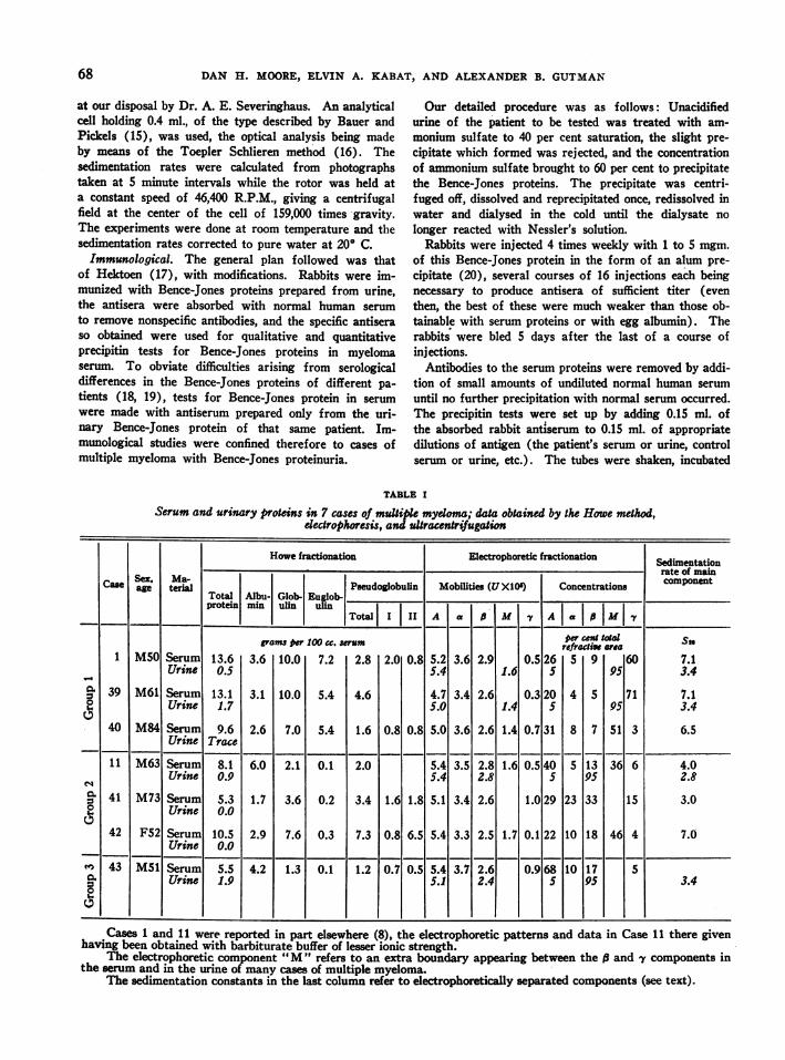
Serum and urinary proteins in 7 cases of muliple myedoma; data obtained by the Howe method,dectrophoresis, and ultracentrifugation
Howe fractionation Electrophoretic fractionation Sedimentation__________________ ramte of main
Case gex, Mterial Pseudoglobulin Mobilties (UX1O') Concentrations componentTotal Albu- Glob- Euglob-protein min ulin ulin _tIi I__ . _ ~~~Tota_IIllA a P M 'y AI|aI MI__p___J___
per cet kw~a St$grams per 100 cc. serum refracti area1 M50 Serum 13.6 3.6 10.0 7.2 2.8 2.0 0.8 5.2 3.6 2.9 0.5 26 5 9 60 7.1
Urine 0.5 5.4 1.6 5 95 3.4
ff 39 M61 Serum 13.1 3.1 10.0 5.4 4.6 4.7 3.4 2.6 0.3 20 4 5 71 7.12 Urine 1.7 5.0 1.4 5 95 3.4
40 M84 Serum 9.6 2.6 7.0 5.4 1.6 0.8 0.8 5.0 3.6 2.6 1.4 0.7 31 8 7 51 3 6.5Urine Trace
11 M63 Serum 8.1 6.0 2.1 0.1 2.0 5.4 3.5 2.8 1.6 0.5 40 5 13 36 6 4.0Urine 0.9 5.4 2.8 5 95 2.8
a 41 M73 Serum 5.3 1.7 3.6 0.2 3.4 1.6 1.8 5.1 3.4 2.6 1.0 29 23 33 15 3.02 Urine 0.0
42 F52 Serum 10.5 2.9 7.6 0.3 7.3 0.8 6.5 5.4 3.3 2.5 1.7 0.1 22 10 18 46 4 7.0Urine 0.0
X 43 M51 Serum 5.5 4.2 1.3 0.1 1.2 0.7 0.5 5.4 3.7 2.6 0.9 68 10 17 5Urine 1.9 5.1 2.4 5 95 3.4
( _ _ __ L__ ______Cases 1 and 11 were reported in part elsewhere (8), the electrophoretic patterns and data in Case 11 there given
having been obtained with barbiturate buffer of lesser ionic strength.The electrophoretic component "M" refers to an extra boundary appeanng between the , and 'y components in
the serum and in the urine of many cases of multiple myeloma.The sedimentation constants in the last column refer to electrophoretically separated compOnents (see text).
68
BENCE-JONES PROTEINEMIA IN MYELOMA
r -I B-J
AR 9RA
sERum 39 URINE
smm 4 2SERUM 42
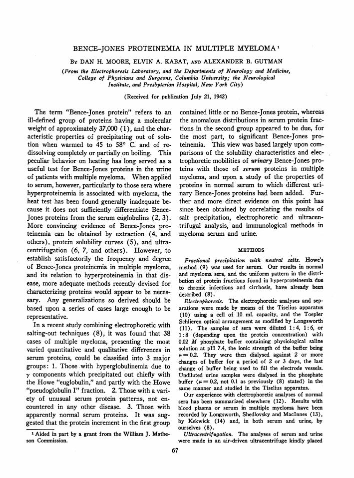
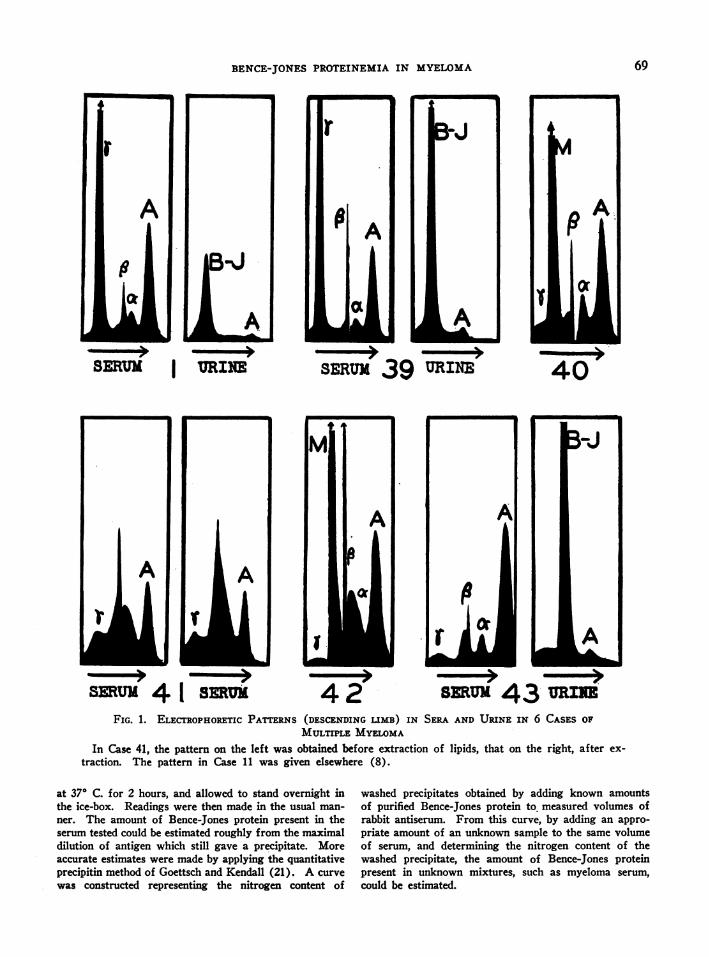
FIG. 1. ELECTROPHORETICPATTERNS (DESCENDING LIMB)MULTIPLE MYELOMA
AA
. r or A~.
INSERA 43-i6
IN SERA AND URINE IN 6 CASES OF
In Case 41, the pattern on the left was obtained before extraction of lipids, that on the right, after ex-traction. The pattern in Case 11 was given elsewhere (8).
at 37° C. for 2 hours, and allowed to stand overnight inthe ice-box. Readings were then made in the usual man-ner. The amount of Bence-Jones protein present in theserum tested could be estimated roughly from the maximaldilution of antigen which still gave a precipitate. Moreaccurate estimates were made by applying the quantitativeprecipitin method of Goettsch and Kendall (21). A curvewas constructed representing the nitrogen content of
washed precipitates obtained by adding known amountsof purified Bence-Jones protein to. measured volumes ofrabbit antiserum. From this curve, by adding an appro-priate amount of an unknown sample to the same volumeof serum, and determining the nitrogen content of thewashed precipitate, the amount of Bence-Jones proteinpresent in unknown mixtures, such as myeloma serum,could be estimated.
1tI A
I
I
1*1SEUSBU A
URIIE 40
SERUMl 4 1
I I I69
4
DAN H. MOORE, ELVIN A. KABAT, AND ALEXANDERB. GUTMAN
RESULTS
In Table I are summarized the results of serum
protein partitions by the Howe and electrophoretictechniques in 7 cases of multiple myeloma. Thediagnosis was established by autopsy or biopsy inall but Case 41. The cases are divided into the 3major groups already described.
Group 1. The serum protein pattern in Cases 1and 39 is representative of that found in a largenumber of patients with multiple myeloma. Thereis marked hyperproteinemia, the protein incrementbeing composed of globulins, the major part ofwhich are thrown down in 13.5 per cent sodiumsulfate. The electrophoretic pattern is character-ized by a large peak representing a marked in-crease in y globulins (Figure 1), the mobilities ofthe main component in these 2 cases being 0.5 and0.3 respectively.
The following evidence that the protein incre-ment in this group of cases includes little or no
Bence-Jones proteins has already been offered: 1.The distribution of serum protein fractions corre-
sponds, in general, to the pattern uniformly foundin hyperglobulinemia due to chronic infections or
cirrhosis, in which it may be presumed that Bence-Jones proteinemia does not occur. 2. Bence-Jonesproteins in native urine rarely if ever have thesolubility characteristics of serum euglobulins ineither ammonium or sodium sulfate solution. 3.Wehave been unable to reproduce a large increasewholly or very largely in the Howe "euglobulin"fraction by adding various urinary Bence-Jonesprotein preparations to normal serum. 4. Theelectrophoretic pattern of normal serum, to whichhas been added urinary Bence-Jones protein frompatients in this group, shows a new peak corre-
sponding to the mobility of the urinary Bence-Jones protein added, but not in the -y range (exceptin the few instances in which the mobility of theurinary Bence-Jones protein is extremely low).
When the y components of the sera in Cases 1and 39 were separated electrophoretically and theirsedimentation constants determined, the value ob-tained for S2O was 7.1 in both instances (TableI). The sedimentation constant of the urinaryBence-Jones proteins in both patients was 3.4 S(Svedberg (22) reports values of 2.8 S and 3.7 Sin different Bence-Jones protein preparations). Itis evident that the main component in the sera was
BLE II
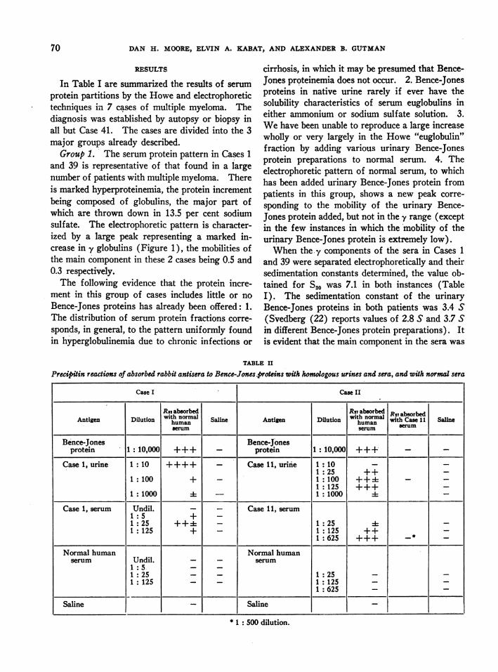
Precipitin reactions of absorbed rabbit antisera to Bence-Jones.proteins with homologous urines and sera, and with normal sera
Case I Case II
Rs bobdwith normal *~zua R27 absorbedAntigen Dilution ihseumn Saline Antigen Dilution wit with Case 11 Salineserum serum seu
Bence-Jones Bence-Jonesprotein 1:10,000 +++ _ protein 1:10,000 +++ _
Case 1 urine 1 :10 ++++ - Case l urine 1 : 101 :25 ++-
1: 100 + I1 100 +++ _ _1 :125 +++-
1:1000 1 - 1:1000 - _Case 1, serum Undil. - - Case 11, serum
1:5 + -1:25 - 1:I25 4-
1:125 + - 1:125 ++ -1:625 +++ _* _
Normal human Normal humanserum Undil. _ _ serum
1:25. - - 1:25 --1 :125 I1125 --I_:125 1 :625 - -
Saline l Saline
* 1: 500 dilution.
70
BENCE-JONES PROTEINEMIA IN MYELOMA
of far greater molecular size than Bence-Jonesprotein, of the order of magnitude of normal y
globulins, which have been found to have a sedi-mentation constant of 7.1 S (23). Similar re-
sults were obtained in 4 of 5 myeloma sera sub-jected to ultracentrifugal analysis by Kekwick(14), and in isolated cases by others. The avail-able data suggest that Bence-Jones protein doesnot constitute the main protein increment in themajority of cases of multiple myeloma with markedhyperproteinemia.
While it was thus shown that the chief proteinconstituent of these sera was not Bence-Jonesprotein, it was possible by serological methods todemonstrate that Bence-Jones protein was presentin small amount in the serum of Case 1. As indi-cated in Table II, rabbit antiserum to this patient'surinary Bence-Jones protein gave a strong pre-
cipitin reaction with a 1: 25 dilution of the pa-
tient's serum, and a definite reaction was obtain-able with a dilution of 1: 125. These reactionscannot be attributed to normal serum protein com-
ponents because normal serum failed to give any
test with the (absorbed) antiserum.It was further possible, by serological methods,
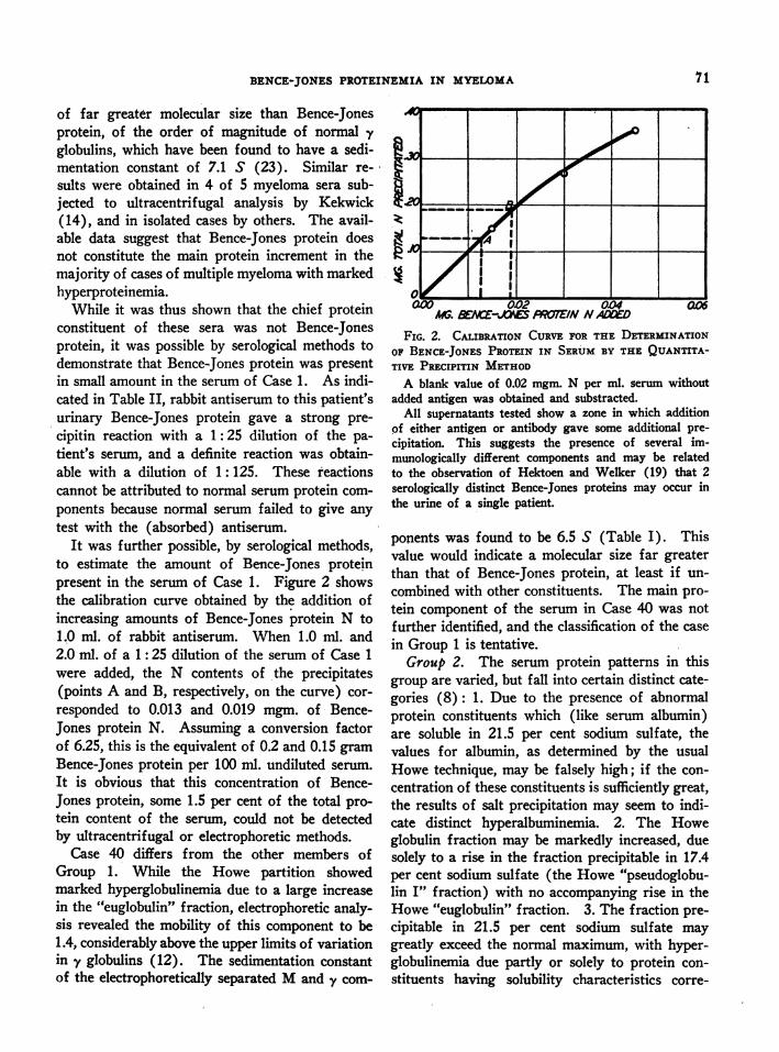
to estimate the amount of Bence-Jones proteinpresent in the serum of Case 1. Figure 2 showsthe calibration curve obtained by the addition ofincreasing amounts of Bence-Jones protein N to1.0 ml. of rabbit antiserum. When 1.0 ml. and2.0 ml. of a 1: 25 dilution of the serum of Case 1
were added, the N contents of the precipitates(points A and B, respectively, on the curve) cor-
responded to 0.013 and 0.019 mgm. of Bence-Jones protein N. Assuming a conversion factorof 6.25, this is the equivalent of 0.2 and 0.15 gram
Bence-Jones protein per 100 ml. undiluted serum.
It is obvious that this concentration of Bence-Jones protein, some 1.5 per cent of the total pro-
tein content of the serum, could not be detectedby ultracentrifugal or electrophoretic methods.
Case 40 differs from the other members ofGroup 1. While the Howe partition showedmarked hyperglobulinemia due to a large increasein the "euglobulin" fraction, electrophoretic analy-sis revealed the mobility of this component to be1.4, considerably above the upper limits of variationin y globulins (12). The sedimentation constant
of the electrophoretically separated Mand y com-
aO0 -- 002 0.04A$. a E-JOYE PROTEIN NAAMED
FIG. 2. CALIBRATION CURVEFOR THE DETERMINATIONOF BENCE-JONES PROTEIN IN SERUMBY THE QUANTITA-TIVE PRECIPITIN METHOD
A blank value of 0.02 mgm. N per ml. serum withoutadded antigen was obtained and substracted.
All supernatants tested show a zone in which additionpf either antigen or antibody gave some additional pre-cipitation. This suggests the presence of several im-munologically different components and may be relatedto the observation of Hektoen and Welker (19) that 2serologically distinct Bence-Jones proteins may occur inthe urine of a single patient.
ponents was found to be 6.5 S (Table I). Thisvalue would indicate a molecular size far greaterthan that of Bence-Jones protein, at least if un-
combined with other constituents. The main pro-tein component of the serum in Case 40 was notfurther identified, and the classification of the case
in Group 1 is tentative.Group 2. The serum protein patterns in this
group are varied, but fall into certain distinct cate-gories (8): 1. Due to the presence of abnormalprotein constituents which (like serum albumin)are soluble in 21.5 per cent sodium sulfate, thevalues for albumin, as determined by the usualHowe technique, may be falsely high; if the con-
centration of these constituents is sufficiently great,the results of salt precipitation may seem to indi-cate distinct hyperalbuminemia. 2. The Howeglobulin fraction may be markedly increased, duesolely to a rise in the fraction precipitable in 17.4per cent sodium sulfate (the Howe "pseudoglobu-lin I" fraction) with no accompanying rise in theHowe "euglobulin" fraction. 3. The fraction pre-
cipitable in 21.5 per cent sodium sulfate may
greatly exceed the normal maximum, with hyper-globulinemia due partly or solely to protein con-
stituents having solubility characteristics corre-
A~~~~~~~~~~~~~~~~~~
-- 0. I3 II I_
.9
zt
I
71
DAN H. MOORE, ELVIN A. KABAT, AND ALEXANDERB. GUTMAN
sponding with those of the Howe "pseudoglobulinII" fraction.
The electrophoretic patterns in this group, like-wise varied, indicate the presence of abnormalcomponents with the mobility of /8 globulins, of yglobulins, or with intermediate mobilities forminga distinct intermediate boundary which we havedesignated as M.
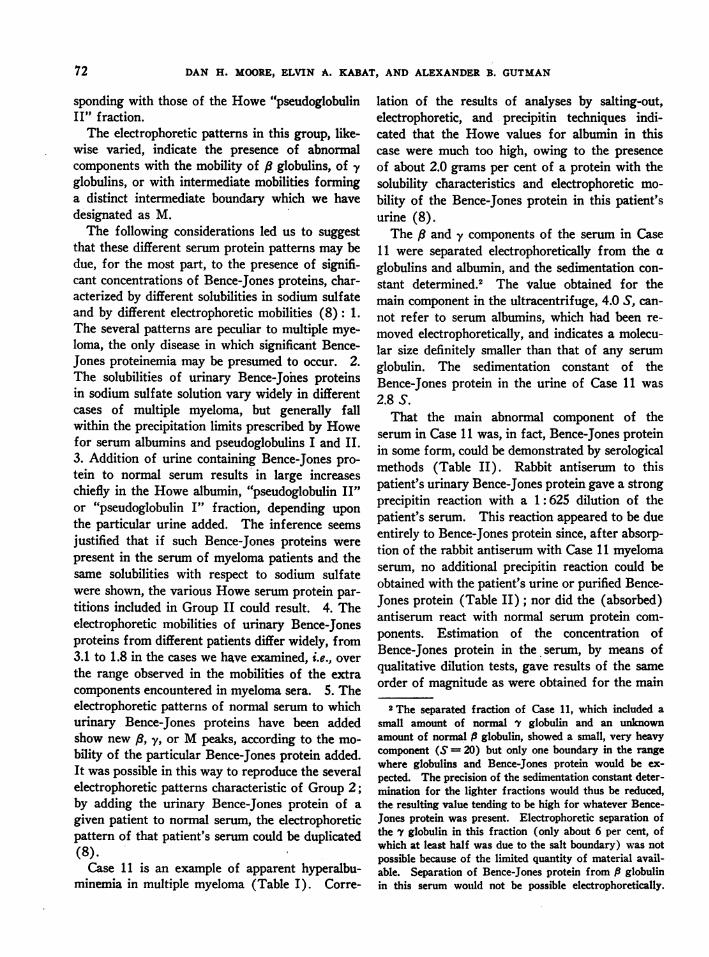
The following considerations led us to suggestthat these different serum protein patterns may bedue, for the most part, to the presence of signifi-cant concentrations of Bence-Jones proteins, char-acterized by different solubilities in sodium sulfateand by different electrophoretic mobilities (8): 1.The several patterns are peculiar to multiple mye-loma, the only disease in which significant Bence-Jones proteinemia may be presumed to occur. 2.The solubilities of urinary Bence-Jones proteinsin sodium sulfate solution vary widely in differentcases of multiple myeloma, but generally fallwithin the precipitation limits prescribed by Howefor serum albumins and pseudoglobulins I and II.3. Addition of urine containing Bence-Jones pro-tein to normal serum results in large increaseschiefly in the Howe albumin, "pseudoglobulin II"or "pseudoglobulin I" fraction, depending uponthe particular urine added. The inference seemsjustified that if such Bence-Jones proteins werepresent in the serum of myeloma patients and thesame solubilities with respect to sodium sulfatewere shown, the various Howe serum protein par-titions included in Group II could result. 4. Theelectrophoretic mobilities of urinary Bence-Jonesproteins from different patients differ widely, from3.1 to 1.8 in the cases we have examined, i.e., overthe range observed in the mobilities of the extracomponents encountered in myeloma sera. 5. Theelectrophoretic patterns of normal serum to whichurinary Bence-Jones proteins have been addedshow new /3, -y, or Mpeaks, according to the mo-bility of the particular Bence-Jones protein added.It was possible in this way to reproduce the severalelectrophoretic patterns characteristic of Group 2;by adding the urinary Bence-Jones protein of agiven patient to normal serum, the electrophoreticpattern of that patient's serum could be duplicated(8).
Case 11 is an example of apparent hyperalbu-minemia in multiple myeloma (Table I). Corre-
lation of the results of analyses by salting-out,electrophoretic, and precipitin techniques indi-cated that the Howe values for albumin in thiscase were much too high, owing to the presenceof about 2.0 grams per cent of a protein with thesolubility characteristics and electrophoretic mo-bility of the Bence-Jones protein in this patient'surine (8).
The /8 and y components of the serum in Case11 were separated electrophoretically from the aglobulins and albumin, and the sedimentation con-stant determined.2 The value obtained for themain component in the ultracentrifuge, 4.0 S, can-not refer to serum albumins, which had been re-moved electrophoretically, and indicates a molecu-lar size definitely smaller than that of any serumglobulin. The sedimentation constant of theBence-Jones protein in the urine of Case 11 was2.8 S.
That the main abnormal component of theserum in Case 11 was, in fact, Bence-Jones proteinin some form, could be demonstrated by serologicalmethods (Table II). Rabbit antiserum to thispatient's urinary Bence-Jones protein gave a strongprecipitin reaction with a 1: 625 dilution of thepatient's serum. This reaction appeared to be dueentirely to Bence-Jones protein since, after absorp-tion of the rabbit antiserum with Case 11 myelomaserum, no additional precipitin reaction could beobtained with the patient's urine or purified Bence-Jones protein (Table II); nor did the (absorbed)antiserum react with normal serum protein com-ponents. Estimation of the concentration ofBence-Jones protein in the serum, by means ofqualitative dilution tests, gave results of the sameorder of magnitude as were obtained for the main
2 The separated fraction of Case 11, which included asmall amount of normal -y globulin and an unknownamount of normal l globulin, showed a small, very heavycomponent (S = 20) but only one boundary in the rangewhere globulins and Bence-Jones protein would be ex-pected. The precision of the sedimentation constant deter-mination for the lighter fractions would thus be reduced,the resulting value tending to be high for whatever Bence-Jones protein was present. Electrophoretic separation ofthe yv globulin in this fraction (only about 6 per cent, ofwhich at least half was due to the salt boundary) was notpossible because of the limited quantity of material avail-able. Separation of Bence-Jones protein from P globulinin this serum would not be possible electrophoretically.
72

BENCE-JONES PROTEINEMIA IN MYELOMA
abnormal component by other methods. Quanti-tative precipitin determinations could not be madein this case because of the weak rabbit antiseraproduced and the limited amounts of urinaryBence-Jones protein available.
Case 41 showed moderate hyperglobulinemia,due to protein constituents which had the solu-bility characteristics in sodium sulfate of Howe's"pseudoglobulin II" fraction (Table I). Theelectrophoretic pattern revealed a large componentmoving with the mobility of f3 globulin (Figure 1).To eliminate the possibility that this might be alipoidal substance, the serum was extracted withalcohol-ether mixture at - 120 (24). The re-sulting electrophoretic pattern showed no signifi-cant change (Figure 1). The cholesterol contentof the serum did not exceed 118 mgm. per cent.
The ,8 and -y components of the serum in case41 were separated out electrophoretically and thesedimentation constant determined to be 3.0 S, afigure subject to the limits in precision already in-dicated. This value is consistent with a proteinof the molecular size of Bence-Jones protein.
No Bence-Jones protein was found in the urineof Case 41 on repeated examination. The absenceof Bence-Jones proteinuria in this patient withBence-Jones proteinemia suggests that the serumBence-Jones protein might have been present inthe circulating fluids in the form of some complex.Bott and Richards (25) have shown that the "in-tact" amphibian glomerular membrane is partiallypermeable to proteins of Svedberg's 35,200 molec-ular weight group; in the case of several Bence-Jones preparations tested, the glomerular filtrationvalues were 20 to 48 per cent. It is likely that themaximal pore diameter of the "intact" humanglomerular membrane is similarly sufficient forpartial passage of uncombined Bence-Jones pro-tein in serum, since Bence-Jones proteinuria with-out significant albuminuria is commonly observed.If so, the absence of Bence-Jones protein in theurine of patients who give evidence of appreciableconcentrations in the serum, and who have nor-mally permeable glomerular membranes (nomarked nitrogen retention), would imply that theBence-Jones protein might occur in the serum incomplexes of larger molecular size.
In the course of certain routine determinations
in the serum of Case 41, it was. observed that al-though the serum itself was clear, the trichloro-acetic acid filtrates obtained were cloudy, due tothe presence of a substance which could not beremoved by multiple filtration. (This phenome-non indeed gave the first intimation of anythingunusual about the then undiagnosed case.) Itdeveloped that if undiluted serum were used, per-fectly clear filtrates could be obtained with eithertungstic or trichloroacetic acids; whereas, if theserum were first brought to the usual dilutionswith water, none of the commonprotein precipita-tion gave clear filtrates. This unusual behaviorwould not be inconsistent with the presence of acomplex dissociable on dilution. The extreme la-bility of this complex precluded its further studyin an undissociated state.
The serum in Case 42 is unique in our experi-ence. It contained very large amounts of aglobulin which precipitated out with the Howe"pseudoglobulin II" fraction and which, migratingwith the mobility of 1.7, produced an M peakintermediate between the 8 and y components.The sedimentation constant of the electrophoretic-ally separated Mand -v components however was7.0 S, indicating a molecular size of normal yglobulins. Further studies of this constituent ofthe serum in Case 42 have been carried out byShapiro, Ross, and Moore (26). The main com-ponent of the serum in this instance was clearlynot Bence-Jones protein and the classification ofCase 42 in Group 2 is tentative.
Group 3. Case 43 presented normal Howe andelectrophoretic serum protein patterns (Table I)and is representative of the large number of casesof multiple myeloma that show no abnormalitieswhen studied by these methods. The urine inCase 43 contained large amounts of a Bence-Jonesprotein which was found to have a sedimentationconstant of 3.4 S.
It was pointed out elsewhere (8) that somemyeloma sera with normal Howe partitions mayshow abnormal electrophoretic patterns, charac-terized by a small M peak, presumably due toBence-Jones proteinemia. Immunological studies,which might,disclose further evidences of Bence-Jones proteinemia in apparently normal myelomasera, have not yet been made.
73
DAN H. MOORE, ELVIN A. KABAT, AND ALEXANDERB. GUTMAN
DISCUSSION
Bence-Jones proteins evidently must be presentin the serum of at least those cases of multiplemyeloma with Bence-Jones proteinuria, since, inthe absence of any indication of a renal origin, theBence-Jones protein must be transported by theblood from the source of origin to the kidneys.Even in cases with no excretion in the urine, thepossibility of significant Bence-Jones proteinemiais not excluded, because interaction in the serummay result in the formation of non-filterable com-plexes. The problem therefore is not so much todetermine whether Bence-Jones proteinemia everoccurs in myeloma, as to devise methods adequatefor its detection and measurement.
A major difficulty in the way of this objectivelies in the multiplicity of Bence-Jones proteins(18, 19, 22, 27, 28) and in their correspondinglyvaried properties, as illustrated by the differencesin solubility, electrophoretic mobility, sedimenta-tion rate, and serological properties observed inthe urinary Bence-Jones proteins in our cases ofmyeloma (8, and the present study). These dif-ferences, reflected in the varied serum proteinpatterns of patients with Bence-Jones proteinemia,necessitate a broad and flexible analytical ap-proach. This is particularly true in view of addeddifficulties due to the presence in myeloma serumof normal and abnormal proteins, usually in greatexcess, to the probable formation of various moreor less labile complexes or-tombinations in somecases, and to a variety of other causes.
It would appear, nevertheless, that the problemis not beyond the reach of integrated studies bysalting-out, electrophoretic, and ultracentrifugalmethods, supplemented by the quantitative pre-cipitin technique. Such, at least, is the indicationof the preliminary results recorded here.
With regard to the proposed classification ofserum protein patterns in multiple myeloma (8),.the present data support its general validity andusefulness. It need hardly be pointed out thatthe classification falls far short of the ideal analy-sis into homogenous components, and that it isincomplete. Too little is known about the occur-rence in myeloma serum of abnormal proteinsother than Bence-Jones protein, however, to war-rant further subdivision at this time.
SUMMARY
The serum and urinary proteins in 7 cases ofmultiple myeloma were investigated by correlatedsalting-out, electrophoretic, and ultracentrifugaltechniques, supplemented by immunological meth-ods in 2 cases.
In 2 cases with marked hyperglobulinemia dueto y components which precipitated out chieflywith the Howe "euglobulin" fraction, sedimenta-tion constants of the main component of theserum indicated a molecular size of the order ofmagnitude of normal -y globulins. Application ofthe quantitative precipitin technique to the serumof one of these cases further revealed the presenceof approximately 0.2 gram per cent Bence-Jonesprotein, a concentration too low for detection byother methods. A review of the available dataindicates that only a very small proportion of theprotein increment is Bence-Jones protein in many(probably the majority of) cases of multiple mye-loma with marked hyperproteinemia.
Two cases with sera showing abnormal /8 orM components, and unusual Howe partitions notencountered in diseases other than multiple mye-loma, were found by ultracentrifugal and sero-logical analysis to have Bence-Jones protein as thechief abnormal protein component of the serum.The available data suggest that marked Bence-Jones proteinemia does occur in multiple myeloma,probably in a lesser but clinically significant pro-portion of cases.
Two cases of multiple myeloma with markedhyperproteinemia were found to have large con-centrations of abnormal, not readily classifiableproteins with sedimentation constants indicatingan approximate molecular size of normal globulins.The serum in one case of multiple myeloma wasfound to have apparently normal Howe and elec-trophoretic serum protein patterns.
In spite of the difficulties involved in the detec-tion and measurement of Bence-Jones proteins inserum, integrated studies of the kind indicatedappear to afford a promising approach to theproblem.
BIBLIOGRAPHY
1. Bull, H. B., Protein structure. Advances in Enzy-mol., 1941, 1, 1.
2. Perlzweig, W. A., Delrue, G., and Geschickter, C.,
74
BENCE-JONES PROTEINEMIA IN MYELOMA
Hyperproteinemia associated with multiple mye-lomas. J. A. M. A., 1928, 90, 755.
3. Magnus-Levy, A., Multiple Myelome. VII. Euglobu-linamie. Zur Klinik und Pathologie. Amyloido-sis. Ztschr. f. klin. Med., 1933, 126, 62.
4. Cantarow, A., Bence-Jones proteinemia in multiplemyeloma. Am. J. M. Sc., 1935, 189, 425.
5. Kydd, D. M., Bence-Jones protein in serum. J. Biol.Chem., 1934, 107, 747.
6. McFarlane, A. S., The behaviour of pathological sera
in the ultracentrifuge. Biochem. J., 1935, 29, 1175.7. Packalen, T., A case of myeloma with spontaneously
crystallizing protein in blood serum and urine.Acta med. Scandinav., 1939, 100, 1.
8. Gutman, A. B., Moore, D. H., Gutman, E. B., Mc-Clellan, V., and Kabat, E. A., Fractionation ofserum proteins in hyperproteinemia, with specialreference to multiple myeloma. J. Clin. Invest.,1941, 20, 765.
9. Howe, P. E., The determination of proteins in blood-a micro method. J. Biol. Chem., 1921, 49, 109.
10. Tiselius, A., A new apparatus for electrophoreticanalysis of colloidal mixtures. Trans. FaradaySoc., 1937, 33, 524.
11. Longsworth, L. G., A modification of the Schlierenmethod for use in electrophoretic analysis. J. Am.Chem. Soc., 1939, 61, 529.
12. Moore, D. H., and Lynn, J., Electrophoretic measure-
ments on normal human plasma. J. Biol. Chem.,1941, 141, 819.
13. Longsworth, L. G., Shedlovsky, T., and MacInnes,D. A., Electrophoretic patterns of normal andpathological human blood serum and plasma. J.Exper. Med., 1939, 70, 399.
14. Kekwick, R. A., The serum proteins in multiple mye-
lomatosis. Biochem. J., 1940, 34, 1248.15. Bauer, J. H., and Pickels, E. G., An improved air-
driven type of ultracentrifuge for molecular sedi-mentation. J. Exper. Med., 1937, 65, 565.
16. Chiles, J. A., and Severinghaus, A. E., Hormone stud-ies with the ultracentrifuge. III. An applicationof Toepler's Schlieren method to the analyticalultracentrifuge. Rev. Scientific Instruments, 1940,11, 71.
17. Hektoen, L., Specific precipitin for Bence-Jones pro-tein. J. A. M. A., 1921, 76, 929.
18. Bayne-Jones, S., and Wilson, D. W., Immunologicalreactions of Bence-Jones proteins. II. Differencesbetween Bence-Jones proteins from various sources.Bull. Johns Hopkins Hosp., 1922, 33, 119.
19. Hektoen, L., and Welker, W. H., Immunological dif-ferences of crystalline Bence-Jones proteins. Bio-chem. J., 1940, 34, 487.
20. Heidelberger, M., and Kendall, F. E., A quantitativetheory of the precipitin reaction. III. The reactionbetween crystalline egg albumin and its homologousantibody. J. Exper. Med., 1935, 62, 697.
21. Goettsch, E., and Kendall, F. E., Analysis of albuminand globulin in biological fluids by the quantitativeprecipitin method. J. Biol. Chem., 1935, 109, 221.
22. Svedberg, T., and Pederson, K. O., The Ultracentri-fuge. Clarendon Press, Oxford, 1940.
23. Kabat, E. A., The molecular weight of antibodies.J. Exper. Med., 1939, 69, 103.
24. Wu, H., Effect of removal of lipoids on precipitabilityof serum proteins by neutral salts. Chinese J.Physiol., 1933, 7, 125.
25. Bott, P. A., and Richards, A. N., The passage ofprotein molecules through the glomerular mem-branes. J. Biol. Chem., 1941, 141, 291.
26. Shapiro, S., Ross, V., and Moore, D. H., A viscousprotein obtained in large amount from the serumof a patient with multiple myeloma. J. Clin. In-vest. (In press.)
27. Magnus-Levy, A., tYber krystallisiertes und amorphesBence-Jones Eiweiss. Multiple Myelome. Ztschr.f. physiol. Chem., 1936, 243, 173.
28. Devine, J., An analysis of Bence-Jones protein. Bio-chem. J., 1941, 35, 433.
75