MYELODYSPLASTIC AND MYELOPROLIFERATIVE DISORDERS
26
MYELODYSPLASTIC AND MYELOPROLIFERATIVE DISORDERS DISORDERS Pediatric Hemato-Oncology Division Medical Faculty Medical Faculty University of Sumatera Utara 1
Transcript of MYELODYSPLASTIC AND MYELOPROLIFERATIVE DISORDERS

MYELODYSPLASTIC AND MYELOPROLIFERATIVE
DISORDERSDISORDERS
Pediatric Hemato-Oncology DivisionMedical FacultyMedical Faculty
University of Sumatera Utara
1

MYELODYSPLASIA SYNDROME
• A group of disorder defect in hematopoetic cell development
• Progresses from dysplastic ineffective hematopoesis to aggressive overt myelogenous leukemia
2
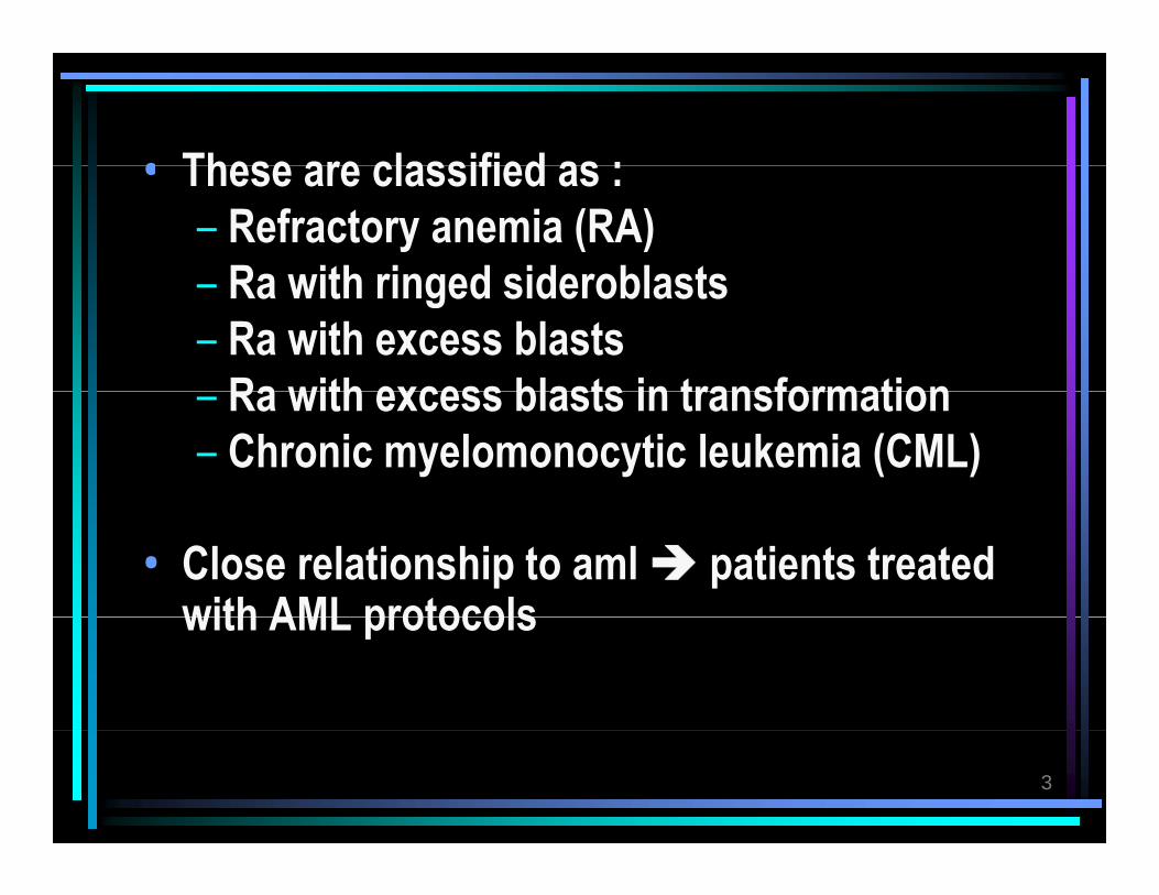
• These are classified as :• These are classified as :– Refractory anemia (RA)
Ra with ringed sideroblasts– Ra with ringed sideroblasts– Ra with excess blasts
Ra with excess blasts in transformation– Ra with excess blasts in transformation– Chronic myelomonocytic leukemia (CML)
• Close relationship to aml patients treated with AML protocolswith AML protocols
3

NORMAL BONE MARROW SMEAR, MAY-GIEMSA STAIN, X100
4
, ,TAKEN FROM: bcl.med.harvard.edu/.../proj/raspap/bm-rars.jpg
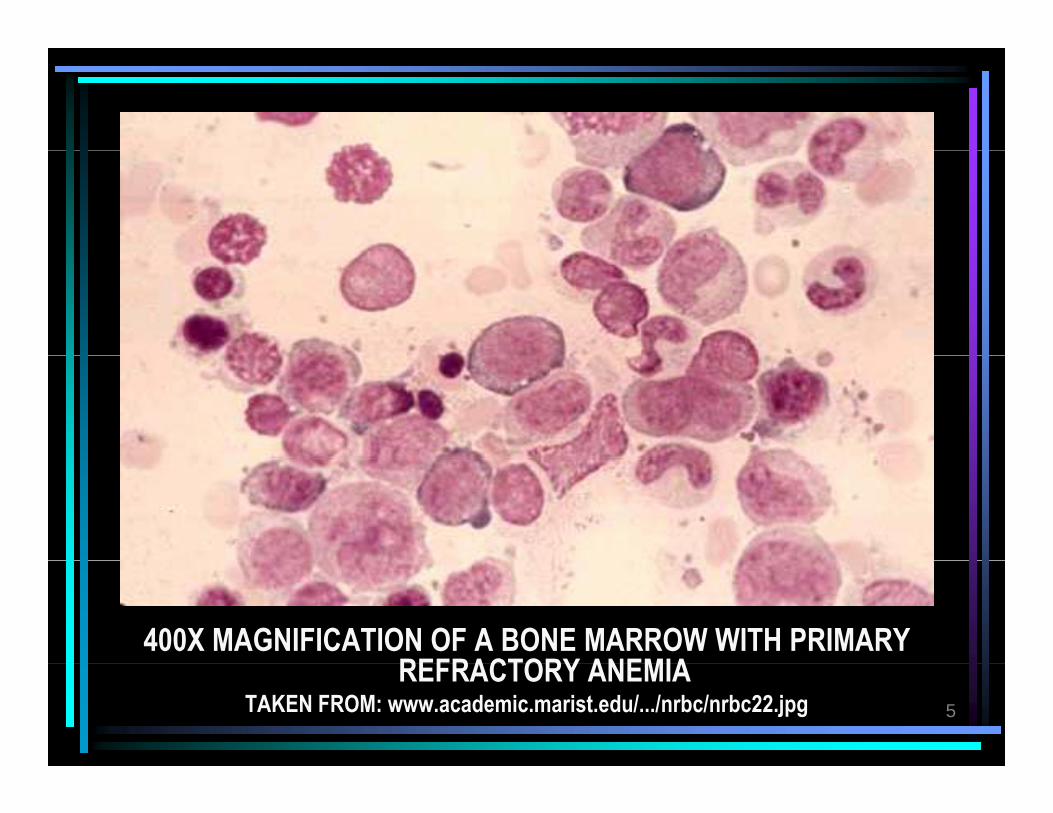
400X MAGNIFICATION OF A BONE MARROW WITH PRIMARY REFRACTORY ANEMIA
5
REFRACTORY ANEMIATAKEN FROM: www.academic.marist.edu/.../nrbc/nrbc22.jpg

REFRACTORY ANEMIA WITH RINGED SIDEROBLASTS (RARS) BONE MARROW SMEAR IRON STAIN X1000
6
(RARS) BONE MARROW SMEAR, IRON STAIN, X1000TAKEN FROM: bcl.med.harvard.edu/.../proj/raspap/bm-rars.jpg

REFRACTORY ANEMIA WITH EXCESS OF BLASTS,
7BONE MARROW SMEAR
TAKEN FROM: citylightsnews.com/.../raeb-excess_blasts.jpeg

REFRACTORY ANEMIA WITH EXCESS OF BLASTS IN TRANSFORMATION (RAEB-T) BONE MARROW SMEAR,
MAY GIEMSA STAIN X2008
MAY-GIEMSA STAIN, X200TAKEN FROM: bcl.med.harvard.edu/.../proj/raspap/bm-rars.jpg

CHRONIC MYELOMONOCYTIC LEUKEMIA PERIPHERAL BLOOD
9
PERIPHERAL BLOODTAKEN FROM: www.bekkoame.ne.jp/.../WBC/CMMoL/CMMoL-PB-H.jpg

ACUTE MYELOMONOCYTIC LEUKEMIA MICROSCOPIC View10
ACUTE MYELOMONOCYTIC LEUKEMIA - MICROSCOPIC ViewTAKEN FROM:medicalimages.allrefer.com/large/acute-myelom.

• These disorder are characterized by a single lineage myeloid proliferation that can progress to lineage myeloid proliferation that can progress to AML-like leukemia, include :– CMLCML– Essential thrombocytopenia (ET)– Policythemia Vera (PV)– Policythemia Vera (PV)– Agnogenic myeloid metaplasia
Juvenile myelomonocytic leukemia– Juvenile myelomonocytic leukemia
11

• Treatment :• Treatment :– Conservative management – If failed or diseases progress to AML-like
leukemic consider Stem cell transplantation
12

THROMBOCYTOSIS
• Thrombocyte count above the normal value for age
• Varies between platelet counts of > 400 - > 1000 x 109/l
• Thrombocytosis are classified as :– Mild : > 500 - < 700 x 109/ lMild : > 500 - < 700 x 10 / l– Moderate : 700 - 900 x 109/ l– Severe : > 900 x 109/ lSevere : > 900 x 10 / l– Extreme : > 1000 x 109/ l
13

• Cause of an increase in platelet count :• Cause of an increase in platelet count :
1 A primary disorder such as myelo proliferative 1. A primary disorder, such as myelo-proliferative or dysplastic syndrome ( An essential or primary thrombocytosis)thrombocytosis)
2 Increased production due to stimuli2. Increased production due to stimuli
3 A shift in platelets from the splenic Reactive or secondarythrombocytosis3. A shift in platelets from the splenic
pool into the peripheral circulationthrombocytosis
14

Table 1. differences between essential and reactive thrombocytosis
Essential (primary) Reactive (secondary)Age ( years)Duration
Mostly > 20, often > 40Over 2 years
Mostly < 20Days or weeks, sometimes
Origin
y
Stem cell defect
y ,monthsReaction to hypoxemia, infection, platelet loss; shift of platelet pool
Microvascular symptomsThrombosisBleeding
OftenOftenOften
of platelet poolExtremely rareExtremely rareExtremely rareBleeding
SplenomegalyPlatelet count ( x 109/l ) Platelet morphology
OftenOftenMostly > 1000Large, dysmorphic
Extremely rareRareMostly < 1000Large, normal appearance
Platelet functionPlatelet distributionIron storesAcute phase reagents such
DisturbedElevatedNormelNormal
NormalNormal widthLowHigh if thrombocytosis
15
Acute phase reagents such as IL-6, CRP, fibrinogen
Normal High, if thrombocytosis caused by infection

ESSENTIAL THROMBOCYTOSIS
• Rare in children and young or middle-aged adultsM t i th fifth i th d d f lif• Most common in the fifth or sixth decade of life
Cli i l if t tiClinical manifestation– persistent elevated platelet count > 1000x109 /l
S l li– Splenomegali– Recurrent bleeding
Microcirculatory disturbances : acrocyanosis – Microcirculatory disturbances : acrocyanosis, myocard infarction, transient ischemic attack (TIA)
– asypmtomatic16
asypmtomatic

Treatment
• Platelet lowering therapy ( e.g. hydoxyurea, busulfan, anagrelide, interferon, radioactive , g , ,phosphorous, platelet apheresis)
• Platelet aggregation inhibition ( e.g. aspirin and dipyridamole)
17

REACTIVE THROMBOCYTOSIS
Pathophysiology
• Stimulation of thrombocyte production after peripheral loss of thrombocyte loss of thrombocyte ( e.g. after immunologic, septic, oncogenic or traumatic events, blood loss or hypoxemia, of respiratory or events, blood loss or hypoxemia, of respiratory or cardiac origin )
• Shift of pool into the peripheral blood (exercise, stress, inj. Epinephrine and isoprenaline,
18
( j p p pasplenia)

Incidence Incidence
• More common than essential thrombocytosisMore common than essential thrombocytosis• Higher incidence in neonates, infants, and young
children children • Incidence between 6-13% in hospitalized children,
and 15% in pediatric outpatientsp p• ♂ = ♀• 78% mild thrombocytosis78% mild thrombocytosis• 15% moderate thrombocytosis• 7% severe thrombocytosis
19
7% severe thrombocytosis

Table 2. Conditions associated with reactive thrombocytosis ( predominantly in infants and young children)( predominantly in infants and young children)• Infection
RespiratoryM i itiMeningitisGastrointestinal
• Tissue damage ( surgery trauma)• Tissue damage ( surgery, trauma)
• Splenectomy
• HypoxemiaAnemia
Iron-deficiency anemiaIron deficiency anemiahemolytic anemiaAnemia due to blood lossAnemia caused by nephrotic disease
20
y pRespiratory diseaseCardiac hypoxemia

Table 2. Conditions associated with reactive thrombocytosis ( predominantly in infants and young children)( predominantly in infants and young children)
• Autoimmune diseaseJuvenile rheumatoid arthritisKawasaki syndromeHenoch-schoenlein disease
R l di• Renal disease
• MalignancyhepatoblastomahepatoblastomaHodgkin’s diseaseHistiocytosisSarcomaSarcomaAcute lymphoblastic leukemia and Non-hodgkin lymphoma
21• Prematurity

Table 2. Conditions associated with reactive thrombocytosis ( predominantly in infants and young children)( predominantly in infants and young children)
• Stress situation
• MedicationEpinephrineCorticosteroidVi lk l idVinca alkaloidsMiconazolePenicillamineMethadone ( during pregnancy)Methadone ( during pregnancy)Hydantoin ( during pregnancy )
• Miscellaneousgastroesophegeal refluxcafley’s disease
22
cafley s disease

ComplicationsComplications• Thrombosis• Headache• Confusion • Convulsions• Cerebral Infarction• Cerebral Infarction• Intracranial hemorrhage• Hemiparesis
23

Indications for prophylaxisIndications for prophylaxis
• Prophylaxis with anticoagulants or platelet p y g paggregation inhibitors if risk factors exist :– Immobilization in a castImmobilization in a cast– Leukemia
Alt ti f th l ti th b hili – Alterations of other plasmatic thrombophilic factors
24

Indications for prophylaxis– Iron-deficiency anemia
Indications for prophylaxis
– Cyanotic heart disease– Cardiac arrythmias after fontan surgeryy g y– Splenectomy for a myeloproliferative
syndrome or hematologic diseasesyndrome or hematologic disease– Post operative thrombocytosis after pancreas
transplantationtransplantation
25

26