
Mycotoxic nephropathy in pigs* - WHO
8
Bull. Org. nond. Sante 11973, 49, 411-418 Bull. Wld Hlth Org. Mycotoxic nephropathy in pigs* F. ELLING1 & T. M0LLER 2 In Denmark a nephropathy in pigs characterized by tubular atrophy and interstitial fibrosis has been identified frequently during the last S decades in the course of meat inspection in slaughterhouses. The disease was first described by Larsen, who recognized the connexion between feeding mouldy rye to pigs and the development ofthe nephropathy. In this study kidneys were examined from 19 pigs coming from a farm with an outbreak of nephropathy. The barley fed to the pigs was contaminated with the mycotoxin ochra- toxin A. Histological examination revealed different degrees of change ranging from slight regressive changes in the tubular epithelium and periglomerular and interstitial fibrosis to tubular atrophy, thickened basement membranes, glomerular sclerosis, and marked fibrosis. These differences were considered to be due to differences in the length of time of exposure to the mouldy barley and differences in the amount of mycotoxin consumed by the individual pig. However, it will be necessary to carry out experiments using crystal- line ochratoxin A in order to prove such a relationship. Mycotoxins have also been suggested as etiological factors in Balkan nephropathy in man, which in the initial stages is charac- terized by tubular lesions similar to those seen in mycotoxic nephropathy in pigs. A morphologically characteristic nephropathy in Danish Landrace pigs, characterized by tubular atrophy and interstitial fibrosis, was described by Larsen in 1928 (7). Since then this disease has been identified frequently in the course of routine meat inspection of the approximately 10 million pigs slaughtered in Denmark each year. The frequency of this disease has been found to vary from year to year, being especially high following wet harvesting periods. Recently, a central register of this porcine nephro- pathy has been set up, covering all slaughterhouses in Denmark. The registration data indicate an inci- dence of 67 per 100 000 for 1971 but only 27 per 100 000 for 1972. In addition, considerable district- to-district variation was observed (P. Krogh, person- al communication, 1972). Recently, a similar neph- ropathy in pigs was described in Ixeland by Buckley (2). Svenkerud (personal communication, 1972), and Bj6rklund (personal communication, 1972) suggested that a similar condition occurred in Nor- way and Sweden respectively but presumably with a lower frequency. In Germany a chronic nepbro- * From the Royal Veterinary and Agricultural Univer- sity, Bulowsvej 13, 1870 Copenhagen V, Denmark. Department of Pathology. 'Professor, Department of Pathology. pathy, nephritis fibrovesiculosa, was described by Kitt (3); Larsen (7) suggested that this condition represented an advanced stage of the same disease that he had described in the Danish pigs. Clinical observations of field cases have been scarce and imprecisely reported. Failure to thrive was the most commonly observed clinical sign of the condition, which results in retarded growth. In older sows and boars the kidney lesions occasionally cause clinical symptoms suggesting renal insufficiency. MATERIAL AND METHODS The description given here of the morphology of this porcine nephropathy is based on a study of field cases from a herd in which an outbreak had occurred. After weaning, the pigs were fed mouldy barley for a period of unknown duration until reaching the normal weight at slaughter, i.e., approximately 90 kg. Chemical analysis revealed that the mouldy barley was contaminated with a mycotoxin, ochratoxin A.a Kidneys from 19 pigs were collected at the abattoir. For histological exami- nation the kidneys were fixed in 10% buffered for- malin within 3 h of slaughter. The material for a (R)-N-[(5-chloro-3,4-dihydro-8-hydroxy-3-methyl-1-oxo- 1H-2-benzopyran-7-yl)carbonyl]phenylalanine. 3135 - 411 - 7
Transcript of Mycotoxic nephropathy in pigs* - WHO

Bull. Org. nond. Sante 11973, 49, 411-418Bull. Wld Hlth Org.
Mycotoxic nephropathy in pigs*F. ELLING1 & T. M0LLER 2
In Denmark a nephropathy in pigs characterized by tubular atrophy and interstitialfibrosis has been identified frequently during the last S decades in the course of meatinspection in slaughterhouses. The disease was first described by Larsen, who recognizedthe connexion betweenfeeding mouldy rye to pigs and the development ofthe nephropathy. Inthis study kidneys were examined from 19 pigs coming from a farm with an outbreakof nephropathy. The barley fed to the pigs was contaminated with the mycotoxin ochra-toxin A. Histological examination revealed different degrees of change rangingfrom slightregressive changes in the tubular epithelium and periglomerular and interstitial fibrosisto tubular atrophy, thickened basement membranes, glomerular sclerosis, and markedfibrosis. These differences were considered to be due to differences in the length of timeof exposure to the mouldy barley and differences in the amount of mycotoxin consumedby the individual pig. However, it will be necessary to carry out experiments using crystal-line ochratoxin A in order to prove such a relationship. Mycotoxins have also been suggestedas etiological factors in Balkan nephropathy in man, which in the initial stages is charac-terized by tubular lesions similar to those seen in mycotoxic nephropathy in pigs.
A morphologically characteristic nephropathy inDanish Landrace pigs, characterized by tubularatrophy and interstitial fibrosis, was described byLarsen in 1928 (7). Since then this disease has beenidentified frequently in the course of routine meatinspection of the approximately 10 million pigsslaughtered in Denmark each year. The frequencyof this disease has been found to vary from year toyear, being especially high following wet harvestingperiods.
Recently, a central register of this porcine nephro-pathy has been set up, covering all slaughterhousesin Denmark. The registration data indicate an inci-dence of 67 per 100 000 for 1971 but only 27 per100 000 for 1972. In addition, considerable district-to-district variation was observed (P. Krogh, person-al communication, 1972). Recently, a similar neph-ropathy in pigs was described in Ixeland by Buckley(2). Svenkerud (personal communication, 1972),and Bj6rklund (personal communication, 1972)suggested that a similar condition occurred in Nor-way and Sweden respectively but presumably witha lower frequency. In Germany a chronic nepbro-
* From the Royal Veterinary and Agricultural Univer-sity, Bulowsvej 13, 1870 Copenhagen V, Denmark.
Department of Pathology.'Professor, Department of Pathology.
pathy, nephritis fibrovesiculosa, was described byKitt (3); Larsen (7) suggested that this conditionrepresented an advanced stage of the same diseasethat he had described in the Danish pigs. Clinicalobservations of field cases have been scarce andimprecisely reported. Failure to thrive was the mostcommonly observed clinical sign of the condition,which results in retarded growth. In older sows andboars the kidney lesions occasionally cause clinicalsymptoms suggesting renal insufficiency.
MATERIAL AND METHODS
The description given here of the morphology ofthis porcine nephropathy is based on a study offield cases from a herd in which an outbreak hadoccurred. After weaning, the pigs were fed mouldybarley for a period of unknown duration untilreaching the normal weight at slaughter, i.e.,approximately 90 kg. Chemical analysis revealedthat the mouldy barley was contaminated with amycotoxin, ochratoxin A.a Kidneys from 19 pigswere collected at the abattoir. For histological exami-nation the kidneys were fixed in 10% buffered for-malin within 3 h of slaughter. The material for
a (R)-N-[(5-chloro-3,4-dihydro-8-hydroxy-3-methyl-1-oxo-1H-2-benzopyran-7-yl)carbonyl]phenylalanine.
3135 - 411 - 7
412 F. ELLING & T. M0LLER
histological examination was embedded in paraffin,sectioned (5 ,tm), and stained with haematoxylin-eosin, iron haematoxylin-van Gieson, and PAS.
RESULTS
The kidneys were divided into 3 groups accordingto the degree of interstitial fibrosis of the cortex.
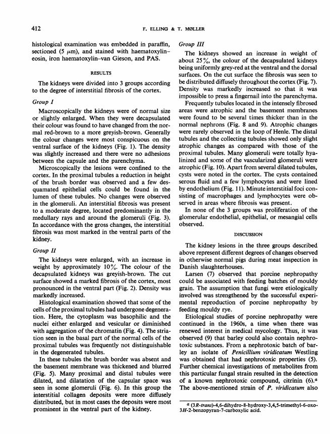
Group IMacroscopically the kidneys were of normal size
or slightly enlarged. When they were decapsulatedtheir colour was found to have changed from the nor-mal red-brown to a more greyish-brown. Generallythe colour changes were most conspicuous on theventral surface of the kidneys (Fig. 1). The densitywas slightly increased and there were no adhesionsbetween the capsule and the parenchyma.
Microscopically the lesions were confined to thecortex. In the proximal tubules a reduction in heightof the brush border was observed and a few des-quamated epithelial cells could be found in thelumen of these tubules. No changes were observedin the glomeruli. An interstitial fibrosis was presentto a moderate degree, located predominantly in themedullary rays and around the glomeruli (Fig. 3).In accordance with the gross changes, the interstitialfibrosis was most marked in the ventral parts of thekidney.
Group IIThe kidneys were enlarged, with an increase in
weight by approximately 10%. The colour of thedecapsulated kidneys was greyish-brown. The cutsurface showed a marked fibrosis of the cortex, mostpronounced in the ventral part (Fig. 2). Density wasmarkedly increased.
Histological examination showed that some of thecells ofthe proximal tubules had undergone degenera-tion. Here, the cytoplasm was basophilic and thenuclei either enlarged and vesicular or diminishedwith aggregation of the chromatin (Fig. 4). The stria-tion seen in the basal part of the normal cells of theproximal tubules was frequently not distinguishablein the degenerated tubules.
In these tubules the brush border was absent andthe basement membrane was thickened and blurred(Fig. 5). Many proximal and distal tubules weredilated, and dilatation of the capsular space wasseen in some glomeruli (Fig. 6). In this group theinterstitial collagen deposits were more diffuselydistributed, but in most cases the deposits were mostprominent in the ventral part of the kidney.
Group IIIThe kidneys showed an increase in weight of
about 25%, the colour of the decapsulated kidneysbeing uniformly grey-red at the ventral and the dorsalsurfaces. On the cut surface the fibrosis was seen tobe distributed diffusely throughout the cortex (Fig. 7).Density was markedly increased so that it wasimpossible to press a fingernail into the parenchyma.
Frequently tubules located in the intensely fibrosedareas were atrophic and the basement membraneswere found to be several times thicker than in thenormal nephrons (Fig. 8 and 9). Atrophic changeswere rarely observed in the loop of Henle. The distaltubules and the collecting tubules showed only slightatrophic changes as compared with those of theproximal tubules. Many glomeruli were totally hya-linized and some of the vascularized glomeruli wereatrophic (Fig. 10). Apart from several dilated tubules,cysts were noted in the cortex. The cysts containedserous fluid and a few lymphocytes and were linedby endothelium (Fig. 1 1). Minute interstitial foci con-sisting of macrophages and lymphocytes were ob-served in areas where fibrosis was present.
In none of the 3 groups was proliferation of theglomerular endothelial, epithelial, or mesangial cellsobserved.
DISCUSSION
The kidney lesions in the three groups describedabove represent different degrees of changes observedin otherwise normal pigs during meat inspection inDanish slaughterhouses.
Larsen (7) observed that porcine nephropathycould be associated with feeding batches of mouldygrain. The assumption that fungi were etiologicallyinvolved was strengthened by the successful experi-mental reproduction of porcine nephropathy byfeeding mouldy rye.
Etiological studies of porcine nephropathy werecontinued in the 1960s, a time when there wasrenewed interest in medical mycology. Thus, it wasobserved (9) that barley could also contain nephro-toxic substances. From a nephrotoxic batch of bar-ley an isolate of Penicillium viridicatum Westlingwas obtained that had nephrotoxic properties (5).Further chemical investigations of metabolites fromthis particular fungal strain resulted in the detectionof a known nephrotoxic compound, citrinin (6)."The above-mentioned strain of P. viridicatum also
a (3R-trans)-4,6-dihydro-8-hydroxy-3,4,5-trimethyl-6-oxo-3H-2-benzopyran-7-carboxylic acid.

Fig. 1. Kidney from Group I. The colour is greyish-brown. The transverse sectionshows the predominance of the interstitial fibrosis in the ventral cortex (transversesection: ventral aspect upwards).
Fig. 2. Kidney from Group II. The colour is greyish-brown and the predominance of theinterstitial fibrosis in the ventral cortex is seen (transverse section: ventral aspectdownwards).
;I,:'

AiFig. 4. Kidney from Group 11. The arrow indicates an epithelial cellwith an enlarged and vesicular nucleus in a proximal tubule (haema-toxylin-eosin).
4Fig. 3. Periglomerular and(PAS).
interstitial fibrosis in kidney from Group I
Fig. 5. Kidney from Group I. Proximal tubule showing thickenedbasement membrane (PAS).
Fig. 6. Kidney from Group I. Interstitial fibrosis in the ventral cortex(haematoxylin -eosin). ....

Fig. 7. Kidney from Group Ill. The colour is greyish-red and the diffusely distributed cortical fibrosis is visible(transverse section: ventral aspect downwards).
Fig. 9. Kidney from Group Ill. Periglomerular and interstitial fibrosis;thickened basement membranes (PAS).
4Fig. 8. Kidney from Group Ill. Interstitial fibrosis and cyst formation(haematoxylin -eosin).

Fig. 10. Kidney from Group Ill. Sclerotic glomerulus (haematoxylin-eosin).
1-M,, _`
1 *I-' -;IFig. 11. Kidney from Group Ill. Cyst containing serous fluid and a few lymphocytesand lined by endothelium (haematoxylin-eosin).
MYCOTOXIC NEPHROPATHY IN PIGS 417
produces ochratoxin A, another known nephrotoxicmycotoxin (11). Citrinin and ochratoxin have beenisolated from many species of Aspergillus and Peni-cillium, as stated in a recent review (4). Citrinin andochratoxin have been reported as natural contami-nants of cereals and other crops in the USA (14),Canada (13), and Europe (15). The last report asso-ciates the natural occurrence of citrinin and ochra-toxin in barley and oats with field cases of porcinenephropathy.The 19 pigs in this study were of approximately
the same weight, and therefore of approximately ofthe same age, when they were slaughtered. As thegrain fed to these pigs was contaminated withochratoxin A, differences in the extent of changescould be due to differences in the length of time ofexposure to the mouldy barley and in the amountof mycotoxin consumed by each pig.aThe changes consisted of a progressive interstitial
fibrosis and regressive tubular changes with thicken-ing of the basement membranes. In the late stages,atrophy of the nephrons and dilatation of tubuleswere observed. In this stage several cysts wereobserved in the cortex; as they contained serousfluid and lymphocytes and were lined with endo-thelium they were considered to be dilated lymphatics.
In the present study it was not possible to clarifywhy, in the lower grades of the nephropathy, thefibrous tissue formation and the atrophic changeswere found mainly in the ventral part of the kidney.The slight infiltration of lymphocytes and histio-
a This suggestion has now been proved experimentally.See: Krogh, P. et al. Experimental porcine nephropathyinduced by ochratoxin A-contaminated feed: changes ofrenal function and structure. Acta path. microbiol. Scand.(Section A), 1974 (in press).
cytes in the later stages was not considered to be ofany significance. Also, vascular lesions or changesin the juxtaglomerular apparatus did not seem toplay any pathogenic role.
Mycotoxins have also been suggested as possibleetiological factors in Balkan nephropathy in man (1).The initial stage of this nephropathy is characterizedby regressive changes in the tubular epithelium (12)morphologically similar to the changes seen in myco-toxic nephropathy in pigs. The lesions in the latestages, however, differ from those found in pigs atthe time of slaughter. Whether pigs would developsimilar lesions, e.g., contracted kidneys, if exposed toochratoxin A for a longer period remains to bedemonstrated. Appropriate experiments are beingplanned.The nephrotoxic properties of ochratoxin A have
also been demonstrated in rats (11) and in chicks (10)and it is reasonable to assume that this substancemay be toxic to man. When pigs are fed horsebeans contaminated with ochratoxin A, residualamounts of the mycotoxin can be found in the kid-neys, liver, and muscles (8). However, ochratoxin Ahas not to date been reported to cause renal lesionsin man.
CONCLUSION
Based on previous observations and the fact thatthe grain fed to the pigs in this study was conta-minated with ochratoxin A, a relationship betweenthis nephrotoxic substance and the renal lesions issuggested. In order to prove such a relationship,however, it will be necessary to carry out furtherexperimental studies including a feeding experimentwith crystalline ochratoxin A. Such experiments arein preparation.
RITSUMEC
NEPHROPATHIE MYCOTOXIQUE CHEZ DES PORCS
Au Danemark, une nephropathie du porc, decrite pourla premiere fois en 1928, et associee A la consommation dec6reales moisies, a ete identifi6e A plusieurs reprises lors del'inspection des viandes dans les abattoirs.La presente etude est consacree A la description des
aspects anatomo-pathologiques de I'affection, basee surl'examen des reins de 19 porcs provenant d'une ferme ou lan6phropathie etait signalee. L'orge moisi donne auxanimaux 6tait contamine par une mycotoxine, l'ochra-toxine A, substance nephrotoxique produite par diversessouches d'Aspergillus et de Penicillium, et notammentPenicillium viridicatum Westling.
L'examen a montr6 trois stades diff6rents d'atteinte
renale, allant de legeres modifications de la structure de1'epithelium tubulaire, avec fibrose interstitielle et peri-glom6rulaire, A I'atrophie tubulaire, avec 6paississementde la couche sous-epitheliale, sclerose glomerulaire etfibrose accentuee. Les animaux etant approximativementdu meme poids et du meme Age lorsqu'ils ont e abattus,ces differences sont attribuees A une dur6e variable de laconsommation d'orge moisi et aux quantites variables demycotoxine absorbees par chaque porc. On envisage, pourconfirmer cette hypothese, de procdder A une etudeexperimentale en utilisant de l'ochratoxine A sous formecristalline.On a aussi attribue A des mycotoxines un role etiolo-

418 F. ELLING & T. M0LLER
gique dans la nephropathie des Balkans, maladie humainepr6sentant, au stade initial, des 1lsions similaires a celles dela nephropathie mycotoxique du porc mais, au stadeterminal, des lesions diff6rentes. Des experiences
sont prevues afin de voir si, exposes i l'ochratoxine Apendant une periode plus longue, les porcs seront por-teurs de l6sions renales analogues A celles observees chezl'homme.
REFERENCES
1. BARNES, J. M. In: Ciba Foundation Study GroupNo. 30, 11 May 1967, London, Ciba Foundation,1967, p. 111.
2. BUCKLEY, H. G. Irish Vet., J. 25, (10): 194 (1971).3. Krrr, TH. Lehrbuch der pathologischen Anatomie
der Haustiere, 1906.4. KROGH, P. J. gen. Microbiol., 73 (3): xxxiv (1972).5. KROGH, P. & HASSELAGER, E. Vet. Agric. Col.
Yearbook, 1968, p. 198.6. KROGH, P. ET AL. Acta path. microbiol. scand. (Sec-
tion B), 78 (4): 401 (1970).7. LARSEN, S. Mdnedsskr. Dyrl., 40: 259 (1928).8. MADSEN, A. ET AL. Ugeskrift for agronomer og horto-
nomer, 6: 100 (1973).
9. NIELSEN, H. E. & HASSELAGER, E. Forsogslab. drbog,1965, p. 91.
10. PECKHAM, J. C. ET AL. Appl. Microbiology, 21: 492(1971).
11. PURCHASE, 1. F. H. & THERON, J. J. Food Cosmet.Toxicol., 6: 479 (1968).
12. SCHOURUP, K. In: Ciba Foundation Study GroupNo. 30, 11 May 1967, London, Ciba Foundation,1967, p. 100.
13. ScoTr, P. M. ET AL. Agr. Food Chem., 20 (6): 1103(1972).
14. SHOTWELL, 0. L. ET AL. Appl. Microbiol., 17 (5):765 (1969).
15. KROGH, P. ET AL. Acta path. microbiol. scand.(Section B), 81 (b): 689 (1973).