Mycobacteria: Tuberculosis and Leprosy Andrew Racette MS IV.
97
Mycobacteria: Tuberculosis and Leprosy Andrew Racette MS IV
-
date post
21-Dec-2015 -
Category
Documents
-
view
218 -
download
0
Transcript of Mycobacteria: Tuberculosis and Leprosy Andrew Racette MS IV.
Mycobacteria: Tuberculosis and Leprosy
Andrew Racette MS IV
TuberculosisEpidemiology
Estimated 1.7 billion infected persons1/3 of world’s population10 million people in US
12 million new cases per year w/ 3 million deaths4 million co-infected with HIV
¾ live in sub-Saharan Africa
Incidence tied to poverty, unemployment, homelessness, AIDS and drug resistanceMulti-drug resistant disease (MDRTB) major problem
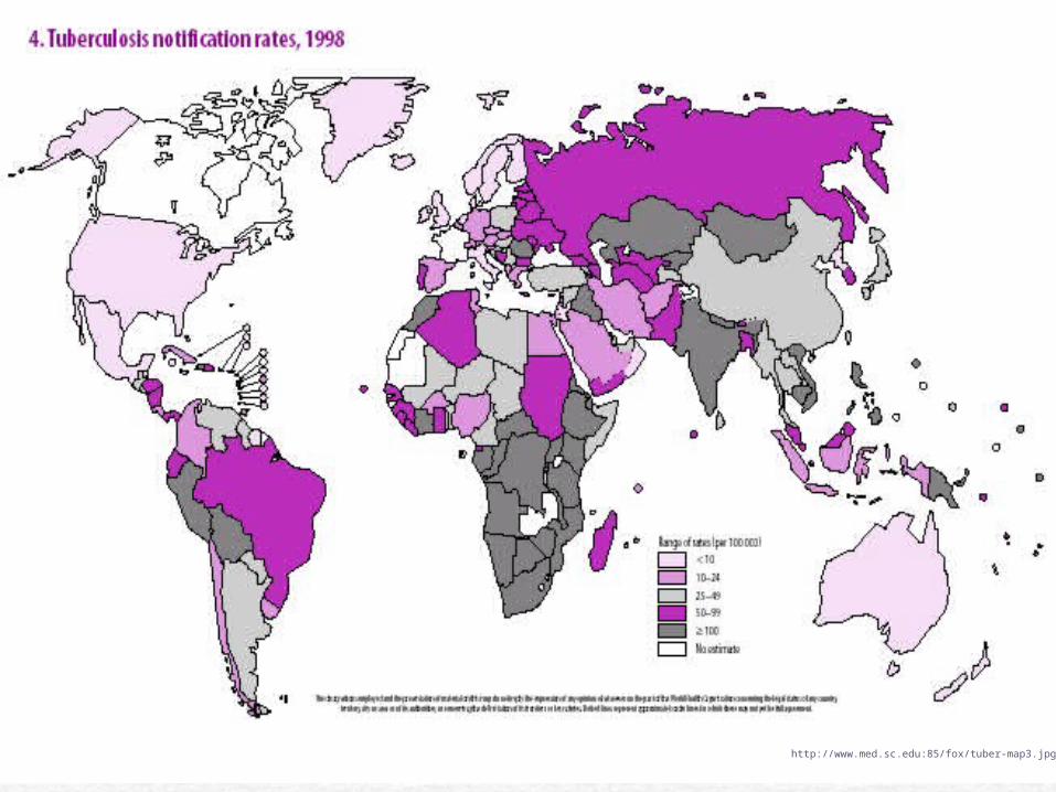
http://www.med.sc.edu:85/fox/tuber-map3.jpg
TuberculosisEtiology
Mycobacterium tuberculosis (Tubercle bacillus, MTB), M. bovis, M. africanum and BCGImmune response contains infection in majority
5-10% of immunocompetent develop clinical disease
Rarely eradicated due to resistance to macrophage destruction, dormancy within granulomasDormant bacilli resistant to antimycobacterialsImmunosuppression often leads to clinical sx
TuberculosisEtiology
MTB Surface CoatMycolic acid
Highly inflammatory
Stimulates Macrophages and T lymphs
Adequate control depends on chronic inflammation and caseating granulomas
Granulomas depends on Interferon (IFN) gamma & IL 12
Genetic component
TuberculosisSymptoms
Pulmonary: SOB
Sputum production
Systemic: Fatigue
Malaise
Fever (in ddx for FUO)
Lethargy
Weight loss
TuberculosisSymptoms
Disseminated Disease: Miliary pattern on CXR
Pancytopenia (BM)
Other Sites: Bones (Potts), GI, brain, meninges
Almost any organ
Asymptomatic in large number of persons90%
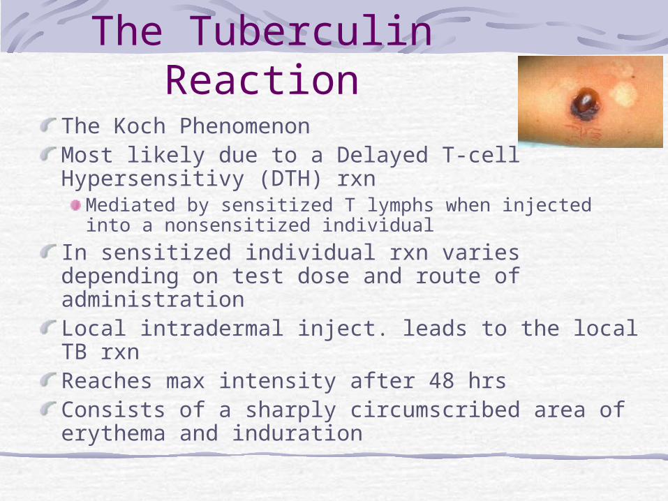
The Tuberculin ReactionThe Koch PhenomenonMost likely due to a Delayed T-cell Hypersensitivy (DTH) rxn
Mediated by sensitized T lymphs when injected into a nonsensitized individual
In sensitized individual rxn varies depending on test dose and route of administrationLocal intradermal inject. leads to the local TB rxnReaches max intensity after 48 hrsConsists of a sharply circumscribed area of erythema and induration
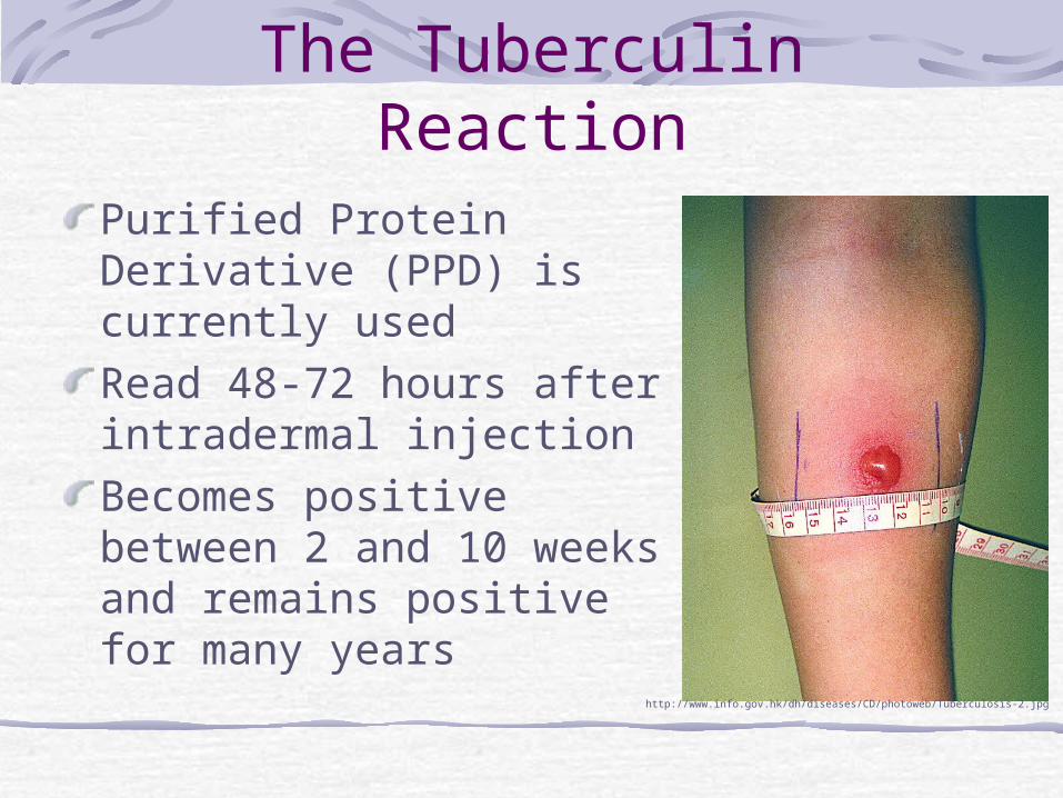
The Tuberculin ReactionPurified Protein Derivative (PPD) is currently usedRead 48-72 hours after intradermal injectionBecomes positive between 2 and 10 weeks and remains positive for many years http://www.info.gov.hk/dh/diseases/CD/photoweb/Tuberculosis-2.jpg
PPD evaluation0.1ml of PPD (5U) placed intradermally to form a wheal
Measure true induration (not erythema) 48-72 hrs >5mm Induration is positive in following hosts:
patients with recent close contact with a person with active TB patients with fibrotic lesions on chest radiograph patients with known or suspected HIV infection
>10mm Induration is positive in:Patients with high risk comorbid conditionsPersons from endemic areasIVDAResidents of long-term (chronic) care facilities
>15mm required for positivity in normal hosts
Previous BCG vaccination does not alter PPD
TB HistopathologyTubercle is the hallmark
Accumulation of epithelioid histiocytes with Langerhans giant cells
Caseation necrosis in the center
Rim of lymphs & monos
The tuberculioid granuloma is characteristic but NOT pathognomonic
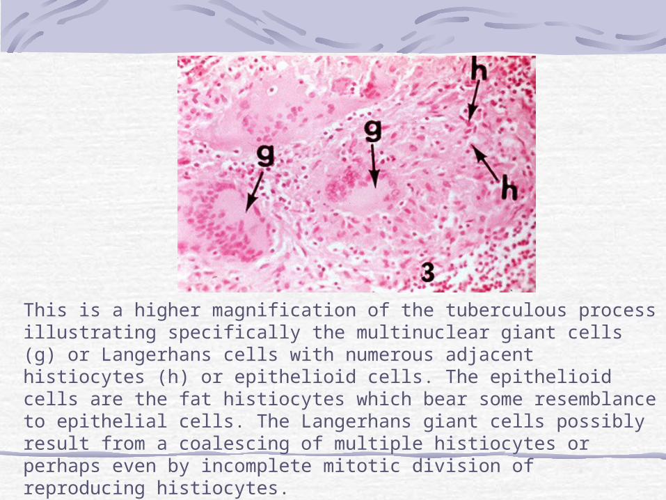
This is a higher magnification of the tuberculous process illustrating specifically the multinuclear giant cells (g) or Langerhans cells with numerous adjacent histiocytes (h) or epithelioid cells. The epithelioid cells are the fat histiocytes which bear some resemblance to epithelial cells. The Langerhans giant cells possibly result from a coalescing of multiple histiocytes or perhaps even by incomplete mitotic division of reproducing histiocytes.
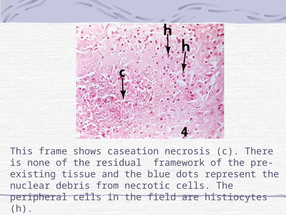
This frame shows caseation necrosis (c). There is none of the residual framework of the pre-existing tissue and the blue dots represent the nuclear debris from necrotic cells. The peripheral cells in the field are histiocytes (h).
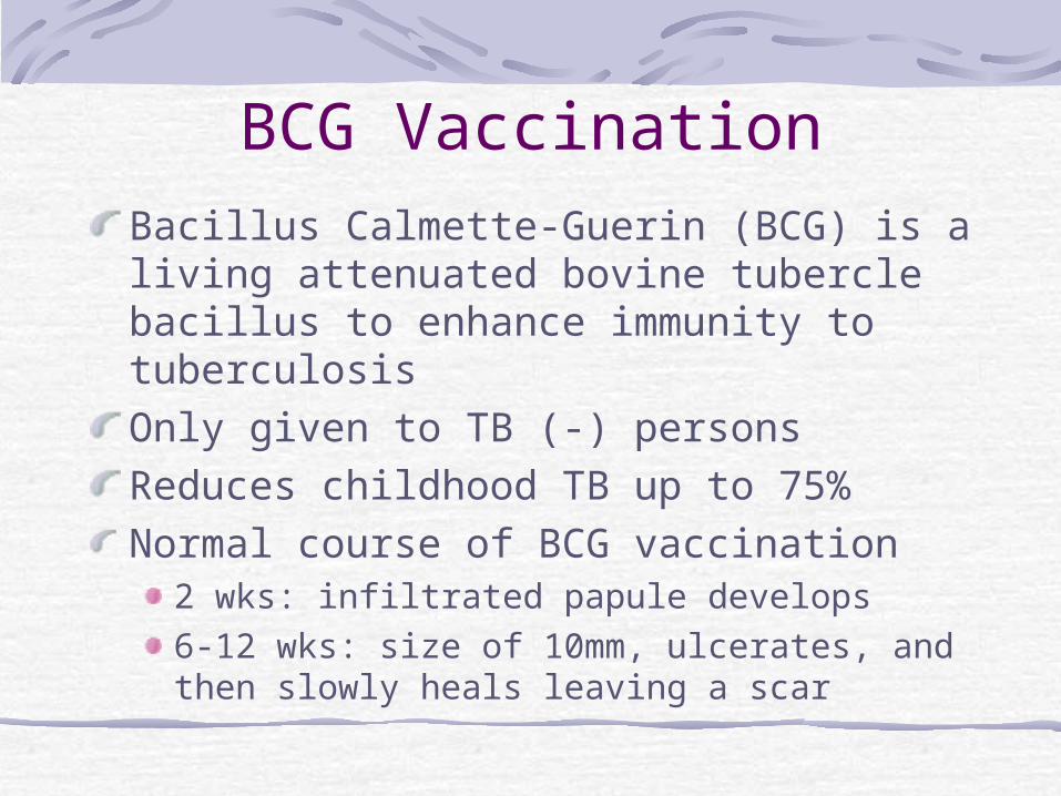
BCG VaccinationBacillus Calmette-Guerin (BCG) is a living attenuated bovine tubercle bacillus to enhance immunity to tuberculosisOnly given to TB (-) personsReduces childhood TB up to 75%Normal course of BCG vaccination
2 wks: infiltrated papule develops
6-12 wks: size of 10mm, ulcerates, and then slowly heals leaving a scar
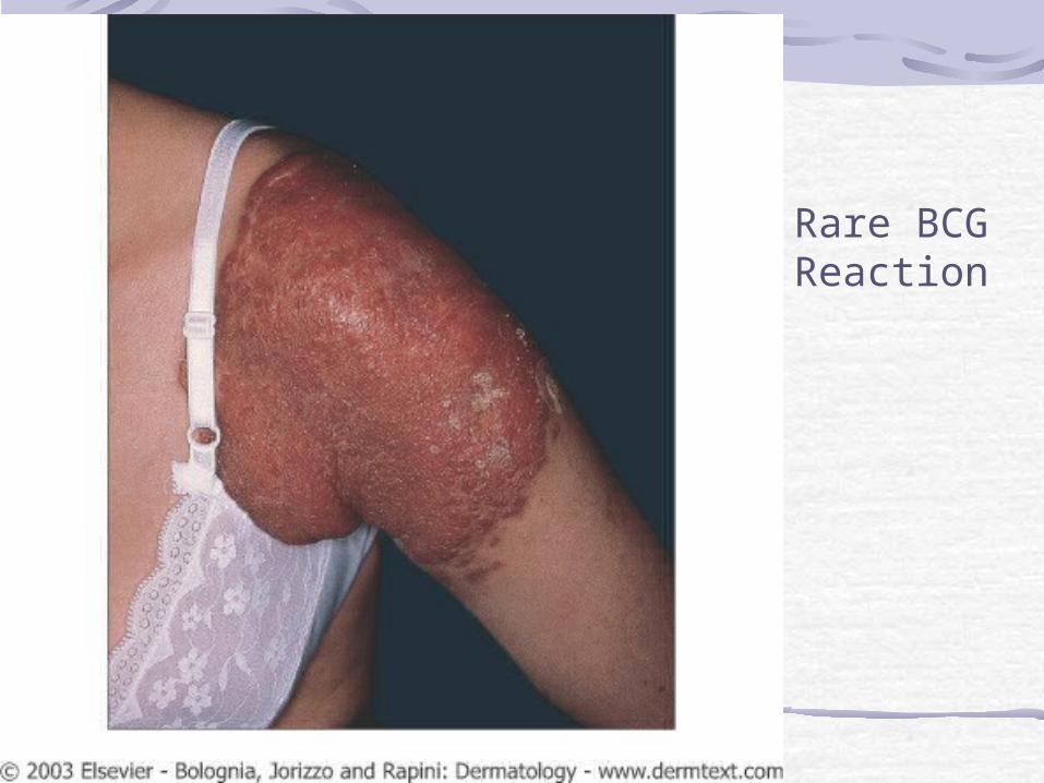
Rare BCG Reaction
Tuberculosis
Four categories of cutaneous tuberculosis
1. Inoculation from an exogenous sourse2. Endogenous cutaneous spread3. Hematogenous spread to the skin4. Tuberculids
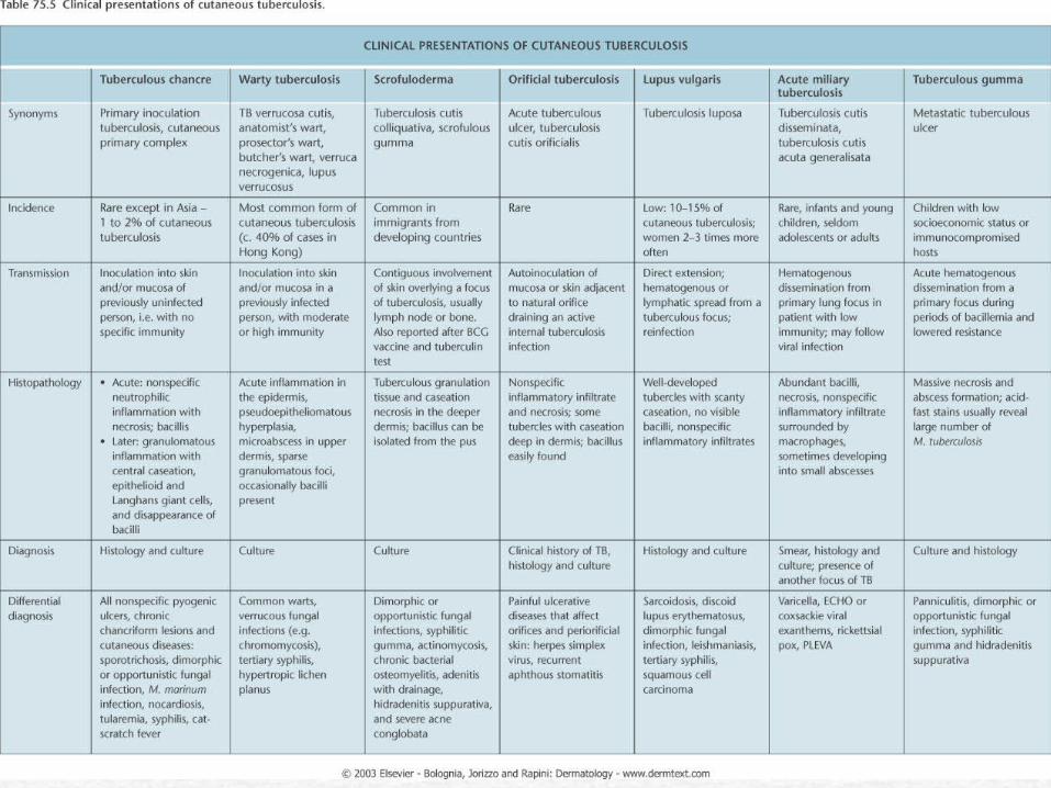
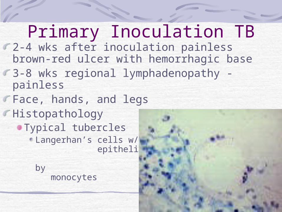
Primary Inoculation TB2-4 wks after inoculation painless brown-red ulcer with hemorrhagic base3-8 wks regional lymphadenopathy - painlessFace, hands, and legsHistopathology
Typical tubercles Langerhan’s cells w/ epithelioid cells surrounded by monocytes
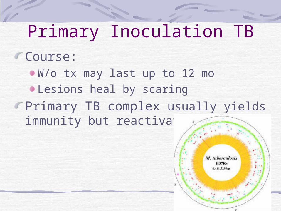
Primary Inoculation TBCourse:
W/o tx may last up to 12 mo
Lesions heal by scaring
Primary TB complex usually yields immunity but reactivation my occur
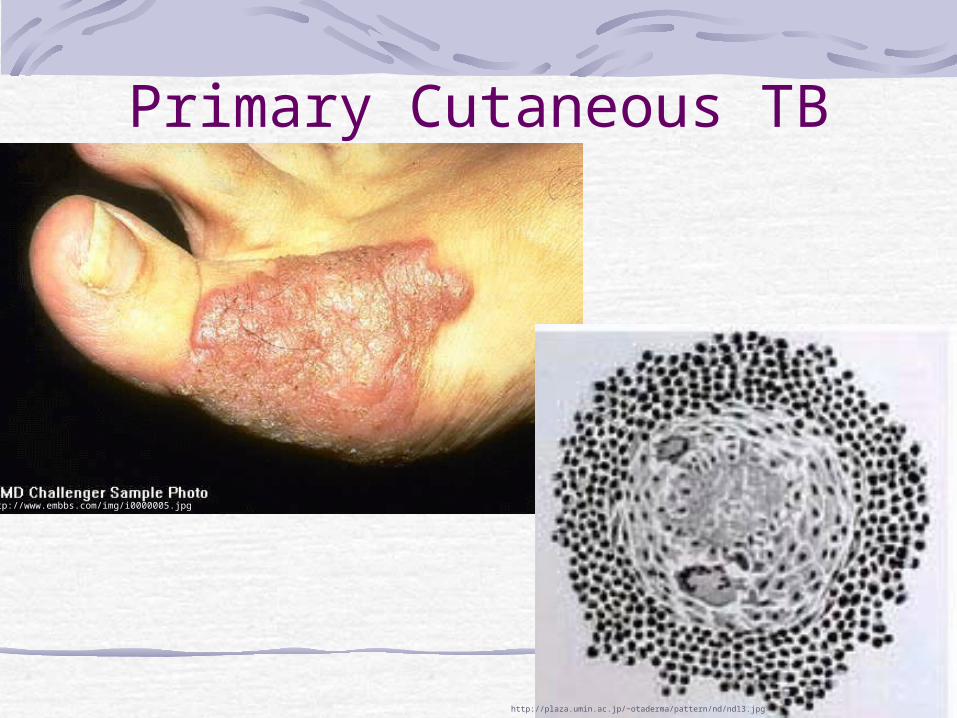
Primary Cutaneous TB
http://www.embbs.com/img/i0000005.jpg
http://plaza.umin.ac.jp/~otaderma/pattern/nd/nd13.jpg
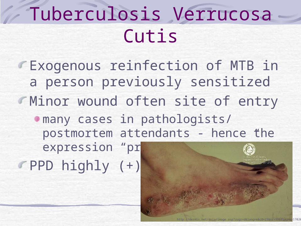
Tuberculosis Verrucosa Cutis
Exogenous reinfection of MTB in a person previously sensitized
Minor wound often site of entrymany cases in pathologists/ postmortem attendants - hence the expression “prosector’s warts”
PPD highly (+)
http://dermis.net/doia/image.asp?zugr=d&lang=e&cd=21&nr=99&diagnr=17020
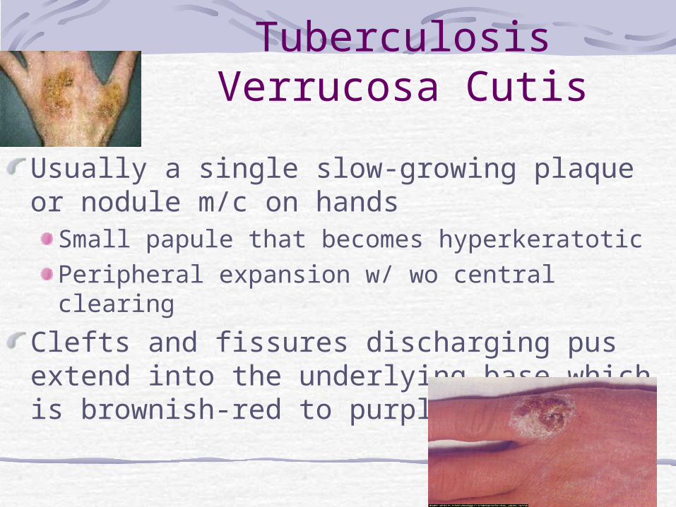
Tuberculosis Verrucosa Cutis
Usually a single slow-growing plaque or nodule m/c on hands
Small papule that becomes hyperkeratotic
Peripheral expansion w/ wo central clearing
Clefts and fissures discharging pus extend into the underlying base which is brownish-red to purplish
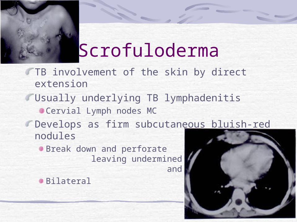
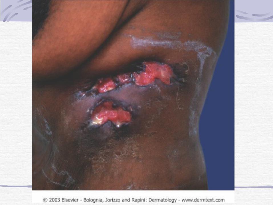
ScrofulodermaTB involvement of the skin by direct extensionUsually underlying TB lymphadenitis
Cervial Lymph nodes MC
Develops as firm subcutaneous bluish-red nodules Break down and perforate leaving undermined ulcers and discharging sinuses
Bilateral
http://www.indianpediatrics.net/jan2002/images/7.jpg
ScrofulodermaHistopathology:
Massive necrosis and abscess formation in the center
The periphery of the abscess or the margins of the sinuses contain tuberculoid granulomas and true tubercles
Acid-fast bacilli
MTB can be found
Tuberculosis OrificialisTB of mucous membranes and skin surrounding orifices
Usually by autoinoculation
Seen in pts with TB of internal organsGI Tract or Lungs
Mouth most commonly affected siteTongue and palate
Prognosis poor – advanced internal diseasePresents as painful yellow or red nodule that ulcerates to form punched-out ulcer
Tuberculosis Orificialis
Histopath: Massive nonspecific inflammatory infiltrate and necrosis
Tubercles with caseation may be found deep in the dermis
Numerous bacilli
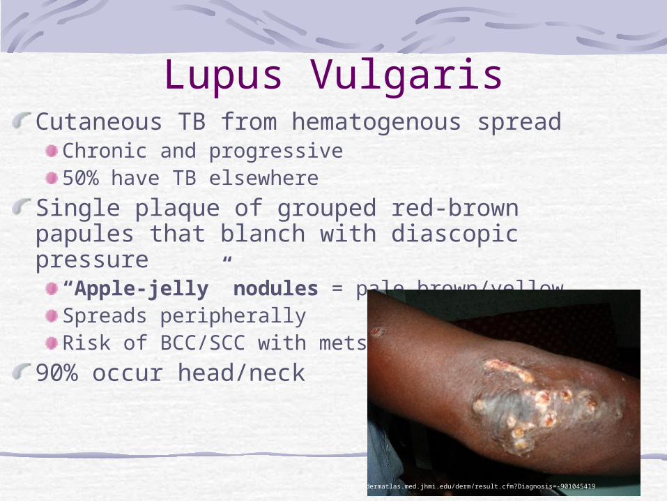
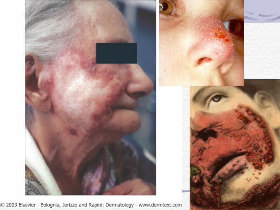
Lupus VulgarisCutaneous TB from hematogenous spread
Chronic and progressive50% have TB elsewhere
Single plaque of grouped red-brown papules that blanch with diascopic pressure
“Apple-jelly” nodules = pale brown/yellowSpreads peripherallyRisk of BCC/SCC with mets
90% occur head/neck
http://dermatlas.med.jhmi.edu/derm/result.cfm?Diagnosis=-901045419
Lupus VulgarisHistopath
Hallmark: Classic Tubercles

Metastatic Tuberculous Abscess
Tuberculous GummaHematogenous dissemination from primary focus during a period of lowered resistance leading to distant abscess/ulcer
SubQ abcessesNontenderFluctuantSingly or as multiples on the trunk, ext, or headUsually occurs in undernourished children or the immunodeficient or immuosuppressed
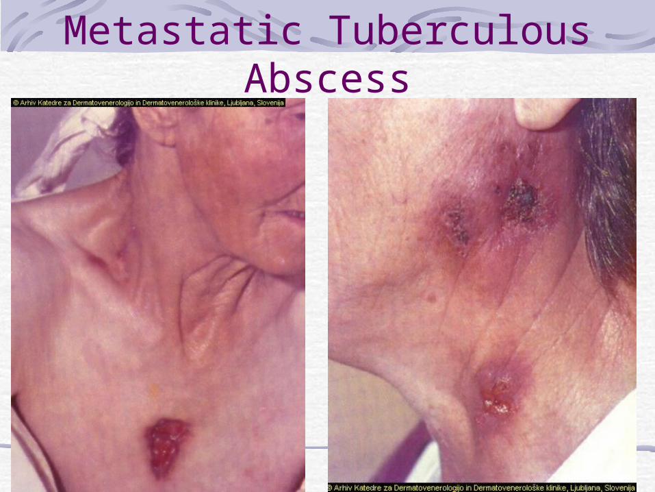
Metastatic Tuberculous Abscess
Metastatic Tuberculous Abscess
Histo:Similar to scrofuloderma
Massive necrosis and abcess formation
Acid fast stains = copious amounts of myocbacteria
Miliary TB (Miliaris Disseminata)
Hematogenous dissemination of MTB
Infants / young children
Focus of infection typically meningeal/pulmonary
May follow infections such as measles and HIV
Presentation:Minute erythematous macules or papules and purpuric lesions
Sometimes umbilicated vesicles or a central necrosis and crust develop in severely ill patients
Miliary TB (Miliaris Disseminata)
Histopath: Initially:
Necrosis and nonspecific inflam infiltrates and abcesses
Occasionally signs of vasculitis
MTB are present in and around vessels
Later stages (if the pt. develops immunity):Lymphocytic cuffing of vessels and even tubercles
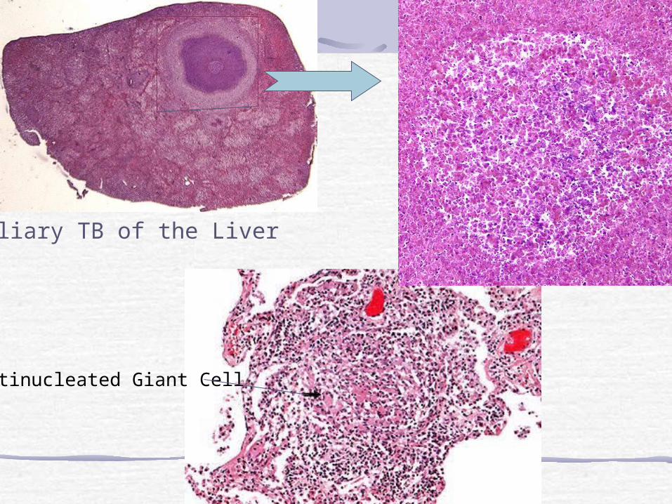
Multinucleated Giant Cell
Miliary TB of the Liver
TuberculidsCutaneous immunologic rxn to TB elsewhereBy definition cx and stains negativeMost likely the result of hematogenous dissemination in pts with high degree of immunity
With PCR, mycobacterial DNA demonstrated in both papulonecrotic tuberculid and erythema induratum of Bazin
All demonstrate rapid response to antiTB txStrongly positive PPDMost exhibit tuberculois features histologically
TuberculidsLichen Scrofulosorum
Rare eruption of asymptomatic, minute, flat-topped yellow to pink follicular or parafollicular papules
May have a minute horny spine or fine scales
Occurs m/c on trunk of children and adolescents with TB in lymph nodes/bone
PPD (+)
Persist for months but spontaneous involution ensues
AntiTB tx results in resolution w/in weeks
TuberculidsLichen ScrofulosorumHistopath:
Superficial noncaseating tuberculoid granulomas develop around hair follicles
Mycobacterium are not seen and can't be cultured
TuberculidsPapulonecrotic Tuberculid
Symmetric, necrotic papules that occur in crops over the extremities and heal by scarringDusky red, symptomless, pea-sized papulesUsually seen in children or young adultsMTB DNA has been detected in about 50% of pts
TuberculidsPapulonecrotic Tuberculid
Histopath:Wedge-shaped necrosis of the upper dermis extending into the epidermis
Involvement of blood vessels is a cardinal feature
Consists of an obliterative and sometimes granulomatous vasculitis leading to thrombosis and complete occlusion
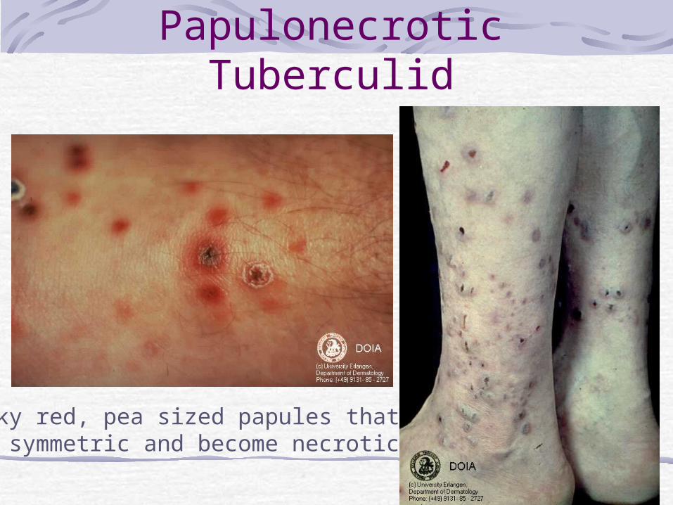
Papulonecrotic Tuberculid
Dusky red, pea sized papules thatare symmetric and become necrotic
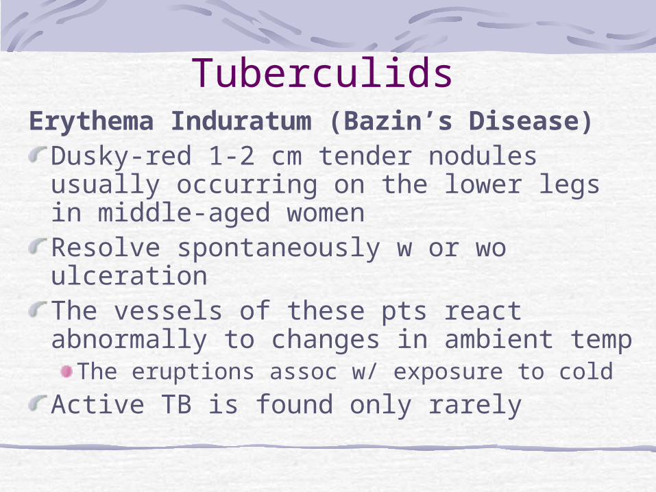
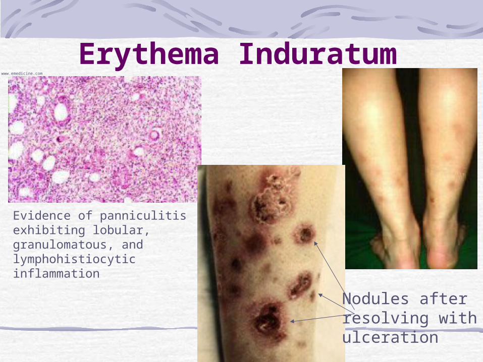
TuberculidsErythema Induratum (Bazin’s
Disease)Dusky-red 1-2 cm tender nodules usually occurring on the lower legs in middle-aged womenResolve spontaneously w or wo ulcerationThe vessels of these pts react abnormally to changes in ambient temp
The eruptions assoc w/ exposure to cold
Active TB is found only rarely
Erythema Induratum
Evidence of panniculitis exhibiting lobular, granulomatous, and lymphohistiocytic inflammation
www.emedicine.com
Nodules after resolving withulceration

Atypical MycobacteriaMycobacterium marinum
“Swimming pool/fish tank” granulomaUlcerating lesions in skin at site of abrasions incurred in swimming pools about 2-3 wks. after inoculationSingle nodules, typically on hands, may ulcerate and suppurate with sporotricoid ascending spreadFresh and salt waterTx with Minocycline 100 mg bidHeals spont. w.in 1-2 yrs. w/residual scarring
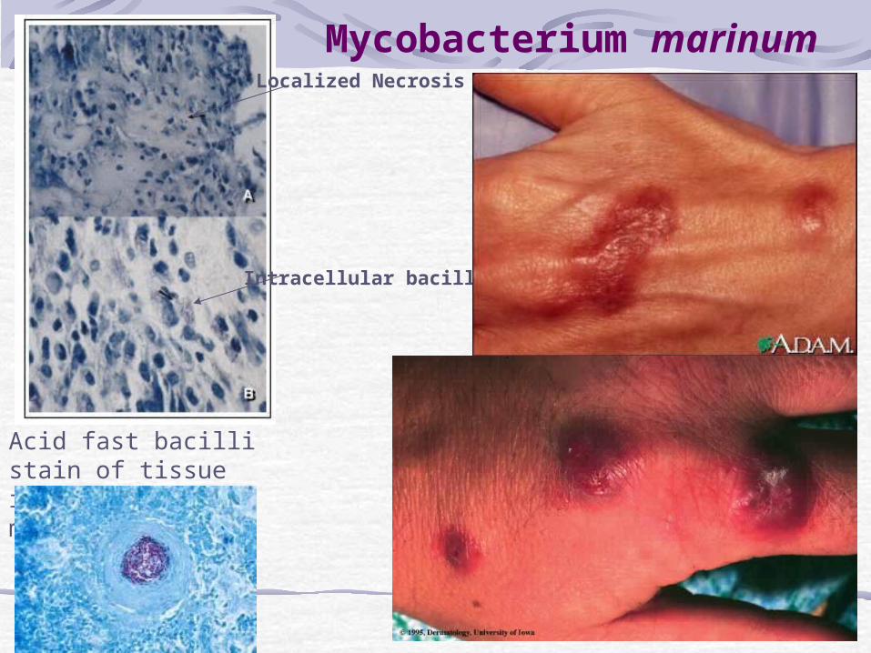
Acid fast bacilli stain of tissue infected with M. marinum
Localized Necrosis
Intracellular bacilli
Mycobacterium marinum
Atypical MycobacteriaMycobacterium ulcerans infection
Buruli ulcer, Bairnsdale ulcer, Searl ulcerSubequatorial regions of Africa, wet, marshy, swampy areasNever found outside the human bodyIncubation period of ~3 moPainless subq swelling which enlarges to a nodule that ulceratesUlcer is deeply undermined and necrotic fat is exposed exposing muscle and tendon
Atypical MycobacteriaMycobacterium ulcerans infection
Histo- Central necrosis in the interlobular septa of the subcut. fat, surrounded by granulation tissue w/giant cells but no typical caseation necrosis or tubercles. AF orgs. can always be demonstrated.TX- Excision of early lesion. Local heat, hyperbaric oxygen and chemo w/RIF and Bactrim.
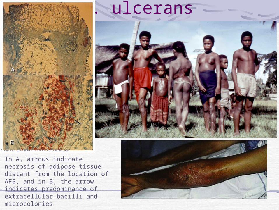
M. ulcerans
In A, arrows indicate necrosis of adipose tissue distant from the location of AFB, and in B, the arrow indicates predominance of extracellular bacilli and microcolonies
http://www.cdc.gov/ncidod/eid/vol5no3/dobos.htm
Atypical MycobacteriaMycobacterium kansasaii
Unusual skin pathogen more commonly associated with pulmonary disease in middle-aged men
Infections localized to Midwestern states and Texas
Acquired from the environmentVariable skin presentations:
NodulesPlaquesCrusted ulcers m/c in immuno-suppressed
Responsive to anti-TB tx: Streptomycin, Rif, Emb
Atypical mycobacterium most closely related to MTB
Atypical MycobacteriaMycobacterium avium complex (MAI/MAC)M. avium and M. intracellulare infects lungs and lymph nodes but occasionally causes cutaneous lesions with disseminationSingle or multiple painless, scaling, yellowish plaques w/ a tendency to ulcerateCommon in AIDSHighly resistant to anti-TB drugs requiring several in combination:
Azithromycin, Rifampin, Ethambutol
Where feasible surgical tx is advisableRifampin used for prophylaxis
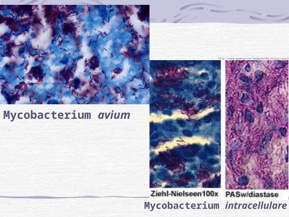
Mycobacterium avium
Mycobacterium intracellulare
http://meds.queensu.ca/~medpalm/PDA_Portal/case11.html
Atypical Mycobacteria
Mycobacterium szulgaiAssociated with:
Cervical lymphadenitisCellulitis Draining nodules and plaques
Can also cause bursitis and pneumoniaMore susceptible to antiTB drugs than most other atypical mycobacterium
Atypical Mycobacteria
Mycobacterium haemophilumSubQ granulomatous eruptions
Immunosuppressed - HIV
Histo:mixed polymorphonuclear and granulomatous inflam“Dimorphic inflammatory response”
No caseation necrosis
May be sensitive to p-aminosaliclyic acid and Rifampin
Atypical Mycobacteria
Mycobacterium genavese
Little is known about this organism
Causes disseminated dzSimilar to M. avium intracellulare in HIV infected pts
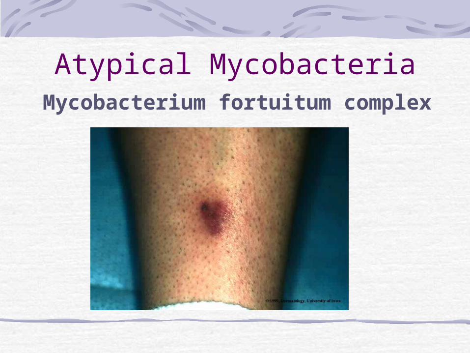
Atypical MycobacteriaMycobacterium fortuitum complex
Three similar species:1. M. fortuitum2. M. chelonei3. M. abscessus
Saprophytes, found chiefly in soil and waterRarely cause human disease
Immunocompromised Prosthetic heart valves and joints
Usually follows puncture wound or surgery
Atypical MycobacteriaMycobacterium fortuitum complex
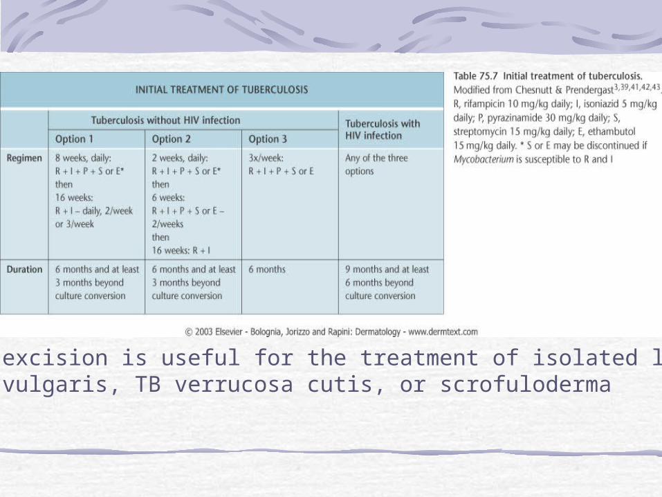
Surgical excision is useful for the treatment of isolated lesionsof lupus vulgaris, TB verrucosa cutis, or scrofuloderma
Initial Treatment of TB
LeprosyEtiology
Dreaded, chronic, poorly-transmissible granulomatous disease of the skin and nerves caused by acid-fast M. leprae Probably least infectious of all diseases:
Strong cell-mediated immunity keeps organism at bay in most people Humans only natural host but reservoirs:
9-banded armadillo (Texas)3 species of monkey
LeprosyEtiology
Pregnancy is a precipitating factor in 10-25% of female patients
Due to altered immunity
Approx 1/3 of newly dx'ed pts w/leprosy will eventually have some chronic disability
Secondary to irreversible nerve injury
M/C hands or feet
LeprosyLepromin skin test
Analogous to the tuberculin testPositive at 48 hours = Fernandez reactionPositive again at 3-4 weeks = Mitusda reaction
Late reaction indicative of immune status of patientStrongly (+) in TTIntermediate in BBAbsent in LL
Clinical presentation complexLittle is known about why different people respond differently to leprosy bacillus
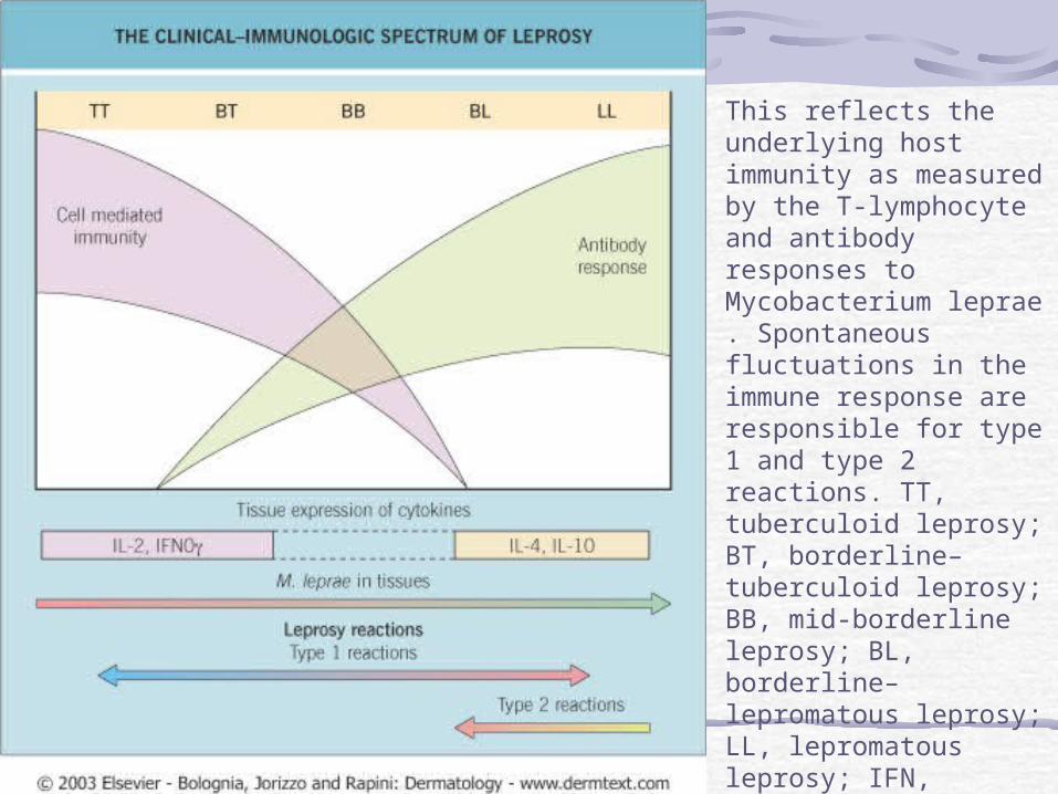
This reflects the underlying host immunity as measured by the T-lymphocyte and antibody responses to Mycobacterium leprae . Spontaneous fluctuations in the immune response are responsible for type 1 and type 2 reactions. TT, tuberculoid leprosy; BT, borderline–tuberculoid leprosy; BB, mid-borderline leprosy; BL, borderline–lepromatous leprosy; LL, lepromatous leprosy; IFN, interferon; IL, interleukin.
LeprosyEpidemiology
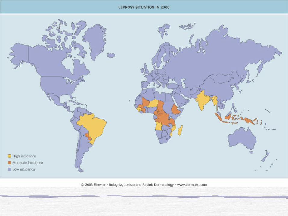
5 million persons worldwide7 thousand active cases in USA250 new cases /year620,000 new cases worldwide/year.80% in 6 countries: Bangladesh, Brazil, India, Indonesia, Myanmar, NigeriaEndemic in SE Asia, Far East, Africa, South/Central AmericaCases in Puerto Rico, Cuba, USA
LeprosyBiological behavior and transmission
Cell-mediated immune responseLow antigenicity
Obligate intracellular parasite
Grows only in colder areas: skin, cutaneous nerves, testes, hands, feet
Multiplies in neurons in macrophages and keratinocytes causing nerve damage/disability
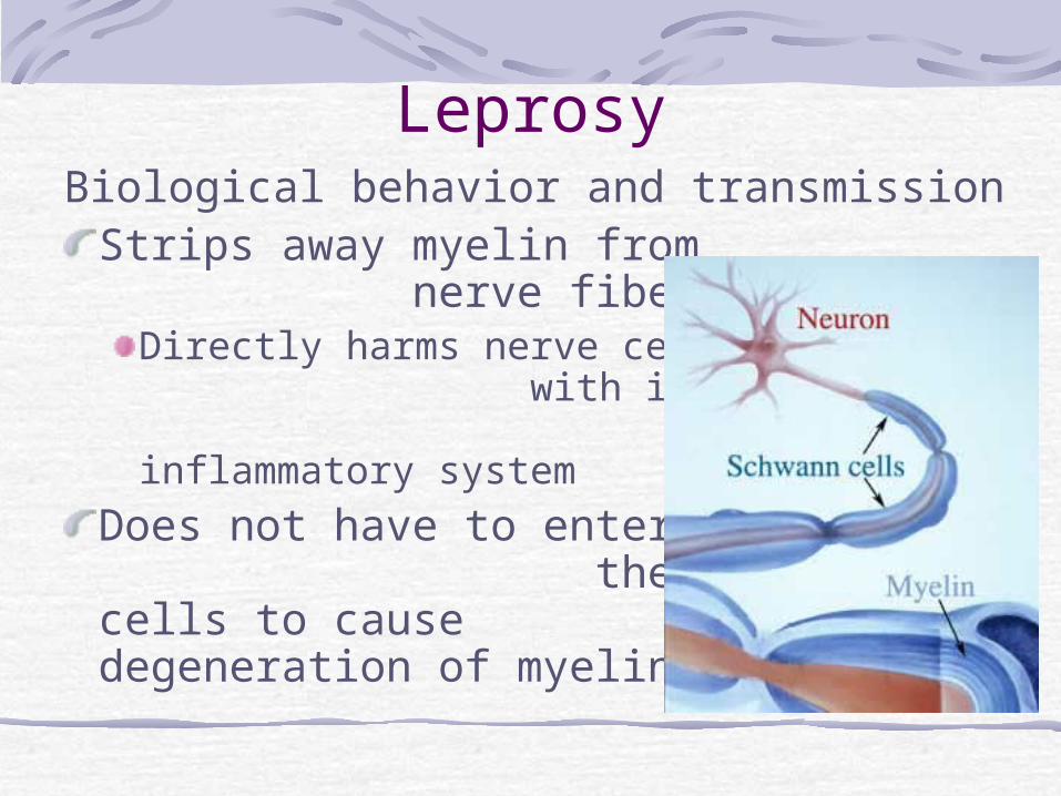
LeprosyBiological behavior and transmission
Strips away myelin from nerve fibers
Directly harms nerve cells with involving the inflammatory system
Does not have to enter the schwann cells to cause degeneration of myelin
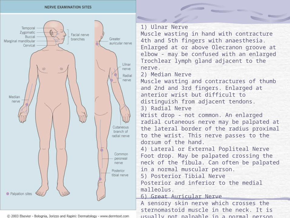
1) Ulnar Nerve Muscle wasting in hand with contracture 4th and 5th fingers with anaesthesia. Enlarged at or above Olecranon groove at elbow - may be confused with an enlarged Trochlear lymph gland adjacent to the nerve. 2) Median Nerve Muscle wasting and contractures of thumb and 2nd and 3rd fingers. Enlarged at anterior wrist but difficult to distinguish from adjacent tendons. 3) Radial Nerve Wrist drop - not common. An enlarged radial cutaneous nerve may be palpated at the lateral border of the radius proximal to the wrist. This nerve passes to the dorsum of the hand. 4) Lateral or External Popliteal Nerve Foot drop. May be palpated crossing the neck of the fibula. Can often be palpated in a normal muscular person. 5) Posterior Tibial Nerve Posterior and inferior to the medial malleolus. 6) Great Auricular Nerve A sensory skin nerve which crosses the sternomastoid muscle in the neck. It is usually not palpable in a normal person. 7) Skin Sensory Nerves near skin lesions may be enlarged. 8) 7th Cranial Nerve It is not palpable but damage to the nerve leads to facial paralysis and lagophthalmos. 9) 5th Cranial Nerve Sensory Fibers If it is damaged, it leads to anaesthesia of cornea.
Nerve Examination Sites
LeprosyBiological behavior and transmission
Transmission similar to TBRespiratory “Globi”
Nasal mucosa
Typically requires extensive contact
Incubation for Tuberculoid leprosy is up to 5 yrs and may be > 20 yrs for LL
LeprosyDiagnosis
2 of 3 clinical criteria1. Anesthesia of the skin2. Thickened peripheral nerves3. Typical skin lesions
Slit-skin smear (Abroad)Tissue fluid exudate examined with Fite stain to determine bacterial index
Punch bx of skin lesion (USA) Fite stain reveals intracellular bacilliPCR
LeprosyDiagnosis
Histologic changes helpful but are not diagnostic
One exception to this rule:Presence of epitheloid cell granulomas w/in nerves = Tuberculoid leprosy or a severe reversal reaction.

LeprosyIdentification and Quantification of Bacilli
AFB in tissue are best shown by carbolfuschin staining using modifications of the Ziehl-Neelson method collectively called Fite-Farraco stainsM. leprae are weekly acid fastRod shaped bacilli
Found in macrophages and nervesQuantified logarithmically by the bacillary index (BI): the numbers of bacilli per oil-immersion field or the numbers of OIFs sought to find 1 bacilli
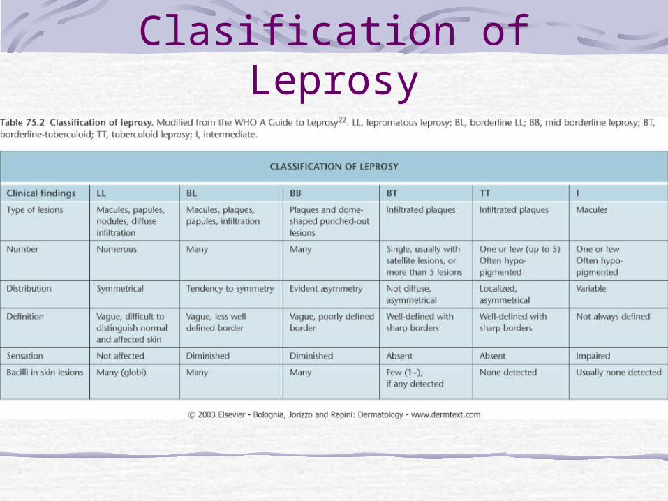
Clasification of Leprosy
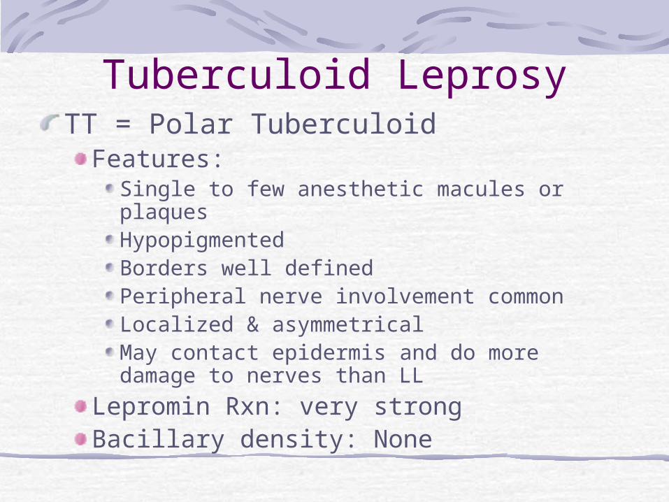
Tuberculoid LeprosyTT = Polar Tuberculoid
Features:Single to few anesthetic macules or plaquesHypopigmentedBorders well definedPeripheral nerve involvement common Localized & asymmetrical May contact epidermis and do more damage to nerves than LL
Lepromin Rxn: very strongBacillary density: None
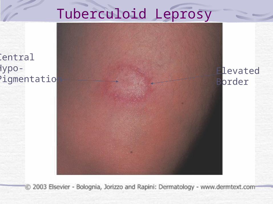
Elevated Border
Central Hypo- Pigmentation
Tuberculoid Leprosy
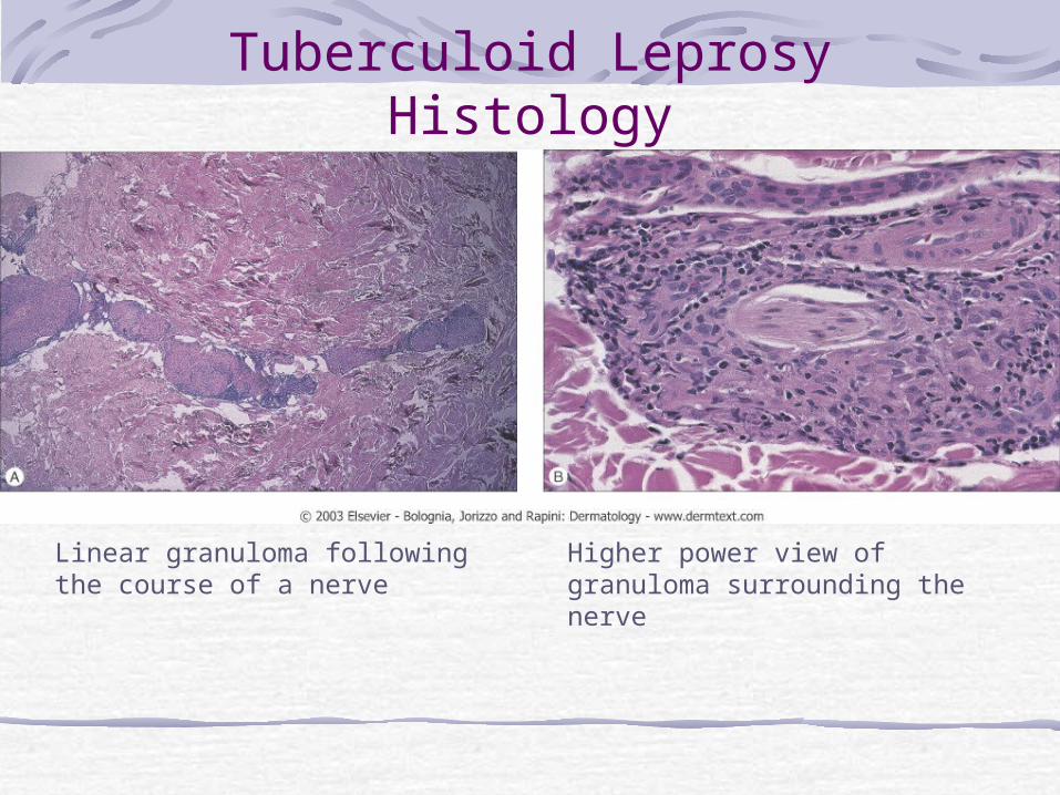
Linear granuloma following the course of a nerve
Higher power view of granuloma surrounding the nerve
Tuberculoid Leprosy Histology
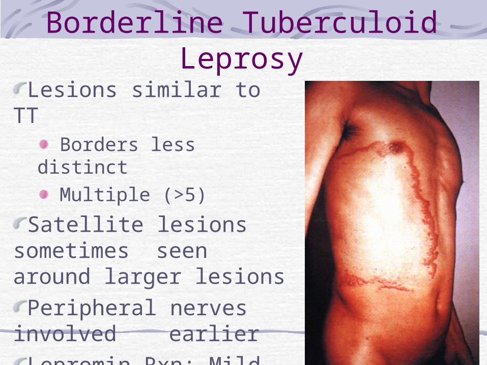
Borderline Tuberculoid Leprosy
Lesions similar to TT Borders less distinct
Multiple (>5)
Satellite lesions sometimes seen around larger lesions
Peripheral nerves involved earlier
Lepromin Rxn: MildBacillary Density: Scant
Borderline LeprosyStill more lesions that BTBorders more vagueAsymmetricBizarre punched-out lesionsHair loss Anhydrosis Most common type Lepromin Rxn: WeakBacillary Density: Moderate
Borderline Lepromatous Leprosy
Multiple macular/papular/plaquesSymmetric lesionsVague borders Neuritis late then neural lesionsSurface smooth and shiny with ill-defined borderMixed granulomasLeprae in neurons = enlargementLepromin Rxn: NoneBacillary Density: Heavy
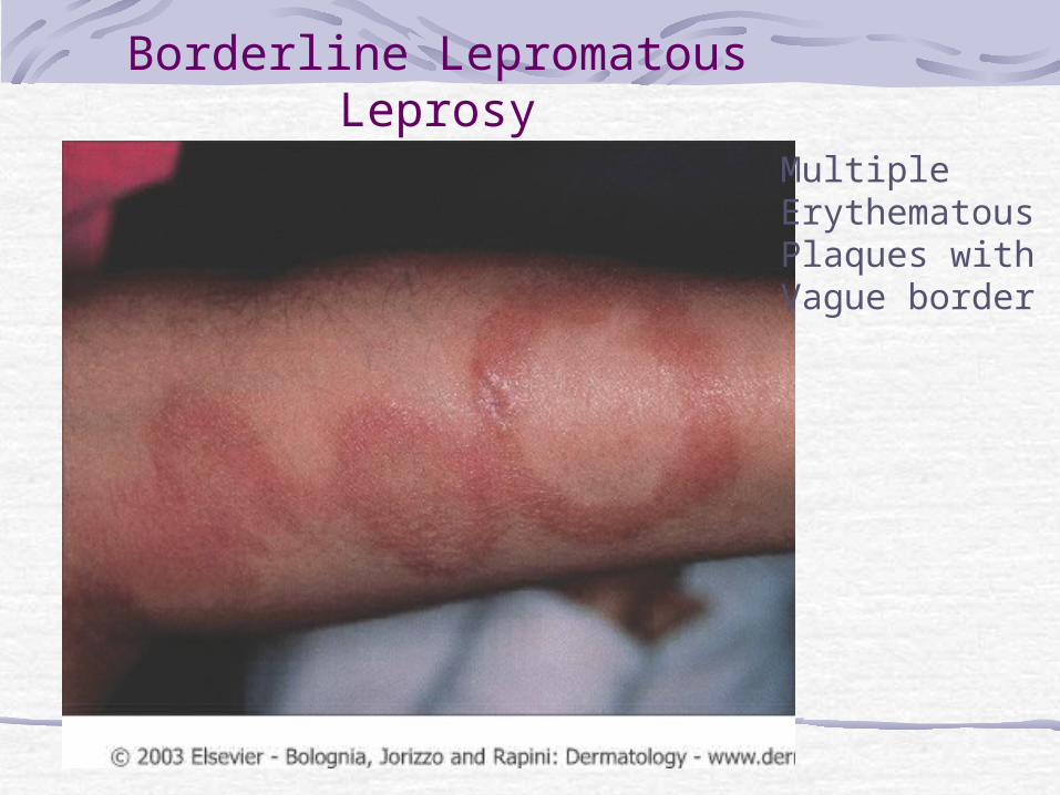
MultipleErythematous Plaques withVague border
Borderline Lepromatous Leprosy
Lepromatous LeprosyMultiple, non-anesthetic, macular and papular lesionsNo neural lesions until very lateLate complications:
MadarosisLeonine faciesTesticular damage
Lepromin Rxn: NoneBacillary Density: Heavy
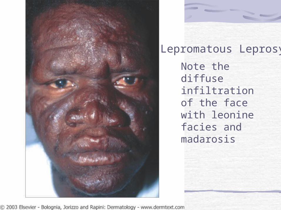
Note the diffuse infiltration of the face with leonine facies and madarosis
Lepromatous Leprosy
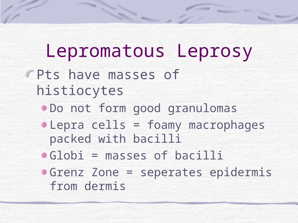
Lepromatous LeprosyPts have masses of histiocytes
Do not form good granulomas
Lepra cells = foamy macrophages packed with bacilli
Globi = masses of bacilli
Grenz Zone = seperates epidermis from dermis
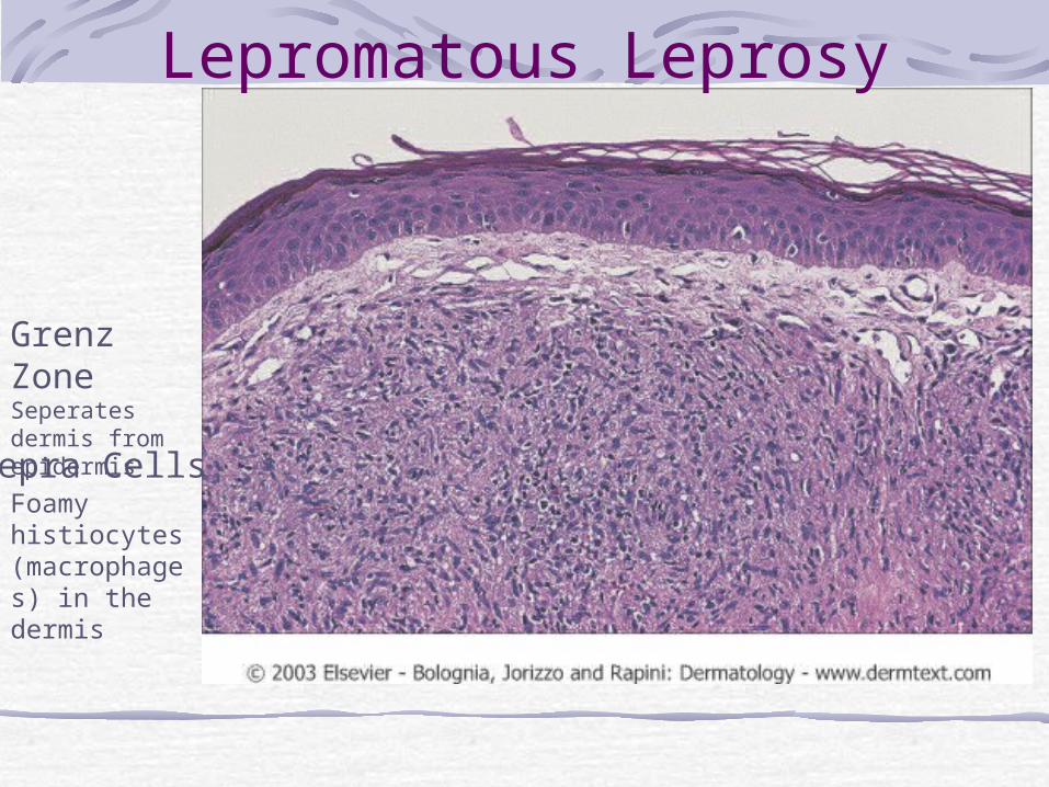
Lepra Cells
Grenz ZoneSeperates dermis from epidermis
Foamy histiocytes (macrophages) in the dermis
Lepromatous Leprosy
Indeterminate LeprosyVaguely defined hypopigmented or red macules
With or without sensory deficit
Lepromin Rxn: Weak
Bacillary Density: Rare
Lucio LeprosyScleroderma-like with hair loss and telangiectasias
Diffusely seen in Mexican/LA patients
May give rise to obstructive vasculitisAka Lucio phenomenon
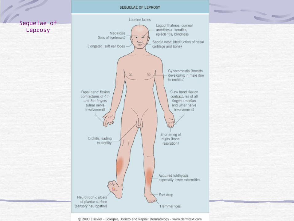
Sequelae of Leprosy
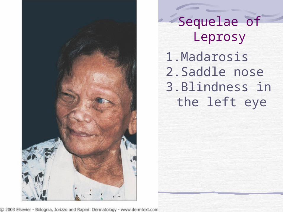
1.Madarosis2.Saddle nose3.Blindness in the
left eye
Sequelae of Leprosy

Reactional States
50% of patients after initiation of therapy
Causes considerable morbidity
Immune response-destructive, inflammatory process

Reactional StatesType 1 Lepra Reactions (upgrade)
Jopling's type 1 Reaction
Affects individuals with borderline disease
Type IV hypersensitivity – Cell-mediated change
Major Complication: Nerve swelling, pain and damage
Cutaneous lesions become tender, erythematous
Accelerated destruction of bacilli
Treat promptly with prednisone 40–60 mg/daily
Note downgrading reactions occur before the initiation of tx and represent shift to LL
Reactional StatesErythema Nodosum Leprosum (Type II lepra rxn)
Josling's type 2 reactionOccurs in 50% of patients with LL and BLImmune complex reaction (type III) between M. leprae antigens and host IgWidely distributed dermal nodules
Do not occur at previous skin lesions
IC precipitate in skin, endothelium, nerves, eyesSystemic Sx’s: Fever, malaise, ulceration, neuritis, uveitis, glaucoma, acute inflammationTx with Thalidomide 400 mg daily
Reactional StatesLucio Phenomenon (Type III Lepra Reaction)
Latin Americans - MexicansPts have La bonita's form of leprosy
Diffuse Lepromatosis
Lucio reaction results in large bullous lesions that ulcerate usually below knees
Due to deep cutaneous vasculitis (hemorrhagic infarcts) Complications: sepsis and deathTx:
Unresponsive to steroids or thalidomideAntimicrobial chemo for leprosy Wound care of ulcers
Treatment of LeprosyMedications of choice
Dapsone: 100mg/d in adults 1mg/kg/d in children
Clofazimine (Lamprene): 50-100mg/d in adultsunestablished in children
Rifampin: 600mg/mo in adults

Treatment of LeprosyType of Leprosy Monthly Daily Duration
Paucibacillary
(I, TT, BT) Rifampin 600mg Dapsone 100mg 6 months
Multibacillary
(LL,BL,BB) Rifampin 600mg Clofazimine 50mg 24 months
Clofazimine 300mg Dapsone 100mg
Treatment of Leprosy
Effective 2nd-line drugsOfloxacin
Minocycline
Clarithromycin
Treatment of Leprosy
MonitoringDapsone:
Baseline G6PD and Hgb
Rifampin: Baseline LFTs and platelets
Baseline and q 2 week PE of sensation and motor nerve function first months of therapy
Opthalmology baseline and periodic exam
Repeat slit-skin, Bx, PCR for response to tx
High Resistance Tuberculoid Leprosy
Characterized by:Few lesionsRare organismsEpitheloid cell granulomas w/ tendency to self-curePlaques w/ sharp margins are the inscription of anti-M. leprae DTH on the skinNerve trunk palsies are its inscription on the peripheral nerves
Low Resistance Lepromatous Leprosy
Characterized by:Wide dissemination
Abundant orgs
Foamy macrophages
Untreated relentless progression