Mycobacteria sp. - UAB School of Optometry year/Micro/powerpoint/Mycobacteria.pdf · 2 3...
28
1 1 Mycobacteriology William H. Benjamin, Jr. William H. Benjamin, PhD Department of Pathology UAB 2 Mycobacteria sp. • Acid Fast Bacilli (AFB) • Mycolic acids (C78 - 91) • Waxes • Obligate aerobes • Slow growing – days to weeks to form colonies – 18 hour doubling time for M. tuberculosis
Transcript of Mycobacteria sp. - UAB School of Optometry year/Micro/powerpoint/Mycobacteria.pdf · 2 3...

1
1
MycobacteriologyWilliam H. Benjamin, Jr.
William H. Benjamin, PhDDepartment of PathologyUAB
2
Mycobacteria sp.
• Acid Fast Bacilli (AFB)• Mycolic acids (C78 - 91)• Waxes• Obligate aerobes• Slow growing
– days to weeks to form colonies– 18 hour doubling time for M. tuberculosis
2
3
Identification of Acid FastBacilli
• Mycobacterium sp. - are identified by the acid faststain
• Mycobacteria predate animal life
• 100 named Mycobacterial species– More than 40 have infected humans
• AIDS• other immunocompromised
4

3
5
Obligate PathogenicMycobacteria
• Mycobacterium tuberculosis– First bacteria shown to cause disease– 1882 Koch’s postulates
• M. leprae– causes Hansen’s disease or leprosy
6
Mycobacterium tuberculosis
• Humans are the only natural host
• 1/3 of the world population is infected
• 9.2 million cases of tuberculosis/year– disease
• 1.7 million deaths caused by tuberculosis/year

4
7
Prehistory of Tuberculosis• 17,000 BPE (before present era) bison in
Wyoming USA– IS6110 and Spoligotype confirmed M. tuberculosis
• 10,000 BPE in Germany skeletal evidence• 3500 - 3000 BPE Egypt Potts disease
– PCR positive for Mtb sequence• 1300 BPE 8 year old Inca boy
– Pott’s disease, AFB smear positive, IS6110 PCRpositive
8

5
Tuberculosis in the United States
0
5,000
10,000
15,000
20,000
25,000
30,000
1980
1982
1984
1986
1988
1990
1992
1994
1996
1998
2000
2002
2004
2006
Cases
Deaths
646
13,299
10
U. S. Tuberculosis Cases
0
2,000
4,000
6,000
8,000
10,000
12,000
14,000
16,000
18,000
20,000
1991 1992 1993 1994 1995 1996 1997 1998 1999 2000 2001 2002 2003
U. S. Born
Foreign Born
2.7 23.6

6
11
12
544
588
541524
464
484
430418
487
433420423
405
380
318314
265
233
258
211216196
175
225
275
325
375
425
475
525
575
625
1985 1987 1989 1991 1993 1995 1997 1999 2001 2003 2005
Alabama Tuberculosis Cases

7
13
0.22?61Tuberculosis death rate4.2680822Tuberculosis rate/100,000
13,67678,00016,156Tuberculosis cases30011.51.8Population (million)
USZambiaNamibia
Examples of high prevalence countries
Transmission of Tuberculosis
• Transmission is by infectious droplet– droplet diameter 1 - 5 µm– droplet contains 1 - 3 bacilli– droplets settle 9 mm/min in still air– infectious dose is 5 - 200 infectious droplets– average patient exhales 1.25 infectious droplets/hour– some cases produce 150 - 200 infectious droplets/hour

8
15
Exposureclose contact
No infection >50%
Infection 50%
Primary active TB 5%
Latent TB 95%
Never reactivate90% Reactivation TB
5% lifetime
HIV+5-10% peryear
Tuberculosis Risk Factors• AIDS - CD4 < 400• Iatrogenic immunosuppression - corticosteroids• Age
– young– old
• Alcoholism/malnourishment• Diabetes• Genetics

9
17
Mantoux skin testmm of induration
18
Significant Induration on Mantoux• 15 mm Always indicates infection• 10 mm population at risk
– low income, minority races, IV drug users– foreign born - from high prevalence countries– Institution populations - prisons, nursing homes, mental
institutions– silicosis, diabetes mellitus, malignancies, immunosuppressive
agents• 5 mm Early, immunosuppressed
– HIV positive or HIV risk factors HIV status unknown– Chest film consistent with old nonreactive tuberculosis– Recent close contact with infectious tuberculosis case

10
Types of Tuberculous disease
• Childhood tuberculosis• “Adult” or reactivation disease• Acute tuberculous pneumonia (AIDS)• Miliary tuberculosis• Cold abscess• Addison’s disease (Adrenal insufficiency)
20
Types of Disease caused by M. tuberculosis

11
M. tuberculosis infection withoutdisease
• Inhalation of infectious droplet• Hilar and peribronchiolar lymph nodes
– 4 - 6 weeks• Lymphohematogenous dissemination
– 6 - 8 weeks• Tubercle formation• Granulomatous inflammation: caseous necrosis• Dystrophic calcification (Ghon complex)
Diagnosis of Tuberculosis
• AFB smear• Tuberculin skin test (Mantoux test)• Chest radiograph• AFB culture

12
Tuberculosis prevention
• Environmental - decrease exposure– Avoid crowded conditions– Air changes– UV irradiation
• Chemoprophylaxis – after positive skin test– INH - after Tuberculin skin test conversion
• 6 months of daily oral INH
• BCG vaccine (Bacille-Calmette-Guerin)– Causes positive skin test– Used in much of the world, except US
24
M. tuberculosis direct smear

13
25
Environmental Resistance ofM. tuberculosis
• Survives drying• Susceptible to UV irradiation (2 hours in
sunlight)• Resistant to many disinfectants
– susceptible to chlorine and phenols• Pasteurization kills (62oC 30 min or 71.7oC 15
sec)• HEPA filters
26
Germicidal Ultraviolet Light
• UV light at 254 nm is effective in killinginfectious agents– In air ducts– In upper room irradiation
• Also used in biological safety cabinets– Demonstration of effectiveness of killing M.
tuberculosis on Middlebrook 7H11 plate

14
27
UV Exposed Culture Plate0 min 1 min
28
AFB Cultures at UAB
total cultures
Positive cultures
Positive for Mtb
1988 2637 80 (3.0%) 42 (1.6%)
1989 2698 76 (2.8%) 23 (0.9%)
1990 2848 126 (4.4%) 24 (0.8%)
1991 2931 120 (4.1%) 23 (0.8%)
1992 3226 145 (4.5%) 35 (1.1%)
1993 3418 177 (5.2%) 39 (1.1%)
1994 4330 259 (5.9%) 89 (2.1%)
1995 4126 219 (5.3%) 98 (2.4%)
1996 3970 224 (5.6%) 82 (2.1%)
1997 4389 308 (7.0%) 114 (2.7%)
1998 5234 293 (5.6%) 77 (1.5%)
1999 4984 256 (5.1%) 73 (1.5%)
2000 4932 278 (5.6%) 99 (2.0%)
2001 5609 271 (4.8%) 69 (1.2%)
2002 5228 223 (4.2%) 50 (1%)
2003 4626 275 (5.9%) 57 (1.2%)
2004 4025 252 (6.2%) 29 (0.7%)
2005 3972 230 5.8%) 31 (0.8%
2006 4742 248 (5.2%) 38 (0.8%)

15
29
Diagnostic M. tuberculosis Cultures
1999 2000 2001 2002 2003 2004 2005
Patients 32 40 24 16 15 10 15
Days to growth 13.6 15.5 16 14 17.6 20 17
Days to
Identification
16.8 19.8 21.3 17 21 30 20
AFB positive
Respiratory
17/28
60%
20/28
71%
14/20
70%
12/15
80%
7/12
58%
5/7
71%
6/11
55%
Positive AFB
M. tuberculosis
17/35
48%
20/31
65%
14/34
41%
12/28
43%
7/29
24%
5/22
23%
6/18
33%
30
Number of Patients Each Species of Mycobacteria Were Isolated from at UAB 1990 to 2007
8M. mucogenicum 12M. scrofulaceum 13M. marinum 18M. lentiflavum 18M. xenopi 19Rapid pigmented115M. abscessus132M. kansasii391M. fortuitum441M. tuberculosis771M. avium cx
1M. simiae1M. peregrinum1M. thermoresistable1M. haemophilum1M. flavescens2M. smegmatis2M. austroafricanum3M. triplex3M. brumae3M. gastri5M. szulgai

16
31
M. tuberculosis concentrated smear
32
Microbiological Diagnosis ofTuberculosis
• Digestion - mucolytic agents• Decontamination• Concentration• Acid fast Stain• Cultivation of Mycobacteria
– solid media– liquid culture
• Anti-Mycobacterial susceptibilities
17
33
34
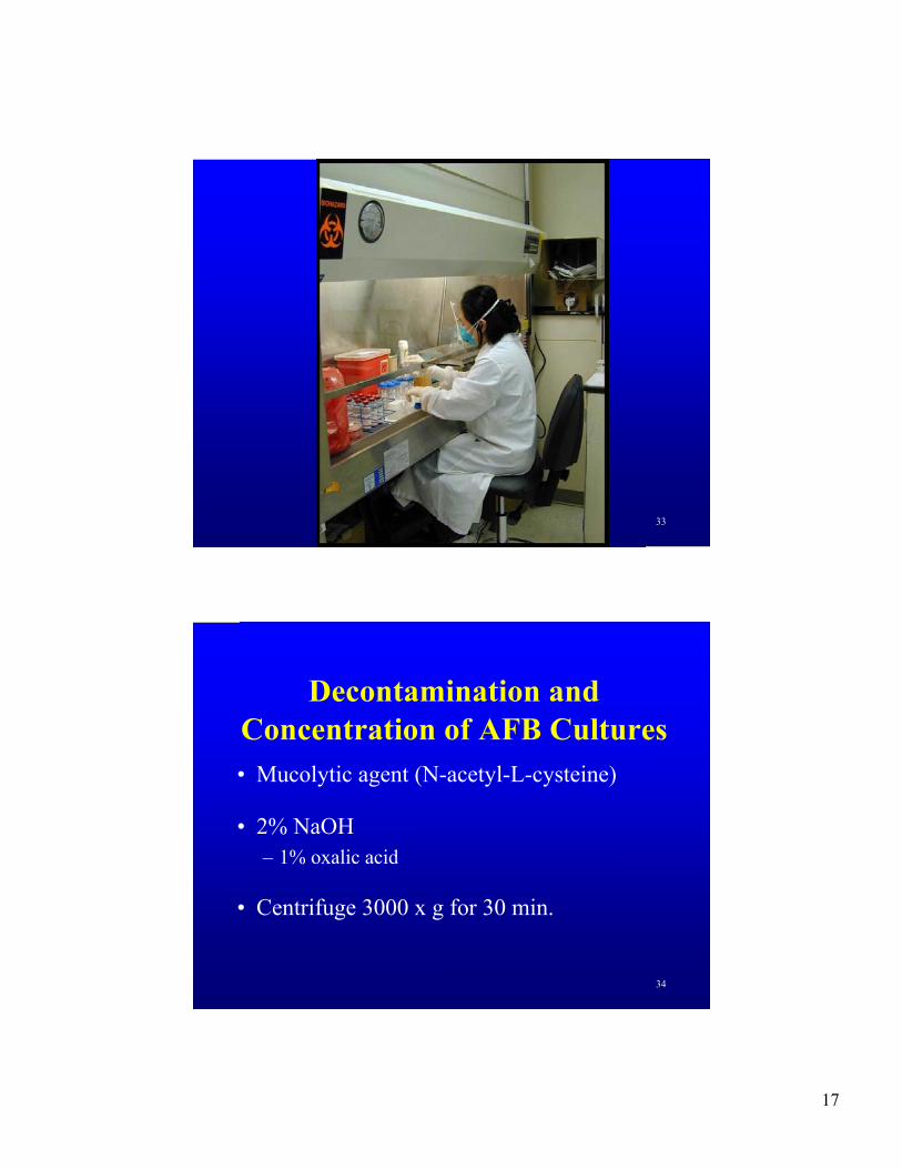
Decontamination andConcentration of AFB Cultures• Mucolytic agent (N-acetyl-L-cysteine)
• 2% NaOH– 1% oxalic acid
• Centrifuge 3000 x g for 30 min.

18
35
Mycobacteria Culture Media• Solid media - 21 - 26 days to detection
– Loewenstein-Jensen (egg based)– Middlebrook 7H11 (agar based)
• Liquid media - 8 to 14 days to detection– BACTEC 460 (14CO2 release from 14C palmitic acid)– MB/BacT (CO2 production)– ESP (O2 utilization - pressure change)– MIDGIT (O2 utilization - quenching of fluorescence)
36
Colony Morphology ofM. gordonae and M. tuberculosis

19
37
VersaTREK® Mycobacteria
MGIT BD
Liquid Culture Systems
MB/BacT
38
Growth in Liquid Medium:M. tuberculosis and M.
fortuitum

20
39
M. tuberculosis cords in liquid culture
40
Mycobacterium abscessus directsputum smear
21
41
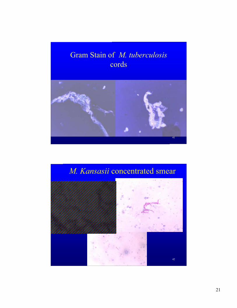
Gram Stain of M. tuberculosiscords
42
M. Kansasii concentrated smear

22
43
Identification of Mycobacteria
• Biochemical tests– 2 to 3 weeks
• GenProbe - DNA - RNA hybridization– 2 hours
• HPLC - high performance liquidchromatography– 1 hour
44
Hybridization Protection Assay(GenProbe)
Denatured (heat)
Acridinium-labeled probe
Alkaline hydrolysis
Substrate light
Inactivated probe

23
Mycobacterium avium complex(MAC)
• found in soil and water - tap water• transmission through either respiratory or GI tract• pulmonary disease like tuberculosis• disseminated disease in AIDS patients
– 50% of autopsies• resistant to many anti-mycobacterial drugs• slow growing non-pigmented colonies
Other Important MOTT• M. kansasii - Photochromogen
– Tuberculosis like disease• M. marinum - Photochromogen
– Found in water - fish tanks and surface water– 30 to 33oC optimum temperature
• M. scrofulaceum -– granulomatous cervical lymphadenitis in children
• M. fortuitum - M. chelonei complex– Rapid growers - colonies in less than 7 days– Skin infections, pulmonary disease

24
47
M. marinumlymphocutaneous
48
M. marinum• Fresh or salt water or no water
exposure• Photochromogen• 1-2 patients/year at UAB• 9 finger patients• 30oC optimum temperature• colonies form in 10-14 days
25
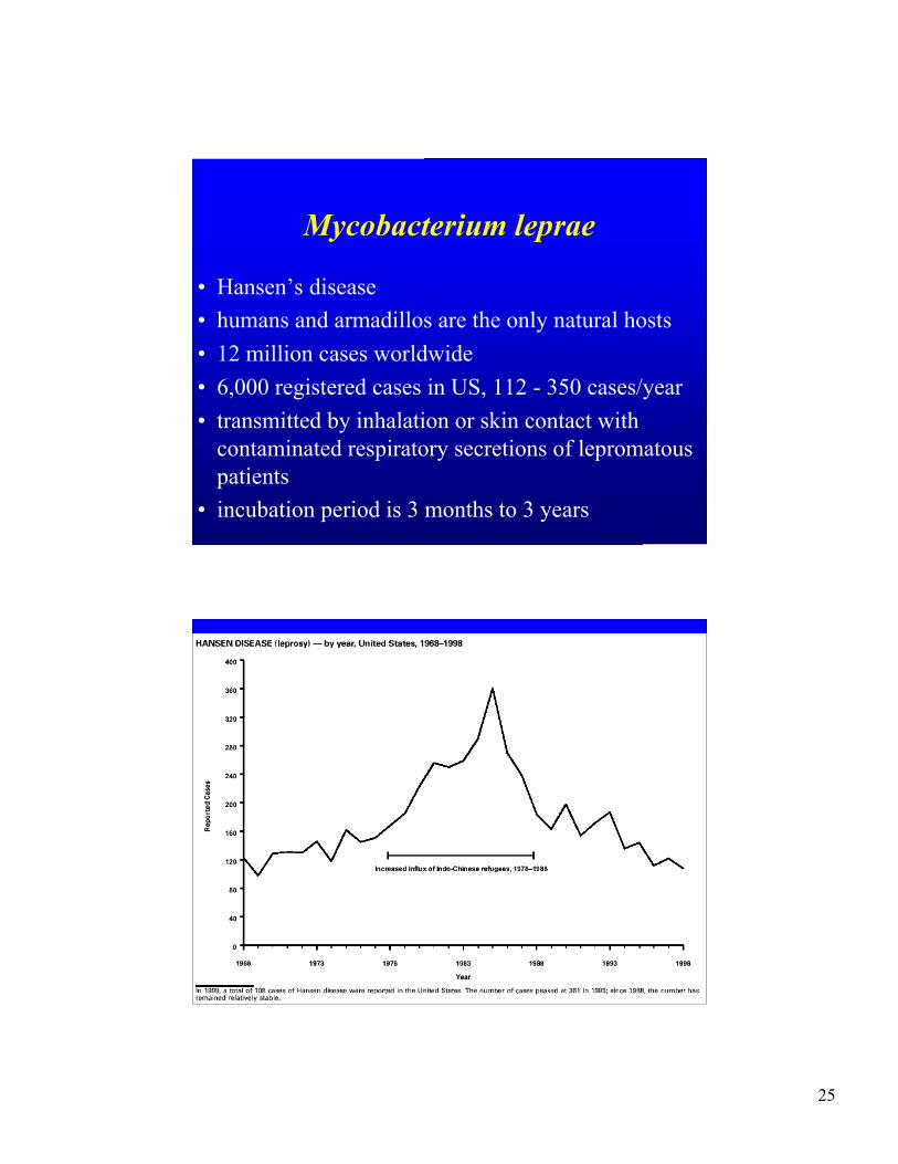
Mycobacterium leprae
• Hansen’s disease• humans and armadillos are the only natural hosts• 12 million cases worldwide• 6,000 registered cases in US, 112 - 350 cases/year• transmitted by inhalation or skin contact with
contaminated respiratory secretions of lepromatouspatients
• incubation period is 3 months to 3 years
50

26
Clinical Types of Leprosy
• Tuberculoid leprosy– intact cell mediated response to M. leprae– organisms rare in tissue– organisms grow in nerves in cooler parts of the
body– cutaneous loss of sensation - nerve damage due
to cell mediated immunity
Leprosy• diagnosis
– does not grow on artificial media– will grow in nude mice or armadillo– AFB stain of nasal secretions– lepromin test - skin test
• treatment– dapsone and rifampicin - at least 1 year
• prevention– isolation of acute lepromatous cases– vaccines under development

27
Lepromatous Leprosy
• depressed CMI response specific for M. leprae• bacteremia with localization in nerves and skin• high numbers of organisms in macrophages• less loss of nerve function• leonine facies• other organs involved - testes, spleen and liver
54
Lepromatous leprosy

28
55
Tuberculoid leprosy
• Non-progressive disease• intact cell mediated response• organisms rare in tissue macular lesions
predominate• organisms invade nerves and form
granulomas• cutaneous loss of sensation - nerve damage
due to CMI
56
Tuberculous leprosy