The crystal structure of TGF-E33 and comparison to TGF-P2 ...
OR I G INA L ART I C L E
Mutations in the latent TGF-beta binding protein 3(LTBP3) gene cause brachyolmia with amelogenesisimperfectaMathilde Huckert1,2,7,†, Corinne Stoetzel1,†, Supawich Morkmued2,3,11,Virginie Laugel-Haushalter3, Véronique Geoffroy1, Jean Muller3,4,8,François Clauss2,5,7, Megana K. Prasad1, Frédéric Obry2,7, Jean Louis Raymond2,Marzena Switala2,7, Yves Alembik9, Sylvie Soskin10, Eric Mathieu6,Joseph Hemmerlé6, Jean-Luc Weickert3, Branka Brukner Dabovic12,Daniel B. Rifkin12, Annelies Dheedene13, Eveline Boudin14, Oana Caluseriu15,Marie-Claude Cholette15, Ross Mcleod15, Reynaldo Antequera16,Marie-Paule Gellé17,18, Jean-Louis Coeuriot17, Louis-Frédéric Jacquelin17,Isabelle Bailleul-Forestier19, Marie-Cécile Manière2,7, Wim Van Hul14,Debora Bertola20, Pascal Dollé3,‡, Alain Verloes21,‡, Geert Mortier13,14,‡,Hélène Dollfus1,9,‡ and Agnès Bloch-Zupan2,3,7,*1Université de Strasbourg, Laboratoire de Génétique Médicale, INSERM UMR 1112, Faculté de Médecine, FMTS, 11rue Humann 67000 Strasbourg, France, 2Université de Strasbourg, Faculté de Chirurgie Dentaire, 8 rue StElisabeth, 67000 Strasbourg, France, 3Université de Strasbourg, Institut de Génétique et de Biologie Moléculaire etCellulaire (IGBMC), CERBM, INSERM U 964, CNRS UMR 7104, 1 rue Laurent Fries, BP 10142, Illkirch 67404, France,4Université de Strasbourg, Laboratoire ICube UMR 7357, CNRS, LBGI, Strasbourg, France, 5Université deStrasbourg, Osteoarticular and Dental Regenerative NanoMedicine, Inserm UMR 1109, 11 rue Humann 67000Strasbourg, France, 6Université de Strasbourg, Biomaterials and Bioengineering, Inserm UMR 1121, 11 rueHumann, 67000 Strasbourg, France, 7Hôpitaux Universitaires de Strasbourg, Pôle deMédecine et Chirurgie Bucco-Dentaires, Reference Centre for Orodental Manifestations of Rare Diseases, CRMR, 1 place de l’Hôpital, 67000Strasbourg, France, 8Hôpitaux Universitaires de Strasbourg, Laboratoire de Diagnostic Génétique, 1 place del’Hôpital, 67000 Strasbourg, France, 9Hôpitaux Universitaires de Strasbourg, Service de Génétique Médicale, 1place de l’Hôpital, 67000 Strasbourg, France, 10Hôpitaux Universitaires de Strasbourg, Service de Pédiatrie 1,Endocrinologie Pédiatrique, 1 place de l’Hôpital, 67000 Strasbourg, France, 11Faculty of Dentistry, Khon Kaen
† These authors contributed equally to the work.‡ Senior authors.Received: November 15, 2014. Revised and Accepted: February 6, 2015
© The Author 2015. Published by Oxford University Press.This is an Open Access article distributed under the terms of the Creative Commons Attribution Non-Commercial License (http://creativecommons.org/licenses/by-nc/4.0/), which permits non-commercial re-use, distribution, and reproduction in any medium, provided the original work is properly cited.For commercial re-use, please contact [email protected]
Human Molecular Genetics, 2015, Vol. 24, No. 11 3038–3049
doi: 10.1093/hmg/ddv053Advance Access Publication Date: 10 February 2015Original Article
3038
Downloaded from https://academic.oup.com/hmg/article-abstract/24/11/3038/719569by gueston 10 February 2018
University, Khon Kaen, Thailand, 12Department of Cell Biology, NYU Langone Medical Centre, New York, USA,13Center for Medical Genetics, Ghent University, Ghent University Hospital, De Pintelaan 185, Ghent 9000,Belgium, 14Department of Medical Genetics, University of Antwerp and Antwerp University Hospital, PrinsBoudewijnlaan 43, Edegem 2650, Belgium, 15Department of Medical Genetics, Faculty of Medicine and Dentistry,University of Calgary, Alberta Children’s Hospital, Calgary, AB, Canada, 16Dentistry Division, HC/FMUSP, SãoPaulo, Brazil, 17Faculté d’Odontologie, Université de Reims Champagne-Ardenne, 2 rue du Général Koenig, Reims51100, France, 18Laboratoire EA 4691 ‘BIOS’, 1, rue du Maréchal Juin, Reims 51100, France, 19Faculty of Dentistry,Paul Sabatier University, LU51, Pôle Odontologie, Hôpitaux de Toulouse, 3 Chemin des Maraîchers, Toulouse,France, 20Unidade de Genética do Instituto da Criança, Hospital das Clínicas da Faculdade de Medicina daUniversidade de São Paulo – Instituto de Biociências, Universidade de São Paulo, São Paulo, Brazil and21Département de Génétique – Hôpital Robert Debré, CRMR ‘Anomalies du Développement & SyndromesMalformatifs’, CRMR ‘Déficiences Intellectuelles de Causes Rares’, 48 bd Sérurier, Paris 75019, France
*To whom correspondence should be addressed at: Faculty of Dentistry, University of Strasbourg, 8 rue St Elisabeth, 67000 Strasbourg, France.Tel: +33 368853919; Fax: +33 368853900; Email: [email protected]
AbstractInherited dentalmalformations constitute a clinically and genetically heterogeneous group of disorders. Here,we report on fourfamilies, three of them consanguineous, with an identical phenotype, characterized by significant short stature withbrachyolmia and hypoplastic amelogenesis imperfecta (AI) with almost absent enamel. This phenotype was first described in1996 by Verloes et al. as an autosomal recessive form of brachyolmia associated with AI. Whole-exome sequencing resulted inthe identification of recessive hypomorphic mutations including deletion, nonsense and splice mutations, in the LTBP3 gene,which is involved in the TGF-beta signaling pathway. We further investigated gene expression duringmouse development andtooth formation. Differentiated ameloblasts synthesizing enamel matrix proteins and odontoblasts expressed the gene. Studyof an available knockoutmousemodel showed that themutantmice displayed very thin to absent enamel in both incisors andmolars, hereby recapitulating the AI phenotype in the human disorder.
IntroductionBrachyolmia (from the greek ‘short trunk’) refers to a heterogeneousgroup of skeletal dysplasias with as major clinical feature a dispro-portionate short stature with short trunk. Radiographic abnormal-ities are predominantly present in the axial skeleton and includegeneralized platyspondyly (i.e. flattened vertebral bodies). Amelo-genesis imperfecta (AI) is a defect in enamel formationandmineral-ization (1). AI can be an isolated finding or occur in association withotheranomalies (syndromicAI) (2). In1996,Verloes et al. (3)describedan autosomal recessive formof platyspondylywithAI [MIM601216].Absence of enamel and oligodontia were themajor dental findings.Bertola et al. (4) subsequently published two other families, one ofthem with abnormal yellow coloration of primary and permanentteeth, as well as retarded dental eruption compatible with a diag-nosis of AI. Here we report on four families, three of them beingconsanguineous, with an identical phenotype, characterized byplatyspondyly (brachyolmia) and AI. Affected individuals havevery thin or almost absent enamel. By using a combined strategyof homozygosity mapping and whole-exome sequencing, recessivemutations in the latentTGF-betabindingprotein 3 (LTBP3) genewereidentified. Analysis of ltbp3 expression during mouse developmentand the studyof dental anomalies observed in the Ltbp3−/−knockoutmousemodel underscored the key role of the latent TGF-beta bind-ing protein 3 in amelogenesis and skeletal development.
ResultsPatients’ phenotype
The index family 1 presented with AI and short stature. The al-most complete absence of enamel in bothprimaryandpermanent
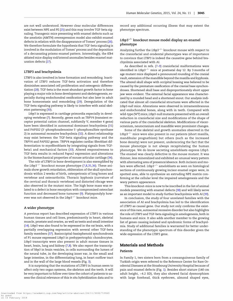
dentitions (Fig. 1A and B) led to the diagnosis of hypoplastic AI,thereby explaining the yellow, small and spaced appearance ofthe teeth. The panoramic radiographs confirmed the absence ofenamel associatedwith large pulp chambers and taurodonticmo-lars (Fig. 1C). Class III mandibular prognathism encountered infamily 1 was due to maxillary underdevelopment (Fig. 1D). Radio-graphs of the skeleton revealed brachyolmia (Fig. 1E and F)
Subsequently, three additional families with a similar pheno-typewere identified. Additional bone anomalies such as osteope-nia and scoliosis were present in family 4. Missing teeth (family4) and retarded teeth eruption (family 3) were also reported.
Enamel shows quantitative and qualitative defects
The enamel structure of a permanent tooth, the left upper secondpremolar (25) extracted within the course of treatment of patientIV-1 of family 1 was further analyzed by scanning electron mi-croscopy (Fig. 1G and H). With this evaluation the enamel hypo-plasia was confirmed and very thin or absent enamel was noted.Dentin was normal. The initial aprismatic enamel layer was ab-sent. A very thin shell of irregular prismatic enamel (PE), with areduced thickness thinner than 150 µm (instead of 300 µm com-paratively at the same site on a control tooth), was deposited cov-ering the dentin scaffold. In this layer, a Hunter-Schreger bandpattern, featuring the arrangement of enamel prisms, was pre-sent. In these areas, no aprismatic outer layer was deposited.However in some areas, amelogenesis continued and some ‘bub-bling’ of non-prismatic enamel (NPE) occurred on top of this basalfirst enamel layer. Waves of aprismatic and prismatic enamel al-ternated. The outermost layer was always aprismatic in areaswhere enamel formation continued.
Human Molecular Genetics, 2015, Vol. 24, No. 11 | 3039
Downloaded from https://academic.oup.com/hmg/article-abstract/24/11/3038/719569by gueston 10 February 2018

Mutations in LTBP3 underlie syndromic AIwith brachyolmia
Whole-exome sequencing was performed independently (in twodifferent labs) in families 1 and 2 and in families 3 and 4. Coverageand variant calling data for families 1 and 2 are provided inSupplementary Material, Table S1.
A single gene, LTBP3, was found to carry bi-allelic mutationsin all affected individuals from the four families (Fig. 2A and B).
In family 1, we identified a homozygous 14 bp deletion, c.[2071_2084delTACCGGCTCAAAGC] (Table 1 and Fig. 2A). This mutationlies within a zone of homozygosity that is shared between thetwo affected individuals, but that is absent in the unaffected sib-ling and parents (Supplementary Material, Fig. S1).
In family 2 we identified compound heterozygosity for a non-sense and a splice donor sitemutation c.[421C>T];[1531+1G>T]; infamilies 3 and 4 homozygosity for a single nucleotide deletionwas found (c.[2216_2217delG] in family 3 and c.[2356_2357delG]
Figure 1. Phenotypic data on family 1. (A) Intraoral viewof individual IV.1 (at age 5 years). All primary teeth are smaller andyellowish showing thin, almost absent, enamel. The
dentition is spaced. (B) Intraoral viewof individual IV.2 (at age 7years). Theeruptingpermanent incisors areyellowandsmall due toabsent enamel. Primary teethwere lost after
recurrent infections. (C) Panoramic radiograph of individual IV.2 (at age 9 years) showing erupted andnon-eruptedpermanent teethwith no enamel. No teetharemissing. Pulp
chambers appear large. Infection as a consequence of microbial contamination of pulp spaces is visible around the 36 (radiolucent area around the roots of the lower left first
permanentmolar filled star) in the absence of the protective enamel layer. The upward arrow points toward a second right lower permanentmolar not yet erupted presenting
with taurodontism and a large pulp chamber. (D–F) Radiographs taken from individual IV.2 (skull at age 9 years and spine at age 14 years). Skull radiograph reveals absent
pneumatization of sinuses and mandibular prognathism secondary to underdevelopment of the maxilla. The spine radiographs show platyspondyly with indentations of
both upper and lower vertebral endplates. (G) The enamel phenotype analyzed at the ultra-structural level through SEM revealed a thin PE layer directly starting at the
dentino-enamel junction (DEJ). In some areas enamel formation continued as an aprismatic layer (NPE). Dentin (DE) was normal. (H) Close up of the PE and NPE thin layer.
3040 | Human Molecular Genetics, 2015, Vol. 24, No. 11
Downloaded from https://academic.oup.com/hmg/article-abstract/24/11/3038/719569by gueston 10 February 2018
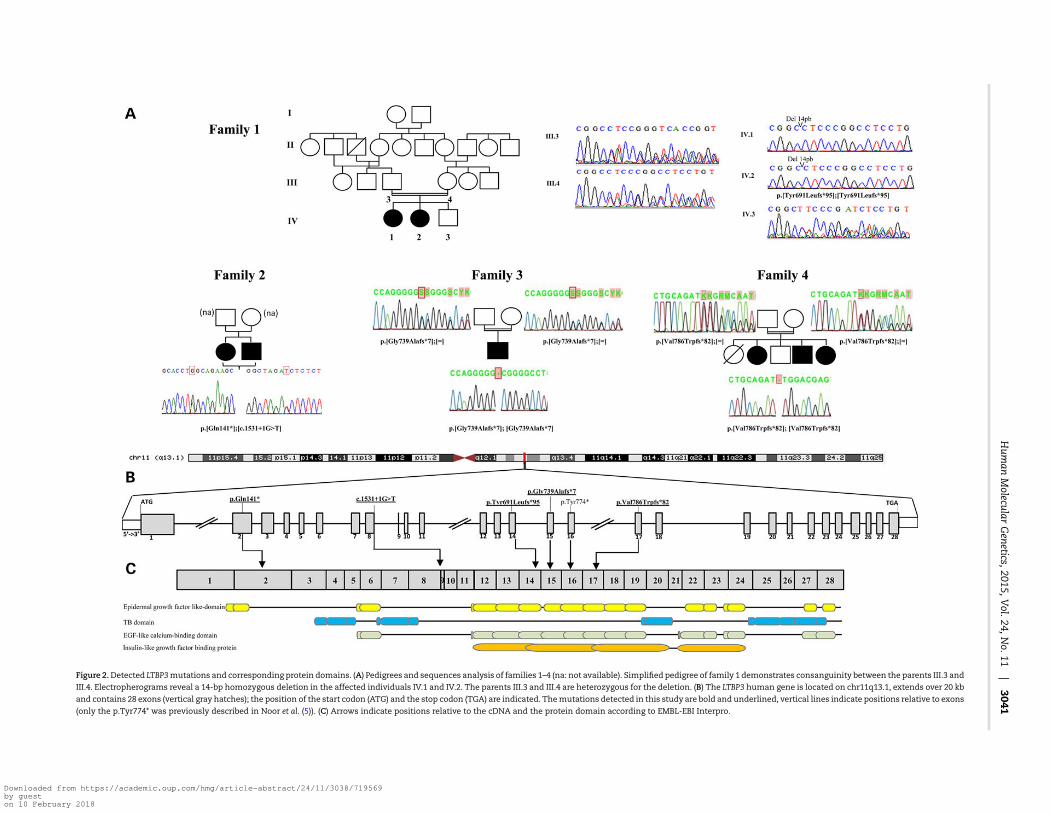
Figure 2.Detected LTBP3mutations and corresponding protein domains. (A) Pedigrees and sequences analysis of families 1–4 (na: not available). Simplified pedigree of family 1 demonstrates consanguinity between the parents III.3 and
III.4. Electropherograms reveal a 14-bp homozygous deletion in the affected individuals IV.1 and IV.2. The parents III.3 and III.4 are heterozygous for the deletion. (B) The LTBP3 human gene is located on chr11q13.1, extends over 20 kb
and contains 28 exons (vertical gray hatches); the position of the start codon (ATG) and the stop codon (TGA) are indicated. Themutations detected in this study are bold and underlined, vertical lines indicate positions relative to exons
(only the p.Tyr774* was previously described in Noor et al. (5)). (C) Arrows indicate positions relative to the cDNA and the protein domain according to EMBL-EBI Interpro.
Hum
anMolecular
Genetics,2015,V
ol.24,No.11
|3041
Downloaded from https://academic.oup.com/hmg/article-abstract/24/11/3038/719569by gueston 10 February 2018
in family 4). All mutations segregated with the disease phenotypein each family and were confirmed by Sanger sequencing (Fig. 2).In addition, they were absent in the Exome Variant Server (EVS)and the Thousand Genomes Project Catalog. Interestingly a singlenucleotide insertion (c.2216_2217insG; p.Gly740Argfs*51) wastabulated in EVS in the homozygous state in 7 out of more than6098 individuals. However, coverage information was not availablefor these individuals toverify if this is a falseor truepositivevariant.
The LTBP3mutations identified in our families are most likelyhypomorphic. The 14-bp deletion found in family 1 does notseem to result in nonsense-mediated decay because it waspresent in RNA extracted from a gingival biopsy of patient IV.1(Supplementary Material, Fig. S2). The mutation most likelygives rise to a truncated protein that lacks the terminal 612amino acids,which encode essential functional domains (epider-mal growth factor like-domain, TB domain, EGF-like calcium-binding domain and insulin-like growth factor binding proteindomain) of the protein (Fig. 2C). The splice-site mutation in fam-ily 2 is predicted to cause an in-frame skipping of exon 8. Theremainingmutations, onenonsensemutation and two single nu-cleotide deletions, are expected to result in nonsense-mediateddecay through the creation of a premature stop codon.
Expression pattern of Ltbp3 in developing mousebone and tooth
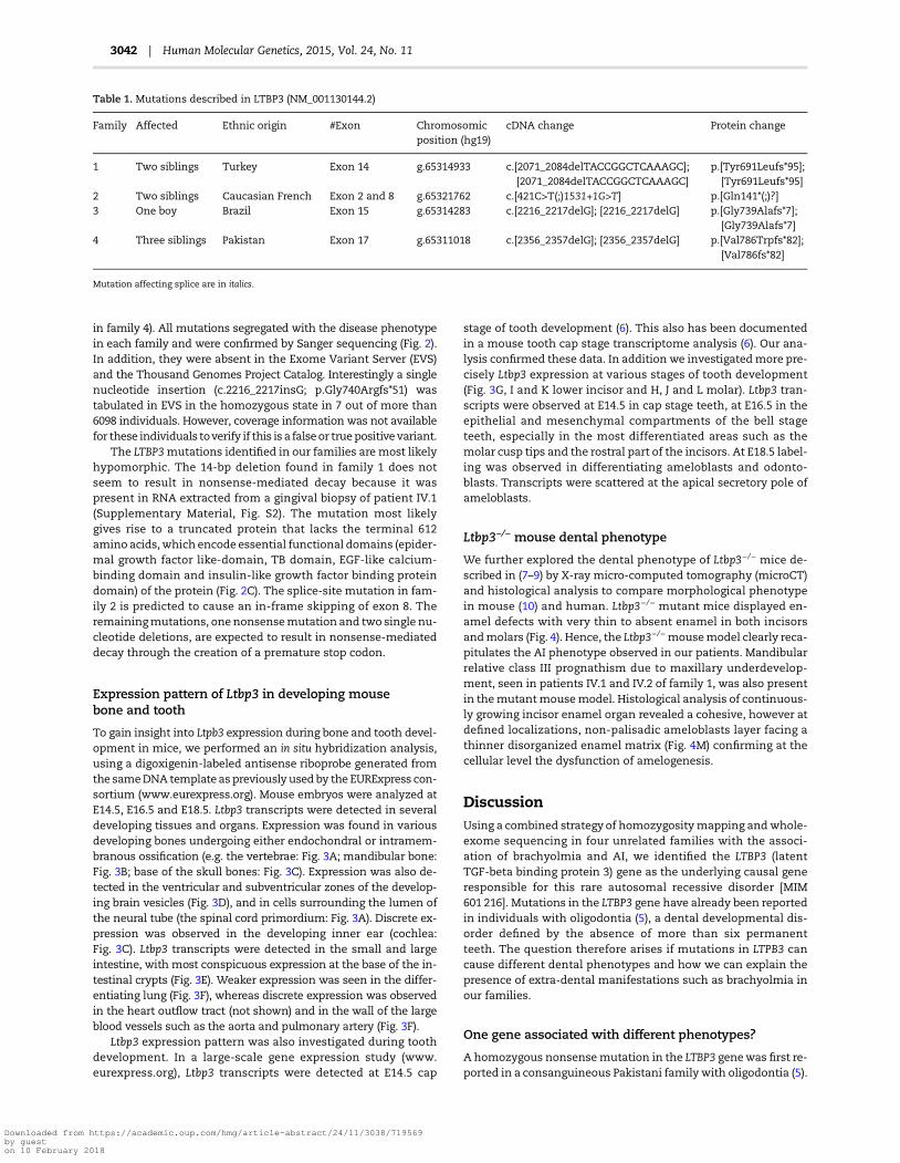
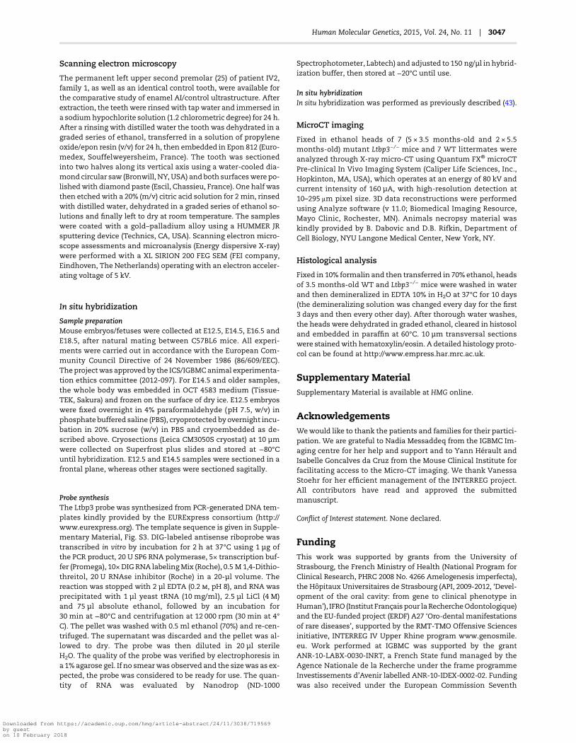
To gain insight into Ltpb3 expression during bone and tooth devel-opment in mice, we performed an in situ hybridization analysis,using a digoxigenin-labeled antisense riboprobe generated fromthe sameDNA template as previously used by the EURExpress con-sortium (www.eurexpress.org). Mouse embryos were analyzed atE14.5, E16.5 and E18.5. Ltbp3 transcripts were detected in severaldeveloping tissues and organs. Expression was found in variousdeveloping bones undergoing either endochondral or intramem-branous ossification (e.g. the vertebrae: Fig. 3A; mandibular bone:Fig. 3B; base of the skull bones: Fig. 3C). Expression was also de-tected in the ventricular and subventricular zones of the develop-ing brain vesicles (Fig. 3D), and in cells surrounding the lumen ofthe neural tube (the spinal cord primordium: Fig. 3A). Discrete ex-pression was observed in the developing inner ear (cochlea:Fig. 3C). Ltbp3 transcripts were detected in the small and largeintestine, with most conspicuous expression at the base of the in-testinal crypts (Fig. 3E). Weaker expression was seen in the differ-entiating lung (Fig. 3F), whereas discrete expression was observedin the heart outflow tract (not shown) and in the wall of the largeblood vessels such as the aorta and pulmonary artery (Fig. 3F).
Ltbp3 expression pattern was also investigated during toothdevelopment. In a large-scale gene expression study (www.eurexpress.org), Ltbp3 transcripts were detected at E14.5 cap
stage of tooth development (6). This also has been documentedin a mouse tooth cap stage transcriptome analysis (6). Our ana-lysis confirmed these data. In addition we investigatedmore pre-cisely Ltbp3 expression at various stages of tooth development(Fig. 3G, I and K lower incisor and H, J and L molar). Ltbp3 tran-scripts were observed at E14.5 in cap stage teeth, at E16.5 in theepithelial and mesenchymal compartments of the bell stageteeth, especially in the most differentiated areas such as themolar cusp tips and the rostral part of the incisors. At E18.5 label-ing was observed in differentiating ameloblasts and odonto-blasts. Transcripts were scattered at the apical secretory pole ofameloblasts.
Ltbp3−/− mouse dental phenotype
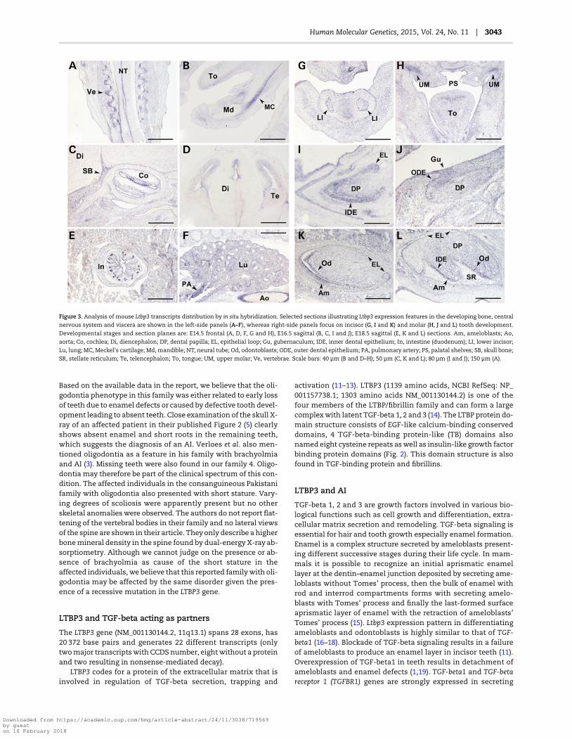
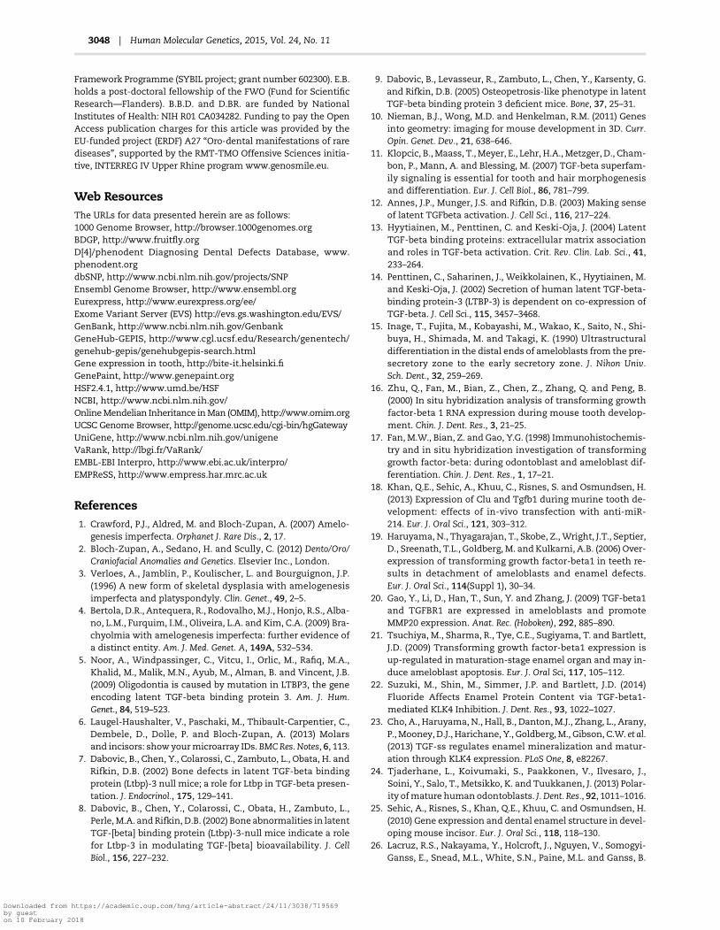
We further explored the dental phenotype of Ltbp3−/− mice de-scribed in (7–9) by X-ray micro-computed tomography (microCT)and histological analysis to compare morphological phenotypein mouse (10) and human. Ltbp3−/− mutant mice displayed en-amel defects with very thin to absent enamel in both incisorsandmolars (Fig. 4). Hence, the Ltbp3−/−mousemodel clearly reca-pitulates the AI phenotype observed in our patients. Mandibularrelative class III prognathism due to maxillary underdevelop-ment, seen in patients IV.1 and IV.2 of family 1, was also presentin themutantmousemodel. Histological analysis of continuous-ly growing incisor enamel organ revealed a cohesive, however atdefined localizations, non-palisadic ameloblasts layer facing athinner disorganized enamel matrix (Fig. 4M) confirming at thecellular level the dysfunction of amelogenesis.
DiscussionUsing a combined strategy of homozygositymapping andwhole-exome sequencing in four unrelated families with the associ-ation of brachyolmia and AI, we identified the LTBP3 (latentTGF-beta binding protein 3) gene as the underlying causal generesponsible for this rare autosomal recessive disorder [MIM601 216]. Mutations in the LTBP3 gene have already been reportedin individuals with oligodontia (5), a dental developmental dis-order defined by the absence of more than six permanentteeth. The question therefore arises if mutations in LTPB3 cancause different dental phenotypes and how we can explain thepresence of extra-dental manifestations such as brachyolmia inour families.
One gene associated with different phenotypes?
A homozygous nonsensemutation in the LTBP3 genewas first re-ported in a consanguineous Pakistani family with oligodontia (5).
Table 1. Mutations described in LTBP3 (NM_001130144.2)
Family Affected Ethnic origin #Exon Chromosomicposition (hg19)
cDNA change Protein change
1 Two siblings Turkey Exon 14 g.65314933 c.[2071_2084delTACCGGCTCAAAGC];[2071_2084delTACCGGCTCAAAGC]
p.[Tyr691Leufs*95];[Tyr691Leufs*95]
2 Two siblings Caucasian French Exon 2 and 8 g.65321762 c.[421C>T(;)1531+1G>T] p.[Gln141*(;)?]3 One boy Brazil Exon 15 g.65314283 c.[2216_2217delG]; [2216_2217delG] p.[Gly739Alafs*7];
[Gly739Alafs*7]4 Three siblings Pakistan Exon 17 g.65311018 c.[2356_2357delG]; [2356_2357delG] p.[Val786Trpfs*82];
[Val786fs*82]
Mutation affecting splice are in italics.
3042 | Human Molecular Genetics, 2015, Vol. 24, No. 11
Downloaded from https://academic.oup.com/hmg/article-abstract/24/11/3038/719569by gueston 10 February 2018
Based on the available data in the report, we believe that the oli-godontia phenotype in this family was either related to early lossof teeth due to enamel defects or caused by defective tooth devel-opment leading to absent teeth. Close examination of the skull X-ray of an affected patient in their published Figure 2 (5) clearlyshows absent enamel and short roots in the remaining teeth,which suggests the diagnosis of an AI. Verloes et al. also men-tioned oligodontia as a feature in his family with brachyolmiaand AI (3). Missing teeth were also found in our family 4. Oligo-dontia may therefore be part of the clinical spectrum of this con-dition. The affected individuals in the consanguineous Pakistanifamily with oligodontia also presented with short stature. Vary-ing degrees of scoliosis were apparently present but no otherskeletal anomalies were observed. The authors do not report flat-tening of the vertebral bodies in their family and no lateral viewsof the spine are shown in their article. Theyonly describe a higherbonemineral density in the spine found by dual-energy X-ray ab-sorptiometry. Although we cannot judge on the presence or ab-sence of brachyolmia as cause of the short stature in theaffected individuals, we believe that this reported familywith oli-godontia may be affected by the same disorder given the pres-ence of a recessive mutation in the LTBP3 gene.
LTBP3 and TGF-beta acting as partners
The LTBP3 gene (NM_001130144.2, 11q13.1) spans 28 exons, has20 372 base pairs and generates 22 different transcripts (onlytwomajor transcriptswithCCDSnumber, eightwithout a proteinand two resulting in nonsense-mediated decay).
LTBP3 codes for a protein of the extracellular matrix that isinvolved in regulation of TGF-beta secretion, trapping and
activation (11–13). LTBP3 (1139 amino acids, NCBI RefSeq: NP_001157738.1; 1303 amino acids NM_001130144.2) is one of thefour members of the LTBP/fibrillin family and can form a largecomplexwith latent TGF-beta 1, 2 and 3 (14). The LTBP protein do-main structure consists of EGF-like calcium-binding conserveddomains, 4 TGF-beta-binding protein-like (TB) domains alsonamed eight cysteine repeats aswell as insulin-like growth factorbinding protein domains (Fig. 2). This domain structure is alsofound in TGF-binding protein and fibrillins.
LTBP3 and AI
TGF-beta 1, 2 and 3 are growth factors involved in various bio-logical functions such as cell growth and differentiation, extra-cellular matrix secretion and remodeling. TGF-beta signaling isessential for hair and tooth growth especially enamel formation.Enamel is a complex structure secreted by ameloblasts present-ing different successive stages during their life cycle. In mam-mals it is possible to recognize an initial aprismatic enamellayer at the dentin–enamel junction deposited by secreting ame-loblasts without Tomes’ process, then the bulk of enamel withrod and interrod compartments forms with secreting amelo-blasts with Tomes’ process and finally the last-formed surfaceaprismatic layer of enamel with the retraction of ameloblasts’Tomes’ process (15). Ltbp3 expression pattern in differentiatingameloblasts and odontoblasts is highly similar to that of TGF-beta1 (16–18). Blockade of TGF-beta signaling results in a failureof ameloblasts to produce an enamel layer in incisor teeth (11).Overexpression of TGF-beta1 in teeth results in detachment ofameloblasts and enamel defects (1,19). TGF-beta1 and TGF-betareceptor 1 (TGFBR1) genes are strongly expressed in secreting
Figure 3. Analysis of mouse Ltbp3 transcripts distribution by in situ hybridization. Selected sections illustrating Ltbp3 expression features in the developing bone, central
nervous system and viscera are shown in the left-side panels (A–F), whereas right-side panels focus on incisor (G, I and K) and molar (H, J and L) tooth development.
Developmental stages and section planes are: E14.5 frontal (A, D, F, G and H), E16.5 sagittal (B, C, I and J); E18.5 sagittal (E, K and L) sections. Am, ameloblasts; Ao,
aorta; Co, cochlea; Di, diencephalon; DP, dental papilla; EL, epithelial loop; Gu, gubernaculum; IDE, inner dental epithelium; In, intestine (duodenum); LI, lower incisor;
Lu, lung; MC, Meckel’s cartilage; Md, mandible; NT, neural tube; Od, odontoblasts; ODE, outer dental epithelium; PA, pulmonary artery; PS, palatal shelves; SB, skull bone;
SR, stellate reticulum; Te, telencephalon; To, tongue; UM, upper molar; Ve, vertebrae. Scale bars: 40 µm (B and D–H); 50 µm (C, K and L); 80 µm (I and J); 150 µm (A).
Human Molecular Genetics, 2015, Vol. 24, No. 11 | 3043
Downloaded from https://academic.oup.com/hmg/article-abstract/24/11/3038/719569by gueston 10 February 2018
ameloblasts where they promote the expression of MMP20, anenamel matrix protease (20). TGF-beta1 is also expressed laterin the maturation-stage ameloblasts and seems to play an im-portant role in ameloblast apoptosis (21). In addition, TGF-β1 isable to induce KLK4 (a protease degrading enamel proteins to in-crease mineralization) expression (22,23). TGF-beta1 may alsocontrol cell layer integrity for odontoblasts (24).
The enamel defects, observed in our families, at the ultra-structural level (Fig. 1) with both the absence of the initial apris-matic enamel layer and the abnormal secretion of non-prismaticbulk enamel, strongly suggest a role of LTBP3 during the life cycleof ameloblasts especially at the secretory stages with Tomes pro-cess formation/modulation. The molecular mechanisms thatcontrol the formation, function and retraction of Tomes’ process
Figure 4. MicroCT and histological analysis of Ltbp3−/− mice. The Quantum FX® microCT Pre-clinical In Vivo Imaging System is a low X-ray dose system devised for
longitudinal microCT in preclinical studies (Caliper Life Sciences, Inc., Hopkinton, MA, USA). Seven Ltbp3−/− mouse heads and seven WT littermates were analyzed.
(A–C) Normal macroscopic aspect of the mouse upper (A face, B side view) and lower incisors (C: side view). Note that the teeth are naturally colored yellow that
enamel is smooth and present only on the labial side. The lower incisors are twice as long as the upper. (D–F) Enamel defects encountered in Ltbp3−/− mouse. The
colored area is decreased and an irregular indented enamel surface is present. (G) Appearance of lower incisor from Ltbp3−/− mouse as observed by X-ray imaging.
Note irregular contour of thin enamel, a hypoplastic area, with limited or no enamel is seen on the labial side. (H–I) 3D reconstruction of microCT imaging
demonstrating the outer surface appearance and contact between continuously growing incisors in a WT mouse (H) or Ltbp3−/− mouse (I). The enamel surface is
reduced and the tooth surface irregular. (J–K) Normal (J: WT) and reduced (in size and density) enamel layer (K: Ltbp3−/− mouse), as observed when isolated from
dentin through its highest mineralization and therefore X-ray density degree. (L) Normal ameloblasts (Am) layer facing a highly organized decalcified enamel (E)
matrix. (M) In the Ltbp3−/− mouse, in certain area, the ameloblast layer became erratic, like blubbing and laid out a differently organized thinner enamel matrix.
3044 | Human Molecular Genetics, 2015, Vol. 24, No. 11
Downloaded from https://academic.oup.com/hmg/article-abstract/24/11/3038/719569by gueston 10 February 2018
are not well understood. However clear molecular differencesexist between NPE and PE (25) and this may involve TGF-beta sig-naling. Transgenic mice presenting with enamel defects such asthe amelotin (AMTN) overexpression model also exhibit enameldefects in relationwith the disappearance of Tomes’ process (26).We therefore formulate the hypothesis that TGF-beta signaling isinvolved in themodulation of Tomes’ process and the depositionof a decussating prisms enamel pattern. Interestingly, the Klk4ablatedmice display rod/interod anomalies besides enamelmat-uration defects (27).
LTBP3 and brachyolmia
LTBP3 is also involved in bone formation and remodeling. Inacti-vation of LTBP3 reduces TGF-beta activation and thereforediminishes associated cell proliferation and osteogenic differen-tiation (28). TGF-beta is the most abundant growth factor in boneplaying amajor role in bone development and skeletogenesis, es-pecially during endochondral ossification but also later in adultbone homeostasis and remodeling (29). Deregulation of theTGF-beta signaling pathway is likely to interfere with axial skel-eton patterning (30).
Ltbp3 is expressed in cartilage primordia especially in devel-oping vertebrae (7). Recently, genes such as TRPV4 (transient re-ceptor potential cation channel, subfamily V, member 4 gene)have been identified in autosomal dominant brachyolmia (31)and PAPSS2 (3′-phosphoadenosine 5′-phosphosulfate synthase2) in autosomal recessive brachyolmia (32). A direct relationshipmay exist between the TGF-beta signaling pathway and theTRPV4 and PAPSS2 genes. TRPV4 regulates cardiac fibroblast dif-ferentiation to myofibroblasts by integrating signals from TGF-beta1 and mechanical factors (33). Altered responsiveness toTGF-beta results in reduced Papss2 expression and alterationsin the biomechanical properties of mouse articular cartilage (34).
The role of LTBP3 in bone development is also exemplified bythe Ltbp3−/− knockout mouse phenotype (7,8,35,36). These ani-mals show growth retardation, obliteration of the skull synchon-drosis within 2 weeks of birth, osteopetrosis of long bones andvertebrae and osteoarthritis. Thoracic kyphosis (curvature ofthe cervical and thoracic vertebrae) and distorted ribcage werealso observed in the mutant mice. The high bone mass was re-lated to a defect in bone resorption with compromised osteoclastfunction and decreased bone turnover (9). Platyspondyly how-ever was not observed in the Ltbp3−/− knockout mice.
Awider phenotype
A previous report has described expression of LTBP3 in varioushuman tissues and cell lines, predominantly in heart, skeletalmuscle, prostate and ovaries, as well as testis and small intestine(14). Ltbp3 was also found to be expressed in the rat brain, with apartially overlapping expression with several other TGF-betafamily members (37). Basioccipital-basisphenoid synchondrosisof P1 mouse expressed Ltbp3 in prehypertrophic chondrocytes.Ltbp3 transcripts were also present in adult mouse tissues inheart, brain, lung and kidney (7,8). We also report the transcrip-tion of ltbp3 in brain vesicles, in cells surrounding the lumen ofthe neural tube, in the developing inner ear, in the small andlarge intestine, in the differentiating lung, in heart outflow tractand in the wall of the large blood vessels (Fig. 3).
It is surprising that the mutations of LTBP3 in human seem toaffect only two organ systems, the skeleton and the teeth. It willbe very important to follow over time the cohort of patients to as-sess the clinical relevance of this in situ hybridization data and to
record any additional occurring illness that may extent thephenotype spectrum.
Ltbp3−/− knockout mouse model display an enamelphenotype
Analyzing further the Ltbp3−/− knockout mouse with respect tothe craniofacial and orodental phenotypes was of importanceto convince that LTBP3 is indeed the causative gene behind bra-chyolmia associated with AI.
As described in refs. (7–9) craniofacial malformations wereidentified in Ltbp3−/− mice at postnatal day 12. By 3 months ofage mutant mice displayed a pronounced rounding of the cranialvault, extensionof themandible beyond themaxilla andkyphosis.The altered skull shape with occipital bossing was believed to becaused by the premature ossification of the cranial base synchon-droses. Shortened skull base and disproportionately short upperjaw were evident. The external facial appearance was character-ized by a rounded head and a shortened snout. Our analysis indi-cated that almost all craniofacial structures were affected in theLtbp3-null mice. Alterations were observed in intramembranousand endochondral bones, along with in teeth. Compared withwild-type (WT)mice, Ltbp3-nullmutants presentedwith anoverallreduction in craniofacial size and modifications of the shape ofvarious parts of the craniofacial skeleton. Modifications of viscer-ocranium, neurocranium andmandiblewere prominent changes.
Some of the skeletal and growth anomalies observed in theLtbp3−/− mice were also present in our patients (short maxilla,mandibular prognathism) but others (such as the increasedbone density) were not present, underscoring the fact that themouse phenotype is not always recapitulating the humanphenotype. We do know secreting ameloblasts express Ltbp3.The enamel was clearly defective in the mouse mutant. It wasthinner, lessmineralized and exhibited an unusual wavy patternwith alternating area of presence/absence. Both incisors andmo-lars were affected. Ltbp3−/− ameloblasts as seen on histologicalsections of continuously growing incisor enamel organ were, inspotted area, able to synthesize an extruding NPE matrix con-firming at the cellular level the impaired amelogenesis and thewavy pattern appearance.
This knockout-mice is now to be inscribed in the list of animalmodels presenting with enamel defects (38) and will likely serveas an important model to study further brachyolmia with AI (39).
In conclusion, the study of four unrelated families with theassociation of AI and brachyolmia has led to the identificationof LTBP3 as causal gene. Our study not only confirms the exist-ence of this rare, autosomal recessive disorder but also highlightsthe role of LTBP3 and TGF-beta signaling in amelogenesis, both inhumans and mice. It also adds another member to the growinglist of genes causing isolated and syndromic forms of brachyol-mia. Study of additional families is warranted for better under-standing of the phenotypic spectrum of this disorder given thewide expression of the LTBP3 gene.
Materials and MethodsPatients
In Family 1, two sisters born from a consanguineous family ofTurkish origin were referred to the Reference Center for Rare Or-odental Diseases at the StrasbourgUniversity Hospital because ofpain and enamel defects (Fig. 1). Besides short stature (140 cmadult height, −4.2 SD), they also showed facial dysmorphismwith large forehead, thick eyebrows, almond shaped eyes,
Human Molecular Genetics, 2015, Vol. 24, No. 11 | 3045
Downloaded from https://academic.oup.com/hmg/article-abstract/24/11/3038/719569by gueston 10 February 2018
myopia and mild learning difficulties. Radiographs of the skel-eton revealed brachyolmia (Fig. 1E and F) and there were nosigns of a generalized skeletal dysplasia. Bone age was consid-ered as normal.
Their orodental findings were documented using the D[4]/phenodent Diagnosing Dental Defects Database registry (www.phenodent.org).
Enamel was almost absent (hypoplastic AI) in both primaryand permanent dentitions (Fig. 1A and B). The teeth were yellow,small and spaced. Several teethwere surgically extracted becauseof recurrent infections. On orthopantomogram, no enamel wasobserved, pulp chamberswere large andmolarswere taurodontic(Fig. 1C). Lateral cephalogram showed somewhat thickened cor-tical plates of the frontal bone and an absence of pneumatizationof the frontal and sphenoid sinuses. Posterior clinoid apophysesof the sphenoid were abnormally shaped. Class III mandibularprognathism was due to maxillary underdevelopment (Fig. 1D).
Family 2 consisted of two sibs born to non-consanguineous,unrelated Caucasian parents. The female proband (III.6) is thesixth child. Birth weight was 2.760 g, birth length 46 cm andhead circumference was 33 cm at 39 weeks of gestation. She hadnormal psychomotor development and no general health pro-blems. Primary teethwere small and yellowishwith a poor squaremorphology and rounded cusps without pronounced fissures.The enamel was smooth and showed external crown resorptionbeforemolars erupted. She suffered of repeated dental abscesses,leading to the extraction of several teeth at ages 2½ and 5 years.When seen at age 8, she had clearly hypoplasic AI, microdontiaand taurodontism. At age 13, she was 149 cm tall (−1.5 SD),weighed 42 kg and had an occipital-frontal circumference of51 cm. She had a triangular face with retraction of the midface.She had short hands with stubby interphalangeal joints. Pro-and supination movements were limited. Osteotendinousreflexes in the lower limbs were jerky. Single nucleotide poly-morphism (SNP)-array was normal (Illumina CytoSNP-12 v2)and did not reveal homozygosity stretches. Radiographs showedmild platyspondyly. Her brother (III.7) was the seventh sib of thefamily. He was prematurely born at 28 weeks of gestation and re-mained in the intensive neonatal care unit for 3 months, withoutmajor cardiopulmonary or neurological complication. Inguinalherniae were surgically corrected. He walked at age 14 monthsand his first words were delayed to 2½ years. He required speechtherapy. When examined at age 5½ year-old, he was 101 cm tall(−3 SD), weighed 16 kg and had a head circumference of48.5 cm. His teeth were small and yellow lacking enamel. Hehad a triangular face, converging squint, a narrow thorax withprominent sternum and hypermobile small joints. The familyhistory was unremarkable. Parents had normal height (175 cmfor the father and 170 for the mother) and teeth. The six othersibs were normal.
Family 3 has been described previously by Bertola et al. (4). Theproband is a 12-year-old boy from consanguineous and healthyparents. The patient sat unsupported at 8 months of age, walkedat 12 months, said his first words at 2 years. Although themotherrefers some difficulties in learning, he attends a regular school(currently last year of high school). He presented with short stat-ure (height at −3 SD for age) and enamel defects in the primaryand permanent dentition. A panoramic radiograph revealedtaurodontic pulp chambers of the permanent teeth. Skeletal sur-vey showed mild flattening of the vertebral bodies with minimalposterior scalloping and no evidence of a generalized epi- or me-taphyseal dysplasia. Mother and father have normal height (145and 162.5 cm, respectively) and dental status. This was con-firmed by clinical and X-ray examinations.
Family 4 includes three affected sibs in a sibship of five chil-dren born to first cousin parents of Pakistani origin. The three af-fected children, two girls and one boy of, respectively, 16, 9 and12 years of age, presented with short stature (height rangingfrom −2 to −5 SD). They were disproportionate with short trunkdue to a generalized platyspondyly. Radiographs showed osteo-penia and mild vertebral flattening with posterior scallopingand superior and inferior indentations in the posterior third ofthe lumbar vertebrae. The oldest girl underwent surgery for anS-shaped scoliosis. The scoliosis in the two other childrenwas ra-ther mild. Recurrent dental abscesses were noted and the dentalpractitioner confirmed a diagnosis of AI with missing teeth.
None of the family members had intellectual disability. Thefather’s height was 165.2 cm (3rd–10th centile), and the mother’sheight was 149.4 cm (>3rd centile). None of the unaffected mem-ber of the family have skeletal nor dental anomalies.
Genetic analyses
This study was approved by the ethics committee of the Stras-bourg University Hospital (ClinicalTrials.gov Identifier:NCT01746121). Informed consent and DNA samples were ob-tained from all participating individuals. Genomic DNAwas iso-lated either from blood using the Flexigene DNA kit (Qiagen,Courtaboeuf, France) or from saliva using the prepIT-L2P OG-250 Oragene®DNA kit (DNA Genotek Inc., ON, Canada). Whole-exome sequencing was performed by IntegraGen (Evry, France).The coding parts of the genomewere captured using the SureSe-lect Human All Exon Kits V5+UTR 70 Mb (Agilent, Massy, France)and the resulting libraries were sequenced as paired-end 75 basepair reads on a HiSeq 2000 (Illumina, San Diego, USA). Image ana-lysis and base calling were performed using the Real Time Ana-lysis (RTA) Pipeline version 1.9 with default parameters(Illumina). The bioinformatic analysis of sequencing data wasbased on the CASAVA1.8 pipeline (Illumina). CASAVA performsalignment anddetects variants (SNPs and indels) based on the al-lele calls and read depth. The variants were annotated and prior-itized using an in-house pipeline VaRank (http://lbgi.fr/VaRank/,(40) (41).
Sanger sequencing (GATC Biotech, Applied Biosystems ABI3730xl™, Konstanz, Germany) was used to validate the muta-tions and verify segregation using the primers shown in Supple-mentary Material, Table S2.
Homozygositymapping via GeneChipHuman 250 K SNPAffy-metrix was performed as previously described (42) in family 1 onaffected individuals IV.1, IV.2, on the unaffected individual IV.3and both parents III.3 and III.4 (Supplementary Material, Fig. S1).
cDNA analysis
RNA was extracted from fibroblasts of patient IV.1 (gingival bi-opsy) and one unrelated control by using a RiboPure™ Kit, fol-lowed by a DNAse treatment with the TURBO DNA-free™ Kit(Life Technologies, Carlsbad, CA). RNA integrity was assessed bygel electrophoresis and RNA concentration was measured withthe Eppendorf Biophotometer Plus™with the Hellma® TrayCell™(Eppendorf, Hamburg, Germany). Reverse transcription of 1 µgtotal RNA to cDNAwas performed using the iScript™ cDNA Syn-thesis Kit (BioRad, Hercules, CA). Reverse transcription polymer-ase chain reaction was performed to determine the exoncontent of the cDNA from the patient. Primers used for reversetranscription polymerase chain reaction are shown in Supple-mentary Material, Table S2.
3046 | Human Molecular Genetics, 2015, Vol. 24, No. 11
Downloaded from https://academic.oup.com/hmg/article-abstract/24/11/3038/719569by gueston 10 February 2018
Scanning electron microscopy
The permanent left upper second premolar (25) of patient IV2,family 1, as well as an identical control tooth, were available forthe comparative study of enamel AI/control ultrastructure. Afterextraction, the teethwere rinsedwith tapwater and immersed ina sodiumhypochlorite solution (1.2 chlorometric degree) for 24 h.After a rinsing with distilled water the tooth was dehydrated in agraded series of ethanol, transferred in a solution of propyleneoxide/epon resin (v/v) for 24 h, then embedded in Epon 812 (Euro-medex, Souffelweyersheim, France). The tooth was sectionedinto two halves along its vertical axis using a water-cooled dia-mond circular saw (Bronwill, NY, USA) and both surfaceswere po-lishedwith diamond paste (Escil, Chassieu, France). One half wasthen etchedwith a 20% (m/v) citric acid solution for 2 min, rinsedwith distilled water, dehydrated in a graded series of ethanol so-lutions and finally left to dry at room temperature. The sampleswere coated with a gold–palladium alloy using a HUMMER JRsputtering device (Technics, CA, USA). Scanning electron micro-scope assessments and microanalysis (Energy dispersive X-ray)were performed with a XL SIRION 200 FEG SEM (FEI company,Eindhoven, The Netherlands) operating with an electron acceler-ating voltage of 5 kV.
In situ hybridization
Sample preparationMouse embryos/fetuses were collected at E12.5, E14.5, E16.5 andE18.5, after natural mating between C57BL6 mice. All experi-ments were carried out in accordance with the European Com-munity Council Directive of 24 November 1986 (86/609/EEC).The projectwas approved by the ICS/IGBMCanimal experimenta-tion ethics committee (2012-097). For E14.5 and older samples,the whole body was embedded in OCT 4583 medium (Tissue-TEK, Sakura) and frozen on the surface of dry ice. E12.5 embryoswere fixed overnight in 4% paraformaldehyde (pH 7.5, w/v) inphosphate buffered saline (PBS), cryoprotected byovernight incu-bation in 20% sucrose (w/v) in PBS and cryoembedded as de-scribed above. Cryosections (Leica CM3050S cryostat) at 10 µmwere collected on Superfrost plus slides and stored at −80°Cuntil hybridization. E12.5 and E14.5 samples were sectioned in afrontal plane, whereas other stages were sectioned sagitally.
Probe synthesisThe Ltbp3 probe was synthesized from PCR-generated DNA tem-plates kindly provided by the EURExpress consortium (http://www.eurexpress.org). The template sequence is given in Supple-mentary Material, Fig. S3. DIG-labeled antisense riboprobe wastranscribed in vitro by incubation for 2 h at 37°C using 1 µg ofthe PCR product, 20 U SP6 RNA polymerase, 5× transcription buf-fer (Promega), 10× DIG RNA labelingMix (Roche), 0.5 M 1,4-Dithio-threitol, 20 U RNAse inhibitor (Roche) in a 20-µl volume. Thereaction was stopped with 2 µl EDTA (0.2 , pH 8), and RNA wasprecipitated with 1 µl yeast tRNA (10 mg/ml), 2.5 µl LiCl (4 M)and 75 µl absolute ethanol, followed by an incubation for30 min at −80°C and centrifugation at 12 000 rpm (30 min at 4°C). The pellet was washed with 0.5 ml ethanol (70%) and re-cen-trifuged. The supernatant was discarded and the pellet was al-lowed to dry. The probe was then diluted in 20 µl sterileH2O. The quality of the probe was verified by electrophoresis ina 1% agarose gel. If no smearwas observed and the sizewas as ex-pected, the probe was considered to be ready for use. The quan-tity of RNA was evaluated by Nanodrop (ND-1000
Spectrophotometer, Labtech) and adjusted to 150 ng/µl in hybrid-ization buffer, then stored at −20°C until use.
In situ hybridizationIn situ hybridization was performed as previously described (43).
MicroCT imaging
Fixed in ethanol heads of 7 (5 × 3.5 months-old and 2 × 5.5months-old) mutant Ltbp3−/− mice and 7 WT littermates wereanalyzed through X-ray micro-CT using Quantum FX® microCTPre-clinical In Vivo Imaging System (Caliper Life Sciences, Inc.,Hopkinton, MA, USA), which operates at an energy of 80 kV andcurrent intensity of 160 µA, with high-resolution detection at10–295 μm pixel size. 3D data reconstructions were performedusing Analyze software (v 11.0; Biomedical Imaging Resource,Mayo Clinic, Rochester, MN). Animals necropsy material waskindly provided by B. Dabovic and D.B. Rifkin, Department ofCell Biology, NYU Langone Medical Center, New York, NY.
Histological analysis
Fixed in 10% formalin and then transferred in 70% ethanol, headsof 3.5 months-old WT and Ltbp3−/− mice were washed in waterand then demineralized in EDTA 10% in H2O at 37°C for 10 days(the demineralizing solution was changed every day for the first3 days and then every other day). After thorough water washes,the heads were dehydrated in graded ethanol, cleared in histosoland embedded in paraffin at 60°C. 10 µm transversal sectionswere stainedwith hematoxylin/eosin. A detailed histology proto-col can be found at http://www.empress.har.mrc.ac.uk.
Supplementary MaterialSupplementary Material is available at HMG online.
AcknowledgementsWewould like to thank the patients and families for their partici-pation. We are grateful to Nadia Messaddeq from the IGBMC Im-aging centre for her help and support and to Yann Hérault andIsabelle Goncalves da Cruz from the Mouse Clinical Institute forfacilitating access to the Micro-CT imaging. We thank VanessaStoehr for her efficient management of the INTERREG project.All contributors have read and approved the submittedmanuscript.
Conflict of Interest statement. None declared.
FundingThis work was supported by grants from the University ofStrasbourg, the French Ministry of Health (National Program forClinical Research, PHRC 2008 No. 4266 Amelogenesis imperfecta),the Hôpitaux Universitaires de Strasbourg (API, 2009-2012, ‘Devel-opment of the oral cavity: from gene to clinical phenotype inHuman’), IFRO (Institut Français pour laRechercheOdontologique)and the EU-funded project (ERDF) A27 ‘Oro-dental manifestationsof rare diseases’, supported by the RMT-TMO Offensive Sciencesinitiative, INTERREG IV Upper Rhine program www.genosmile.eu. Work performed at IGBMC was supported by the grantANR-10-LABX-0030-INRT, a French State fund managed by theAgence Nationale de la Recherche under the frame programmeInvestissements d’Avenir labelled ANR-10-IDEX-0002-02. Fundingwas also received under the European Commission Seventh
Human Molecular Genetics, 2015, Vol. 24, No. 11 | 3047
Downloaded from https://academic.oup.com/hmg/article-abstract/24/11/3038/719569by gueston 10 February 2018
Framework Programme (SYBIL project; grant number 602300). E.B.holds a post-doctoral fellowship of the FWO (Fund for ScientificResearch—Flanders). B.B.D. and D.BR. are funded by NationalInstitutes of Health: NIH R01 CA034282. Funding to pay the OpenAccess publication charges for this article was provided by theEU-funded project (ERDF) A27 “Oro-dental manifestations of rarediseases”, supported by the RMT-TMO Offensive Sciences initia-tive, INTERREG IV Upper Rhine program www.genosmile.eu.
Web ResourcesThe URLs for data presented herein are as follows:1000 Genome Browser, http://browser.1000genomes.orgBDGP, http://www.fruitfly.orgD[4]/phenodent Diagnosing Dental Defects Database, www.phenodent.orgdbSNP, http://www.ncbi.nlm.nih.gov/projects/SNPEnsembl Genome Browser, http://www.ensembl.orgEurexpress, http://www.eurexpress.org/ee/Exome Variant Server (EVS) http://evs.gs.washington.edu/EVS/GenBank, http://www.ncbi.nlm.nih.gov/GenbankGeneHub-GEPIS, http://www.cgl.ucsf.edu/Research/genentech/genehub-gepis/genehubgepis-search.htmlGene expression in tooth, http://bite-it.helsinki.fiGenePaint, http://www.genepaint.orgHSF2.4.1, http://www.umd.be/HSFNCBI, http://www.ncbi.nlm.nih.gov/OnlineMendelian Inheritance inMan (OMIM), http://www.omim.orgUCSC Genome Browser, http://genome.ucsc.edu/cgi-bin/hgGatewayUniGene, http://www.ncbi.nlm.nih.gov/unigeneVaRank, http://lbgi.fr/VaRank/EMBL-EBI Interpro, http://www.ebi.ac.uk/interpro/EMPReSS, http://www.empress.har.mrc.ac.uk
References1. Crawford, P.J., Aldred, M. and Bloch-Zupan, A. (2007) Amelo-
genesis imperfecta. Orphanet J. Rare Dis., 2, 17.2. Bloch-Zupan, A., Sedano, H. and Scully, C. (2012) Dento/Oro/
Craniofacial Anomalies and Genetics. Elsevier Inc., London.3. Verloes, A., Jamblin, P., Koulischer, L. and Bourguignon, J.P.
(1996) A new form of skeletal dysplasia with amelogenesisimperfecta and platyspondyly. Clin. Genet., 49, 2–5.
4. Bertola, D.R., Antequera, R., Rodovalho,M.J., Honjo, R.S., Alba-no, L.M., Furquim, I.M., Oliveira, L.A. and Kim, C.A. (2009) Bra-chyolmia with amelogenesis imperfecta: further evidence ofa distinct entity. Am. J. Med. Genet. A, 149A, 532–534.
5. Noor, A., Windpassinger, C., Vitcu, I., Orlic, M., Rafiq, M.A.,Khalid, M., Malik, M.N., Ayub, M., Alman, B. and Vincent, J.B.(2009) Oligodontia is caused by mutation in LTBP3, the geneencoding latent TGF-beta binding protein 3. Am. J. Hum.Genet., 84, 519–523.
6. Laugel-Haushalter, V., Paschaki, M., Thibault-Carpentier, C.,Dembele, D., Dolle, P. and Bloch-Zupan, A. (2013) Molarsand incisors: show yourmicroarray IDs. BMCRes. Notes, 6, 113.
7. Dabovic, B., Chen, Y., Colarossi, C., Zambuto, L., Obata, H. andRifkin, D.B. (2002) Bone defects in latent TGF-beta bindingprotein (Ltbp)-3 null mice; a role for Ltbp in TGF-beta presen-tation. J. Endocrinol., 175, 129–141.
8. Dabovic, B., Chen, Y., Colarossi, C., Obata, H., Zambuto, L.,Perle,M.A. andRifkin, D.B. (2002) Bone abnormalities in latentTGF-[beta] binding protein (Ltbp)-3-null mice indicate a rolefor Ltbp-3 in modulating TGF-[beta] bioavailability. J. CellBiol., 156, 227–232.
9. Dabovic, B., Levasseur, R., Zambuto, L., Chen, Y., Karsenty, G.and Rifkin, D.B. (2005) Osteopetrosis-like phenotype in latentTGF-beta binding protein 3 deficient mice. Bone, 37, 25–31.
10. Nieman, B.J., Wong, M.D. and Henkelman, R.M. (2011) Genesinto geometry: imaging for mouse development in 3D. Curr.Opin. Genet. Dev., 21, 638–646.
11. Klopcic, B., Maass, T.,Meyer, E., Lehr, H.A., Metzger, D., Cham-bon, P., Mann, A. and Blessing, M. (2007) TGF-beta superfam-ily signaling is essential for tooth and hair morphogenesisand differentiation. Eur. J. Cell Biol., 86, 781–799.
12. Annes, J.P., Munger, J.S. and Rifkin, D.B. (2003) Making senseof latent TGFbeta activation. J. Cell Sci., 116, 217–224.
13. Hyytiainen, M., Penttinen, C. and Keski-Oja, J. (2004) LatentTGF-beta binding proteins: extracellular matrix associationand roles in TGF-beta activation. Crit. Rev. Clin. Lab. Sci., 41,233–264.
14. Penttinen, C., Saharinen, J., Weikkolainen, K., Hyytiainen, M.and Keski-Oja, J. (2002) Secretion of human latent TGF-beta-binding protein-3 (LTBP-3) is dependent on co-expression ofTGF-beta. J. Cell Sci., 115, 3457–3468.
15. Inage, T., Fujita, M., Kobayashi, M., Wakao, K., Saito, N., Shi-buya, H., Shimada, M. and Takagi, K. (1990) Ultrastructuraldifferentiation in the distal ends of ameloblasts from the pre-secretory zone to the early secretory zone. J. Nihon Univ.Sch. Dent., 32, 259–269.
16. Zhu, Q., Fan, M., Bian, Z., Chen, Z., Zhang, Q. and Peng, B.(2000) In situ hybridization analysis of transforming growthfactor-beta 1 RNA expression during mouse tooth develop-ment. Chin. J. Dent. Res., 3, 21–25.
17. Fan, M.W., Bian, Z. and Gao, Y.G. (1998) Immunohistochemis-try and in situ hybridization investigation of transforminggrowth factor-beta: during odontoblast and ameloblast dif-ferentiation. Chin. J. Dent. Res., 1, 17–21.
18. Khan, Q.E., Sehic, A., Khuu, C., Risnes, S. and Osmundsen, H.(2013) Expression of Clu and Tgfb1 during murine tooth de-velopment: effects of in-vivo transfection with anti-miR-214. Eur. J. Oral Sci., 121, 303–312.
19. Haruyama, N., Thyagarajan, T., Skobe, Z., Wright, J.T., Septier,D., Sreenath, T.L., Goldberg, M. and Kulkarni, A.B. (2006) Over-expression of transforming growth factor-beta1 in teeth re-sults in detachment of ameloblasts and enamel defects.Eur. J. Oral Sci., 114(Suppl 1), 30–34.
20. Gao, Y., Li, D., Han, T., Sun, Y. and Zhang, J. (2009) TGF-beta1and TGFBR1 are expressed in ameloblasts and promoteMMP20 expression. Anat. Rec. (Hoboken), 292, 885–890.
21. Tsuchiya, M., Sharma, R., Tye, C.E., Sugiyama, T. and Bartlett,J.D. (2009) Transforming growth factor-beta1 expression isup-regulated in maturation-stage enamel organ and may in-duce ameloblast apoptosis. Eur. J. Oral Sci, 117, 105–112.
22. Suzuki, M., Shin, M., Simmer, J.P. and Bartlett, J.D. (2014)Fluoride Affects Enamel Protein Content via TGF-beta1-mediated KLK4 Inhibition. J. Dent. Res., 93, 1022–1027.
23. Cho, A., Haruyama, N., Hall, B., Danton, M.J., Zhang, L., Arany,P.,Mooney,D.J., Harichane, Y., Goldberg,M., Gibson, C.W. et al.(2013) TGF-ss regulates enamel mineralization and matur-ation through KLK4 expression. PLoS One, 8, e82267.
24. Tjaderhane, L., Koivumaki, S., Paakkonen, V., Ilvesaro, J.,Soini, Y., Salo, T., Metsikko, K. and Tuukkanen, J. (2013) Polar-ity ofmature human odontoblasts. J. Dent. Res., 92, 1011–1016.
25. Sehic, A., Risnes, S., Khan, Q.E., Khuu, C. and Osmundsen, H.(2010) Gene expression and dental enamel structure in devel-oping mouse incisor. Eur. J. Oral Sci., 118, 118–130.
26. Lacruz, R.S., Nakayama, Y., Holcroft, J., Nguyen, V., Somogyi-Ganss, E., Snead, M.L., White, S.N., Paine, M.L. and Ganss, B.
3048 | Human Molecular Genetics, 2015, Vol. 24, No. 11
Downloaded from https://academic.oup.com/hmg/article-abstract/24/11/3038/719569by gueston 10 February 2018
(2012) Targeted overexpression of amelotin disrupts themicrostructure of dental enamel. PLoS One, 7, e35200.
27. Bartlett, J.D. and Simmer, J.P. (2014) Kallikrein-related peptid-ase-4 (KLK4): role in enamel formation and revelations fromablated mice. Front. Physiol., 5, 240.
28. Koli, K., Ryynanen, M.J. and Keski-Oja, J. (2008) Latent TGF-beta binding proteins (LTBPs)-1 and -3 coordinate prolifer-ation and osteogenic differentiation of humanmesenchymalstem cells. Bone, 43, 679–688.
29. Kanaan, R.A. and Kanaan, L.A. (2006) Transforming growthfactor beta1, bone connection. Med. Sci. Monit., 12, RA164–RA169.
30. Alliston, T., Piek, E. and Derynck, R. (2008) 22 TGF-βFamily Signaling in Skeletal Development, Maintenance,and Disease. In Derynck, R. and Miyazono, K. (eds), TheTGF-Beta Family. Cold Spring Harbor Monograph Archive,North America, 50. Available at: https://cshmonographs.org/index.php/monographs/article/view/3854 (last accessed, 17February 2015).
31. Grigelioniene, G., Geiberger, S., Horemuzova, E., Mostrom,E., Jantti, N., Neumeyer, L., Astrom, E., Nordenskjold, M.,Nordgren, A. and Makitie, O. (2014) Autosomal dominantbrachyolmia in a large Swedish family: phenotypic spec-trum and natural course. Am. J. Med. Genet. A, 164, 1635–1641.
32. Iida, A., Simsek-Kiper, P.O., Mizumoto, S., Hoshino, T., Elcio-glu, N., Horemuzova, E., Geiberger, S., Yesil, G., Kayserili, H.,Utine, G.E. et al. (2013) Clinical and radiographic features ofthe autosomal recessive form of brachyolmia caused byPAPSS2 mutations. Hum. Mutat., 34, 1381–1386.
33. Adapala, R.K., Thoppil, R.J., Luther, D.J., Paruchuri, S., Mes-zaros, J.G., Chilian, W.M. and Thodeti, C.K. (2013) TRPV4channels mediate cardiac fibroblast differentiation by inte-grating mechanical and soluble signals. J. Mol. Cell. Cardiol.,54, 45–52.
34. Ramaswamy, G., Sohn, P., Eberhardt, A. and Serra, R. (2012)Altered responsiveness to TGF-beta results in reducedPapss2 expression and alterations in the biomechanical
properties of mouse articular cartilage. Arthritis Res. Ther.,14, R49.
35. Colarossi, C., Chen, Y., Obata, H., Jurukovski, V., Fontana, L.,Dabovic, B. and Rifkin, D.B. (2005) Lung alveolar septation de-fects in Ltbp-3-null mice. Am. J. Pathol., 167, 419–428.
36. Chen, Y., Dabovic, B., Colarossi, C., Santori, F.R., Lilic, M., Vuk-manovic, S. and Rifkin, D.B. (2003) Growth retardation as wellas spleen and thymus involution in latent TGF-beta bindingprotein (Ltbp)-3 null mice. J. Cell. Physiol., 196, 319–325.
37. Dobolyi, A. and Palkovits, M. (2008) Expression of latent trans-forming growth factor beta binding proteins in the rat brain.J. Comp. Neurol., 507, 1393–1408.
38. Pugach, M.K. and Gibson, C.W. (2014) Analysis of enamel de-velopment using murine model systems: approaches andlimitations. Front. Physiol., 5, 313.
39. Fleischmannova, J., Matalova, E., Tucker, A.S. and Sharpe, P.T.(2008) Mouse models of tooth abnormalities. Eur. J. Oral Sci.,116, 1–10.
40. Geoffroy, V., Pizot, C., Redin, C., Piton, A., Vasli, N., Stoetzel, C.,Blavier, A., Laporte, J. and Muller, J. (2015) VaRank: a simpleand powerful tool for ranking genetic variants. PeerJ. in press.
41. Redin, C., Le Gras, S., Mhamdi, O., Geoffroy, V., Stoetzel, C.,Vincent, M.C., Chiurazzi, P., Lacombe, D., Ouertani, I., Petit,F. et al. (2012) Targeted high-throughput sequencing for diag-nosis of genetically heterogeneous diseases: efficient muta-tion detection in Bardet-Biedl and Alstrom syndromes.J. Med. Genet., 49, 502–512.
42. Bloch-Zupan, A., Jamet, X., Etard, C., Laugel, V., Muller, J.,Geoffroy, V., Strauss, J.P., Pelletier, V., Marion, V., Poch, O.et al. (2011) Homozygosity mapping and candidate prioritiza-tion identify mutations, missed by whole-exome sequen-cing, in SMOC2, causing major dental developmentaldefects. Am. J. Hum. Genet., 89, 773–781.
43. Laugel-Haushalter, V., Langer, A., Marrie, J., Fraulob, V.,Schuhbaur, B., Koch-Phillips, M., Dolle, P. and Bloch-Zupan,A. (2012) From the transcription of genes involved in ectoder-mal dysplasias to the understanding of associated dentalanomalies. Mol. Syndromol., 3, 158–168.
Human Molecular Genetics, 2015, Vol. 24, No. 11 | 3049
Downloaded from https://academic.oup.com/hmg/article-abstract/24/11/3038/719569by gueston 10 February 2018