Mutant Superoxide Dismutase-1-Linked Familial Amyotrophic ... · Mutant Superoxide...
11
Mutant Superoxide Dismutase-1-Linked Familial Amyotrophic Lateral Sclerosis: Molecular Mechanisms of Neuronal Death and Protection G. D. Ghadge, 1 J. P. Lee, 2 V. P. Bindokas, 2 J. Jordan, 2 L. Ma, 1 R. J. Miller, 2 and R. P. Roos 1 Departments of 1 Neurology and 2 Pharmacological and Physiological Sciences, The University of Chicago, Chicago, Illinois 60637 Mutations in human Cu/Zn superoxide dismutase-1 (SOD) cause ;20% of cases of familial amyotrophic lateral sclerosis (FALS). We investigated the mechanism of mutant SOD- induced neuronal degeneration by expressing wild-type and mutant SODs in neuronal cells by means of infection with replication-deficient recombinant adenoviruses. Expression of two FALS-related mutant SODs (A4V and V148G) caused death of differentiated PC12 cells, superior cervical ganglion neurons, and hippocampal pyramidal neurons. Cell death included many features typical of apoptosis. Death could be prevented by copper (Cu 21 ) chelators, Bcl-2, glutathione, vitamin E, and inhibitors of caspases. Mutant SOD-expressing PC12 cells had higher rates of superoxide (O 2 2 ) production under a variety of conditions. The results support the hypothesis that mutant SOD induced-neurodegeneration is associated with disturbances of neuronal free radical homeostasis. Key words: familial amyotrophic lateral sclerosis; superoxide dismutase-1; apoptosis; recombinant adenovirus; neurodegen- eration; oxidative stress Amyotrophic lateral sclerosis (ALS) is a fatal progressive para- lytic disorder of unknown cause involving motor neurons of the brain and spinal cord. Approximately 10–15% of ALS cases are autosomal dominantly inherited. More than 30 sites for mutations in a ubiquitously occurring cytoplasmic enzyme, Cu/Zn super- oxide dismutase-1 (SOD), have been identified in ;20% of pa- tients with dominantly inherited familial ALS (FALS) (Brown, 1995). Identification of these mutations suggested that free radi- cals play a critical role in the pathogenesis of the disease (Deng et al., 1993). It remained unclear, however, whether SOD mutation produced motor neuron death because of loss of SOD enzymatic activity or gain of an adverse f unction. Subsequent studies involv- ing the transduction of mutant SOD genes into non-neuronal cells (Borchelt et al., 1994) and involving transgenic mice (Gurney et al., 1994; Ripps et al., 1995) demonstrated no correlation between SOD activity and the frequency or severity of the disease, sug- gesting that mutant SOD does not cause FALS because of a deficiency in SOD activity. The latter hypothesis was also sup- ported by studies demonstrating the viability of motor neurons in SOD knock-out mice (Reaume et al., 1996). These observations suggested that mutant SOD produces mo- tor neuron death because of gain of a new adverse function or enhancement of a nondismutase activity of SOD that is normally present. The latter hypotheses were bolstered by experiments in yeast as well as in a continuous rat nigral cell line that had been permanently transfected with wild-type or mutant SOD cDNA (Rabizadeh et al., 1995). Expression of FALS-associated SOD mutants promoted rat nigral cell death after serum withdrawal or application of a Ca 21 ionophore, despite the fact that they had significant SOD enzyme activity. In contrast, overexpressed wild- type SOD inhibited cell death. A potential drawback of the latter studies is that the permanently transfected cells may have mani- fested additional phenotypes besides the one specifically associ- ated with mutant SOD, as a result of selection. The new or enhanced function of FALS-linked mutant SODs remains unclear. Beckman and colleagues (Beckman et al., 1993) have proposed that peroxynitrite, a product of superoxide (O 2 2 ) and nitric oxide (NO), reacts with the Cu 21 of mutant SODs, producing nitronium ions, which lead to nitration of proteins and subsequent neurotoxicity. This hypothesis was supported by stud- ies demonstrating that motor neurons of ALS patients exhibit increased immunoreactivity for nitrotyrosine (Abe et al., 1995; Chou et al., 1996). An alternative hypothesis was proposed by Wiedau-Pazos et al. (1996) and Yim et al. (1996), who reported enhanced peroxidase activity of mutant SOD compared with wild type on the basis of spin trap studies. The enhanced peroxidase activity may increase production of hydroxyl radicals, which could damage neurons. These in vitro spin trap studies, however, may not accurately reflect the situation within neural cells expressing mutant SOD. Wong et al. (1995) recently proposed that the FAL S-linked mutant SOD failed to bind or shield copper (Cu 21 ) as effectively as the wild-type enzyme. This change could lead to an enhancement of Cu 21 -catalyzed oxidative reactions. In this report, we describe a unique approach for studying the mechanisms underlying neuronal death induced by mutant SOD. We used replication-deficient recombinant adenoviruses (AdVs) to deliver and express human wild-type or mutant SOD genes into primary neurons as well as differentiated rat pheochromocy- toma cells (PC12 cells). We then determined the effect of this overexpression on cell viability. Using an imaging assay (Bindo- kas et al., 1996), we were able to determine the O 2 2 content of single cells. Our results indicate that expression of mutant SOD Received May 23, 1997; revised Aug. 18, 1997; accepted Aug. 26, 1997. This work was supported by grants from the National Institutes of Health (R.J.M. and R.P.R.) and the ALS Association (G.D.G. and R.P.R.). J.J. was supported by the Spanish Ministry of Education and Science. G.D.G and J.P.L. contributed equally to this study. Correspondence should be addressed to Dr. Raymond P. Roos, Department of Neurology (MC 2030), The University of Chicago, 5841 South Maryland Avenue, Chicago, IL 60637. Copyright © 1997 Society for Neuroscience 0270-6474/97/178756-11$05.00/0 The Journal of Neuroscience, November 15, 1997, 17(22):8756–8766
Transcript of Mutant Superoxide Dismutase-1-Linked Familial Amyotrophic ... · Mutant Superoxide...
Mutant Superoxide Dismutase-1-Linked Familial AmyotrophicLateral Sclerosis: Molecular Mechanisms of NeuronalDeath and Protection
G. D. Ghadge,1 J. P. Lee,2 V. P. Bindokas,2 J. Jordan,2 L. Ma,1 R. J. Miller,2 and R. P. Roos1
Departments of 1Neurology and 2Pharmacological and Physiological Sciences, The University of Chicago,Chicago, Illinois 60637
Mutations in human Cu/Zn superoxide dismutase-1 (SOD)cause ;20% of cases of familial amyotrophic lateral sclerosis(FALS). We investigated the mechanism of mutant SOD-induced neuronal degeneration by expressing wild-type andmutant SODs in neuronal cells by means of infection withreplication-deficient recombinant adenoviruses. Expression oftwo FALS-related mutant SODs (A4V and V148G) caused deathof differentiated PC12 cells, superior cervical ganglion neurons,and hippocampal pyramidal neurons. Cell death included manyfeatures typical of apoptosis. Death could be prevented by
copper (Cu21) chelators, Bcl-2, glutathione, vitamin E, andinhibitors of caspases. Mutant SOD-expressing PC12 cells hadhigher rates of superoxide (O2
2) production under a variety ofconditions. The results support the hypothesis that mutant SODinduced-neurodegeneration is associated with disturbances ofneuronal free radical homeostasis.
Key words: familial amyotrophic lateral sclerosis; superoxidedismutase-1; apoptosis; recombinant adenovirus; neurodegen-eration; oxidative stress
Amyotrophic lateral sclerosis (ALS) is a fatal progressive para-lytic disorder of unknown cause involving motor neurons of thebrain and spinal cord. Approximately 10–15% of ALS cases areautosomal dominantly inherited. More than 30 sites for mutationsin a ubiquitously occurring cytoplasmic enzyme, Cu/Zn super-oxide dismutase-1 (SOD), have been identified in ;20% of pa-tients with dominantly inherited familial ALS (FALS) (Brown,1995). Identification of these mutations suggested that free radi-cals play a critical role in the pathogenesis of the disease (Deng etal., 1993). It remained unclear, however, whether SOD mutationproduced motor neuron death because of loss of SOD enzymaticactivity or gain of an adverse function. Subsequent studies involv-ing the transduction of mutant SOD genes into non-neuronal cells(Borchelt et al., 1994) and involving transgenic mice (Gurney etal., 1994; Ripps et al., 1995) demonstrated no correlation betweenSOD activity and the frequency or severity of the disease, sug-gesting that mutant SOD does not cause FALS because of adeficiency in SOD activity. The latter hypothesis was also sup-ported by studies demonstrating the viability of motor neurons inSOD knock-out mice (Reaume et al., 1996).
These observations suggested that mutant SOD produces mo-tor neuron death because of gain of a new adverse function orenhancement of a nondismutase activity of SOD that is normallypresent. The latter hypotheses were bolstered by experiments inyeast as well as in a continuous rat nigral cell line that had beenpermanently transfected with wild-type or mutant SOD cDNA(Rabizadeh et al., 1995). Expression of FALS-associated SOD
mutants promoted rat nigral cell death after serum withdrawal orapplication of a Ca 21 ionophore, despite the fact that they hadsignificant SOD enzyme activity. In contrast, overexpressed wild-type SOD inhibited cell death. A potential drawback of the latterstudies is that the permanently transfected cells may have mani-fested additional phenotypes besides the one specifically associ-ated with mutant SOD, as a result of selection.
The new or enhanced function of FALS-linked mutant SODsremains unclear. Beckman and colleagues (Beckman et al., 1993)have proposed that peroxynitrite, a product of superoxide (O2
2)and nitric oxide (NO), reacts with the Cu21 of mutant SODs,producing nitronium ions, which lead to nitration of proteins andsubsequent neurotoxicity. This hypothesis was supported by stud-ies demonstrating that motor neurons of ALS patients exhibitincreased immunoreactivity for nitrotyrosine (Abe et al., 1995;Chou et al., 1996). An alternative hypothesis was proposed byWiedau-Pazos et al. (1996) and Yim et al. (1996), who reportedenhanced peroxidase activity of mutant SOD compared with wildtype on the basis of spin trap studies. The enhanced peroxidaseactivity may increase production of hydroxyl radicals, which coulddamage neurons. These in vitro spin trap studies, however, maynot accurately reflect the situation within neural cells expressingmutant SOD. Wong et al. (1995) recently proposed that theFALS-linked mutant SOD failed to bind or shield copper (Cu21)as effectively as the wild-type enzyme. This change could lead toan enhancement of Cu21-catalyzed oxidative reactions.
In this report, we describe a unique approach for studying themechanisms underlying neuronal death induced by mutant SOD.We used replication-deficient recombinant adenoviruses (AdVs)to deliver and express human wild-type or mutant SOD genesinto primary neurons as well as differentiated rat pheochromocy-toma cells (PC12 cells). We then determined the effect of thisoverexpression on cell viability. Using an imaging assay (Bindo-kas et al., 1996), we were able to determine the O2
2 content ofsingle cells. Our results indicate that expression of mutant SOD
Received May 23, 1997; revised Aug. 18, 1997; accepted Aug. 26, 1997.This work was supported by grants from the National Institutes of Health (R.J.M.
and R.P.R.) and the ALS Association (G.D.G. and R.P.R.). J.J. was supported bythe Spanish Ministry of Education and Science.
G.D.G and J.P.L. contributed equally to this study.Correspondence should be addressed to Dr. Raymond P. Roos, Department of
Neurology (MC 2030), The University of Chicago, 5841 South Maryland Avenue,Chicago, IL 60637.Copyright © 1997 Society for Neuroscience 0270-6474/97/178756-11$05.00/0
The Journal of Neuroscience, November 15, 1997, 17(22):8756–8766
induces neural cell death that bears the hallmarks of apoptosis, issensitive to metal chelators, antioxidants, and antiapoptoticagents, and is associated with abnormalities in free radicalproduction.
MATERIALS AND METHODSCell culture. Human embryonic kidney (HEK) 293 and baby hamsterkidney (BHK-21) monolayer cell cultures were grown in DMEM sup-plemented with 10% fetal bovine serum and 0.01% gentamycin (LifeTechnologies, Gaithersburg, MD). Rat pheochromocytoma PC12 cellswere plated on poly-L-lysine-coated glass coverslips at a density of 10,000cells per coverslip in DMEM supplemented with 10% bovine calf serumand 10 mg/ml penicillin /streptomycin (Sigma, St. Louis, MO). Differen-tiation of PC12 cells was induced within 24 hr after the addition ofDMEM containing 100 ng/ml of nerve growth factor (NGF) (Collabo-rative Biomedical Products, Inc.) and no serum. Seven days later, cellswere infected with recombinant AdVs as described below. Primarysympathetic neurons were isolated from superior cervical ganglia of 3- to5-d-old Holtzman rats by previously described methods (Jordan et al.,1995). Dissociated cells were maintained on coverslips in L-15 mediumwith 1 mg/ml NGF and 5% rat serum (Life Technologies, Grand Island,NY). E17 hippocampal neuronal cultures were isolated according to themethod of Banker (1980) with some modifications (Abele et al., 1990;Scholz and Miller, 1991) and grown on coverslips. Rat cortical astrocyteswere cultured following the method of Landis and Weinstein (1983). Theastrocytes were used for virus infection when they had reached conflu-ency in DMEM with 5% horse serum and maintained in DMEM withN2.1 in the presence of AraC to prevent cell proliferation.
Preparation of recombinant replication-deficient AdVs expressing wild-type or mutant SOD. The preparation of AdVs expressing wild-type SODhas been described previously (Jordan et al., 1995). The SOD cDNA wasplaced downstream from elongation factor 1-a promotor (EF-1a) andupstream of cellular heavy chain enhancer (4F2) and the bovine growthhormone polyadenylation site. We also prepared recombinant AdVsexpressing SOD that carry a mutation in exon 1 changing alanine tovaline at amino acid position 4 (the most common FALS-linked mutationin North America) (Deng et al., 1993) and one with a mutation of valineto glycine at amino acid position 148 in exon 5. Each of the mutations wasseparately engineered into a shuttle vector, pAdKN (Jordan et al., 1995)as follows. Briefly, a PstI–PstI fragment of SOD cDNA was first clonedinto the PstI site of pTZ18R phagemid (Pharmacia, Piscataway, NJ) andthen mutated using a Bio-Rad (Hercules, CA) in vitro mutagenesis kitwith the following mutant oligonucleotide primers: 59-CAC GCA CACGAC CTT CGT CGC-39 (Ala3Val) and 59-GAT CCC AAT TCC ACCACA AGC-39 (Val3Gly). A blunt-ended PstI–NheI fragment of mutantSOD cDNA (PstI–NheI) was then cloned into the EcoRV site of pAdKNto generate pAdKN.SODA4V and pAdKN.SODV148G. These shuttlevectors were then used to generate recombinant replication-deficientwild-type AdSODWT and two mutants, AdSODA4V andAdSODV148G, following previously described methods (Jordan et al.,1995). Viruses used for neural cell infections were plaque-purified threetimes to isolate a homogeneous population and sequenced to confirm themutations in the SOD gene. Viruses were gradient-purified by CsClisopycnic centrifugation, dialyzed against HEPES-buffered saline (inmM: 10 HEPES, 140 NaCl, and 2 MgCl2 , pH 7.5) containing 10%glycerol, stored at 270°C in small aliquots, and titered by plaque assay.In some experiments, we infected cells with an AdV that expressescalbindin D28k (AdCABP virus) (Chard et al., 1995) and, as an additionalcontrol, with AdLacZ virus (Barr et al., 1994).
Recombinant virus infections and treatment with drugs. BHK-21 andHEK 293 cells were infected for 1 hr with recombinant AdVs in asufficient volume of postinfection media (culture media containing 2%serum) to just cover the cells, washed once, and then incubated with thepostinfection media. An aliquot (1–10 ml) of purified virus was added tothe medium to achieve a multiplicity of infection (MOI) of 100–1000plaque-forming units per cell. The supernatant was then removed andreplaced with postinfection medium. In the case of PC12 cells, 7 d afterdifferentiation in DMEM with NGF, the medium was removed andreplaced with virus (in the same medium) at a concentration similar tothat noted above. Two hours later, the supernatant was removed, andnew medium with or without drug was added. In the case of primarysympathetic neurons, the cultures were infected after 3 d in culture. Insome experiments, NGF was withdrawn from neurons 3 d after infection,and a 1:1000 dilution of anti-NGF serum (Sigma) was added. Coverslips
containing hippocampal neurons were removed from the glial feederplate after 5–7 d in culture and placed face up in a 60 mm tissue culturedish containing 2 ml of glial-conditioned medium and virus at a dosagenoted above. Two hours later, coverslips were transferred back into theoriginal plates, facing down as before, and incubated for up to 8 d.
Drugs were added to cells immediately after infection and replenishedin fresh media every 3 d. The drugs that were used included: tetraethyl-enepentamine (TEPA) (50 mM; Sigma), a Cu21 chelator; EUK-8 (100 nM;Eukarion, Inc., Bedford, MA), which is an SOD mimic, and an analog ofthe latter with improved catalase activity, EUK-134 (100 nM; Eukarion);benzyloxycarbonyl-Val-Ala-Asp(O-methyl)-fluoromethylketone (ZVAD-FMK) (50 mM; Enzyme Systems Products, Inc., Dublin, CA) and acetyl-Tyr-Val-Ala-Asp-chloromethylketone (Ac-YVAD-CMK) (100 mM;Bachem Bioscience, Inc., Torrance, CA), which are caspase inhibitors;Nv-nitro-L-arginine (1 mM; Sigma), a nitric oxide synthase (NOS) inhib-itor; glutathione ethyl ester (1 mM; Sigma); vitamin E (100 mM; Sigma);and S-nitroso-N-penicillamine (SNP) (10 mM; Sigma). The Ac-YVAD-CMK and ZVAD-FMK were stored in aliquots in DMSO at 280°C anddiluted before use.
For experiments involving Bcl-2, PC12 cells were transduced with apCMV7 retrovirus vector containing cDNA encoding Bcl-2 or withpCMV7 without an insert and then selected with geneticin, as describedpreviously (Wagner et al., 1993); these retrovirus vectors were kindlyprovided by Dr. Nissim Hay (University of Chicago). Expression of thetransduced Bcl-2 was confirmed by immunohistochemical staining andWestern blot (data not shown). The control transduced PC12 cells and thePC12 cells expressing Bcl-2 cells were infected with recombinant AdVs totest the protective effect of Bcl-2.
Western blot analysis. BHK-21 cells were either mock-infected orinfected with AdLacZ, AdSODWT, AdSODA4V, or AdSODV148Gviruses at an MOI of 10 and incubated for 72 hr. Cells were scraped,washed with cold PBS, swollen for 15 min on ice with hypotonic buffer (inmM: 10 HEPES, 10 KCl, and 1 dithiothreitol, pH 7.5) containing pro-tease inhibitors (1 mM phenylmethylsulfonyl fluoride, 500 U/ml aproti-nin, and 1 mg/ml leupeptin), and then lysed for 15 min with 1.0% NonidetP-40. Cytoplasmic proteins were obtained by centrifugation at 12,000rpm for 15 min at 4°C. The proteins (50 mg) were separated on 12.5%SDS-PAGE. Separated proteins were transferred onto nitrocellulosepaper and immunostained with horseradish peroxidase-conjugated anti-SOD polyclonal antibody (The Binding Site, catalog #PP077) using anenhanced chemiluminescence (ECL) Western blotting detection system(Amersham, Arlington Heights, IL). Nitration of proteins was analyzedon the protein extracts prepared from differentiated PC12 cells. Cyto-plasmic extracts were prepared in a similar manner as described above.Western blot of proteins (100 mg) was probed with rabbit anti-nitrotyrosine polyclonal antibody (Upstate Biotechnology, Lake Placid,NY) and detected by using horseradish peroxidase-conjugated donkeyanti-rabbit antibody (Amersham) followed by an ECL Western blottingdetection system.
Immunocytochemical analysis. Neurons and PC12 cells on coverslipswere fixed at 37°C for 15 min with 4% paraformaldehyde, washed threetimes with 0.1 M PBS, and permeabilized with 0.1% Triton X-100 in PBSfor 2.5 min. Cells were treated at 25°C for 1 hr with blocking medium(0.1% Tween 20, 4% bovine serum albumin, and 0.1 M PBS) and thenincubated at 4°C overnight with a 1:300 dilution of murine anti-humanSOD monoclonal antibody (Sigma). Immunoreacted primary antibodywas detected by a 1:500 dilution of anti-mouse IgG antibody–alkalinephosphatase (Boehringer Mannheim, Indianapolis, IN) and X-phosphate as a chromogen in blocking medium (Jackson ImmunoRe-search, West Grove, PA).
Cell viabilit y. The effect of wild-type or FALS-linked mutant SODgene expression on viability of cells was determined using fluoresceindiacetate (Rotman and Papermaster, 1966), propidium iodide (MolecularProbes, Inc., Eugene, OR) (Krishan, 1975) double staining. The stainedcells were examined immediately under a fluorescence microscope(Leitz, Wetzlar, Germany). Cells with red nuclei staining for propidiumiodide represent dead cells, whereas cells positive for fluorescein stainingrepresent living cells. Five random microscopic fields were counted foreach coverslip, and a total of 15 fields were examined for each drugtreatment. In addition, Hoechst 33342 (Molecular Probes) (Telford et al.,1992) and terminal deoxynucleotidyl transferase-mediated deoxyuridinetriphosphate nick end labeling (TUNEL) stain (Gavrieli et al., 1992)were used to assess chromatin condensation and DNA nicking as pub-lished (Prehn et al., 1996).
Ghadge et al. • Mutant Superoxide Dismutase-1-Linked FALS J. Neurosci., November 15, 1997, 17(22):8756–8766 8757
The percentage of cells surviving at the time indicated in the text wascalculated after infection with mutant SOD as the average number ofliving cells after a particular treatment compared with the number ofviable cells without treatment from at least three different experiments.In the case of sympathetic neurons, morphological changes were used asthe criterion for viability and were assessed at different times afterinfection.
Measurement of cellular superoxide production. O22 generation in indi-
vidual cells was monitored using hydroethidine (HEt) as describedpreviously (Bindokas et al., 1996). In this assay, HEt (Molecular Probes)is oxidized by O2
2 anions to red fluorescent ethidium. Ethidium fluores-cence was measured over time using digital-imaging microfluorimetryand Fluor software (Universal Imaging, Inc., West Chester, PA). Regionsof interest were positioned over cell somas in a phase-contrast image ofthe field, background was subtracted, dye (3 mM) was added, and fluo-rescence (in nm: excitation, 510/25; dichroic mirror, 580; emission, 590;Nikon) increase was recorded in images (16 frame average) taken every10 sec over a 7 min period in each treatment. After collection of basalrates, the solution was replaced with the mitochondrial uncouplercarbonylcyanide-p-(trifluoromethoxy)phenyl hydrazone (FCCP; Sigma;1 mM; 1HEt) and, finally, with one containing 1 mM H2O2 (1HEt).Linear regressions were fit to the rises obtained during each treatmentfor each cell and used as the measure of O2
2 production. Statisticalcomparisons of slopes were made with the Kolmogorov–Smirnov test.
Total SOD activity in PC12 cell lysates was determined either by useof a colorimetric assay kit (Calbiochem, San Diego, CA) or by directmeasurement of O2
2 disproportionation (Marklund, 1976). Preparationof cell lysates and protein quantitation were performed as describedabove for Western blot analysis. SOD activity in equal quantities of totallysate protein was determined in triplicate for two to three different cellpreparations. SOD activity in wild-type SOD- and mutant SOD-expressing cells was normalized to that of mock-infected controls.
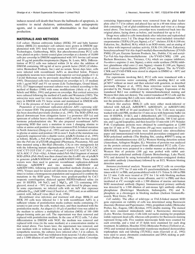
RESULTSOverexpression of wild-type and FALS-associatedmutant SOD using replication-deficient adenovirusesWe prepared recombinant AdVs with wild-type and mutantSODs as vectors for gene delivery into primary neural cells (Fig.1A). The presence of the mutation in the SOD cDNA wasconfirmed by PCR and sequencing (data not shown). We thentested whether expression of the SOD transgene resulted from theAdV infection. A Western blot of BHK-21 cells demonstrated thepresence of an immunostained protein product in the mock-infected (Fig. 1B, lane 1) as well as the AdV-infected cells (Fig.1B, lanes 2–5), corresponding to the electrophoretic mobility ofrodent SOD. This immunostaining was a result of cross-reactivityof the anti-human SOD polyclonal antibody with endogenousrodent SOD. An additional immunostained band of slower elec-trophoretic mobility was present in extracts from cells infectedwith AdSODWT, AdSODA4V, and AdSODV148G viruses (Fig.1B, lanes 3–5, respectively), corresponding to the electrophoreticmobility of human SOD. The levels of human SOD that wereexpressed in these cells were approximately similar to those ofendogenous SOD. A previous study has demonstrated functionalactivity of the AdSODWT virus (Jordan et al., 1995). Total SODactivity at 3 d after infection was similar in mock-infected PC12cells as well as cells infected with AdSODWT and AdSODV148Gvirus [relative activities were 100, 103 6 11, and 103 6 3% formock, wild-type (WT), and V148G expression, respectively].
Effects of overexpression of mutant SODs on neuralcell viabilityWe determined the effects of expression of wild-type and mutantSODs on differentiated PC12 cells, because they are frequentlyused as a model system for differentiated neural cells. Under ourexperimental conditions, infection with AdSODWT led to expres-sion in ;60 6 8% (n 5 7) of the cells, as demonstrated byimmunopositive staining with human SOD-specific antiserum (Fig.
2B). Comparable results were obtained after AdSODV148G in-fection (Fig. 2C) and after infection of primary neuronal cells (datanot shown; see below). We were unable to test for expression of theSODA4V mutant transgene with immunohistochemical stains be-cause of the lack of an available antibody that can differentiate theA4V mutant SOD from endogenous SOD; expression of this mu-tant was verified by Western blot (see above).
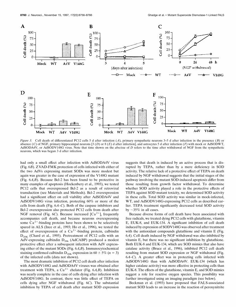
We compared PC12 cell viability after infection with AdVsexpressing mutant SODs with that seen after infection with a virusexpressing wild-type SOD. Compared to mock-infected controls,approximately half of the PC12 cells died 5 d after infection withAdSODA4V and AdSODV148G (Fig. 3A). In contrast, there wasno decline in survival of cells 5 d after infection with AdV express-ing wild-type SOD (AdSODWT) when compared with mock-infected cells (Fig. 3A). It is also clear that cells that died and thoseexpressing mutant SODs were the same population. Three daysafter infection ;60% of cells showed immunoreactivity for WTSOD or SODV148G. After 5 d, however, 54 6 4% of cells werestained positive for WT SOD (n 5 3), whereas only 14 6 3% werenow positive for SODV148G (n 5 3).
Figure 1. A, Schematic representation of the AdV-expressing wild-typeor mutant SOD. The virus contains a deletion in the early region 3 (DE3),replacement of early region 1 with EF-1a, Cu/Zn superoxide dismutase-1cDNA (SOD1cDNA), cellular 4F2 heavy chain enhancer (4F2), bovinegrowth hormone polyadenylation site [poly(A)], and adenovirus type 5map units (m.u.). The gene lengths shown are not to scale. B, Western blotof BHK-21 cells after mock infection (lane 1) or infection with AdLacZ(lane 2), AdSODWT (lane 3), AdSODA4V (lane 4 ), or AdSODV148Gvirus (lane 5). The cells were lysed, and the lysates were then subjected toSDS-PAGE, blotted onto nitrocellulose, and incubated with an anti-SODpolyclonal antibody. There is evidence of immunostaining of a proteinspecies corresponding to the electrophoretic mobility of rodent SOD(R-SOD) in all lanes and of human SOD (H-SOD) after infection with thewild-type and mutant SOD recombinant AdVs (lanes 3–5). The otherhigher molecular weight proteins that are immunostained are nonspecific.
8758 J. Neurosci., November 15, 1997, 17(22):8756–8766 Ghadge et al. • Mutant Superoxide Dismutase-1-Linked FALS

We then studied the effect of wild-type or mutant SOD expres-sion on primary sympathetic neurons during normal conditions aswell as during the added stress of growth factor withdrawal. Asshown in Figure 3B, there was no decline in viability of primaryneural cells grown in the presence of NGF after infection withAdSODWT virus. In contrast, AdSODV148G and AdSODA4V
induced cell death, so that only 60 and 45% of cells, respectively,remained after 5 d (Fig. 3B). As expected, uninfected primarysympathetic neurons died after NGF withdrawal and applicationof anti-NGF antiserum, a procedure known to induce apoptosisof these cells (Fig. 3C) (Martin et al., 1988). Infection withAdSODWT virus 3 d before NGF withdrawal almost completelyprotected these cells from further cell death (Fig. 3C), consistentwith our previously published studies (Jordan et al., 1995). Incontrast, infection with AdSODV148G and AdSODA4V led to anenhancement of cell death (Fig. 3C).
We also examined the effect of mutant SOD expression oncultured hippocampal pyramidal neurons. Three days after infec-tion there was little evidence of cell death (Fig. 3D). By 5 d celldeath increased after infection with AdSODV148G andAdSODA4V compared with mock-infected cells or cells infectedwith AdSODWT (data not shown), and by 8 d there was a veryrobust decrease in the number of viable cells after infection withthe viruses expressing mutant SODs compared with the mockinfection or after infection with AdSODWT virus (Fig. 3E).
To examine whether the cell death induced by mutant SOD wasspecific for neural cells, we also tested the effect of the recombi-nant AdVs on primary astrocytes. In contrast to neurons, astro-cytes proved to be resistant to the effects of SOD mutants (Fig.3F). Only 5–6% of the cells died by 5 d after infection, which wasnot significantly different from that after expression of WT SOD.
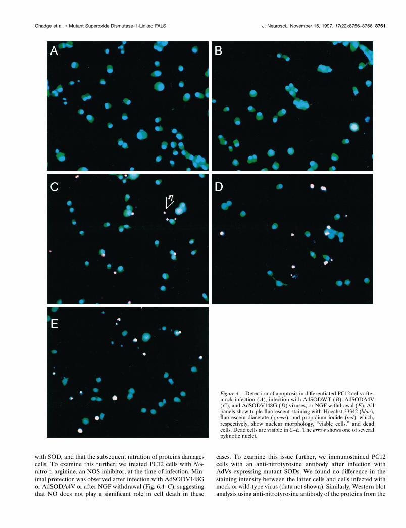
Characteristics of mutant SOD-induced neuralcell deathWe examined cells dying after expression of mutant SOD forevidence of apoptosis (Figs. 4, 5). Apoptotic changes were fre-quently found in these and detectable in many microscope fieldsexamined; in contrast, these changes were rarely seen in cellsinfected with AdSODWT. Dying PC12 cells (Fig. 4), hippocam-pal pyramidal neurons (Fig. 5), or sympathetic neurons (data notshown) exhibited histological and morphological features of ap-optosis, including shrunken cell soma, chromatin condensation(assessed using the Hoechst 33342–fluorescent diacetate–pro-pidium iodide triple staining), and DNA nicking (assessed usingTUNEL staining). As a comparison, we also performed experi-ments involving the death of differentiated PC12 cells and sym-pathetic neurons induced by removal of NGF (Martin et al.,1988). Cells dying after growth factor withdrawal showed fea-tures similar to those of the same cell types dying after mutantSOD expression (Fig. 4E).
In summary, we observed similar toxic effects of mutant SODsin differentiated PC12 cells and primary hippocampal and pri-mary sympathetic neurons. For this reason, and because of thegreater ease in performing studies with differentiated PC12 cells,we used these cells for future characterization of mutant SOD-induced cell death.
Inhibition of SOD mutant-induced neural cell deathTo clarify the mechanism(s) by which mutant SODs induceddeath of PC12 cells, the cells were treated with different agentsafter recombinant AdV infection (Fig. 6A,B). These results werecompared with the effects of the same agents on the death ofPC12 cells caused by removal of NGF (Fig. 6C). We tested theeffect of ZVAD-FMK and Ac-YVAD-CMK, two irreversibleinhibitors of interleukin 1b-converting enzyme-like proteases(caspases) that have been implicated in apoptosis (Troy et al.,1996b). Ac-YVAD-CMK inhibited PC12 cell death 5 d afterinfection with AdSODV148G virus (Fig. 6A), although the drug
Figure 2. Immunohistochemical staining of PC12 cells (A–C) with anti-human SOD antibody after mock infection ( A) or infection withAdSODWT (B) and AdSODV148G ( C) viruses. Further details aregiven in Materials and Methods.
Ghadge et al. • Mutant Superoxide Dismutase-1-Linked FALS J. Neurosci., November 15, 1997, 17(22):8756–8766 8759
had only a small effect after infection with AdSODA4V virus(Fig. 6B). ZVAD-FMK protection of cells infected with either ofthe two AdVs expressing mutant SODs was more modest butagain was greater in the case of expression of the V148G mutant(Fig. 6A,B). Because Bcl-2 has been found to be protective inmany examples of apoptosis (Hockenbery et al., 1993), we testedPC12 cells that overexpressed Bcl-2 as a result of retroviraltransduction (see Materials and Methods). Bcl-2 overexpressionhad a significant effect on cell viability after AdSODA4V andAdSODV148G virus infection, protecting 60% or more of thecells from death (Fig. 6A–C). Both of the caspase inhibitors andBcl-2 overexpression also protected PC12 cells from death afterNGF removal (Fig. 6C). Because increased [Ca21]i frequentlyaccompanies cell death, and because neurons overexpressingsome Ca 21-binding proteins have been shown to be selectivelyspared in ALS (Ince et al., 1993; Ho et al., 1996), we tested theeffect of overexpression of a Ca 21-binding protein, calbindinD28K (Chard et al., 1995). Pretreatment of PC12 cells with anAdV-expressing calbindin D28k (AdCABP) produced a modestprotective effect after a subsequent infection with AdV express-ing either of the mutant SODs (Fig. 6A,B). Immunocytochemicalstaining confirmed calbindin D28K expression in 60 6 5% (n 5 3)of the infected cells (data not shown).
The most dramatic inhibition of PC12 cell death after infectionwith AdSODV148G and AdSODA4V viruses was obtained aftertreatment with TEPA, a Cu 21 chelator (Fig. 6A,B). Inhibitionwas nearly complete in the case of cells dying after infection withAdSODV148G. In contrast, there was little effect of TEPA oncells dying after NGF withdrawal (Fig. 6C). The substantialinhibition by TEPA of cell death after mutant SOD expression
suggests that death is induced by an active process that is dis-rupted by TEPA, rather than by a mere deficiency in SODactivity. The relative lack of a protective effect of TEPA on deathinduced by NGF withdrawal suggests that the initial stages of thepathway involving the mutant SOD-induced apoptosis differ fromthose resulting from growth factor withdrawal. To determinewhether SOD activity played a role in the protective effects ofTEPA against SOD mutant toxicity, we determined SOD activityin these cells. Total SOD activity was similar in mock-infected,WT, and AdSODV148G-expressing PC12 cells as described ear-lier. TEPA treatment significantly decreased total SOD activityby ;35% in all cases.
Because diverse forms of cell death have been associated withfree radicals, we treated dying PC12 cells with glutathione, vitaminE, EUK-8, and EUK-134. A significant inhibition of cell deathinduced by expression of SODV148G was observed after treatmentwith the antioxidant compounds glutathione and vitamin E (Fig.6A). Cell death induced by NGF withdrawal was also inhibited byvitamin E, but there was no significant inhibition by glutathione.Both EUK-8 and EUK-134, which are SOD mimics that also havecatalase activity (Bruce et al., 1996), inhibited PC12 cell deathresulting from mutant SOD expression or NGF withdrawal (Fig.6A–C). A greater effect was in protecting cells infected withAdSODV148G than with AdSODA4V. EUK-134 (which hashigher catalase activity) was more effective in protecting cells thanEUK-8. The effects of the glutathione, vitamin E, and SOD mimicssuggest a role for reactive oxygen species. This possibility wasfurther investigated using an imaging paradigm (see below).
Beckman et al. (1993) have proposed that FALS-associatedmutant SOD leads to an increase in the reaction of peroxynitrite
Figure 3. Cell death of differentiated PC12 cells 5 d after infection ( A), primary sympathetic neurons 3–5 d after infection in the presence ( B) orabsence (C) of NGF, primary hippocampal neurons [3 (D) or 8 (E) d after infection], and astrocytes 5 d after infection (F) with mock or AdSODWT,AdSODA4V, or AdSODV148G virus. Note that time shown on the abscissa of D refers to the time after withdrawal of NGF from the sympatheticneurons, which was begun 3 d after infection.
8760 J. Neurosci., November 15, 1997, 17(22):8756–8766 Ghadge et al. • Mutant Superoxide Dismutase-1-Linked FALS
with SOD, and that the subsequent nitration of proteins damagescells. To examine this further, we treated PC12 cells with Nv-nitro-L-arginine, an NOS inhibitor, at the time of infection. Min-imal protection was observed after infection with AdSODV148Gor AdSODA4V or after NGF withdrawal (Fig. 6A–C), suggestingthat NO does not play a significant role in cell death in these
cases. To examine this issue further, we immunostained PC12cells with an anti-nitrotyrosine antibody after infection withAdVs expressing mutant SODs. We found no difference in thestaining intensity between the latter cells and cells infected withmock or wild-type virus (data not shown). Similarly, Western blotanalysis using anti-nitrotyrosine antibody of the proteins from the
Figure 4. Detection of apoptosis in differentiated PC12 cells aftermock infection ( A), infection with AdSODWT ( B), AdSODA4V(C), and AdSODV148G ( D) viruses, or NGF withdrawal ( E). Allpanels show triple fluorescent staining with Hoechst 33342 (blue),fluorescein diacetate ( green), and propidium iodide (red), which,respectively, show nuclear morphology, “viable cells,” and deadcells. Dead cells are visible in C–E. The arrow shows one of severalpyknotic nuclei.
Ghadge et al. • Mutant Superoxide Dismutase-1-Linked FALS J. Neurosci., November 15, 1997, 17(22):8756–8766 8761
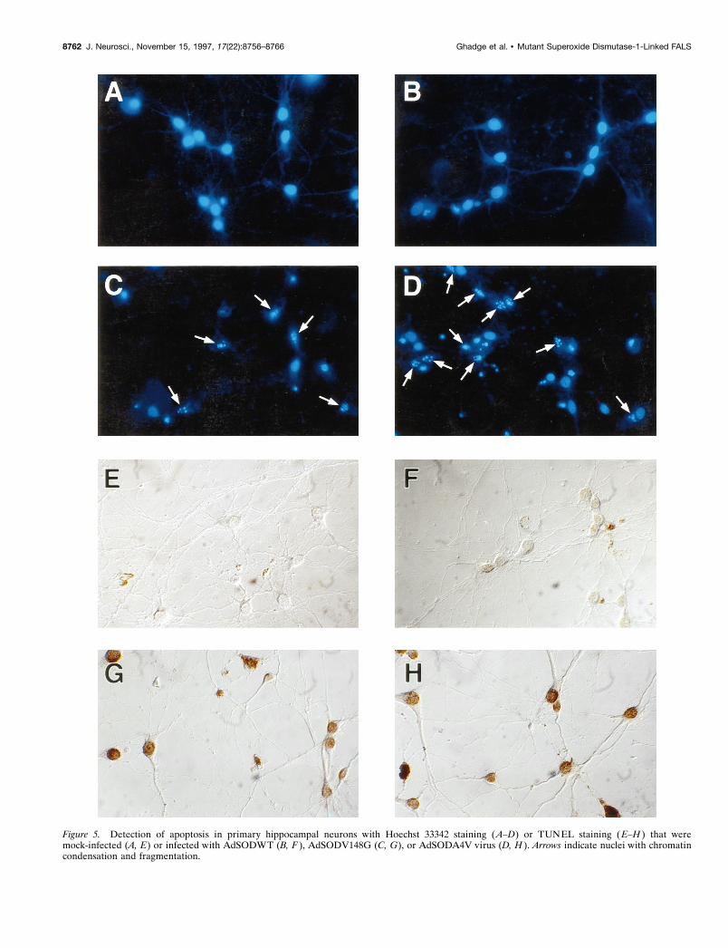
Figure 5. Detection of apoptosis in primary hippocampal neurons with Hoechst 33342 staining (A–D) or TUNEL staining (E–H ) that weremock-infected (A, E) or infected with AdSODWT (B, F ), AdSODV148G (C, G), or AdSODA4V virus (D, H ). Arrows indicate nuclei with chromatincondensation and fragmentation.
8762 J. Neurosci., November 15, 1997, 17(22):8756–8766 Ghadge et al. • Mutant Superoxide Dismutase-1-Linked FALS
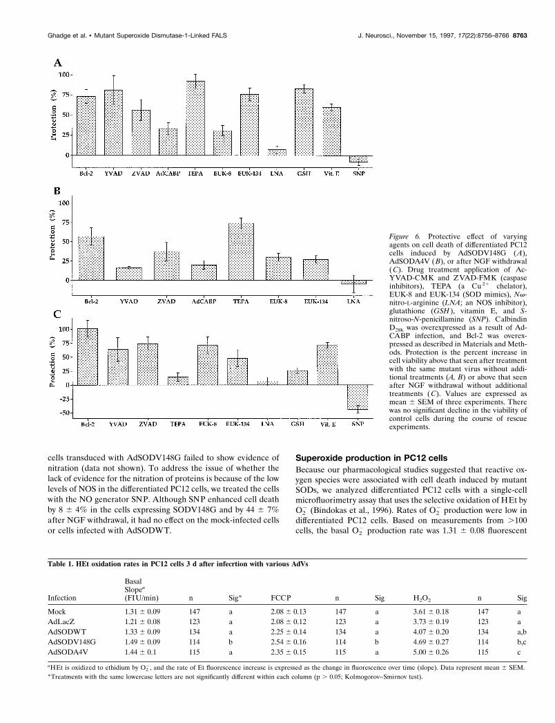
cells transduced with AdSODV148G failed to show evidence ofnitration (data not shown). To address the issue of whether thelack of evidence for the nitration of proteins is because of the lowlevels of NOS in the differentiated PC12 cells, we treated the cellswith the NO generator SNP. Although SNP enhanced cell deathby 8 6 4% in the cells expressing SODV148G and by 44 6 7%after NGF withdrawal, it had no effect on the mock-infected cellsor cells infected with AdSODWT.
Superoxide production in PC12 cellsBecause our pharmacological studies suggested that reactive ox-ygen species were associated with cell death induced by mutantSODs, we analyzed differentiated PC12 cells with a single-cellmicrofluorimetry assay that uses the selective oxidation of HEt byO2
2 (Bindokas et al., 1996). Rates of O22 production were low in
differentiated PC12 cells. Based on measurements from .100cells, the basal O2
2 production rate was 1.31 6 0.08 fluorescent
Table 1. HEt oxidation rates in PC12 cells 3 d after infecrtion with various AdVs
Infection
BasalSlopea
(FIU/min) n Sig* FCCP n Sig H2O2 n Sig
Mock 1.31 6 0.09 147 a 2.08 6 0.13 147 a 3.61 6 0.18 147 aAdLacZ 1.21 6 0.08 123 a 2.08 6 0.12 123 a 3.73 6 0.19 123 aAdSODWT 1.33 6 0.09 134 a 2.25 6 0.14 134 a 4.07 6 0.20 134 a,bAdSODV148G 1.49 6 0.09 114 b 2.54 6 0.16 114 b 4.69 6 0.27 114 b,cAdSODA4V 1.44 6 0.1 115 a 2.35 6 0.15 115 a 5.00 6 0.26 115 c
aHEt is oxidized to ethidium by O22, and the rate of Et fluorescence increase is expressed as the change in fluorescence over time (slope). Data represent mean 6 SEM.
*Treatments with the same lowercase letters are not significantly different within each column (p . 0.05; Kolmogorov–Smirnov test).
Figure 6. Protective effect of varyingagents on cell death of differentiated PC12cells induced by AdSODV148G (A),AdSODA4V ( B), or after NGF withdrawal(C). Drug treatment application of Ac-YVAD-CMK and ZVAD-FMK (caspaseinhibitors), TEPA (a Cu 21 chelator),EUK-8 and EUK-134 (SOD mimics), Nv-nitro-L-arginine (LNA; an NOS inhibitor),glutathione (GSH ), vitamin E, and S-nitroso-N-penicillamine (SNP). CalbindinD28k was overexpressed as a result of Ad-CABP infection, and Bcl-2 was overex-pressed as described in Materials and Meth-ods. Protection is the percent increase incell viability above that seen after treatmentwith the same mutant virus without addi-tional treatments (A, B) or above that seenafter NGF withdrawal without additionaltreatments (C). Values are expressed asmean 6 SEM of three experiments. Therewas no significant decline in the viability ofcontrol cells during the course of rescueexperiments.
Ghadge et al. • Mutant Superoxide Dismutase-1-Linked FALS J. Neurosci., November 15, 1997, 17(22):8756–8766 8763
units (FlU)/min (Table 1). Rates increased slightly in PC12 cellsafter addition of the mitochondrial uncoupler FCCP, consistentwith the electron transport chain being one source of O2
2 gener-ation (Bindokas et al., 1996). Addition of hydrogen peroxide(H2O2) significantly increased the oxidation of HEt, indicating anincrease in O2
2. This action was not attributable to a directinteraction of H2O2 with HEt (Bindokas et al., 1996) but a resultof H2O2 inactivating or overwhelming other O2
2-scavenging path-ways and possibly also enhancing the generation of O2
2.Expression of the two mutant AdSODs tended to increase
basal rates of O22 production slightly, but only after V148G
expression was this significantly higher than mock-treated cells( p 5 0.001) (Table 1). The rate of O2
2 production after infectionwith the AdV-expressing wild-type SOD was nearly identical tothat of mock-treated cells (1.33 6 0.09), whereas there was aslightly lower rate (1.21 6 0.08; p 5 0.48) after infection withAdLacZ virus.
Application of FCCP (1 mM) increased the O22 production rates
;1.5 times to 2.08 6 0.13 FlU/min in mock-treated cells and incells infected with AdLacZ virus. There was a greater increase inslope for cells in which either of the mutant SODs were ex-pressed. The highest rates were observed with the AdSODV148Gmutant ( p , 0.05), with less of an increase with AdSODA4V.These results were in contrast with those seen after expression ofwild-type SOD, in which there was no significant change com-pared with controls.
Application of H2O2 further increased O22 production and
amplified the trends observed. The largest increases occurredwith cells expressing mutant SODV148G ( p 5 0.04 vs mock) andSODA4V ( p 5 0.001). Cells infected with AdSODWT were notsignificantly different from the mock-infected cells ( p 5 0.23).These data are consistent with an enhanced peroxidase activityfor mutant SOD (Wiedau-Pazos et al., 1996; Yim et al., 1996).
DISCUSSIONThe identification of mutations in SOD as a cause of FALSgenerated substantial excitement among neuroscientists, owing tothe possibility that these findings might clarify the pathogenesis ofnot only FALS but sporadic ALS as well. Initially it was supposedthat the FALS-associated mutant enzymes had inadequate SODactivity, thereby leading to an accumulation of O2
2 and other freeradicals with the subsequent death of motor neurons. This hy-pothesis had to be modified, however, because a number ofstudies, including those involving FALS transgenic mice (Gurneyet al., 1994; Ripps et al., 1995) and wild-type SOD knock-out mice(Reaume et al., 1996), suggested that FALS-associated mutantSODs induced neuronal damage by another mechanism ratherthan one involving impaired dismutase enzyme activity.
Past studies exploring the effect of mutant SODs in vitro haveprimarily involved yeast and non-neural cells. The only study thathas investigated neural cells used a continuous rat nigral cell linethat was permanently transfected with wild-type or mutant SODcDNAs (Rabizadeh et al., 1995). A potential drawback of thelatter studies is that continued growth of these permanentlytransfected cells may have led to compensatory changes attribut-able to expression of the deleterious gene. Experiments involvingtransient expression in primary cultured neurons have not previ-ously been feasible because of the inefficiency of conventionalmethods for gene transduction into such cells. In the presentreport we circumvented this problem by using AdVs to deliverand express foreign genes efficiently in primary neuronal cells aswell as a differentiated neural cell line.
Our studies demonstrated that mutant SODs induce the deathof several types of differentiated neural cells. We found that celldeath occurred in differentiated PC12 cells, primary sympatheticneurons, and primary hippocampal neurons. In contrast, therewas little death of primary astrocytes, suggesting that neurons aremore sensitive to the effects of mutant SOD. The greater celldeath after the SODA4V expression compared with theSODV148G may be related to the greater toxicity of the formermutant, as suggested by the decreased survival of FALS patientswho carry this mutation (Juneja et al., 1996). We found noevidence of increased cell death after transduction of these cellswith wild-type SOD, demonstrating the differential effects ofmutant versus wild-type SOD (Jordan et al., 1995).
The cell death that occurred after expression of mutant SODshad morphological features typical of apoptosis. This was alsosupported by our finding that antiapoptotic agents, such as Bcl-2and caspase inhibitors, blocked cell death. These results as well asthe finding of a robust protective effect of TEPA suggest thatcaspase inhibitors and TEPA should be tested for their ability todelay the onset or decrease the severity of the neurodegenerationin FALS transgenic mice. It may be that screening drugs thatreverse the cell death in mutant SOD-expressing PC12 cells willprovide a means to identify drugs that are effective in the treat-ment of FALS as well as sporadic ALS.
It should be noted, however, that the cell death data do notnecessarily mean that neurons in FALS normally use an apoptoticmechanism of cell death during the disease state, because virtu-ally any cell can die by apoptosis if prompted to do so by someadverse stimulus (Raff et al., 1994). Rather, the results supportthe idea that mutant SODs are perceived by these cells as noxiousin some way and suggest that apoptotic mechanisms may also beinvolved in the neurodegeneration in FALS.
Oxidative damage and decreased free radical scavenging areboth known to induce apoptotic cell death (Greenlund et al.,1995). In our studies, reactive oxygen species, especially O2
2,appeared to be involved in the mutant SOD-induced cell death,because glutathione and SOD mimics tended to decrease thismortality. We also found that Bcl-2, which is known to affect freeradical generation and cell viability similarly, protected the cellsagainst the apoptosis induced by mutant SOD (Hockenbery et al.,1993). In addition, microfluorimetry demonstrated a slightly in-creased rate of O2
2 accumulation in PC12 cells after expression ofmutant SODs, especially in cells undergoing oxidative stress;these data suggest that expression of mutant SOD increases thesensitivity to oxidative stress. Increased O2
2 can potentially leadto formation of additional reactive species, including hydroxylradicals and peroxynitrite. Studies illustrating a beneficial effectof vitamin E on the clinical course of FALS transgenic mice(Gurney et al., 1996) and our results with vitamin E are alsoconsistent with a role for oxidative stress in disease pathogenesis.
Our data regarding O22 suggest that the free radical content of
cells expressing mutant SODs is disturbed, despite the expressionof endogenous SOD. There are several possible explanations forour findings. A simple, slight decrease in dismutase activity couldexplain the increased O2
2 levels that generate additional reactivespecies. It is possible that the mutant SOD could interfere withthe function of the wild-type SOD; i.e., there is a dominantnegative effect; although there is some evidence for the lattereffect in Drosophila melanogaster that carry a mutant SOD trans-gene (Phillips et al., 1995), there is little support for this activityin vertebrate cells (Borchelt et al., 1994). It may be that the SODactivity is unable to keep up with the enhanced generation of O2
2
8764 J. Neurosci., November 15, 1997, 17(22):8756–8766 Ghadge et al. • Mutant Superoxide Dismutase-1-Linked FALS
as a result, for example, of mitochondrial damage. Although SODknock-out mice do not demonstrate anterior horn cell degenera-tion, they are more sensitive to injury, such as axotomy (Reaumeet al., 1996). We believe, however, that it is unlikely that themechanism of mutant SOD cell death involves a deficiency ofSOD activity. Studies with PC12 cells have demonstrated thatdownregulation of SOD activity kills via an apoptotic pathwaythat is different from the apoptotic pathway that we found aftermutant SOD expression. Thus, we found no protective effect ofNOS inhibitors on the mutant SOD-induced apoptosis, whereasNOS inhibitors decrease cell death after SOD downregulation ofPC12 cells (Troy et al., 1996a,b). These results cannot be ex-plained by postulating a deficiency of SOD activity as the mech-anism of cell death. Furthermore, total SOD activity in the WTand mutant SOD-expressing PC12 cells was similar, and TEPAtreatment decreased this activity by 35% in all the cases.
It has been proposed that mutant SODs may possess a gain infunction or enhancement of an existing toxic nondismutase func-tion. Beckman and colleagues (1993) suggested that the mutantenzyme had an enhanced reactivity for peroxynitrite leading toan increase in the nitration of tyrosines. We tested this hypothesisby subjecting PC12 cells that had been infected by AdV-expressing mutant SOD to immunostaining and Western blotanalysis using an antiserum that reacts with nitrotyrosine. Wefailed to find evidence of increased nitrotyrosine antigenicity inthe virus-infected cells. We also investigated the potential impor-tance of peroxynitrite in the mutant SOD-induced apoptosis byperturbing NOS activity leading to decreased or increased pro-duction of NO. Again, there was no evidence that decreasing NOsynthesis affected cell viability or that increasing NO generationaccelerated cell death; as mentioned above, these results contrastwith experiments in PC12 cells in which downregulation of SODactivity produced NOS-dependent cell death (Troy et al.,1996a,b) and in which generation of NO enhanced cell death afterNGF withdrawal (Fig. 6C) and suggest that mutant SOD mayitself be a source for toxic radicals. These results fail to support arole for peroxynitrite and the subsequent nitration of proteins asa primary cause of cell death after expression of mutant SOD.The nitrotyrosine staining that has been found in motor neuronsof ALS patients (Abe et al., 1995; Chou et al., 1996) may be asecondary and later change that follows a different primary injuryto these cells. Thus, it may be that the O2
2 accumulation that weobserved could lead, in particular cells, to the secondary produc-tion of a number of other free radical species, such as peroxyni-trite. Our findings in mutant SOD-expressing cells are consistentwith other possible mechanisms that have been proposed toexplain the effects of mutant SODs. This includes an increase inperoxidase activity that is normally present in wild-type SOD andcould be enhanced in the mutant form (Wiedau-Pazos et al.,1996; Yim et al., 1996) as well as a decrease in binding orshielding of the mutant enzyme to metals, which results in anincrease in Cu21-catalyzed oxidative reactions.
Because all neuronal cells we tested were susceptible to the celldeath of mutant SOD, the data do little to explain the selectivevulnerability of motor neurons that occurs in FALS and ALS. Itmay be that the extraordinary metabolic activity of motor neu-rons puts them at a heightened risk with respect to damage fromthe free radical species that are generated as a result of themutant SOD expression. Alternatively, there may be other factorspresent in the CNS that confer a selective vulnerability to motorneurons.
REFERENCESAbe K, Pan LH, Watanabe M, Kato T, Itoyama Y (1995) Induction of
nitrotyrosine-like immunoreactivity in the lower motor neuron ofamyotrophic lateral sclerosis. Neurosci Lett 199:152–154.
Abele AE, Scholz KP, Scholz WK, Miller RJ (1990) Excitotoxicityinduced by enhanced excitatory neurotransmission in cultured hip-pocampal pyramidal neurons. Neuron 4:413–419.
Banker GA (1980) Trophic interactions between astroglial cells and hip-pocampal neurons in culture. Science 209:809–810.
Barr E, Carroll J, Kalynych AM, Tripathy SK, Kozarsky K, Wilson JM,Leiden JM (1994) Efficient catheter-mediated gene transfer into theheart using replication-defective adenovirus. Gene Ther 1:51–58.
Beckman JS, Carson M, Smith CD, Koppenol WH (1993) ALS, SODand peroxynitrite [letter]. Nature 364:584.
Bindokas VP, Jordan J, Lee CC, Miller RJ (1996) Superoxide produc-tion in rat hippocampal neurons: selective imaging with hydroethidine.J Neurosci 16:1324–1336.
Borchelt DR, Lee MK, Slunt HS, Guarnieri M, Xu ZS, Wong PC, BrownJr RH, Price DL, Sisodia SS, Cleveland DW (1994) Superoxide dis-mutase 1 with mutations linked to familial amyotrophic lateral sclerosispossesses significant activity. Proc Natl Acad Sci USA 91:8292–8296.
Brown Jr RH (1995) Amyotrophic lateral sclerosis: recent insights fromgenetics and transgenic mice. Cell 80:687–692.
Bruce AJ, Malfroy B, Baudry M (1996) Beta-amyloid toxicity in orga-notypic hippocampal cultures: protection by EUK-9, a synthetic cata-lytic free radical scavenger. Proc Natl Acad Sci USA 93:2312–2316.
Chard PS, Jordan J, Marcuccilli CJ, Miller RJ, Leiden JM, Roos RP,Ghadge GD (1995) Regulation of excitatory transmission at hip-pocampal synapses by calbindin D28k. Proc Natl Acad Sci USA92:5144–5148.
Chou SM, Wang HS, Taniguchi A (1996) Role of SOD-1 and nitricoxide/cyclic GMP cascade on neurofilament aggregation in ALS/MND. J Neurol Sci [Suppl] 139:16–26.
Deng HX, Hentati A, Tainer JA, Iqbal Z, Cayabyab A, Hung WY,Getzoff ED, Hu P, Herzfeldt B, Roos RP, Warner C, Deng G, SorianoE, Smyth C, Parge HE, Ahmed A, Roses AD, Hallewell RA, Pericak-Vance MA, Siddique T (1993) Amyotrophic lateral sclerosis andstructural defects in Cu,Zn superoxide dismutase. Science261:1047–1051.
Gavrieli Y, Sherman Y, Ben-Sasson SA (1992) Identification of pro-grammed cell death in situ via specific labeling of nuclear DNA frag-mentation. J Cell Biol 119:493–501.
Greenlund LJS, Deckwerth TL, Johnson Jr EM (1995) Superoxide dis-mutase delays neuronal apoptosis: a role for reactive oxygen species inprogrammed neuronal death. Neuron 14:303–315.
Gurney ME, Pu H, Chiu AY, Dal Canto MC, Polchow CY, AlexanderDD, Caliendo J, Hentati A, Kwon YW, Deng HX, Chen W, Zhai P,Sufit RL, Siddique T (1994) Motor neuron degeneration in mice thatexpress a human Cu,Zn superoxide dismutase mutation. Science264:1772–1775.
Gurney ME, Cutting FB, Zhai P, Doble A, Taylor CP, Andrus PK, HallED (1996) Benefit of vitamin E, riluzole, and gabapentin in a trans-genic model of familial amyotrophic lateral sclerosis. Ann Neurol39:147–157.
Ho BK, Alexianu ME, Colom LV, Mohamed AH, Serrano F, Appel SH(1996) Expression of calbindin-D28K cDNA prevents amyotrophiclateral sclerosis IgG-mediated cytotoxicity. Proc Natl Acad Sci USA93:6786–6801.
Hockenbery DM, Oltvai ZN, Yin X-M, Milliman CL, Korsmeyer SJ(1993) Bcl-2 functions in an antioxidant pathway. Cell 75:241–251.
Ince P, Stout N, Shaw P, Slade J, Hunziker W, Heizmann CW, Baim-bridge KG (1993) Parvalbumin and calbindin D-28k in the humanmotor system and in motor neuron disease. Neuropathol Appl Neuro-biol 19:291–299.
Jordan J, Ghadge GD, Prehn JH, Toth PT, Roos RP, Miller RJ (1995)Expression of human copper/zinc-superoxide dismutase inhibits thedeath of rat sympathetic neurons caused by withdrawal of nerve growthfactor. Mol Pharmacol 47:1095–1100.
Juneja T, Cardenas Y, Quershi I, Siddique T (1996) Survival predictionin familial amyotrophic lateral sclerosis with superoxide dismutase-1mutations. Neurology [Suppl] 46:A206–A207.
Krishan A (1975) Rapid flow cytofluorometric analysis of mammaliancell cycle by propidium iodide staining. J Cell Biol 66:188–193.
Landis DM, Weinstein LA (1983) Membrane structure in cultured as-trocytes. Brain Res 276:31–41.
Ghadge et al. • Mutant Superoxide Dismutase-1-Linked FALS J. Neurosci., November 15, 1997, 17(22):8756–8766 8765
Marklund S (1976) Spectrophotometric study of spontaneous dispropor-tionation of superoxide anion radical and sensitive direct assay forsuperoxide dismutase. J Biol Chem 251:7504–7507.
Martin DP, Schmidt RE, DiStefano PS, Lowry OH, Carter JG, JohnsonJr EM (1988) Inhibitors of protein synthesis and RNA synthesis pre-vent neuronal death caused by nerve growth factor withdrawal. J CellBiol 106:829–844.
Phillips JP, Tainer JA, Getzoff ED, Boulianne GL, Kirby K, Hilliker AJ(1995) Subunit-destabilizing mutations in Drosophila copper/zinc su-peroxide dismutase: neuropathology and a model of dimer dysequilib-rium. Proc Natl Acad Sci USA 92:8574–8578.
Prehn JH, Bindokas VP, Jordan J, Galindo MF, Ghadge GD, Roos RP,Boise LH, Thompson CB, Krajewski S, Reed JC, Miller RJ (1996)Protective effect of transforming growth factor-beta 1 on beta-amyloidneurotoxicity in rat hippocampal neurons. Mol Pharmacol 49:319–328.
Rabizadeh S, Gralla EB, Borchelt DR, Gwinn R, Valentine JS, Sisodia S,Wong P, Lee M, Hahn H, Bredesen DE (1995) Mutations associatedwith amyotrophic lateral sclerosis convert superoxide dismutase froman antiapoptotic gene to a proapoptotic gene: studies in yeast andneural cells. Proc Natl Acad Sci USA 92:3024–3028.
Raff MC, Barres BA, Burne JF, Coles HS, Ishizaki Y, Jacobson MD(1994) Programmed cell death and the control of cell survival. PhilosTrans R Soc Lond [Biol] 345:265–268.
Reaume AG, Elliott JL, Hoffman EK, Kowall NW, Ferrante RJ, SiwekDF, Wilcox HM, Flood DG, Beal MF, Brown Jr RH, Scott RW, SniderWD (1996) Motor neurons in Cu/Zn superoxide dismutase-deficientmice develop normally but exhibit enhanced cell death after axonalinjury. Nat Genet 13:43–47.
Ripps ME, Huntley GW, Hof PR, Morrison JH, Gordon JW (1995)Transgenic mice expressing an altered murine superoxide dismutasegene provide an animal model of amyotrophic lateral sclerosis. ProcNatl Acad Sci USA 92:689–693.
Rotman B, Papermaster BW (1966) Membrane properties of living
mammalian cells as studied by enzymatic hydrolysis of fluorogenicesters. Proc Natl Acad Sci USA 55:134–141.
Scholz KP, Miller RJ (1991) Analysis of adenosine actions on Ca 21
currents and synaptic transmission in cultured rat hippocampal pyra-midal neurones. J Physiol (Lond) 435:373–393.
Telford WG, King LE, Fraker PJ (1992) Comparative evaluation ofseveral DNA binding dyes in the detection of apoptosis-associatedchromatin degradation by flow cytometry. Cytometry 13:137–143.
Troy CM, Derossi D, Prochiantz A, Greene LA, Shelanski ML (1996a)Downregulation of Cu/Zn superoxide dismutase leads to cell death viathe nitric oxide-peroxynitrite pathway. J Neurosci 16:253–261.
Troy CM, Stefanis L, Prochiantz A, Greene LA, Shelanski ML (1996b)The contrasting roles of ICE family proteases and interleukin-1b inapoptosis induced by trophic factor withdrawal and by copper/zincsuperoxide dismutase down-regulation. Proc Natl Acad Sci USA93:5635–5640.
Wagner AJ, Small MB, Hay N (1993) Myc-mediated apoptosis isblocked by ectopic expression of Bcl-2. Mol Cell Biol 13:2432–2440.
Wiedau-Pazos M, Goto JJ, Rabizadeh S, Gralla EB, Roe JA, Lee MK,Valentine JS, Bredesen DE (1996) Altered reactivity of superoxidedismutase in familial amyotrophic lateral sclerosis. Science271:515–518.
Wong PC, Pardo CA, Borchelt DR, Lee MK, Copeland NG, Jenkins NA,Sisodia SS, Cleveland DW, Price DL (1995) An adverse property of afamilial ALS-linked SOD1 mutation causes motor neuron disease char-acterized by vacuolar degeneration of mitochondria. Neuron14:1105–1116.
Yim MB, Kang J-H, Yim H-S, Kwak H-S, Chock PB, Stadtman ER(1996) A gain of-function of an amyotrophic lateral sclerosis-associated Cu,Zn-superoxide dismutase mutant: an enhancement offree radical formation due to a decrease in Km for hydrogen peroxide.Proc Natl Acad Sci USA 93:5709–5714.
8766 J. Neurosci., November 15, 1997, 17(22):8756–8766 Ghadge et al. • Mutant Superoxide Dismutase-1-Linked FALS