MUSCULAR SYSTEM. Types of muscles SkeletalCardiacSmooth.
26
MUSCULAR SYSTEM
-
Upload
devonte-broxton -
Category
Documents
-
view
239 -
download
0
Transcript of MUSCULAR SYSTEM. Types of muscles SkeletalCardiacSmooth.
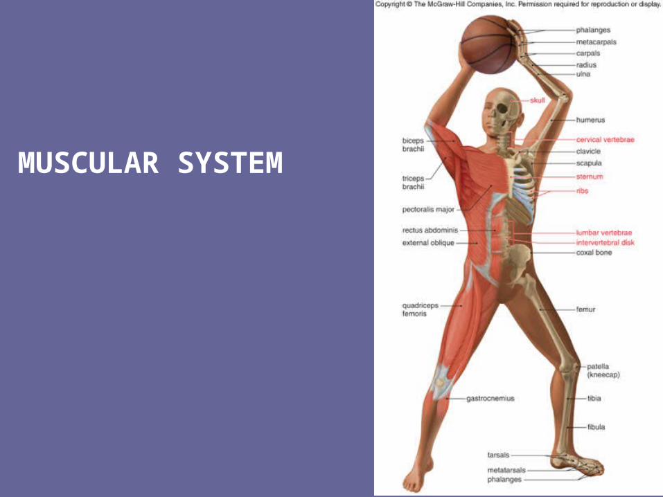
MUSCULAR SYSTEM
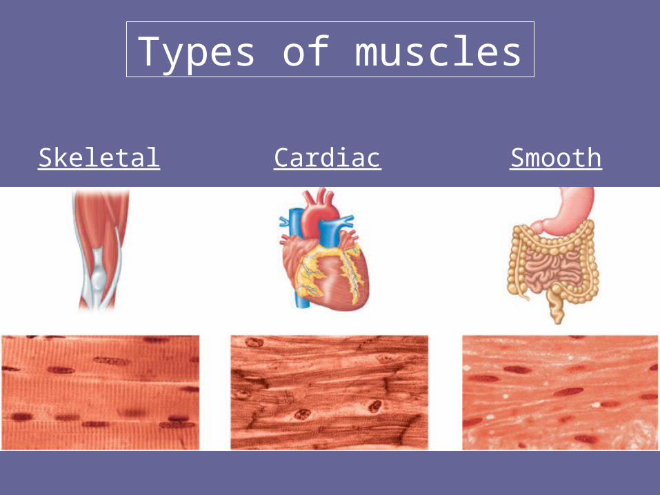
Types of muscles
Skeletal Cardiac Smooth
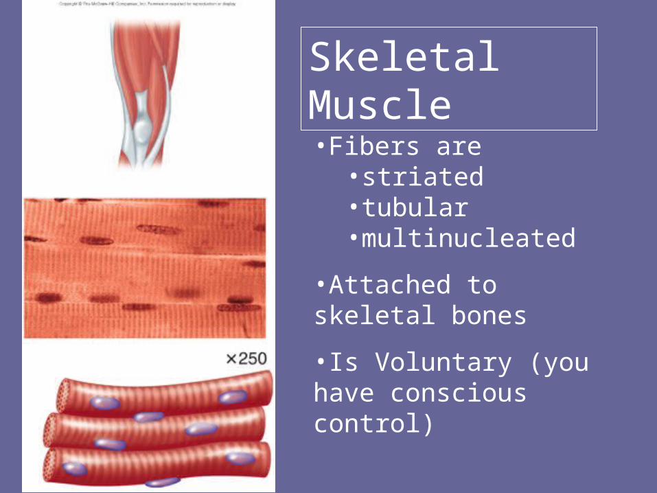
Skeletal Muscle
•Fibers are •striated•tubular•multinucleated
•Attached to skeletal bones
•Is Voluntary (you have conscious control)
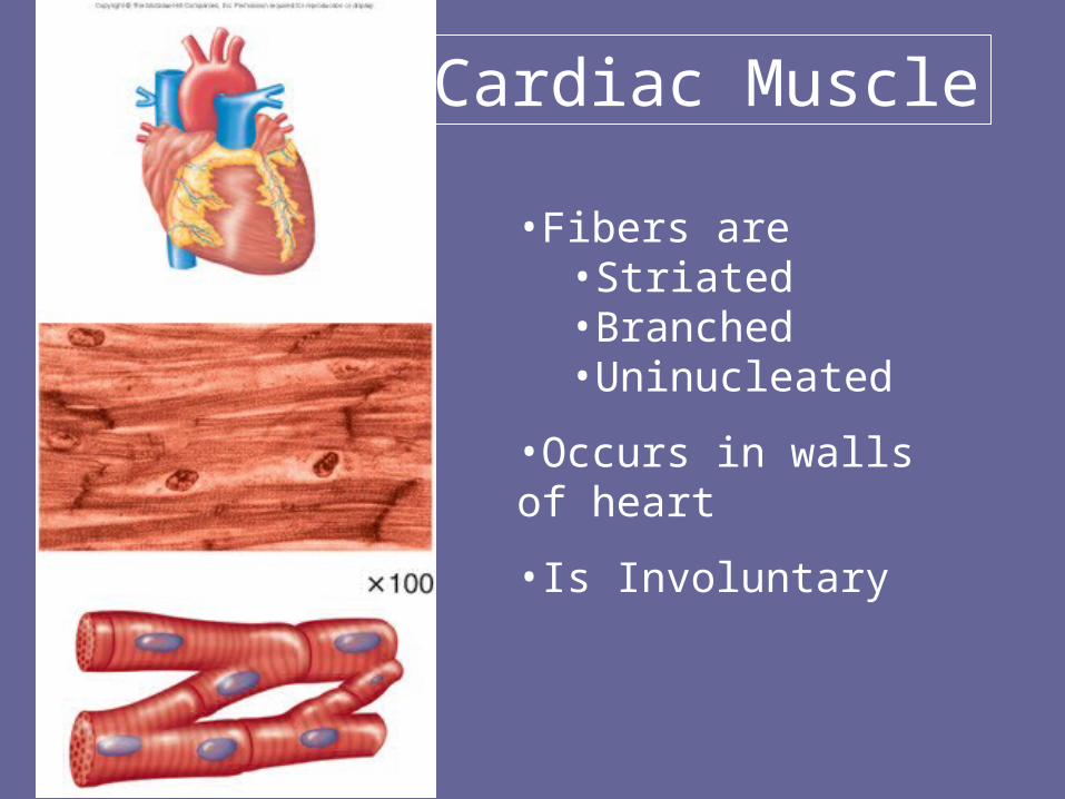
Cardiac Muscle
•Fibers are•Striated•Branched•Uninucleated
•Occurs in walls of heart
•Is Involuntary
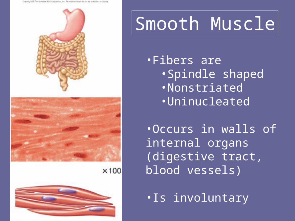
Smooth Muscle
•Fibers are•Spindle shaped•Nonstriated•Uninucleated
•Occurs in walls of internal organs (digestive tract, blood vessels)
•Is involuntary
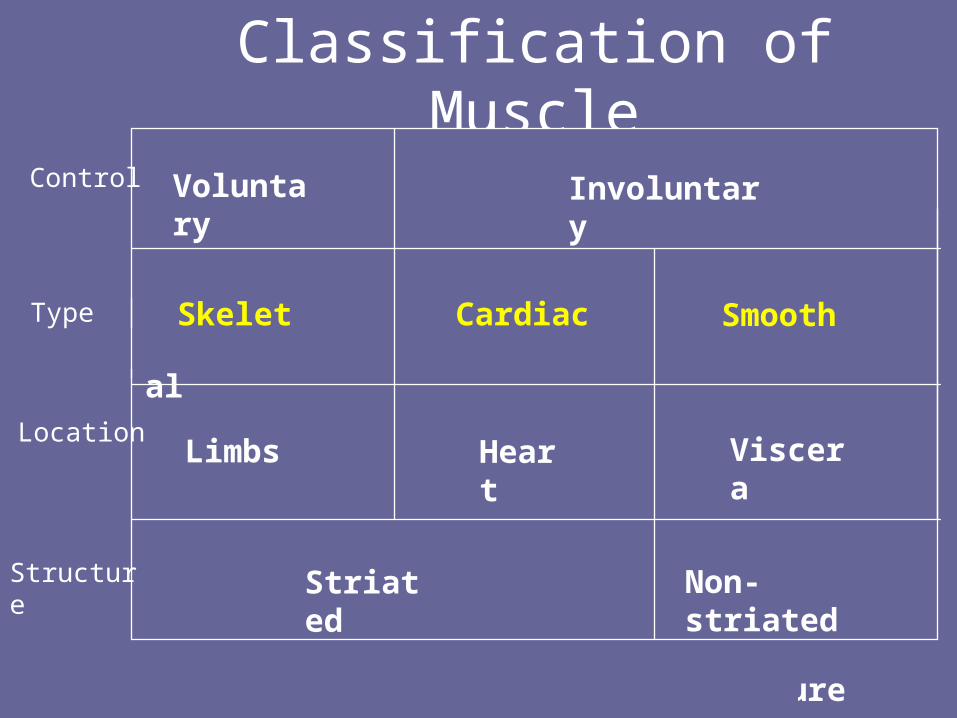
Classification of Muscle
Voluntary Involuntary
Skeletal Cardiac Smooth
Limbs Heart Viscera
Striated Non-striated
Skeletal Cardiac Smooth
Note: Control, Location and Structure
Control
Location
Type
Structure
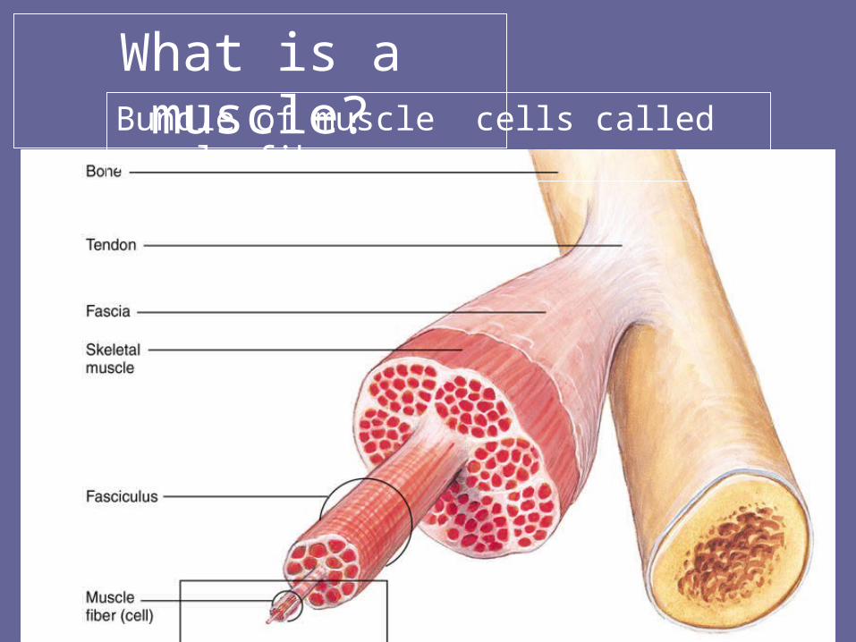
What is a muscle?Bundle of muscle cells called muscle fibers
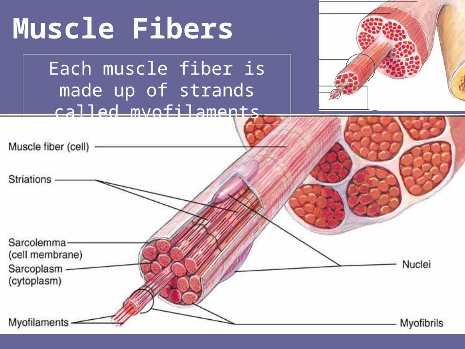
Each muscle fiber is made up of strands called myofilaments
Muscle Fibers
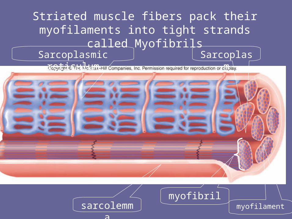
myofibril
sarcolemma
SarcoplasmSarcoplasmic reticulum
Striated muscle fibers pack their myofilaments into tight strands called Myofibrils
myofilament
To contract: to get shorter
Muscle cells
• Contract when stimulated by motor impulses or hormones
• Only contract (tighten up) or relax
• can only pull - never push
Muscle Contraction
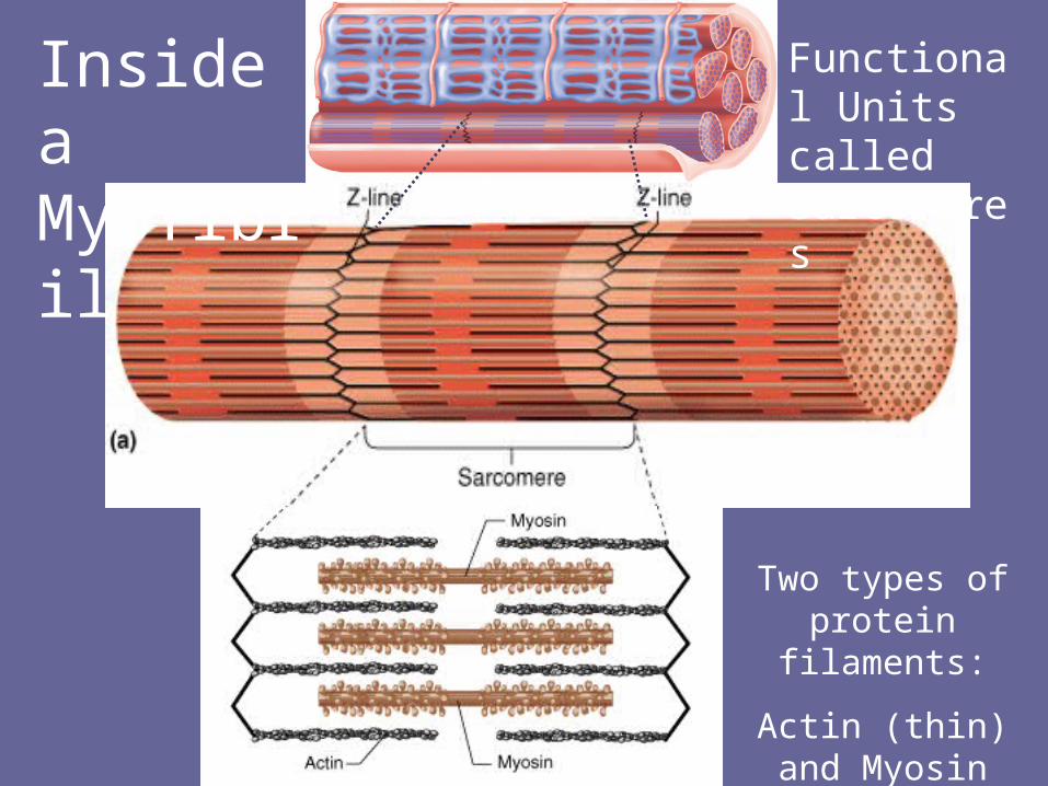
Inside a Myofibril
Functional Units called Sarcomeres
Two types of protein filaments:
Actin (thin) and Myosin
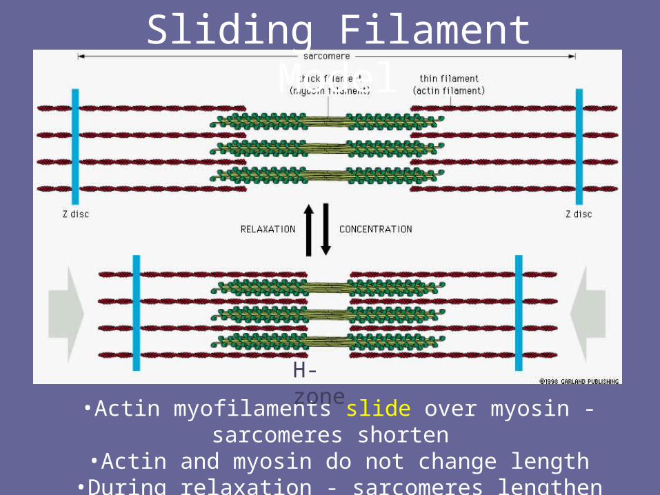
•Actin myofilaments slide over myosin - sarcomeres shorten•Actin and myosin do not change length
•During relaxation - sarcomeres lengthen
H-zone
Sliding Filament Model
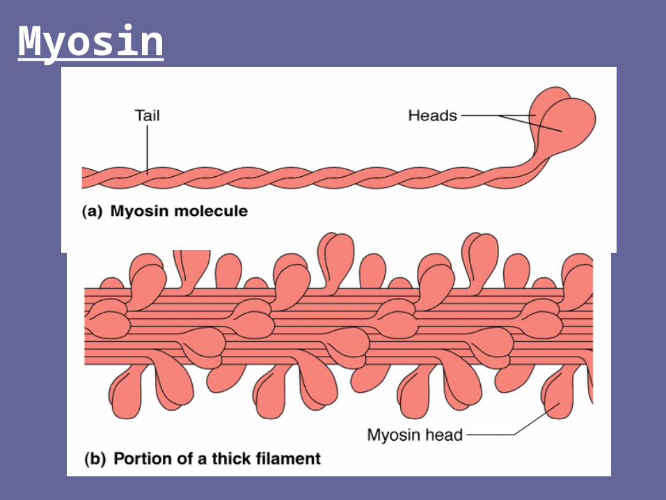
Myosin
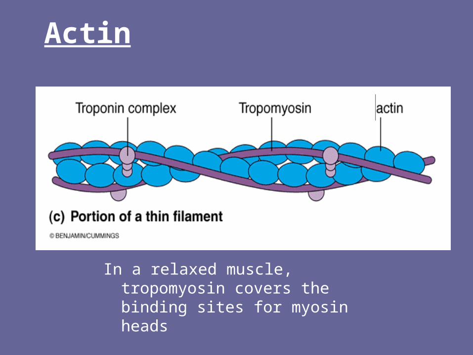
Actin
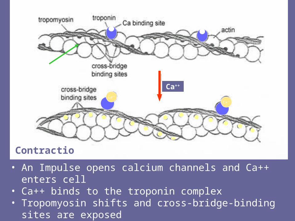
In a relaxed muscle, tropomyosin covers the binding sites for myosin heads
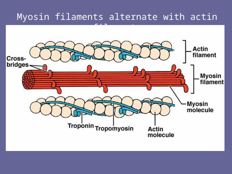
Myosin filaments alternate with actin filaments
Ca++
Ca++
Ca++
• An Impulse opens calcium channels and Ca++ enters cell• Ca++ binds to the troponin complex• Tropomyosin shifts and cross-bridge-binding sites are
exposed
Contraction

Cross-bridge formation
Myosin heads can now attach at the cross bridge binding sites
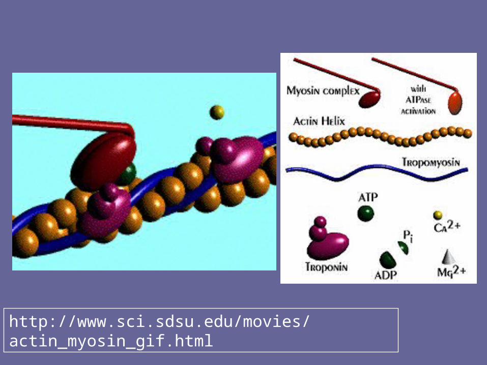
http://www.sci.sdsu.edu/movies/actin_myosin_gif.html
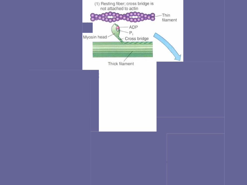
Muscle Contraction Cycle
• In the presence of calcium, myosin binds to the actin filaments.
• The myosin head flexes inward and backward, causing the actin filament to shorten.
• In the presence of ATP, the myosin head detaches and then reattaches at a new position on the actin filament.
• This cycle repeats to continue the shortening of the muscle (contraction).
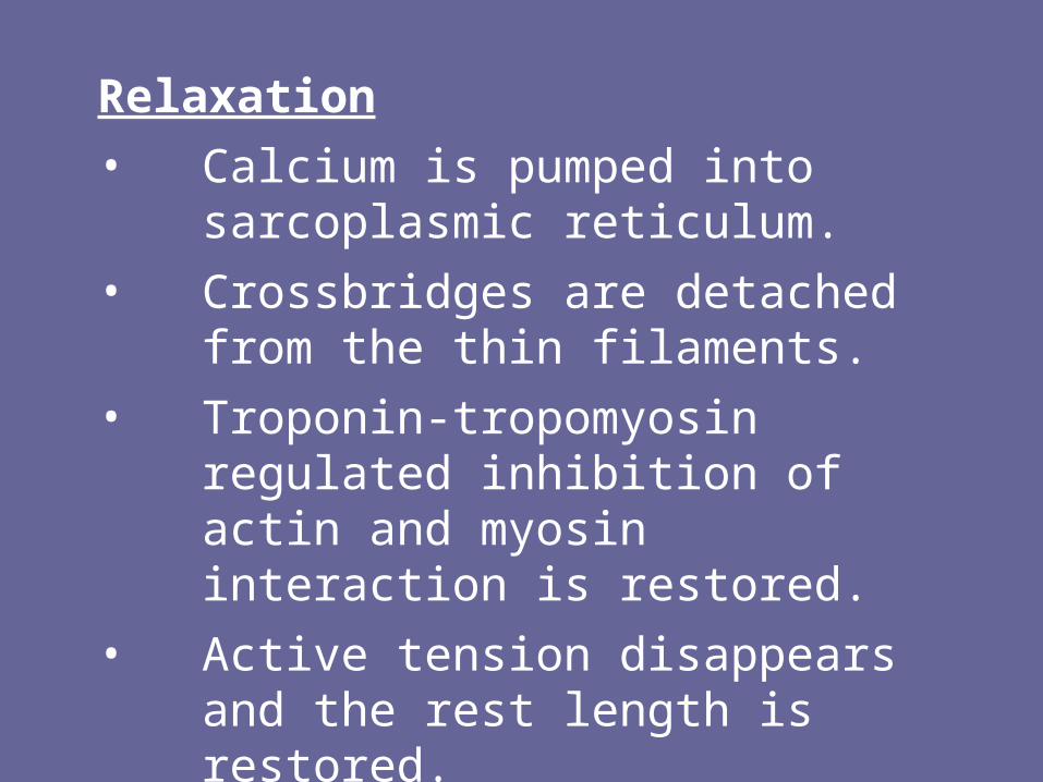
Relaxation
• Calcium is pumped into sarcoplasmic reticulum.
• Crossbridges are detached from the thin filaments.
• Troponin-tropomyosin regulated inhibition of actin and myosin interaction is restored.
• Active tension disappears and the rest length is restored.
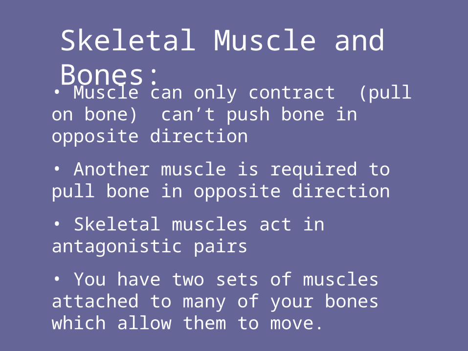
• Muscle can only contract (pull on bone) can’t push bone in opposite direction
• Another muscle is required to pull bone in opposite direction
• Skeletal muscles act in antagonistic pairs
• You have two sets of muscles attached to many of your bones which allow them to move.
Skeletal Muscle and Bones:
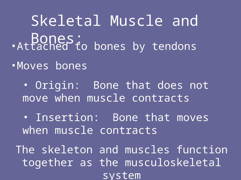
•Attached to bones by tendons
•Moves bones
• Origin: Bone that does not move when muscle contracts
• Insertion: Bone that moves when muscle contracts
The skeleton and muscles function together as the musculoskeletal system
Skeletal Muscle and Bones:
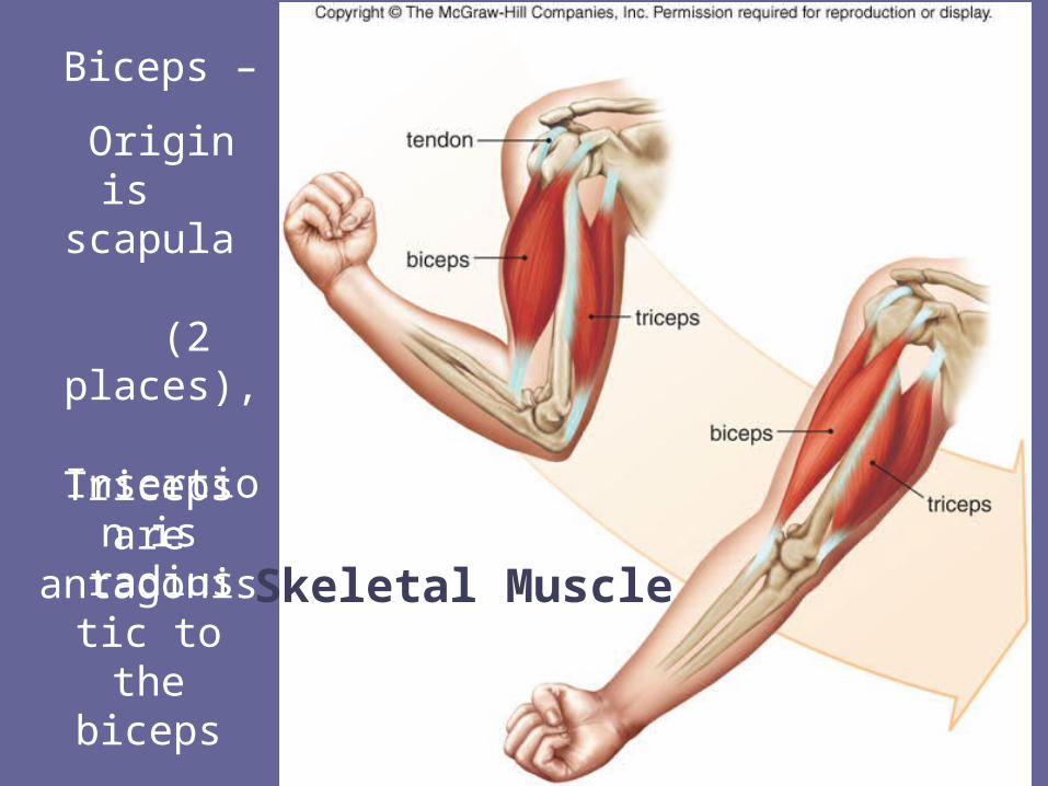
Skeletal Muscle
Biceps –
Origin is scapula
(2 places),
Insertion is radius
Triceps are antagonistic
to the biceps
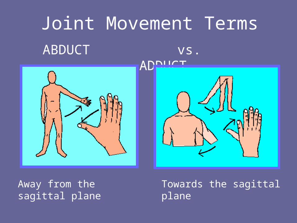
Joint Movement TermsABDUCT vs. ADDUCT
Away from the sagittal plane Towards the sagittal plane
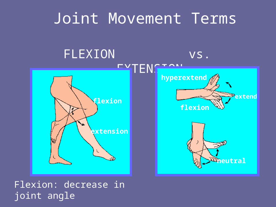
Joint Movement Terms
FLEXION vs. EXTENSION
flexion
extension
hyperextend
flexion
neutral
extend
Flexion: decrease in joint angle