Muscular System: Histology and Physiology. Muscular System Functions Body movement (Locomotion)...
36
Muscular System: Histology and Physiology
-
Upload
augustine-thompson -
Category
Documents
-
view
219 -
download
0
Transcript of Muscular System: Histology and Physiology. Muscular System Functions Body movement (Locomotion)...
Muscular System:Histology and Physiology
Muscular System Functions
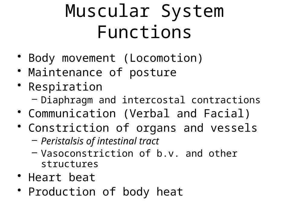
• Body movement (Locomotion)• Maintenance of posture• Respiration
– Diaphragm and intercostal contractions• Communication (Verbal and Facial)• Constriction of organs and vessels
– Peristalsis of intestinal tract– Vasoconstriction of b.v. and other structures
• Heart beat • Production of body heat
Properties of Muscle
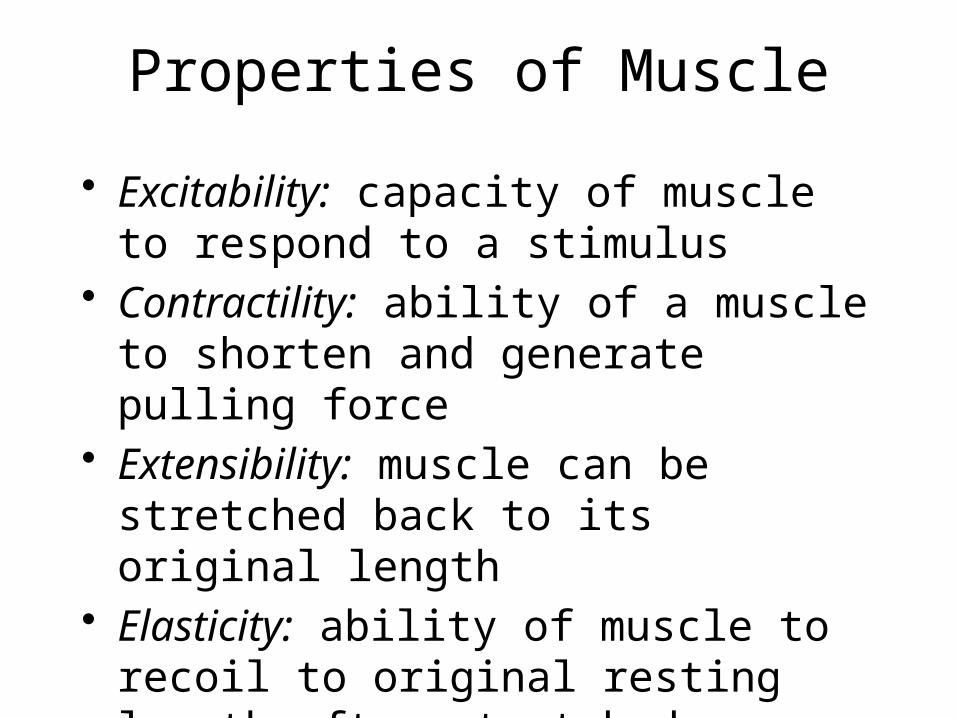
• Excitability: capacity of muscle to respond to a stimulus
• Contractility: ability of a muscle to shorten and generate pulling force
• Extensibility: muscle can be stretched back to its original length
• Elasticity: ability of muscle to recoil to original resting length after stretched
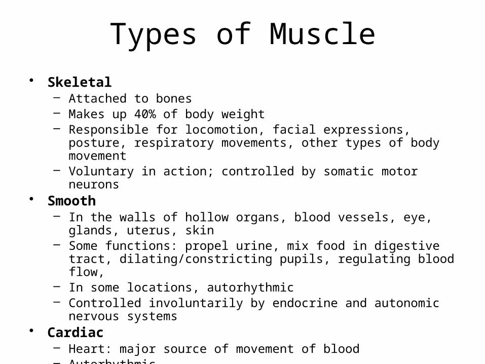
Types of Muscle• Skeletal
– Attached to bones– Makes up 40% of body weight– Responsible for locomotion, facial expressions, posture,
respiratory movements, other types of body movement– Voluntary in action; controlled by somatic motor neurons
• Smooth– In the walls of hollow organs, blood vessels, eye, glands,
uterus, skin– Some functions: propel urine, mix food in digestive tract,
dilating/constricting pupils, regulating blood flow, – In some locations, autorhythmic– Controlled involuntarily by endocrine and autonomic nervous
systems• Cardiac
– Heart: major source of movement of blood– Autorhythmic– Controlled involuntarily by endocrine and autonomic nervous
systems
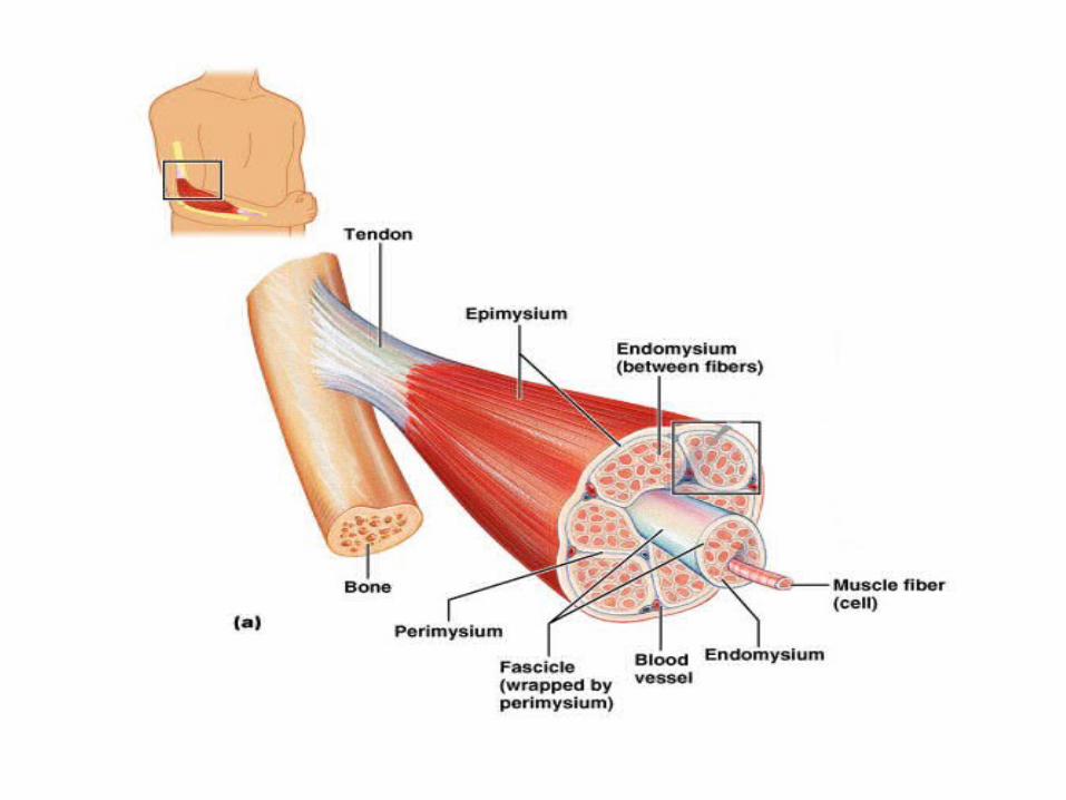
Connective Tissue Sheaths
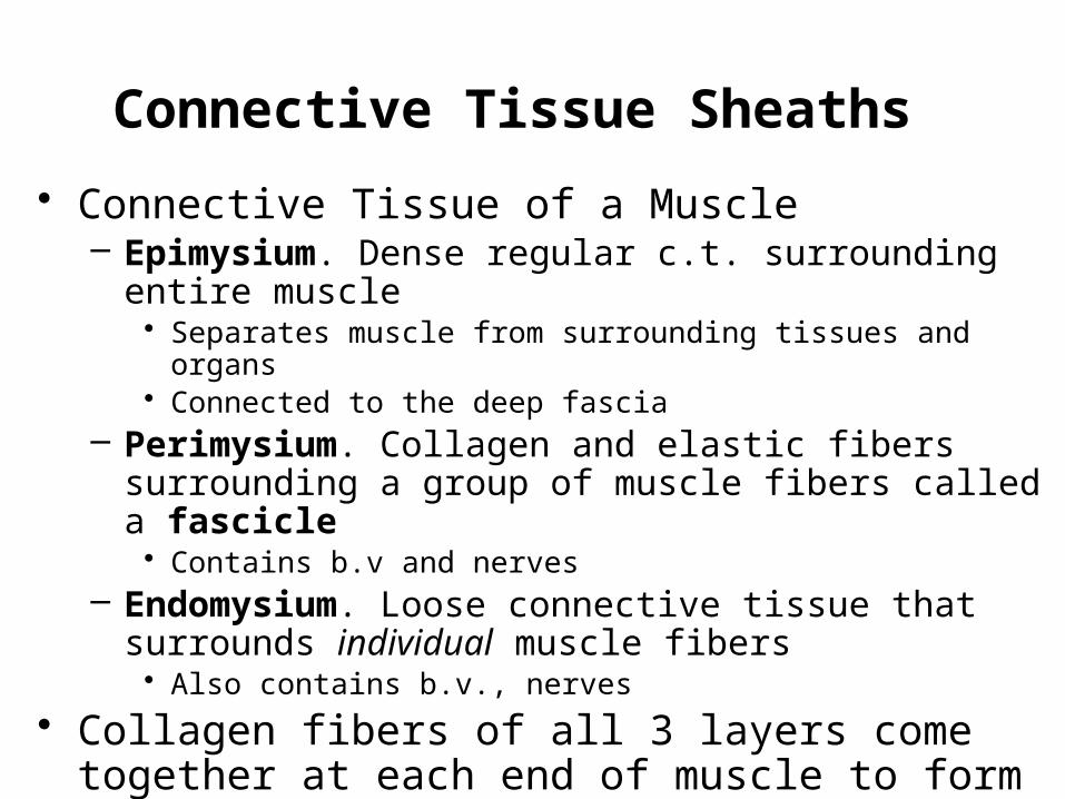
• Connective Tissue of a Muscle– Epimysium. Dense regular c.t. surrounding entire
muscle• Separates muscle from surrounding tissues and organs• Connected to the deep fascia
– Perimysium. Collagen and elastic fibers surrounding a group of muscle fibers called a fascicle• Contains b.v and nerves
– Endomysium. Loose connective tissue that surrounds individual muscle fibers• Also contains b.v., nerves
• Collagen fibers of all 3 layers come together at each end of muscle to form a tendon.
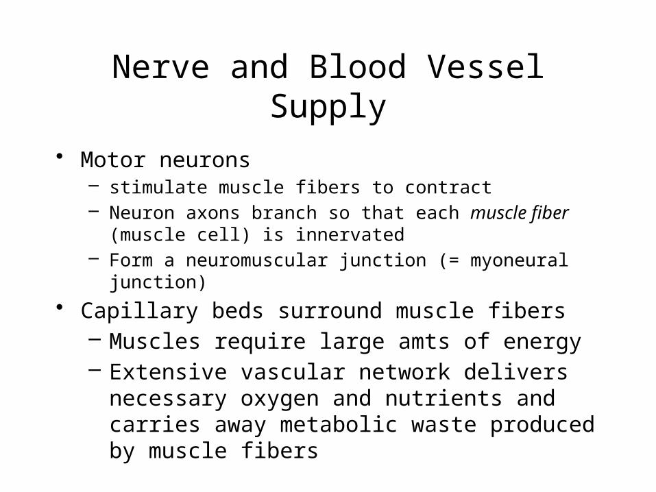
Nerve and Blood Vessel Supply
• Motor neurons– stimulate muscle fibers to contract– Neuron axons branch so that each muscle fiber
(muscle cell) is innervated– Form a neuromuscular junction (= myoneural
junction)• Capillary beds surround muscle fibers
– Muscles require large amts of energy– Extensive vascular network delivers
necessary oxygen and nutrients and carries away metabolic waste produced by muscle fibers
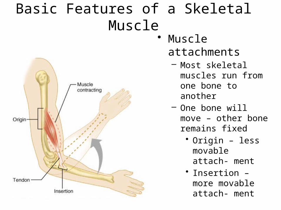
Basic Features of a Skeletal Muscle
• Muscle attachments– Most skeletal
muscles run from one bone to another
– One bone will move – other bone remains fixed• Origin – less
movable attach- ment
• Insertion – more movable attach- ment
Basic Features of a Skeletal Muscle
• Muscle attachments (continued)– Muscles attach to origins and
insertions by connective tissue• Fleshy attachments – connective tissue
fibers are short• Indirect attachments – connective tissue
forms a tendon.

Skeletal Muscle Structure• Composed of muscle cells
(fibers), connective tissue, blood vessels, nerves
• Fibers are long, cylindrical, and multinucleated
• Tend to be smaller diameter in small muscles and larger in large muscles. 1 mm- 4 cm in length
• Develop from myoblasts• Striated appearance• Nuclei are peripherally located
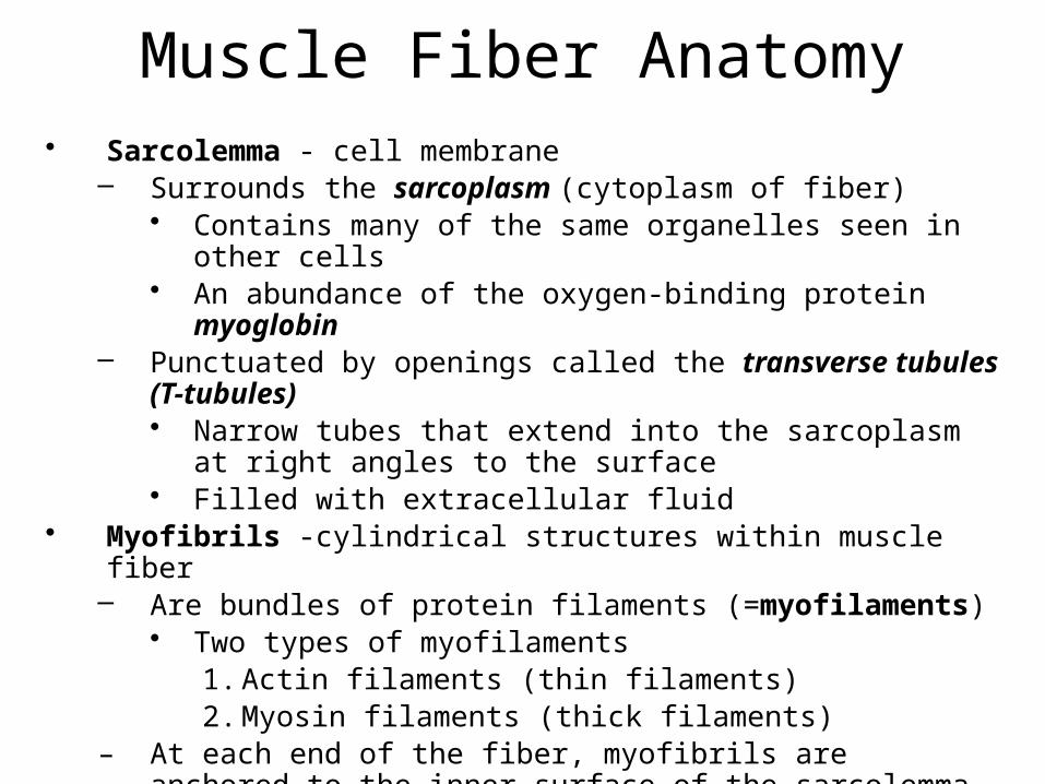
Muscle Fiber Anatomy• Sarcolemma - cell membrane
– Surrounds the sarcoplasm (cytoplasm of fiber)• Contains many of the same organelles seen in other
cells• An abundance of the oxygen-binding protein
myoglobin– Punctuated by openings called the transverse tubules
(T-tubules)• Narrow tubes that extend into the sarcoplasm at right
angles to the surface• Filled with extracellular fluid
• Myofibrils -cylindrical structures within muscle fiber– Are bundles of protein filaments (=myofilaments)
• Two types of myofilaments1. Actin filaments (thin filaments)2. Myosin filaments (thick filaments)
– At each end of the fiber, myofibrils are anchored to the inner surface of the sarcolemma
– When myofibril shortens, muscle shortens (contracts)
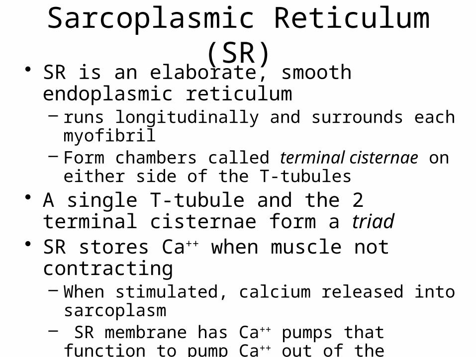
Sarcoplasmic Reticulum (SR)
• SR is an elaborate, smooth endoplasmic reticulum – runs longitudinally and surrounds each
myofibril– Form chambers called terminal cisternae on
either side of the T-tubules• A single T-tubule and the 2 terminal
cisternae form a triad• SR stores Ca++ when muscle not
contracting– When stimulated, calcium released into
sarcoplasm– SR membrane has Ca++ pumps that
function to pump Ca++ out of the sarcoplasm back into the SR after contraction

Sarcoplasmic Reticulum (SR)
Figure 9.5
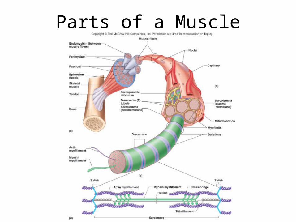
Parts of a Muscle
Sarcomeres: Z Disk to Z
Disk
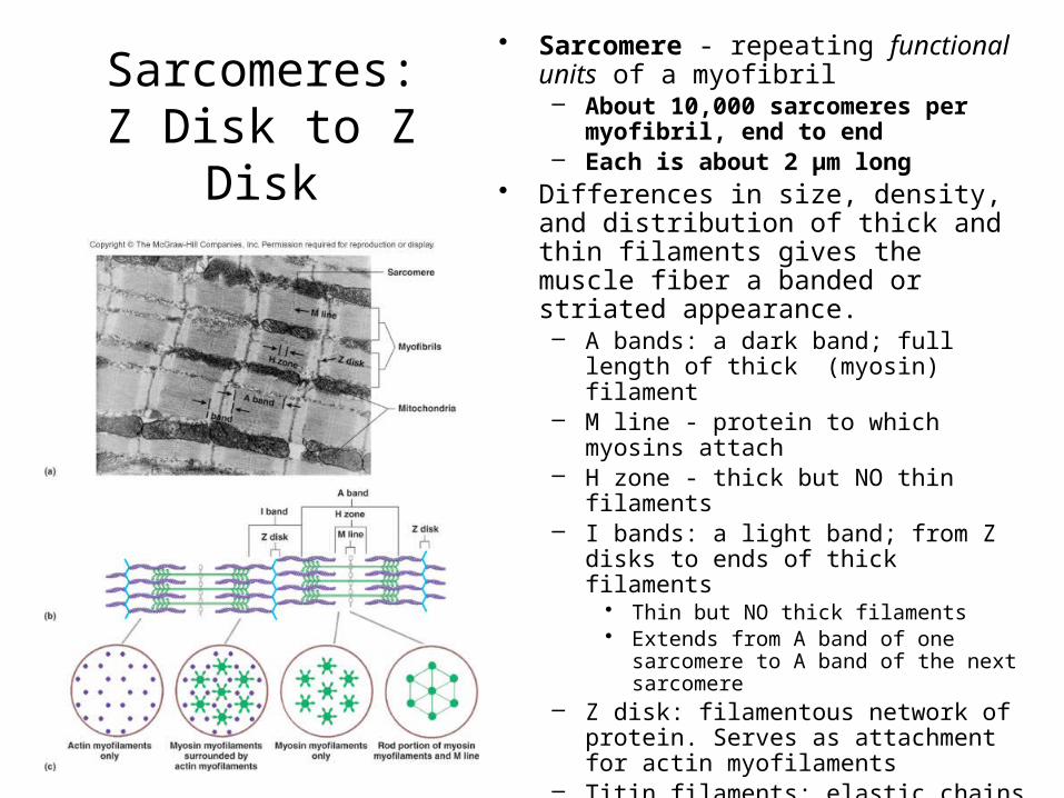
• Sarcomere - repeating functional units of a myofibril– About 10,000 sarcomeres per
myofibril, end to end– Each is about 2 µm long
• Differences in size, density, and distribution of thick and thin filaments gives the muscle fiber a banded or striated appearance.– A bands: a dark band; full length of
thick (myosin) filament– M line - protein to which myosins
attach– H zone - thick but NO thin filaments– I bands: a light band; from Z disks
to ends of thick filaments• Thin but NO thick filaments• Extends from A band of one
sarcomere to A band of the next sarcomere
– Z disk: filamentous network of protein. Serves as attachment for actin myofilaments
– Titin filaments: elastic chains of amino acids; keep thick and thin filaments in proper alignment
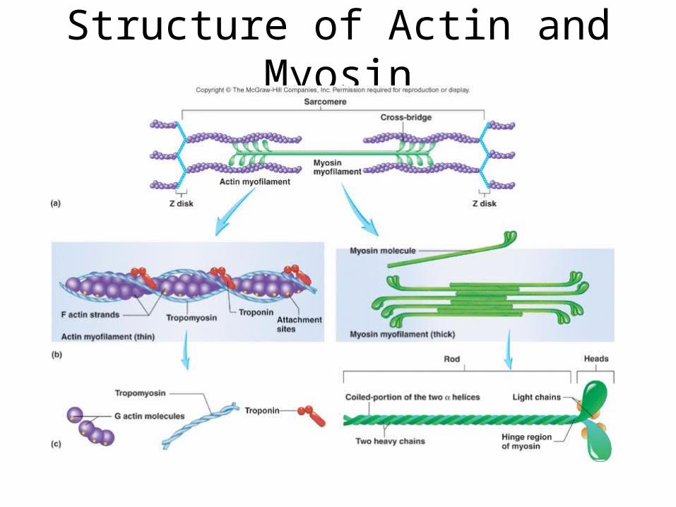
Structure of Actin and Myosin
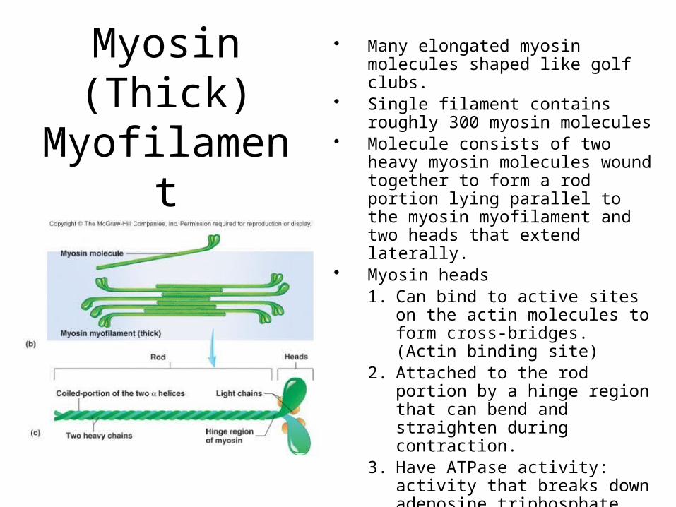
Myosin (Thick)
Myofilament
• Many elongated myosin molecules shaped like golf clubs.
• Single filament contains roughly 300 myosin molecules
• Molecule consists of two heavy myosin molecules wound together to form a rod portion lying parallel to the myosin myofilament and two heads that extend laterally.
• Myosin heads1. Can bind to active sites on the
actin molecules to form cross-bridges. (Actin binding site)
2. Attached to the rod portion by a hinge region that can bend and straighten during contraction.
3. Have ATPase activity: activity that breaks down adenosine triphosphate (ATP), releasing energy. Part of the energy is used to bend the hinge region of the myosin molecule during contraction
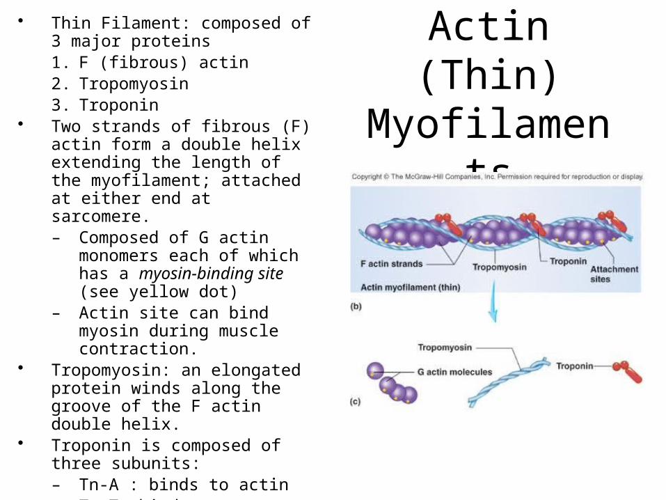
Actin (Thin) Myofilamen
ts
• Thin Filament: composed of 3 major proteins1. F (fibrous) actin2. Tropomyosin3. Troponin
• Two strands of fibrous (F) actin form a double helix extending the length of the myofilament; attached at either end at sarcomere.– Composed of G actin
monomers each of which has a myosin-binding site (see yellow dot)
– Actin site can bind myosin during muscle contraction.
• Tropomyosin: an elongated protein winds along the groove of the F actin double helix.
• Troponin is composed of three subunits: – Tn-A : binds to actin– Tn-T :binds to tropomyosin,– Tn-C :binds to calcium ions.
Sliding Filament Model of Contraction
• Thin filaments slide past the thick ones so that the actin and myosin filaments overlap to a greater degree
• In the relaxed state, thin and thick filaments overlap only slightly
• Upon stimulation, myosin heads bind to actin and sliding begins

Sliding Filament Model of Contraction
• Each myosin head binds and detaches several times during contraction, acting like a ratchet to generate tension and propel the thin filaments to the center of the sarcomere
• As this event occurs throughout the sarcomeres, the muscle shortens
InterActive Physiology®: Muscular System: Sliding Filament TheoryPLAY
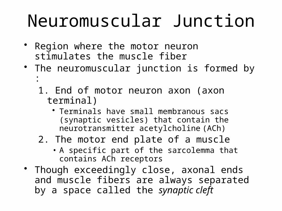
Neuromuscular Junction• Region where the motor neuron stimulates
the muscle fiber• The neuromuscular junction is formed by :
1. End of motor neuron axon (axon terminal)• Terminals have small membranous sacs (synaptic
vesicles) that contain the neurotransmitter acetylcholine (ACh)
2. The motor end plate of a muscle• A specific part of the sarcolemma that contains
ACh receptors• Though exceedingly close, axonal ends and
muscle fibers are always separated by a space called the synaptic cleft
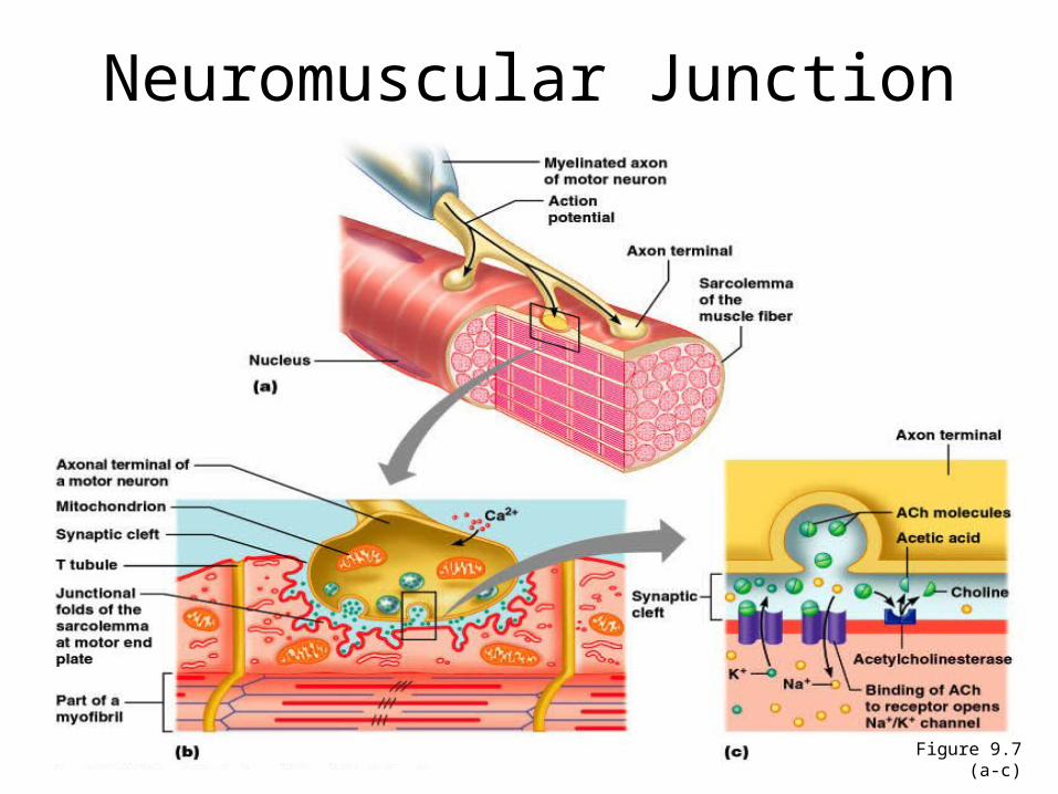
Neuromuscular Junction
Figure 9.7 (a-c)

Motor Unit: The Nerve-Muscle Functional Unit
• A motor unit is a motor neuron and all the muscle fibers it supplies
• The number of muscle fibers per motor unit can vary from a few (4-6) to hundreds (1200-1500)
• Muscles that control fine movements (fingers, eyes) have small motor units
• Large weight-bearing muscles (thighs, hips) have large motor units
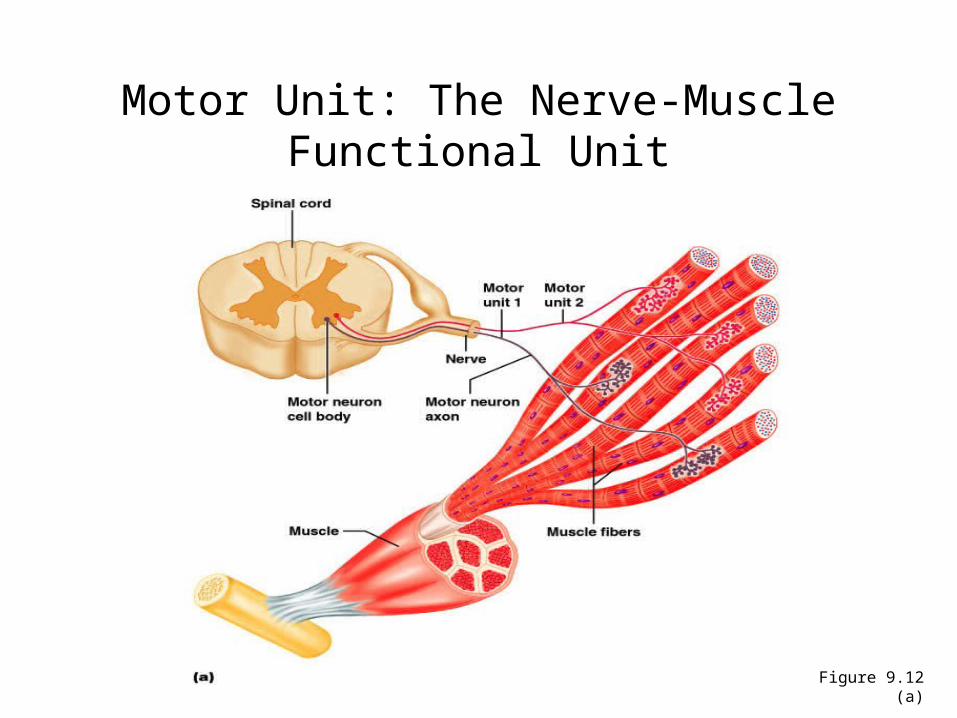
Motor Unit: The Nerve-Muscle Functional Unit
Figure 9.12 (a)
Motor Unit: The Nerve-Muscle Functional Unit
• Muscle fibers from a motor unit are spread throughout the muscle– Not confined to one fascicle
• Therefore, contraction of a single motor unit causes weak contraction of the entire muscle
• Stronger and stronger contractions of a muscle require more and more motor units being stimulated (recruited)
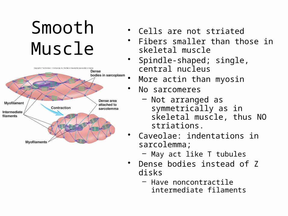
Smooth Muscle
• Cells are not striated• Fibers smaller than those in
skeletal muscle• Spindle-shaped; single, central
nucleus• More actin than myosin• No sarcomeres
– Not arranged as symmetrically as in skeletal muscle, thus NO striations.
• Caveolae: indentations in sarcolemma;– May act like T tubules
• Dense bodies instead of Z disks – Have noncontractile intermediate
filaments
Smooth Muscle
Figure 9.24
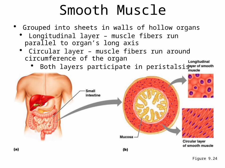
• Grouped into sheets in walls of hollow organs• Longitudinal layer – muscle fibers run parallel to organ’s long axis• Circular layer – muscle fibers run around circumference of the organ
• Both layers participate in peristalsis

Smooth Muscle• Is innervated by autonomic nervous system
(ANS)• Visceral or unitary smooth muscle
– Only a few muscle fibers innervated in each group
– Impulse spreads through gap junctions– Who sheet contracts as a unit– Often autorhythmic
• Multiunit: – Cells or groups of cells act as independent units– Arrector pili of skin and iris of eye
Cardiac Muscle• Found only in heart where it forms a thick layer
called the myocardium• Striated fibers that branch• Each cell usually has one centrally-located
nucleus• Fibers joined by intercalated disks
– IDs are composites of desmosomes and gap junctions– Allow excitation in one fiber to spread quickly to
adjoining fibers• Under control of the ANS (involuntary) and
endocrine system (hormones)• Some cells are autorhythmic
– Fibers spontaneously contract (aka Pacemaker cells)
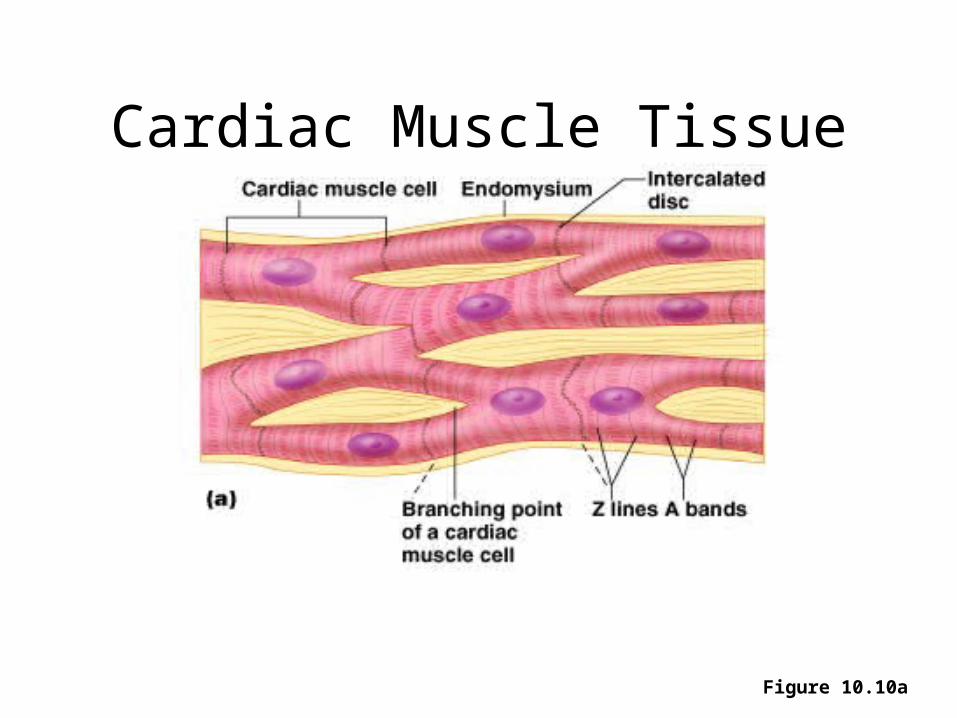
Cardiac Muscle Tissue
Figure 10.10a
Disorders of Muscle Tissue
• Muscle tissues experience few disorders– Heart muscle is the exception– Skeletal muscle – remarkably
resistant to infection– Smooth muscle – problems stem from
external irritants
Disorders of Muscle Tissue
• Muscular dystrophy – a group of inherited muscle destroying disease– Affected muscles enlarge with fat and
connective tissue – Muscles degenerate
• Types of muscular dystrophy– Duchenne muscular dystrophy – Myotonic dystrophy
Disorders of Muscle Tissue
• Myofascial pain syndrome – pain is caused by tightened bands of muscle fibers
• Fibromyalgia – a mysterious chronic-pain syndrome– Affects mostly women– Symptoms – fatigue, sleep
abnormalities, severe musculoskeletal pain, and headache
Developmental Aspects: Regeneration
• Cardiac and skeletal muscle become amitotic, but can lengthen and thicken
• Myoblast-like satellite cells show very limited regenerative ability
• Cardiac cells lack satellite cells• Smooth muscle has good regenerative ability• There is a biological basis for greater strength
in men than in women• Women’s skeletal muscle makes up 36% of
their body mass• Men’s skeletal muscle makes up 42% of their
body mass
Developmental Aspects: Male and Female
• These differences are due primarily to the male sex hormone testosterone
• With more muscle mass, men are generally stronger than women
• Body strength per unit muscle mass, however, is the same in both sexes
Developmental Aspects: Age Related
• With age, connective tissue increases and muscle fibers decrease
• Muscles become stringier and more sinewy
• By age 80, 50% of muscle mass is lost (sarcopenia)
• Decreased density of capillaries in muscle
• Reduced stamina• Increased recovery time• Regular exercise reverses sarcopenia