Muscular System Functions Body movement (Locomotion) Maintenance of posture Respiration *...
22
Physiology of Muscles PHYSIOLOGY I (PHL 215) Dr. Gamal Gabr
-
Upload
chana-boldon -
Category
Documents
-
view
215 -
download
0
Transcript of Muscular System Functions Body movement (Locomotion) Maintenance of posture Respiration *...
Physiology of Muscles
PHYSIOLOGY I (PHL 215)
Dr. Gamal Gabr

Muscular System Functions
Body movement (Locomotion) Maintenance of posture Respiration
*Diaphragm and intercostals contractions Communication (Verbal and Facial) Constriction of organs and vessels Heart beat Production of body heat (Thermogenesis)

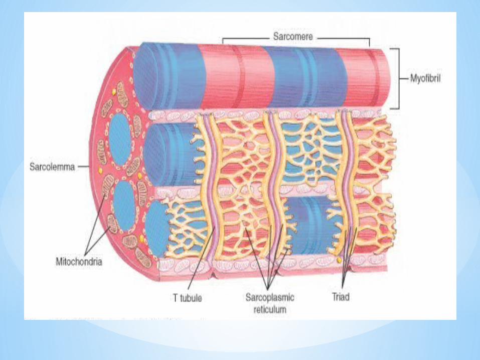
Typical cells Muscle cell=fiber
Plasma membrane Sarcolemma
Cytoplasm Sarcoplasm
Endoplasmic reticulum
Sarcoplasmic reticulum (SR)
Many mitochondria
Multiple nuclei

Muscle cell structures not found in other cells
Myofibrils: bundles of very fine fibers Thick and thin myofilaments: very fine fibers that make
up myofibrils Sarcomere: segment of myofibril between two Z lines;
contractile unit T tubules: transmit electrical impulses through cell

Myofilaments
4 protein molecules that make up myofilaments: Myosin, actin, tropomyosin, troponin
Thin filaments: actin, tropomyosin, troponin
Thick filaments: mostly myosin

Properties of Muscle
Excitability: Capacity of muscle to respond to a stimulus
Contractility: Ability of a muscle to shorten and generate pulling force
Extensibility: Ability stretches when pulled
Elasticity: Ability to return to original shape and length after contraction or extension
Muscle structure
Connective Tissue Sheaths
Connective Tissue (CT) of a Muscle
Epimysium: Dense regular CT surrounding entire muscle
*Separates muscle from surrounding tissues and organs
Perimysium: Collagen and elastic fibers surrounding a group of muscle fibers called a fascicle
*Contains blood vessels and nerves
Endomysium: Loose CT that surrounds individual muscle fibers
*Also contains blood vessels and nerves
*Collagen fibers of all 3 layers come together at each end of muscle to form a tendon or aponeurosis.
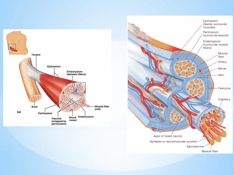

Motor neurons
*Stimulate muscle fibers to contract
*Neuron axons branch so that each muscle fiber (muscle cell) is innervated
*Form a neuromuscular junction
Capillary beds surround muscle fibers
*Muscles require large amount of energy
*Extensive vascular network delivers necessary oxygen and nutrients and carries away metabolic waste produced by muscle fibers

Energy for Muscle Contractions
*ATP: adenosine triphosphate
*CP: creatine phosphate
Glucose & Oxygen
*Glucose stored in form of glycogen in muscle
*Excess oxygen molecules in sarcoplasm bound to myoglobin

Anaerobic respiration
Allows body to avoid use of oxygen in short termProduces lactic acidAccumulation of lactic acid in muscles causes burning sensation

Types of MuscleSkeletal
*Attached to bones
*Makes up 40% of body weight (Women’s skeletal muscle makes up 36% of their body mass, Men’s skeletal muscle makes up 42% of their body mass)
*Responsible for locomotion, facial expressions, posture, respiratory movements, other types of body movement
*Voluntary in action; controlled by somatic motor neurons
Smooth
*In the walls of hollow organs, blood vessels, eye, glands, uterus, skin
*Functions: propel urine, mix food in digestive tract, regulating blood flow
*Controlled involuntarily by endocrine and autonomic nervous systems
Cardiac
*Heart: major source of movement of blood

Basic Features of a Skeletal Muscle
Muscle attachments*Most skeletal muscles run from one bone to another
*One bone will move – other bone remains fixed
*Origin – less movable attachment
*Insertion – more movable attachment
*Muscles attach to origins and insertions by connective tissue
Fleshy attachments – connective tissue fibers are short
Indirect attachments – connective tissue forms a tendon

Skeletal Muscle Structure
*Composed of muscle cells (fibers), connective tissue, blood vessels, nerves
*Fibers are long, cylindrical, and multinucleated
*Tend to be smaller diameter in small muscles and larger in large muscles. 1 mm - 4 cm in length
*Striated appearance
*Nuclei are peripherally located

Muscle Fiber Anatomy
Sarcolemma - cell membrane
* Surrounds the Sarcoplasm (cytoplasm of fiber)
* Punctuated by openings called the transverse tubules (T-tubules)
Myofibrils -cylindrical structures within muscle fiber
* Are bundles of protein filaments (=myofilaments)
* Two types of myofilaments
1. Actin filaments (thin filaments)
2.Myosin filaments (thick filaments)
– When myofibril shortens, muscle shortens (contracts)

Sarcoplasmic Reticulum (SR)
SR is an elaborate, smooth endoplasmic reticulum Runs longitudinally and surrounds each myofibrilSR stores Ca++ when muscle not contractingWhen stimulated, calcium released into sarcoplasm SR membrane has Ca++ pumps that function to pump Ca++ out of the sarcoplasm back into the SR after contraction
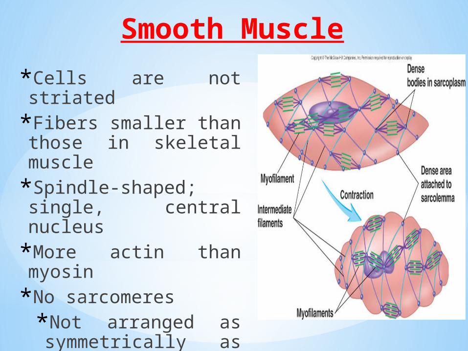
Smooth Muscle
*Cells are not striated
*Fibers smaller than those in skeletal muscle
*Spindle-shaped; single, central nucleus
*More actin than myosin
*No sarcomeres
*Not arranged as symmetrically as in skeletal muscle, thus no striations.

Smooth Muscle
• Grouped into sheets in walls of hollow organs• Longitudinal layer: muscle fibers run parallel to organ’s long
axis• Circular layer: muscle fibers run around circumference of the
organ• Both layers participate in peristalsis
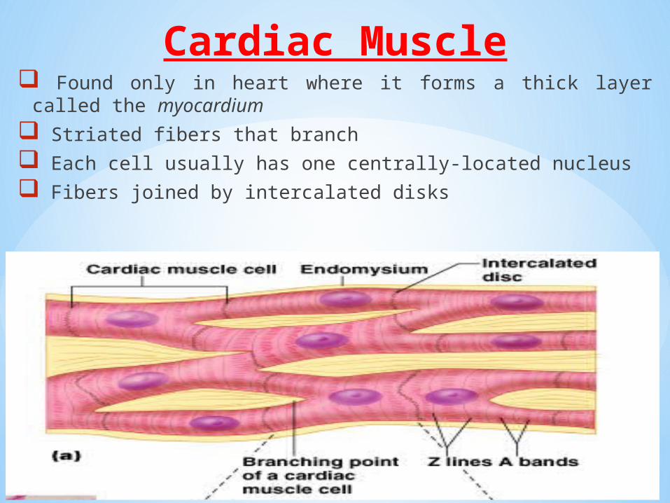
Cardiac Muscle Found only in heart where it forms a thick layer called the
myocardium
Striated fibers that branch
Each cell usually has one centrally-located nucleus
Fibers joined by intercalated disks

Disorders of Muscle Tissue
*Muscle Fatigue
*Lack of oxygen causes ATP deficit
*Lactic acid builds up from anaerobic respiration
*Muscle Atrophy
*a decrease in the mass of the muscle
*Weakening and shrinking of a muscle
*May be caused
*Immobilization
*Loss of neural stimulation

Disorders of Muscle Tissue
*Muscle Hypertrophy
*Enlargement of a muscle
*More capillaries
*More mitochondria
*Caused by*Strenuous exercise
*Steroid hormones
*Muscle Tonus
*Tightness of a muscle
*Some fibers always contracted