Muscles of the leg - MD Connect · Gray’s Anatomy for Students: ... Ch.5 Fig5.4, 13. Figure 6.13...
16
Leg muscles Assoc. Prof. Jenny Hayes
-
Upload
nguyenhanh -
Category
Documents
-
view
217 -
download
0
Transcript of Muscles of the leg - MD Connect · Gray’s Anatomy for Students: ... Ch.5 Fig5.4, 13. Figure 6.13...

Leg muscles
Assoc. Prof. Jenny Hayes

WARNING This material has been provided to you pursuant
to section 49 of the Copyright Act 1968 (the Act) for the purposes of research or study.
The contents of the material may be subject to copyright protection under the Act.
Further dealings by you with this material may be
a copyright infringement. To determine whether such a communication would be an
infringement, it is necessary to have regard to the criteria set out in Part 3, Division 3 of the
Act.

Objectives Major features and surface landmarks of the tibia and fibula. Fascial compartments enclosing the major muscle groups. Component muscles of the major muscle groups • position • joint/s they act upon • movement/s produced
Suggested pre-reading Gray’s Anatomy for Students: Conceptual overview of Bones and joints, Muscles, Chapter 6, Lower Limb, p. 517-520
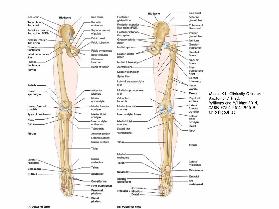
Moore K L. Clinically Oriented Anatomy. 7th ed. Williams and Wilkins; 2014. ISBN 978-1-4511-1945-9. Ch.5 Fig5.4, 13

Figure 6.13 Muscle compartments in the thigh and leg.
Downloaded from: StudentConsult (on 26 August 2011 06:57 AM) © 2005 Elsevier
Muscles in the thigh and leg are separated into compartments by bone, fascia and interosseous membrane:
Thigh: Medial (adductor) compartment with muscles acting mostly on hip Anterior (extensor) compartment with muscles extending knee and flexing hip Posterior (flexor) compartment with muscles that act on hip and knee because they span both joins
Leg: Muscles in anterior compartment dorsiflex the foot and extend the toes Muscles in the posterior compartment plantarflex the foot and flex the toes Muscles in the lateral compartment evert the foot

Netter, F.H. Interactive Atlas of Human Anatomy. 3rd ed. New Jersey, Icon Learning Systems, 2003, ISBN: 1-929007-15-9, Plate #491A
intermuscular septa:
anterior
posterior interosseous membrane
tibia
fibula deep fascia

Netter, F.H. Interactive Atlas of Human Anatomy. 3rd ed. New Jersey, Icon Learning Systems, 2003, ISBN: 1-929007-15-9, Plate #491B
ant
lat
post

Netter, F.H. Interactive Atlas of Human Anatomy. 3rd ed. New Jersey, Icon Learning Systems, 2003, ISBN: 1-929007-15-9, Plate #488
tibialis anterior
EHL EDL
fibularis tertius

Netter, F.H. Interactive Atlas of Human Anatomy. 3rd ed. New Jersey, Icon Learning Systems, 2003, ISBN: 1-929007-15-9, Plate #498
tibialis anterior
EHL
EDL
Extensor retinaculum “Timothy has a nasty, dirty toe”

Netter, F.H. Interactive Atlas of Human Anatomy. 3rd ed. New Jersey, Icon Learning Systems, 2003, ISBN: 1-929007-15-9, Plate #490
fibularis longus
fibularis brevis
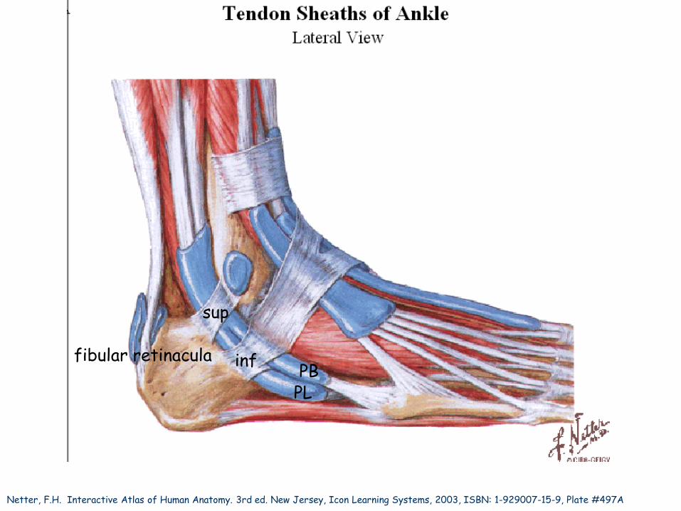
Netter, F.H. Interactive Atlas of Human Anatomy. 3rd ed. New Jersey, Icon Learning Systems, 2003, ISBN: 1-929007-15-9, Plate #497A
fibular retinacula
sup
inf
PL PB

Netter, F.H. Interactive Atlas of Human Anatomy. 3rd ed. New Jersey, Icon Learning Systems, 2003, ISBN: 1-929007-15-9, Plate #485
gastrocnemius

Netter, F.H. Interactive Atlas of Human Anatomy. 3rd ed. New Jersey, Icon Learning Systems, 2003, ISBN: 1-929007-15-9, Plate #486
soleus
plantaris popliteus

Netter, F.H. Interactive Atlas of Human Anatomy. 3rd ed. New Jersey, Icon Learning Systems, 2003, ISBN: 1-929007-15-9, Plate #487
FDL
tibialis posterior
FHL
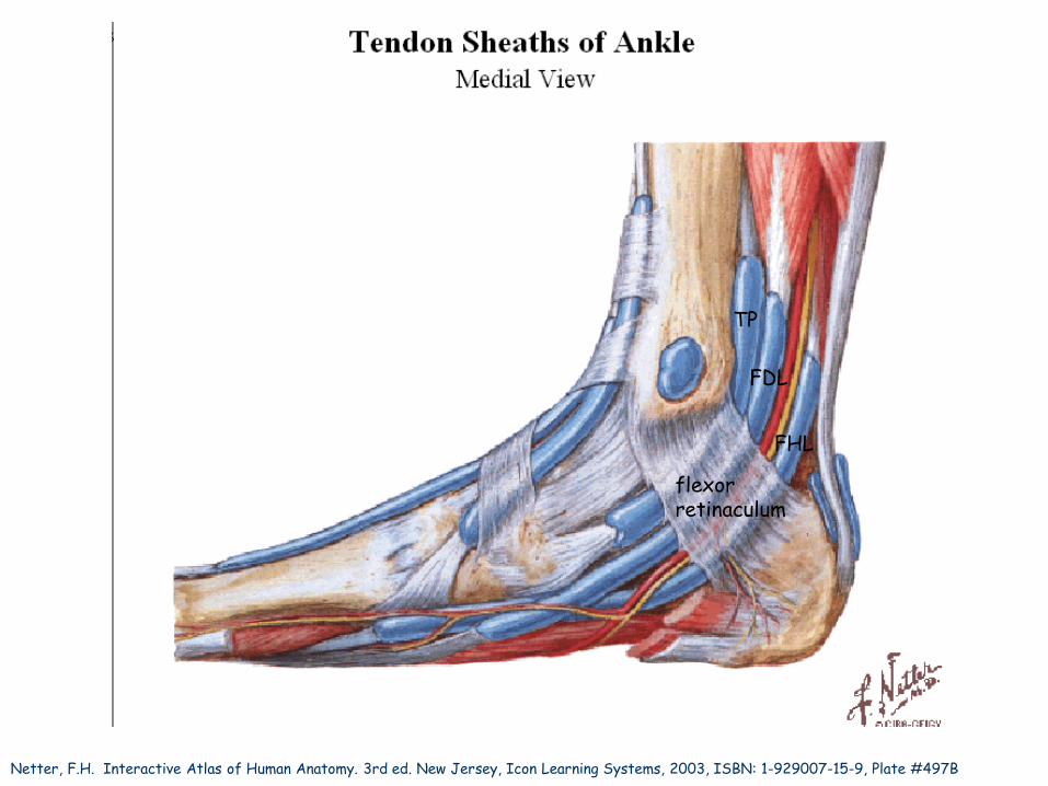
Netter, F.H. Interactive Atlas of Human Anatomy. 3rd ed. New Jersey, Icon Learning Systems, 2003, ISBN: 1-929007-15-9, Plate #497B
flexor retinaculum
“Tom, Dick and very naughty Harry”
TP
FDL
FHL

Barcode Specimen Description Location
516-100333 Left lower limb Three dissections of the muscles on the anterior aspect of the left leg. From left to right: the first displays tibialis anterior, the second displays peroneus longus and peroneus brevis and the third shows the muscles and the anterior tibial nerve with its branches to tibialis anterior.
24C
516-100344 Flexor hallucis longus A dissection showing the flexor hallucis longus muscle. The muscle belly of flexor digitorum longus has been removed and its tendon can be seen in the distal part of the dissection.
24A
516-100327 Lower limb Left male leg and hemipelvis with viscera removed.
Museum annex (front display)
516-100317 Adductor magnus Dissection of the left adductor magnus. The fascial attachment of the hamstring part of the muscle to the femur has been removed.
24A
516-100329 Lower limb Dissection of the medial and posterior relations of the left knee joint.
24A
Images and information on these specimens can also be accessed via the museum online database: http://mdhs.unimelb.edu.au/harrybrookesallenmuseum/catalogue