Muscle, tendon and bone injuries · change. A classic example of mechanotransduction in action is...
34
1 Muscle, tendon and bone injuries Håvard Moksnes, PT PhD Br J Sports Med 2009;43:247–251. http://bjsm.bmj.com/content/43/4/247 Podcast: http://bjsm.bmj.com/site/podcasts/ How does load stimulate the tendon? Mechanotransduction refers to the process by which the body converts mechanical loading into cellular responses. These cellular responses, in turn, promote structural change. A classic example of mechanotransduction in action is bone adapting to load. Khan & Scott; Br J Sports Med 2009;43:4 247-252 Cell communication in mechanotherapy Khan & Scott BJSM 2009;43(4):247-52.
-
Upload
duongkhanh -
Category
Documents
-
view
222 -
download
0
Transcript of Muscle, tendon and bone injuries · change. A classic example of mechanotransduction in action is...

1
Muscle, tendon and
bone injuries
Håvard Moksnes, PT PhD
Br J Sports Med 2009;43:247–251.
http://bjsm.bmj.com/content/43/4/247
Podcast: http://bjsm.bmj.com/site/podcasts/
How does load stimulate the tendon?
Mechanotransduction refers to the process by which the
body converts mechanical loading into cellular
responses.
These cellular responses, in turn, promote structural
change.
A classic example of mechanotransduction in action is
bone adapting to load.
Khan & Scott; Br J Sports Med 2009;43:4 247-252
Cell communication in mechanotherapy
Khan & Scott BJSM 2009;43(4):247-52.

2
Intracellular upregulation
Wolf et al 20142014
Exercise progression model
Blanchard & Glasgow, 2014
Rehabilitation
Brukner & Khan vol.5 2017

3
Over the course of one year
68%of Swedish athletics
athletes experienced a
performance-limiting
overuse injury
Jacobsson et al. 2013
23%of participants at the
2014 FINA World Aquatics
Championships competed
with symptoms of
overuse injury
Mountjoy et al. 2014
"After four years and
hundreds of shots injected
into my knee weekly to
alleviate swelling and pain,
my body is begging me to
stop the pounding"
Former world #2 Li Na
retires 2014, age 32

4
What is an overuse injury?Site Type Common examples
Bone • Bone strain/stress
reaction/stress fracture
• Osteitis, periostitis• Apophysitis
• Metatarsal stress fracture in
runners
• Medial tibial stress syndrome
in runners and dancers
Tendon • Tendinopathy (includes
paratenonitis, tenosynovitis, tendinosis, tendinitis)
• Patellar tendinopathy in
jumpers ("jumper's knee")
Joint • Synovitis
• Labrum injuries• Chondropathy
• SLAP lesions in throwing
athletes
• Hip FAI in footballers
Ligament • Chronic degeneration • Ulnar collateral ligament injury in baseball pitchers
Muscle/Fascia • Fasciitis/fasciosis
• Exertional compartment
syndromes
• Illiotibial band syndrome in running (“runner’s knee”)
Bursa • Bursitis • Trochanteric bursitis in race walkers
Nerve • Altered mechanosensitivity• Entrapment
• Ulnar neuropathy in cyclists (“handlebar palsy”)
MACRO traumaMICRO trauma
Acute Injury
Caused by
instantaneous
energy transfer
Overuse Injury
Caused by
cumulative
energy transfer
Vs
Acute injury
Injury event
Return to
sport
Overuse injury
Repeated episodes
of overload
5
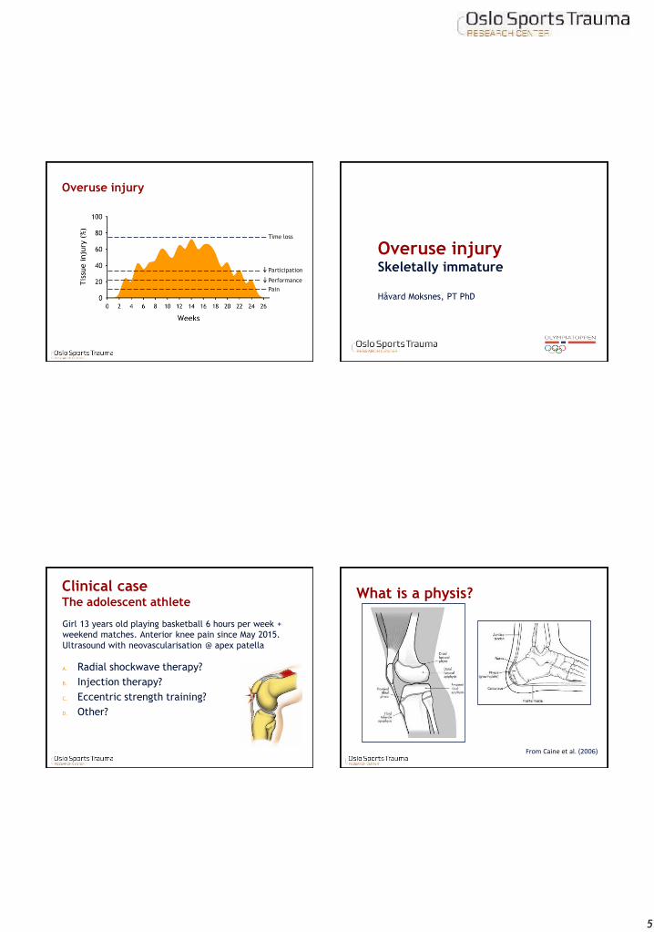
Participation
Pain
Time loss
Performance
Overuse injury
Overuse injurySkeletally immature
Håvard Moksnes, PT PhD
Clinical caseThe adolescent athlete
Girl 13 years old playing basketball 6 hours per week +
weekend matches. Anterior knee pain since May 2015.
Ultrasound with neovascularisation @ apex patella
A. Radial shockwave therapy?
B. Injection therapy?
C. Eccentric strength training?
D. Other?
What is a physis?
From Caine et al. (2006)

6
Overuse injuries«Demise of the fittest; are we
destroying our biggest talents?»
Editorial from
Prof Roald Bahr
May 2014
Envelope of load acceptance
Dye 2005
Normal load
No
pain
Envelope of load acceptance
Growth spurt
Dye 2005
Fatigue
Trauma
Cartilage
injury
Pain
Puberty
Malalignment
Normal load

7
Adolescents with anterior knee
painDiagnosis
➢ Mb.Sinding-Larsen
➢ Mb.Osgood-Schlatter
➢ PFP
➢ Patellar instability
➢ Tendinopathy
➢ Osteochondritis
dissecans
➢ Osteosarcoma
➢ Rheumatoid illness
Adolescents with anterior knee
painDiagnosis
➢ Mb.Sinding-Larsen
➢ Mb.Osgood-Schlatter
➢ PFP
➢ Patellar instability
➢ Tendinopathy
Treatment
Main rule:
✓ Overload of soft tissue
or growth zones
✓ Adjust total load!
Mb.Sinding-Larsen-Johansson
✓ Occurs during
growth spurt
✓ Resembles
“Jumpers knee”
✓ US helpful?
Patellar tendinopathy
Plantar fasciopathy
Lateral epcondyopathy
8
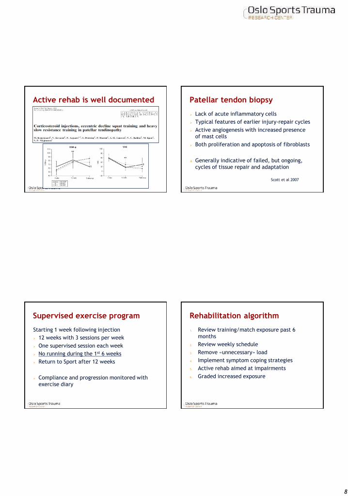
Active rehab is well documented Patellar tendon biopsy
➢ Lack of acute inflammatory cells
➢ Typical features of earlier injury‐repair cycles
➢ Active angiogenesis with increased presence
of mast cells
➢ Both proliferation and apoptosis of fibroblasts
❖ Generally indicative of failed, but ongoing,
cycles of tissue repair and adaptation
Scott et al 2007
Supervised exercise program
Starting 1 week following injection
➢ 12 weeks with 3 sessions per week
➢ One supervised session each week
➢ No running during the 1st 6 weeks
➢ Return to Sport after 12 weeks
➢ Compliance and progression monitored with
exercise diary
Rehabilitation algorithm
1. Review training/match exposure past 6
months
2. Review weekly schedule
3. Remove «unnecessary» load
4. Implement symptom coping strategies
5. Active rehab aimed at impairments
6. Graded increased exposure

9
1.Review training/match
exposure past 6 months
✓ Increased frequency?
✓ Increased intensity?
✓ Changed environment?
✓ Playing several teams?
✓ “Did you have a holiday this summer?”
2. Review weekly schedule
✓ Write it down on paper!
✓ When do you rest?
✓ Which sessions can be removed?
3. Remove «unnecessary» load
✓ Remove “unnecessary” runs and hops
✓ Change from running to cycling
✓ School sessions vs club sessions
✓ Leisure time activities
4. Implement symptom coping
strategies
✓ RICE after loading
✓ Pain monitoring model
✓ Rest days
✓ Less playing time
✓ One game per week
✓ Play and train with only one team!

10
5. Active rehab aimed at
impairments Increase the size of the envelope
Shock absorption
➢ Hip, hamstring and kinetic chain
6. Graded increased exposure
✓ Stabilize load with symptom control
✓ Alternate days team and rehab
✓ Increase frequency OR intensity
✓ Continue coping strategies
Plantar fasciopathy
http://blogs.bmj.com/bjsm/2014/09/15/plantar-fasciitis-
important-new-research-by-michael-rathleff/
Rathleff et al 2014
Effect of Corticosteroid Injection, Physiotherapy,
or Both on Clinical Outcomes in Patients With
Unilateral Lateral Epicondylalgia A RCT
Conclusion and Relevance
Among patients with chronic unilateral lateral
epicondylalgia, the use of corticosteroid injection vs
placebo injection resulted in worse clinical outcomes
after 1 year, and physiotherapy did not result in any
significant differences
Coombes et al JAMA 2013

11
Take home messages RTP with
overuse injuries
✓ Assess and tweak total load
✓ Include the parents and coaches
✓ Removal from play may be necessary
✓ Agree on a structured progression plan
Hamstring injuries
- injury types and rehabilitation
Håvard Moksnes
Sports Physiotherapist
Hamstring muscle group
Semitendinosu
s
Semimembrano
sus
Biceps
Femoris
Biceps
Femoris –
short head

12
Hamstring injuries
Different types of injuries
High-speed running
Slow-speed stretching
High-speed stretching
Overuse (proximal)
Askling 2007,
Heiderschneit 2010
Two distinct acute injuries
van der
Made et al
2016
van der
Made et al
2016

13
Hamstring muscle group
Semitendinosu
s
Semimembrano
sus
Biceps
Femoris
Biceps
Femoris –
short head
van der Made et al
2016
van der
Made et al
2016
Muscle injuries in football
Hamstring 37%
Adductor 23%
Quadriceps 19%
Calf 13%
Ekstrand et al 2011
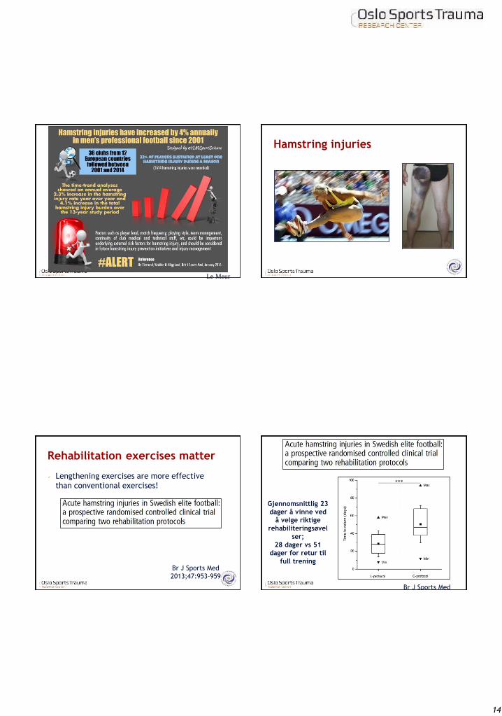
14
Le Meur
2016
Hamstring injuries
Rehabilitation exercises matter
✓ Lengthening exercises are more effective
than conventional exercises!
Br J Sports Med
2013;47:953-959
Gjennomsnittlig 23
dager å vinne ved
å velge riktige
rehabiliteringsøvel
ser;
28 dager vs 51
dager for retur til
full trening
Br J Sports Med
2013;47:953-959

15
Ivolvement of the
proximal tendon
significantly increases
time to return to
sport
Br J Sports Med
2013;47:953-959
Do not forget apophyseal avulsions !
Prediction RTS with MRI
Grade I
➢ Mean 18 days (±19)
➢ 70% = 0 -> 37 days
➢ 95% = 0 -> 56 days
Grade III
➢ Mean 24 days (±13)
➢ 70% = 11 -> 37 days
➢ 95% = 0 -> 50 days

16
Hamstring muscle group
Semitendinosu
s
Semimembrano
sus
Biceps
Femoris
Biceps
Femoris –
short head
Sprinting type injury
Long head of biceps femoris
Askling
2007
Sprinting type injury
Askling
2007
PMT
J
PT
Sprinting type injury
Long head of biceps femoris
Intramuscular tendon involvement
Yes = longer recovery time
Secondary problems
Biceps femoris short head
Askling
2007

17
The intramuscular tendon
Reurink
2016
Stretching type injury
Semimem
.
n.ischiadicu
s.
Quadratus
femoris.
Askling
2007
Hamstring muscle group
Semitendinosu
s
Semimembrano
sus
Biceps
Femoris
Biceps
Femoris –
short head

18
Proximal Hamstring Tendinopathy:
A Real Pain in The Butt For Runners!
Pain during high intensity running
Pain with prolonged sitting
Pain with increased stide length
Pain during uphill running
Must reduce running volume!
Le Meur
2016
Sport specific RTS

19
The Extender
Askling, BJSM 2013
The Diver
Askling, BJSM 2013
The Glider
Askling, BJSM 2013
Advanced lengthening exercise

20
Norges idrettsforbund og olympiske og paralympiske komité
Rehabilitering og testing etter akillesseneruptur
Side 81
The results of the present study demonstrate that
stable surgical repair with accelerated tendon
loading could be performed in all (n = 49) patients without reruptures and major soft tissue–related
complications.
Olsson et al AJSM 2013 Side 82 Olsson et al AJSM 2013
21
Side 83 Olsson et al AJSM 2013
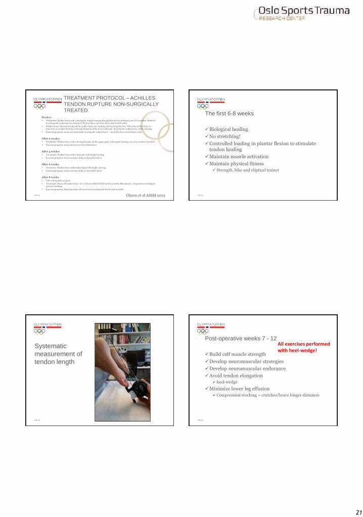
TREATMENT PROTOCOL – ACHILLES
TENDON RUPTURE NON-SURGICALLY
TREATED
Week 0
✓ Treatment: Walker brace with 3 heel pads, weight-bearing through the heel as tolerated, use of 2 crutches. Referral to orthopedic technician for shoe heel-lift (use shoe with heel-lift on the healthy side).
✓ Walker brace: Allowed to take off the walker brace for washing and aerating the foot. When the walker brace is removed, no weight-bearing or dorsal extension of the foot is allowed. Wearing the walker brace while sleeping.
✓ Exercise program: home exercises daily wearing the walker brace – move the toes several times a day
After 2 weeks:
✓ Treatment: Walker brace with 2 heel pads (take off the upper pad), full weight-bearing, use of 2 crutches if needed.
✓ Exercise program: home exercises as described above.
After 4 weeks:
✓ Treatment: Walker brace with 1 heel pad, full weight-bearing
✓ Exercise program: home exercises daily as described above
After 6 weeks
✓ Treatment: Walker brace without heel pad, full weight-bearing
✓ Exercise program: home exercises daily as described above
After 8 weeks
✓ Visit orthopaedic surgeon
✓ Treatment: Wean off walker brace. Use of shoes with heel-lift (until 14 weeks after injury), compression stocking to prevent swelling.
✓ Exercise program: Important that all exercises are performed slowly and carefully
Side 84
The first 6-8 weeks
✓Biological healing
✓No stretching!
✓Controlled loading in plantar flexion to stimulatetendon healing
✓Maintain muscle activation
✓Maintain physical fitness
✓ Strength, bike and eliptical trainer
Side 85
Systematic
measurement of
tendon length
Side 86
Post-operative weeks 7 - 12
✓Build calf muscle strength
✓Develop neuromuscular strategies
✓Develop neuromuscular endurance
✓Avoid tendon elongation
➢ heel-wedge
✓Minimize lower leg effusion
➢ Compression stocking + crutches/brace longer distances
All exercises performedwith heel-wedge!

22
Side 87
Build calf muscle strength
Standing heel-raises
Bilateral
Extended knee
✓Seated heel raises
➢90° knee flexion
✓Leg press machine
➢Extended knee
➢Avoid tendon stretch!
Side 88
Build calf muscle strength
Standing heel raises
Bilateral
Extended knee
Seated heel raises
1. 90° knee flexion
2. Leg press machine
➢Extended knee
➢Not push from hip
➢Push all the way out!
➢Avoid tendon stretch!
Side 89
Develop neuromuscular endurance
Single & double leg stance
Extended knee!
Sagittal sway
Closed eyes
Perturbation?
Side 90
Develop neuromuscular strategies and strength

23
Side 91
Develop neuromuscular endurance
Side 92
Develop neuromuscular strategies and strength
Side 93
Develop neuromuscular strategies and strength
Downhill Sidewalk with rubber band
Side 94
Post-operative weeks 12 →
✓Rebuild energy storage and release
✓Increase velocity in movements
➢ Reduce frequency and reps
✓Slowly increase hopping and running
➢ Think reps – not time or distance!
✓Running, hopping and single-leg hopping

24
Side 95
Post-operative weeks 12 →
Side 96
Post-operative weeks 12 →
Side 97
Post-operative weeks 12 →
Side 98
Physical function tests
✓Single leg CMJ jump
✓25 consecutive single leg hops
✓Heel-raise exhaustion
Average hop height
Jump coefficient (contact/flight time)
Evaluate flow and quality

25
Hopping; jump coefficientContact time/flight time
0,96
0,830,88
0,71
0
0,2
0,4
0,6
0,8
1
1,2
Injured Non-injured
4 months
5 months
Single leg CMJ (cm)
19,5
24,5
42,3
22,525,1
42,7
10
15
20
25
30
35
40
45
Injured Non-injured Both
4 months
5 months
Heel-raise exhaustion (work)
912
2714
1780
3096
0
500
1000
1500
2000
2500
3000
3500
Injured Non-injured
4 months
5 months
Bone stress injuries
Clinical update

26
BSI pathophysiology BSI pathophysiology
Warden et al. 2015
Bone stress injury continuum
Stress
reaction
Stress
fracture
Complete
fracture
Increased bone turnover
Periosteal± marrow oedema
Discernable fracture line
Structural instability
Video

27
Epidemiology
Extremely common in distance runners
33-66% have a history of stress fracture
8-21% per year affected
Up to 20% of all athletics injuries
More common in females
1.5 to 3 times greater risk
Endurance, aesthetic and weight-class sports
a particular problem
High rate of recurrence
Wentz 2011, Snyder 2006
Humerus Throwing and racquet sports
Ribs Rowing and kayak
Pars interarticularis Cricket, diving, gymnastics,
ballet
Femur Distance running, jumping,
ballet
Tibia Distance running, ballet
Navicular Sprinting, jumping, football
Metatarsal Sprinting, jumping, marching
ballet
Brukner & Khan, 2012
Internal risk factors
Age
Sex
Anatomy
Body composition
Health & injury history
Physical Characteristics
- Fitness
- Muscle strength
- Joint ROM
Skill level
Psychological factors
External risk factors
Sports equipment
Environment
Sports factors
(coaching, rules,
referees)
Protective equipment
Inciting event
Playing situation
Player/opponent
behaviour
Gross biomechanical
description
Detailed biomechanical
description
Susceptible
Athlete
Predisposed
Athlete INJURY
A comprehensive model of injury causation
Meeuwisse, 1994
Bahr & Krosshaug, 2005
Risk factors Factors
affecting bone
loading
Bone loading
Bone strain
Factors
affecting bone
load tolerance
Risk factors for bone stress injuries
Warden 2015

28
Factors affecting bone loading
Training factors
Magnitude of load
Rate of load application
Number of loading cycles
Training frequency (recovery)
Factors affecting bone loading
Intrinsic anatomy/biomechanics
Excessive or inadequate subtalar pronation
Increased hip external rotation
Leg length difference
Low muscle strength or endurance
Finestrone 1991, Bennell 1996, Hoffman 1999
Sullivan 1984, Simkin 1989
Factors affecting bone loading
Training surface
Hard or cambered surfaces
Changes to normal surface
Equipment
Shoes
Orthotic inserts
Milgrom 2003, Brukner & Khan, 2012
Factors affecting bone loading
Technique factors
Forefoot/rearfoot strike pattern
Stride length
Mixed bowling action in cricket
Elliot 2000

29
Factors affecting load tolerance
Genetics
Maternal famliy history of osteopenia/osteoporosis
Physcial activity history
Nutrition
Energy balance (intake vs. expenditure)
Calcium and Vitamin D status
Ruohola 2006, Chen 2013, Lappe 2008
Factors affecting load tolerance
Endocrine status
Older age at menarche
Disease
Medication use
Glucocorticoids
Anticonvulsants
Chen 2013
Factors
affecting bone
loading
Bone loading
Bone strain
Factors
affecting bone
load tolerance
Risk factors for bone stress injuries
Warden 2015
Diagnosis of BSI - History
Gradual onset of pain
Recent change in training
Pain does not "warm up" with continued
training
Pain resolves with rest in early phases
Later stages – pain with ADLs, at rest and at
night
30
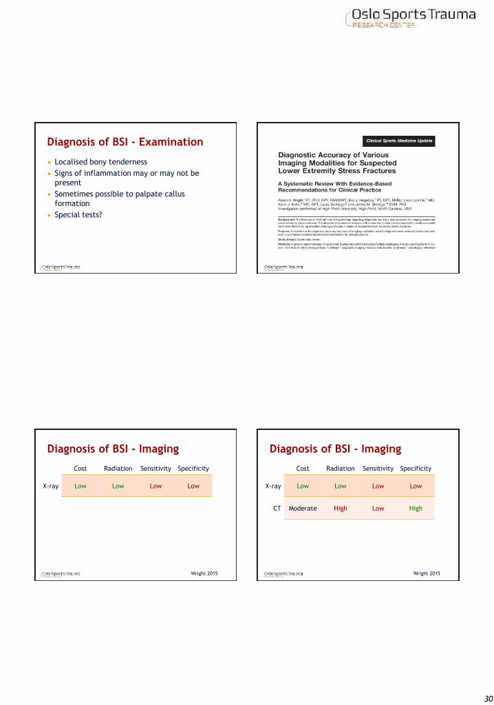
Diagnosis of BSI - Examination
Localised bony tenderness
Signs of inflammation may or may not be
present
Sometimes possible to palpate callus
formation
Special tests?
Diagnosis of BSI - Imaging
Cost Radiation Sensitivity Specificity
X-ray Low Low Low Low
Diagnosis of BSI - Imaging
Wright 2015
Cost Radiation Sensitivity Specificity
X-ray Low Low Low Low
CT Moderate High Low High
Diagnosis of BSI - Imaging
Wright 2015

31
Cost Radiation Sensitivity Specificity
X-ray Low Low Low Low
CT Moderate High Low High
Bone
scanModerate High High Moderate
Diagnosis of BSI - Imaging
Wright 2015
Cost Radiation Sensitivity Specificity
X-ray Low Low Low Low
CT Moderate High Low High
Bone
scanModerate High High Moderate
MRI High None High High
Diagnosis of BSI - Imaging
Wright 2015
High-risk BSIs
More likely to progress to complete fracture,
delayed union or non-union
Those that require surgical repair
Those that require assisted or non-
weightbearing
Often located on the tension side of the
bone's biomechanical axis
Boden 2000
Low-risk High-risk
Femoral shaft Femoral neck
Posteromedial tibia Anterior cortex of tibia
Fibula Medial malleolus
Calcaneus Talus (lateral process)
2nd-4th metatarsal
diaphysis
Navicular
Proximal 5th metatarsal
Base of 2nd metatarsal
Great toe sesamoids
Boden 2000, 2001

32
MRI grading
1Periosteal surface - mild to moderate oedema (T2 images)
Bone marrow normal (T1 &T2 images)
2Periosteal surface - moderate to severe oedema (T2 images)
Bone marrow oedema (T2 images)
3Periosteal surface - moderate to severe oedema (T2 images)
Bone marrow oedema (T1 & T2 images)
4Periosteal surface - moderate to severe oedema (T2 images)
Bone marrow oedema (T1 & T2 images)
Clearly visible fracture line
Fredericson 1995
Management – key principles
Unload, then systematically reintroduce loading
Address all potential causes
Factors affecting bone loading
Factors affecting load tolerance
Avoid NSAIDs – they may affect bone healing
Is there a role for adjunct treatments?
Electrotherapies
Pharmaceutical agents
Ziltener 2010
Loading stress fractures
Unloading necessary for all stress fractures
There is probably an "optimal load"
Maximise speed of recovery
Maximise strength of repair
Reduce risk of recurrence
Unfortunately, we don't know what that is
Little research
Large individual variability
Practical solution – use pain as a guide
Low-risk fractures
If pain-free gait, full weight bearing is
permitted
If gait painful, partial weight bearing with
boot/brace
Maintain conditioning with low-weight-
bearing activities
Swimming
Deep-water running
Antigravity treadmill (when gait is pain-free)

33
Warden 2015
High-risk stress fractures
Minimum 4 weeks non-weight bearing
Immediate surgery considered in some cases
Warden 2015
Osteoclast inhibitors
Bisphosphates often used to treat
osteoporosis, Paget's disease, & bone tumors
Used by some for stress fractures, but no
evidence it accelerates recovery
No evidence that it prevents stress fractures
Side effects - nausea, fatigue, gastro-
intestinal symptoms, joint & muscle pain
Not recommended
Shima 2008
Parathyroid hormone and
antisclerosin antibody therapy
Both have anabolic effect on bone production
Promote osteogenesis, osteoblast proliferation
and survival
Laboratory studies promising
Effect unknown in vivo
Routine use not advised
Sloan 2010

34
Finally, when is a BSI healed?
Brukner & Khan 2012
The Oslo Sports Trauma Research Center
has been established at
the Norwegian School of Sport Sciences
through generous grants from the Royal Norwegian Ministry of
Culture, the South-Eastern Norway Regional Health Authority, the
International Olympic Committee, the Norwegian Olympic
Committee & Confederation of Sport, and Norsk Tipping AS