Multiple transcription factor codes activate epidermal...
6
Multiple transcription factor codes activate epidermal wound–response genes in Drosophila Joseph C. Pearson 1 , Michelle T. Juarez, Myungjin Kim, and William McGinnis 2 Section of Cell and Developmental Biology, Division of Biology, University of California at San Diego, La Jolla, CA 92093 Edited by Kathryn V. Anderson, Sloan–Kettering Institute, New York, NY, and approved December 5, 2008 (received for review October 14, 2008) Wounds in Drosophila and mouse embryos induce similar genetic pathways to repair epidermal barriers. However, the transcription factors that transduce wound signals to repair epidermal barriers are largely unknown. We characterize the transcriptional regulatory enhancers of 4 genes—Ddc, ple, msn, and kkv—that are rapidly activated in epidermal cells surrounding wounds in late Drosophila embryos and early larvae. These epidermal wound enhancers all contain evolutionarily conserved sequences matching binding sites for JUN/FOS and GRH transcription factors, but vary widely in trans- and cis-requirements for these inputs and their binding sites. We propose that the combination of GRH and FOS is part of an ancient wound–response pathway still used in vertebrates and invertebrates, but that other mechanisms have evolved that result in similar tran- scriptional output. A common, but largely untested assumption of bioinformatic analyses of gene regulatory networks is that transcrip- tion units activated in the same spatial and temporal patterns will require the same cis-regulatory codes. Our results indicate that this is an overly simplistic view. cuticle grainy head fos wound repair A nimals have evolved systems to sense and repair epidermal wounds and to fight microbial infections that follow wound- ing. Many wound responses are carried out by homologous genetic pathways in arthropods and vertebrates and thus appar- ently had evolved in a common ancestor of bilateral animals. These responses include pathways mediating reepithelialization (1–9), responses to microbial invasion (10), and regeneration of epidermal barrier layers (11, 12). The outermost epidermal barrier in mammals is the stratum corneum, a constantly regen- erated layer of crosslinked keratinocytes, proteins, and lipids (13). The analogous protective barrier for arthropods is the cuticle, comprised of crosslinked chitin, proteins, and lipids (14). Although mammalian and arthropodal epidermal barriers are constructed in largely different ways, they share a homologous genetic pathway controlling barrier repair that involves tran- scription factors of the Grainy head (GRH) family (11, 12). One central question in the control of epidermal wound repair is how signals are integrated at the level of transcription to activate the large battery of effector genes that mediate wound responses. Although many genes are known to be activated in epidermal cells after wounding (15–17), little is known about the cis-regulatory enhancers that mediate wound-induced gene ac- tivation. In Drosophila, there are only a few known genes activated at epidermal wound sites (11, 14, 17). Two of these genes encode the enzymes Dopa decarboxylase (Ddc) and Tyrosine hydroxylase (encoded by ple) (11). These 2 enzymes are limiting steps in the pathway to produce the highly reactive quinones that crosslink chitin and cuticle proteins during the construction and repair of cuticular barriers (18). To better characterize the transcriptional wound response in Drosophila embryos, we have mutagenized minimal Ddc and ple epidermal wound enhancers, showing that both require AP-1- like and GRH consensus sites. We then searched for AP-1 and GRH consensus binding sites in the regulatory DNA for other epidermal wound–response genes, which led to the identification of epidermal wound enhancers for the genes krotzkopf verkehrt (kkv) and misshapen (msn). Ddc, ple, kkv, and msn are all transcriptionally activated within minutes after epidermal wounding. Three of the genes use overlapping transcriptional codes involving GRH and FOS to activate their epidermal wound enhancers, but kkv uses a fundamentally different code for wound activation. Whereas the common wound–response cis-regulatory codes we describe will be useful in the identifica- tion of new epidermal wound enhancers in both vertebrates and invertebrates, the evidence for alternative wound–response codes provides insight into how genetic control of wound healing has evolved in metazoans. Results Minimal Ddc Epidermal Wound Enhancers. By using reporter gene constructs in Drosophila embryos, we previously identified a 0.47-kb DNA fragment just upstream of the Ddc gene that functioned as an epidermal wound enhancer in late embryos (Fig. 1A) (11). This enhancer can activate GFP reporter expres- sion in a zone around aseptic epidermal punctures in late embryos (Fig. 1B), in larvae just before molts (data not shown), and in very young adults (Fig. 1C). The Ddc .47 wound-enhancer DNA contains 2 regions upstream of the basal promoter that are highly conserved among all sequenced drosophilid species: CR1 (44 nt) and CR2 (13 nt) (Fig. 1 A and SI Appendix). The CR2 sequence consists largely of a high-affinity GRH binding site (ACCGGTT) (12, 19, 20), which is required for wound-enhancer function (11). Ddc .47 CR1 contains a sequence matching the consensus binding site (TGANTCA) for AP-1, a transcription factor consisting of a JUN-FOS dimer (21). To test for AP-1-like site function in the Ddc .47 wound enhancer, we mutated the conserved AP-1-like site in CR1 and a second AP-1-like site that lies between CR1 and CR2 (Fig. 1 A). This construct is unable to activate reporter expression after wounding (Fig. 1D). We also deleted 355 bp between the conserved regions (Ddc Gap) (Fig. 1 A), to test whether any required DNA elements are located within the region between CR1 and CR2. This deletion mutant functions as an epidermal wound enhancer, but the number of cells that activate reporter gene expression is reduced compared with WT Ddc .47 (Fig. 1E). In summary, both AP-1-like and GRH consensus sequences are absolutely required for Ddc .47 wound activation, and a minimal enhancer of 117 bp containing these sites is sufficient for modest activation in cells around wound sites. However, additional sequences within .47 Ddc contribute to the strength of this epidermal wound enhancer. Author contributions: J.C.P., M.T.J., M.K., and W.M. designed research; J.C.P., M.T.J., and M.K. performed research; J.C.P., M.T.J., M.K., and W.M. analyzed data; and J.C.P. and W.M. wrote the paper. The authors declare no conflict of interest. This article is a PNAS Direct Submission. Freely available online through the PNAS open access option. 1 Present address: Program in Molecular Biology and Biotechnology, University of North Carolina, Chapel Hill, NC 27599. 2 To whom correspondence should be addressed. E-mail: [email protected]. This article contains supporting information online at www.pnas.org/cgi/content/full/ 0810219106/DCSupplemental. © 2009 by The National Academy of Sciences of the USA 2224 –2229 PNAS February 17, 2009 vol. 106 no. 7 www.pnas.orgcgidoi10.1073pnas.0810219106
Transcript of Multiple transcription factor codes activate epidermal...
Multiple transcription factor codes activate epidermalwound–response genes in DrosophilaJoseph C. Pearson1, Michelle T. Juarez, Myungjin Kim, and William McGinnis2
Section of Cell and Developmental Biology, Division of Biology, University of California at San Diego, La Jolla, CA 92093
Edited by Kathryn V. Anderson, Sloan–Kettering Institute, New York, NY, and approved December 5, 2008 (received for review October 14, 2008)
Wounds in Drosophila and mouse embryos induce similar geneticpathways to repair epidermal barriers. However, the transcriptionfactors that transduce wound signals to repair epidermal barriers arelargely unknown. We characterize the transcriptional regulatoryenhancers of 4 genes—Ddc, ple, msn, and kkv—that are rapidlyactivated in epidermal cells surrounding wounds in late Drosophilaembryos and early larvae. These epidermal wound enhancers allcontain evolutionarily conserved sequences matching binding sitesfor JUN/FOS and GRH transcription factors, but vary widely in trans-and cis-requirements for these inputs and their binding sites. Wepropose that the combination of GRH and FOS is part of an ancientwound–response pathway still used in vertebrates and invertebrates,but that other mechanisms have evolved that result in similar tran-scriptional output. A common, but largely untested assumption ofbioinformatic analyses of gene regulatory networks is that transcrip-tion units activated in the same spatial and temporal patterns willrequire the same cis-regulatory codes. Our results indicate that this isan overly simplistic view.
cuticle � grainy head � fos � wound repair
Animals have evolved systems to sense and repair epidermalwounds and to fight microbial infections that follow wound-
ing. Many wound responses are carried out by homologousgenetic pathways in arthropods and vertebrates and thus appar-ently had evolved in a common ancestor of bilateral animals.These responses include pathways mediating reepithelialization(1–9), responses to microbial invasion (10), and regeneration ofepidermal barrier layers (11, 12). The outermost epidermalbarrier in mammals is the stratum corneum, a constantly regen-erated layer of crosslinked keratinocytes, proteins, and lipids(13). The analogous protective barrier for arthropods is thecuticle, comprised of crosslinked chitin, proteins, and lipids (14).Although mammalian and arthropodal epidermal barriers areconstructed in largely different ways, they share a homologousgenetic pathway controlling barrier repair that involves tran-scription factors of the Grainy head (GRH) family (11, 12).
One central question in the control of epidermal wound repairis how signals are integrated at the level of transcription toactivate the large battery of effector genes that mediate woundresponses. Although many genes are known to be activated inepidermal cells after wounding (15–17), little is known about thecis-regulatory enhancers that mediate wound-induced gene ac-tivation. In Drosophila, there are only a few known genesactivated at epidermal wound sites (11, 14, 17). Two of thesegenes encode the enzymes Dopa decarboxylase (Ddc) andTyrosine hydroxylase (encoded by ple) (11). These 2 enzymes arelimiting steps in the pathway to produce the highly reactivequinones that crosslink chitin and cuticle proteins during theconstruction and repair of cuticular barriers (18).
To better characterize the transcriptional wound response inDrosophila embryos, we have mutagenized minimal Ddc and pleepidermal wound enhancers, showing that both require AP-1-like and GRH consensus sites. We then searched for AP-1 andGRH consensus binding sites in the regulatory DNA for otherepidermal wound–response genes, which led to the identificationof epidermal wound enhancers for the genes krotzkopf verkehrt
(kkv) and misshapen (msn). Ddc, ple, kkv, and msn are alltranscriptionally activated within minutes after epidermalwounding. Three of the genes use overlapping transcriptionalcodes involving GRH and FOS to activate their epidermalwound enhancers, but kkv uses a fundamentally different codefor wound activation. Whereas the common wound–responsecis-regulatory codes we describe will be useful in the identifica-tion of new epidermal wound enhancers in both vertebrates andinvertebrates, the evidence for alternative wound–responsecodes provides insight into how genetic control of wound healinghas evolved in metazoans.
ResultsMinimal Ddc Epidermal Wound Enhancers. By using reporter geneconstructs in Drosophila embryos, we previously identified a0.47-kb DNA fragment just upstream of the Ddc gene thatfunctioned as an epidermal wound enhancer in late embryos(Fig. 1A) (11). This enhancer can activate GFP reporter expres-sion in a zone around aseptic epidermal punctures in lateembryos (Fig. 1B), in larvae just before molts (data not shown),and in very young adults (Fig. 1C). The Ddc .47 wound-enhancerDNA contains 2 regions upstream of the basal promoter that arehighly conserved among all sequenced drosophilid species: CR1(44 nt) and CR2 (13 nt) (Fig. 1 A and SI Appendix). The CR2sequence consists largely of a high-affinity GRH binding site(ACCGGTT) (12, 19, 20), which is required for wound-enhancerfunction (11). Ddc .47 CR1 contains a sequence matching theconsensus binding site (TGANTCA) for AP-1, a transcriptionfactor consisting of a JUN-FOS dimer (21).
To test for AP-1-like site function in the Ddc .47 woundenhancer, we mutated the conserved AP-1-like site in CR1 anda second AP-1-like site that lies between CR1 and CR2 (Fig. 1A).This construct is unable to activate reporter expression afterwounding (Fig. 1D). We also deleted 355 bp between the conservedregions (Ddc Gap) (Fig. 1A), to test whether any required DNAelements are located within the region between CR1 and CR2. Thisdeletion mutant functions as an epidermal wound enhancer, but thenumber of cells that activate reporter gene expression is reducedcompared with WT Ddc .47 (Fig. 1E). In summary, both AP-1-likeand GRH consensus sequences are absolutely required for Ddc .47wound activation, and a minimal enhancer of 117 bp containingthese sites is sufficient for modest activation in cells around woundsites. However, additional sequences within .47 Ddc contribute tothe strength of this epidermal wound enhancer.
Author contributions: J.C.P., M.T.J., M.K., and W.M. designed research; J.C.P., M.T.J., andM.K. performed research; J.C.P., M.T.J., M.K., and W.M. analyzed data; and J.C.P. and W.M.wrote the paper.
The authors declare no conflict of interest.
This article is a PNAS Direct Submission.
Freely available online through the PNAS open access option.
1Present address: Program in Molecular Biology and Biotechnology, University of NorthCarolina, Chapel Hill, NC 27599.
2To whom correspondence should be addressed. E-mail: [email protected].
This article contains supporting information online at www.pnas.org/cgi/content/full/0810219106/DCSupplemental.
© 2009 by The National Academy of Sciences of the USA
2224–2229 � PNAS � February 17, 2009 � vol. 106 � no. 7 www.pnas.org�cgi�doi�10.1073�pnas.0810219106

An Epidermal Wound Enhancer from the ple Gene Requires GRH andAP-1-Like Sequences. We previously identified 2 epidermal woundenhancers upstream of ple by searching for conserved clusters ofAP-1-like and GRH consensus sites (11). We have refined theboundaries of the distal 3-kb ple fragment that contains a woundenhancer, delimiting the enhancer to a 687-bp DNA fragmentthat resides 3.45 kb upstream of ple (Wound Enhancer 1 or WE1)(Fig. 2 A and B). In addition to conserved AP-1-like and GRHconsensus sites, analysis of this ple wound enhancer revealedconserved regions with consensus binding sites for other tran-scription factors. Of these sites, we noted putative binding sitesfor CREB homodimers (TGACGTMA) (22), EXD/PBX ho-modimers (WGATTGAW) (23–25), and Hox monomers(YMATTA) (26, 27). To test the importance of these sites, wemutated them in the context of ple-WE1 (Fig. 2 A). Mutating theconsensus AP-1-like sites abolished ple wound-enhancer func-tion (Fig. 2C), whereas mutation of the GRH consensus siteresulted in a consistent reduction of wound-enhancer function(Fig. 2D). In contrast, mutations in the CREB, EXD, or HOXconsensus sites had no consistent effect on ple WE1 wound-dependent activation (Fig. 2E–G). Therefore, activation of Ddcand ple wound enhancers depends on AP-1-like and GRHconsensus binding sites.
To test whether sequences matching the GRH and AP-1-likeconsensus sites in the Ddc and ple wound enhancers bound GRHand AP-1 family proteins in vitro, we used EMSAs (SI Text) (28).
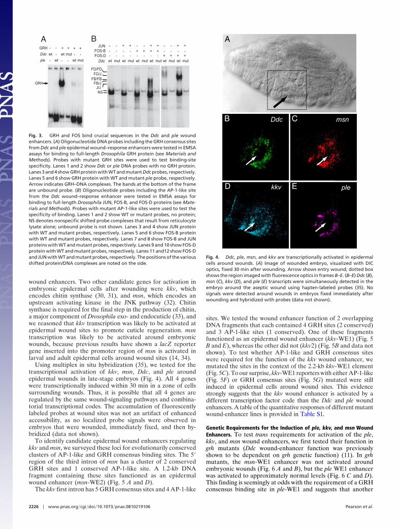
An oligonucleotide including a GRH consensus site (ACCG-GTT) from the Ddc wound enhancer binds full-length GRHprotein with high affinity and specificity (Fig. 3A) (19). TheGRH-like site in the ple WE1 element (ACTCGTTT) is aweaker match to an optimal site (AACCGGTTT) (20), andoligonucleotides with this site bind GRH protein with lowaffinity and specificity (Fig. 3A).
EMSA experiments using full-length Drosophila JUN andFOS-B proteins (the fly kay gene produces 4 isoforms of FOS,A–D), indicate that they bind as heterodimers with high speci-ficity and affinity to AP-1-like sites from CR1 of the Ddc woundenhancer, whereas JUN or FOS-B homodimers had loweraffinities (Fig. 3B). Because the AP-1-like site in the ple en-hancer has the same consensus sequence as the one in the Ddcenhancer, we did not test JUN/FOS binding to ple WE1 AP-1-like sites. In contrast to mammalian FOS, Drosophila FOS-Bprotein can bind as a homodimer to AP-1-like consensus sites,albeit with lower affinity than JUN/FOS heterodimers (Fig. 3B)(29). To explore the possibility that other Drosophila FOS isoformsmight bind wound-enhancer AP-1-like sites, we also tested FOS-Dbinding to an oligonucleotide containing the Ddc AP-1-like site. Asseen in Fig. 3B, FOS-D homodimers apparently have higher affinitywith Ddc AP-1-like sites than FOS-B or JUN homodimers, albeitless than JUN/FOS-B or JUN/FOS-D heterodimers. The relevanceof FOS-D homodimer binding will be seen later in this report. Fromthe above, we concluded that the mutations we introduced into theGRH and AP-1-like consensus sites in the Ddc and ple woundenhancers would eliminate binding of GRH, JUN, or FOS tran-scription factors in vivo.
Identification of Epidermal Wound–Response Enhancers. We nextwished to determine whether clusters of AP-1-like and GRHconsensus binding sites were a common feature of epidermal
Ddc Gap
+WR
-
A
CR
1
CR
2
Ddc .47 APm
Ddc .47
Ddc .47 GRHm -+/-
Ddc .47 embryoB C Ddc .47 adult
D Ddc .47 APm Ddc GapE
Fig. 1. Sequence requirements of the Ddc epidermal wound enhancer. (A)Diagram of WT and mutant Ddc .47 epidermal wound–response enhancers.White blocks indicate conserved regions with other drosophilids; red blocksdenote mutant sequences. Functional wound enhancers are indicated by ‘‘�,’’nonfunctional enhancers by ‘‘�.’’ (B–E) Dashed lines outline embryos, arrowsindicate wound sites, and fluorescent nuclei surrounding wounds show GFP orDsRed reporter gene activation provided by wound enhancers. (B) Ddc .47activated GFP-reporter expression around wounds in late-stage embryonicepidermis. (C) Ddc .47 activated GFP-reporter expression around wounds inearly adult abdominal epidermis. (D) A mutation of AP-1-like consensus sites(APm) in Ddc .47 abolished wound-enhancer function. (E) Ddc Gap hadreduced wound-enhancer function. Previous results showed that a GRH sitemutant (GRHm) in Ddc .47 abolished wound-enhancer function (11).
WR
-+
++/-
CREBm EXDm HOXm
ple-WE1
ple-WE1 APm
ple-WE1 CREBm
ple-WE1 EXDm
ple-WE1 GRHm
ple-WE1 HOXm
++
A
E G
ple-WE1B APmC
GRHm FD
APc - TGANTCA
CREBc - TGACGTMA
EXDc - WGATTGAW
GRHc - ACYNGTT
HOXc - YMATTA
-3.45 kb
-4.14 kb
Fig. 2. ple epidermal wound enhancer binding site requirements. (A) Dia-gram of WT and mutant ple-WE1 enhancers. White boxes indicate regions ofsequence conservation in drosophilids; red blocks denote mutant sequences.Wound responses of the elements are indicated at right. (B–G) Dashed linesoutline the embryos; arrows indicate wound sites. (B) ple-WE1 was activatedat epidermal wounds. (C) Mutation of two AP-1-like consensus sites abolishedple-WE1 wound-enhancer activity. (D) Mutation of a GRH consensus sitestrongly reduced ple-WE1 wound-enhancer activity. (E–G) Mutation of 2CREB-like sites (E), or 2 EXD-like sites (F), or 14 HOX-like binding site had noeffect on ple-WE1 enhancer function (G).
Pearson et al. PNAS � February 17, 2009 � vol. 106 � no. 7 � 2225
DEV
ELO
PMEN
TAL
BIO
LOG
Y
wound enhancers. Two other candidate genes for activation inembryonic epidermal cells after wounding were kkv, whichencodes chitin synthase (30, 31), and msn, which encodes anupstream activating kinase in the JNK pathway (32). Chitinsynthase is required for the final step in the production of chitin,a major component of Drosophila exo- and endocuticle (33), andwe reasoned that kkv transcription was likely to be activated atepidermal wound sites to promote cuticle regeneration. msntranscription was likely to be activated around embryonicwounds, because previous results have shown a lacZ reportergene inserted into the promoter region of msn is activated inlarval and adult epidermal cells around wound sites (14, 34).
Using multiplex in situ hybridization (35), we tested for thetranscriptional activation of kkv, msn, Ddc, and ple aroundepidermal wounds in late-stage embryos (Fig. 4). All 4 geneswere transcriptionally induced within 30 min in a zone of cellssurrounding wounds. Thus, it is possible that all 4 genes areregulated by the same wound-signaling pathways and combina-torial transcriptional codes. The accumulation of fluorescentlylabeled probes at wound sites was not an artifact of enhancedaccessibility, as no localized probe signals were observed inembryos that were wounded, immediately fixed, and then hy-bridized (data not shown).
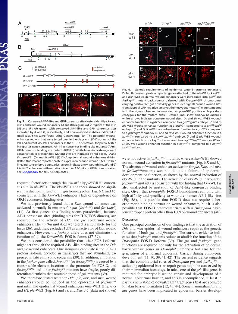
To identify candidate epidermal wound enhancers regulatingkkv and msn, we surveyed these loci for evolutionarily conservedclusters of AP-1-like and GRH consensus binding sites. The 5�region of the third intron of msn has a cluster of 2 conservedGRH sites and 1 conserved AP-1-like site. A 1.2-kb DNAfragment containing these sites functioned as an epidermalwound enhancer (msn-WE2) (Fig. 5 A and D).
The kkv first intron has 5 GRH consensus sites and 4 AP-1-like
sites. We tested the wound enhancer function of 2 overlappingDNA fragments that each contained 4 GRH sites (2 conserved)and 3 AP-1-like sites (1 conserved). One of these fragmentsfunctioned as an epidermal wound enhancer (kkv-WE1) (Fig. 5B and E), whereas the other did not (kkv2) (Fig. 5B and data notshown). To test whether AP-1-like and GRH consensus siteswere required for the function of the kkv wound enhancer, wemutated the sites in the context of the 2.2-kb kkv-WE1 element(Fig. 5C). To our surprise, kkv-WE1 reporters with either AP-1-like(Fig. 5F) or GRH consensus sites (Fig. 5G) mutated were stillinduced in epidermal cells around wound sites. This evidencestrongly suggests that the kkv wound enhancer is activated by adifferent transcription factor code than the Ddc and ple woundenhancers. A table of the quantitative responses of different mutantwound-enhancer lines is provided in Table S1.
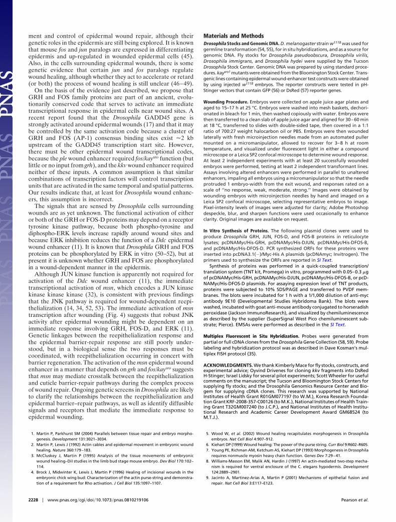
Genetic Requirements for the Induction of ple, kkv, and msn WoundEnhancers. To test trans requirements for activation of the ple,kkv, and msn wound enhancers, we first tested their function ingrh mutants (Ddc wound-enhancer function was previouslyshown to be dependent on grh genetic function) (11). In grhmutants, the msn-WE1 enhancer was not activated aroundembryonic wounds (Fig. 6 A and B), but the ple WE1 enhancerwas activated to approximately normal levels (Fig. 6 C and D).This finding is seemingly at odds with the requirement of a GRHconsensus binding site in ple-WE1 and suggests that another
GRH
Ddc
ple
wt wt mut
mutwtwt-
-
- -
- -
- - + + + +
GRH
JUNFOS-BFOS-DDdc
- -- - - - - - - -- - - - - - - -
+ ++ + + +
+ + + +
- - + + - - + +
mutwt mutwt mutwt mutwt mutwt mutwt
J/JFB/J
FD/JFB/FB
FD/FD
A B
NS
Fig. 3. GRH and FOS bind crucial sequences in the Ddc and ple woundenhancers. (A) Oligonucleotide DNA probes including the GRH consensus sitesfrom Ddc and ple epidermal wound–response enhancers were tested in EMSAassays for binding to full-length Drosophila GRH protein (see Materials andMethods). Probes with mutant GRH sites were used to test binding-sitespecificity. Lanes 1 and 2 show Ddc or ple DNA probes with no GRH protein.Lanes 3 and 4 show GRH protein with WT and mutant Ddc probes, respectively.Lanes 5 and 6 show GRH protein with WT and mutant ple probe, respectively.Arrow indicates GRH–DNA complexes. The bands at the bottom of the frameare unbound probe. (B) Oligonucleotide probes including the AP-1-like sitefrom the Ddc wound–response enhancer were tested in EMSA assays forbinding to full-length Drosophila JUN, FOS-B, and FOS-D proteins (see Mate-rials and Methods). Probes with mutant AP-1-like sites were used to test thespecificity of binding. Lanes 1 and 2 show WT or mutant probes, no protein;NS denotes nonspecific shifted probe complexes that result from reticulocytelysate alone; unbound probe is not shown. Lanes 3 and 4 show JUN proteinwith WT and mutant probes, respectively. Lanes 5 and 6 show FOS-B proteinwith WT and mutant probes, respectively. Lanes 7 and 8 show FOS-B and JUNproteins with WT and mutant probes, respectively. Lanes 9 and 10 show FOS-Dprotein with WT and mutant probes, respectively. Lanes 11 and12 show FOS-Dand JUN with WT and mutant probes, respectively. The positions of the variousshifted protein/DNA complexes are noted on the side.
B
D
A
C
E
msn
plekkv
Ddc
Fig. 4. Ddc, ple, msn, and kkv are transcriptionally activated in epidermalcells around wounds. (A) Image of wounded embryo, visualized with DICoptics, fixed 30 min after wounding. Arrow shows entry wound; dotted boxshows the region imaged with fluorescence optics in frames B–E. (B–E) Ddc (B),msn (C), kkv (D), and ple (E) transcripts were simultaneously detected in theembryo around the aseptic wound using hapten-labeled probes (35). Nosignals were detected around wounds in embryos fixed immediately afterwounding and hybridized with probes (data not shown).
2226 � www.pnas.org�cgi�doi�10.1073�pnas.0810219106 Pearson et al.
required factor acts through the low-affinity ple‘‘GRH’’ consen-sus site in ple-WE1. The kkv-WE1 enhancer showed no signif-icant reduction in function in grh homozygotes (Fig. 6 E and F),consistent with the kkv-WE1 enhancer’s lack of dependence onGRH consensus binding sites.
We had previously found that a Ddc wound enhancer wasactivated normally in mutants for jun (JraIA109) and fos (kay1)(11). At first glance, this finding seems paradoxical, becauseAP-1 consensus sites (binding sites for JUN/FOS dimers), arerequired for the activity of Ddc and ple epidermal woundenhancers. The jun/Jra mutation we tested is a null allele for thelocus (36), and, thus, excludes JUN as an activator of Ddc woundenhancers. However, the fos/kay1 allele does not eliminate thefunction of all the Drosophila FOS isoforms (37–39).
We thus considered the possibility that other FOS isoformsmight act through the required AP-1-like binding sites in the Ddcand ple wound enhancers. One intriguing candidate is the FOS-Dprotein isoform, encoded in transcripts that are abundantly ex-pressed in late embryonic epidermis (39). In addition, a mutationin the fos/kay gene called shroudP54 (or fos/kaysroP54) is caused by atransposable element insertion in the promoter for FOS-D, andfos/kaysroP54 and other fos/kaysro mutants have fragile, poorly dif-ferentiated cuticles that resemble those of grh mutants (39).
We therefore tested whether Ddc, ple, kkv, and msn woundenhancers could be induced in the epidermis of fos/kaysro1
mutants. The epidermal wound enhancers msn-WE1 (Fig. 6 Gand H), ple-WE1 (Fig. 6 I and J), and Ddc .47 (data not shown)
were not active in fos/kaysro1 mutants, whereas kkv-WE1 showednormal wound activation in fos/kaysro1 mutants (Fig. 6 K and L).The absence of wound-enhancer activation for ple, Ddc, and msnin fos/kaysro1mutants was not due to a failure of epidermaldevelopment or function, as shown by the normal induction ofkkv-WE1 in the mutants. The activation of kkv-WE1 in woundedfos/kaysro1 mutants is consistent with the finding that kkv-WE1 isalso unaffected by mutation of AP-1-like consensus bindingsites. Given that Drosophila FOS-D homodimers can bind withhigh affinity and specificity to wound-enhancer AP-1-like sites(Fig. 3B), it is possible that FOS-D does not require a het-erodimeric binding partner on wound enhancers, but it is alsopossible that FOS-D heterodimerizes with a Drosophila basic-leucine zipper protein other than JUN on wound enhancers (40).
DiscussionOne principal conclusion of our findings is that the activation ofDdc and msn epidermal wound enhancers requires the geneticfunction of both grh and fos/kaysro. The current evidence indi-cates that fos/kaysro mutants reduce or abolish the function of theDrosophila FOS-D isoform (39). The grh and fos/kaysro genefunctions are required not only for the activation of epidermalbarrier-repair genes in Drosophila embryos but also for thegeneration of a normal epidermal barrier during embryonicdevelopment (11, 30, 39, 41, 42). The current evidence suggeststhat the combinatorial roles of Drosophila grh and fos/kaysro inactivating epidermal barrier-repair genes might be conserved bytheir mammalian homologs. In mice, one of the grh-like genes isrequired for embryonic wound repair and development of anormal epidermal barrier, and this is accomplished at least inpart via activation of downstream target genes that are requiredfor skin barrier formation (12, 43, 44). Some mammalian fos andjun genes have been implicated in epidermal barrier develop-
kkv-WE1
A
B
conservedsequences
mutatedsequences
kkv-APm
kkv-GRHm
C
kkv-APm kkv-GRHm
kkv2
kkv-WE1
msn2
msn-WE1
msn-WE2msn
1kb
a a
g g
g gGG G G
G GA A
kkv
1kb
a a a a a
g g g g g g g g
g g g g g gG G GA G
msn
D F G
A
msn-WE1 kkv-WE1E
Fig. 5. Conserved AP-1-like and GRH consensus site clusters identify kkv andmsn epidermal wound enhancers. (A and B) Diagrams of 5� regions of the msn(A) and kkv (B) genes, with conserved AP-1-like and GRH consensus sitesindicated by A and G, respectively, and nonconserved matches indicated insmall case. Sites were found using GenePalette (60). The potential wound-enhancer regions that were tested overlie the diagrams. (C) Diagrams of theWT and mutant kkv-WE1 enhancers. In the 5�–3� orientation, they were testedin reporter gene constructs. AP-1-like consensus binding site mutants (APm);GRH consensus binding site mutants (GRHm). White boxes indicate regions ofconservation in drosophilids. Mutant sites are indicated by red boxes. (D andE) msn-WE1 (D) and kkv-WE1 (E) DNA epidermal wound enhancers drivingDsRed fluorescent reporter protein expression around wound sites. Dashedlines indicate embryo boundaries; arrows indicate entry-wound sites. (F and G)kkv-WE1 enhancers with mutations in either AP-1-like or GRH consensus sites.See SI Appendix for all DNA sequences.
I
kkvWE1
K
J
L
BA
E F
grh/+ grh/grh
pleWE1
msnWE1
kaysro/+ kaysro/kaysro
C D
G H
40 uM
Fig. 6. Genetic requirements of epidermal wound–response enhancers.DsRed fluorescent protein reporter genes attached to the ple-WE1, kkv-WE1,and msn-WE1 epidermal wound enhancers were introduced into grhIM andfos/kaysro1 mutant backgrounds balanced with Kruppel-GFP chromosomescarrying positive WT grh or fos/kay genes. DsRed signals around wound sitesfrom Kruppel-GFP negative embryos (homozygous mutants) were comparedwith the signals observed in wounded Kruppel-GFP positive embryos (het-erozygous for the mutant allele). Dashed lines show embryo boundaries;white arrows indicate puncture-wound sites. (A and B) msn-WE1 wound-enhancer function in a grhIM/� compared to a grhIM/grhIM embryo. (C and D)ple-WE1 wound-enhancer function in a grhIM/� compared to a grhIM/grhIM
embryo. (E and F) kkv-WE1 wound-enhancer function in a grhIM/� comparedto a grhIM/grhIM embryo. (G and H) msn-WE1 wound-enhancer function in akaysro1/� compared to a kaysro1/kaysro1 embryo. (I and J) ple-WE1 wound-enhancer function in a kaysro1/� compared to a kaysro1/kaysro1 embryo. (K andL) kkv-WE1 wound-enhancer function in a kaysro1/� compared to a kaysro1/kaysro1 embryo.
Pearson et al. PNAS � February 17, 2009 � vol. 106 � no. 7 � 2227
DEV
ELO
PMEN
TAL
BIO
LOG
Y
ment and control of epidermal wound repair, although theirgenetic roles in the epidermis are still being explored. It is knownthat mouse fos and jun paralogs are expressed in differentiatingepidermis and up-regulated in wounded epidermal cells (45).Also, in the cells surrounding epidermal wounds, there is somegenetic evidence that certain jun and fos paralogs regulatewound healing, although whether they act to accelerate or retard(or both) the process of wound healing is still unclear (46–49).
On the basis of the evidence just described, we propose thatGRH and FOS family proteins are part of an ancient, evolu-tionarily conserved code that serves to activate an immediatetranscriptional response in epidermal cells near wound sites. Arecent report found that the Drosophila GADD45 gene isstrongly activated around epidermal wounds (17) and that it maybe controlled by the same activation code because a cluster ofGRH and FOS (AP-1) consensus binding sites exist �2 kbupstream of the GADD45 transcription start site. However,there must be other epidermal wound transcriptional codes,because the ple wound enhancer required fos/kaysro function (butlittle or no input from grh), and the kkv wound enhancer requiredneither of these inputs. A common assumption is that similarcombinations of transcription factors will control transcriptionunits that are activated in the same temporal and spatial patterns.Our results indicate that, at least for Drosophila wound enhanc-ers, this assumption is incorrect.
The signals that are sensed by Drosophila cells surroundingwounds are as yet unknown. The functional activation of eitheror both of the GRH or FOS-D proteins may depend on a receptortyrosine kinase pathway, because both phospho-tyrosine anddiphospho-ERK levels increase rapidly around wound sites andbecause ERK inhibition reduces the function of a Ddc epidermalwound enhancer (11). It is known that Drosophila GRH and FOSproteins can be phosphorylated by ERK in vitro (50–52), but atpresent it is unknown whether GRH and FOS are phosphorylatedin a wound-dependent manner in the epidermis.
Although JUN kinase function is apparently not required foractivation of the Ddc wound enhancer (11), the immediatetranscriptional activation of msn, which encodes a JUN kinasekinase kinase kinase (32), is consistent with previous findingsthat the JNK pathway is required for wound-dependent reepi-thelialization (14, 34, 52, 53). The immediate activation of msntranscription after wounding (Fig. 4) suggests that robust JNKactivity after epidermal wounding might be dependent on animmediate response involving GRH, FOS-D, and ERK (11).Genetic linkages between the reepithelialization response andthe epidermal barrier-repair response are still poorly under-stood, but in a biological sense the two responses must becoordinated, with reepithelialization occurring in concert withbarrier regeneration. The activation of the msn epidermal woundenhancer in a manner that depends on grh and fos/kaysro suggeststhat msn may mediate crosstalk between the reepithelializationand cuticle barrier-repair pathways during the complex processof wound repair. Ongoing genetic screens in Drosophila are likelyto clarify the relationships between the reepithelialization andepidermal barrier–repair pathways, as well as identify diffusiblesignals and receptors that mediate the immediate response toepidermal wounding.
Materials and MethodsDrosophila Stocks and Genomic DNA. D. melanogaster strain w1118 was used forgermline transformation (54, 55), for in situ hybridizations, and as a source forgenomic DNA. Fly stocks for Drosophila pseudoobscura, Drosophila virilis,Drosophila immigrans, and Drosophila hydei were supplied by the TucsonDrosophila Stock Center. Genomic DNA was prepared by using standard proce-dures. kaysro1 mutants were obtained from the Bloomington Stock Center. Trans-genic lines containing epidermal wound-enhancer test constructs were obtainedby using injected w1118 embryos. The reporter constructs were tested in pH-Stinger vectors that contain GFP (56) or DsRed (57) reporter genes.
Wounding Procedure. Embryos were collected on apple juice agar plates andaged to 15–17 h at 25 °C. Embryos were washed into mesh baskets, dechori-onated in bleach for 1 min, then washed copiously with water. Embryos werethen transferred to a clean slab of apple juice agar and aligned for 30–60 minat 18 °C, transferred to slides with double-sided tape, then covered in a 1:1ratio of 700:27 weight halocarbon oil or PBS. Embryos were then woundedlaterally with fresh microinjection needles made from an automated pullermounted on a micromanipulator, allowed to recover for 3–8 h at roomtemperature, and visualized under fluorescent light in either a compoundmicroscope or a Leica SP2 confocal microscope to determine wound response.At least 2 independent experiments with at least 20 successfully woundedembryos were performed, testing at least 2 independent transformant lines.Assays involving altered enhancers were performed in parallel to unalteredenhancers, impaling all embryos using a micromanipulator so that the needleprotruded 1 embryo-width from the exit wound, and responses rated on ascale of ‘‘no response, weak, moderate, strong.’’ Images were obtained bywounding embryos with microinjection needles by hand and imaged on aLeica SP2 confocal microscope, selecting representative embryos to image.Pixel-intensity levels of images were adjusted for clarity; Adobe Photoshopdespeckle, blur, and sharpen functions were used occasionally to enhanceclarity. Original images are available on request.
In Vitro Synthesis of Proteins. The following plasmid clones were used toproduce Drosophila GRH, JUN, FOS-D, and FOS-B proteins in reticulocytelysates; pcDNAMyc/His-GRH, pcDNAMyc/His-DJUN, pcDNAMyc/His-DFOS-B,and pcDNAMyc/His-DFOS-D. PCR synthesized ORFs for these proteins wereinserted into pcDNA3.1(�)/Myc-His A plasmids (pcDNAmyc; Invitrogen). Theprimers used to synthesize the ORFs are reported in SI Text.
Synthesis of proteins was performed in a quick-coupled transcription/translation system (TNT kit, Promega) in vitro, programmed with 0.05–0.3 �gof pcDNAMyc/His-GRH, pcDNAMyc/His-DJUN, pcDNAMyc/His-DFOS-B, or pcD-NAMyc/His-DFOS-D plasmids. For assaying expression level of TNT products,proteins were subjected to 10% SDS/PAGE and transferred to PVDF mem-branes. The blots were incubated for 1 h with a 1/1,000 dilution of anti-mycantibody 9E10 (Developmental Studies Hybridoma Bank). The blots werewashed, incubated with goat anti-mouse antibody conjugated to horseradishperoxidase (Jackson ImmunoResearch), and visualized by chemiluminescenceas described by the supplier (SuperSignal West Pico chemiluminescent sub-strate; Pierce). EMSAs were performed as described in the SI Text.
Multiplex Fluorescent in Situ Hybridization. Probes were generated frompartial or full cDNA clones from the Drosophila Gene Collection (58, 59). Probelabeling and hybridization protocol was as described in Dave Kosman’s mul-tiplex FISH protocol (35).
ACKNOWLEDGMENTS. We thank Kimberly Mace for fly stocks, constructs, andexperimental advice; Oyvind Drivenes for cloning kkv fragments into DsRedH-Stinger; Israel Lidsky for several pilot experiments; Scott Wheeler for usefulcomments on the manuscript; the Tucson and Bloomington Stock Centers forsupplying fly stocks; and the Drosophila Genomics Resource Center and Bio-gem for supplying cDNA clones. This research was supported by NationalInstitutes of Health Grant R01GM077197 (to W.M.), Korea Research Founda-tion Grant KRF-2008-357-C00126 (to M.K.), National Institutes of Health Train-ing Grant T32GM007240 (to J.C.P.), and National Institutes of Health Institu-tional Research and Academic Career Development Award GM68524 (toM.T.J.).
1. Martin P, Parkhurst SM (2004) Parallels between tissue repair and embryo morpho-genesis. Development 131:3021–3034.
2. Martin P, Lewis J (1992) Actin cables and epidermal movement in embryonic woundhealing. Nature 360:179–183.
3. McCluskey J, Martin P (1995) Analysis of the tissue movements of embryonicwound healing–DiI studies in the limb bud stage mouse embryo. Dev Biol 170:102–114.
4. Brock J, Midwinter K, Lewis J, Martin P (1996) Healing of incisional wounds in theembryonic chick wing bud: Characterization of the actin purse-string and demonstra-tion of a requirement for Rho activation. J Cell Biol 135:1097–1107.
5. Wood W, et al. (2002) Wound healing recapitulates morphogenesis in Drosophilaembryos. Nat Cell Biol 4:907–912.
6. Kiehart DP (1999) Wound healing: The power of the purse string. Curr Biol 9:R602–R605.7. Young PE, Richman AM, Ketchum AS, Kiehart DP (1993) Morphogenesis in Drosophila
requires nonmuscle myosin heavy chain function. Genes Dev 7:29–41.8. Williams-Masson EM, Malik AN, Hardin J (1997) An actin-mediated two-step mecha-
nism is required for ventral enclosure of the C. elegans hypodermis. Development124:2889–2901.
9. Jacinto A, Martinez-Arias A, Martin P (2001) Mechanisms of epithelial fusion andrepair. Nat Cell Biol 3:E117–E123.
2228 � www.pnas.org�cgi�doi�10.1073�pnas.0810219106 Pearson et al.
10. Hoffmann JA, Reichhart JM (2002) Drosophila innate immunity: An evolutionaryperspective. Nat Immunol 3:121–126.
11. Mace KA, Pearson JC, McGinnis W (2005) An epidermal barrier wound repair pathwayin Drosophila is mediated by grainy head. Science 308:381–385.
12. Ting SB, et al. (2005) A homolog of Drosophila grainy head is essential for epidermalintegrity in mice. Science 308:411–413.
13. Alibardi L (2006) Structural and immunocytochemical characterization of keratiniza-tion in vertebrate epidermis and epidermal derivatives. Int Rev Cytol 253:177–259.
14. Galko MJ, Krasnow MA (2004) Cellular and genetic analysis of wound healing inDrosophila larvae. PLoS Biol 2:E239.
15. Cole J, Tsou R, Wallace K, Gibran N, Isik F (2001) Early gene expression profile of humanskin to injury using high-density cDNA microarrays. Wound Repair Regen 9:360–370.
16. Fitsialos G, et al. (2007) Transcriptional signature of epidermal keratinocytes subjectedto in vitro scratch wounding reveals selective roles for ERK1/2, p38, and phosphatidyl-inositol 3-kinase signaling pathways. J Biol Chem 282:15090–15102.
17. Stramer B, et al. (2008) Gene induction following wounding of wild-type versusmacrophage-deficient Drosophila embryos. EMBO Rep 9:465–471.
18. Andersen SO, Hojrup P, Roepstorff P (1995) Insect cuticular proteins. Insect BiochemMol Biol 25:153–176.
19. Uv AE, Thompson CR, Bray SJ (1994) The Drosophila tissue-specific factor Grainyheadcontains novel DNA-binding and dimerization domains which are conserved in thehuman protein CP2. Mol Cell Biol 14:4020–4031.
20. Venkatesan K, McManus HR, Mello CC, Smith TF, Hansen U (2003) Functional conser-vation between members of an ancient duplicated transcription factor family, LSF/Grainyhead. Nucleic Acids Res 31:4304–4316.
21. Pollock R, Treisman R (1990) A sensitive method for the determination of protein-DNAbinding specificities. Nucleic Acids Res 18:6197–6204.
22. Benbrook DM, Jones NC (1994) Different binding specificities and transactivation ofvariant CRE’s by CREB complexes. Nucleic Acids Res 22:1463–1469.
23. van Dijk MA, Murre C (1994) Extradenticle raises the DNA binding specificity ofhomeotic selector gene products. Cell 78:617–624.
24. Van Dijk MA, Voorhoeve PM, Murre C (1993) Pbx1 is converted into a transcriptionalactivator upon acquiring the N-terminal region of E2A in pre-B-cell acute lymphoblas-toid leukemia. Proc Natl Acad Sci USA 90:6061–6065.
25. Neuteboom ST, Murre C (1997) Pbx raises the DNA binding specificity but not theselectivity of antennapedia Hox proteins. Mol Cell Biol 17:4696–4706.
26. Ekker SC, Young KE, von Kessler DP, Beachy PA (1991) Optimal DNA sequence recog-nition by the Ultrabithorax homeodomain of Drosophila. EMBO J 10:1179–1186.
27. Pearson JC, Lemons D, McGinnis W (2005) Modulating Hox gene functions duringanimal body patterning. Nat Rev Genet 6:893–904.
28. Fried M, Crothers DM (1981) Equilibria and kinetics of lac repressor-operator interac-tions by polyacrylamide gel electrophoresis. Nucleic Acids Res 9:6505–6525.
29. Perkins KK, Dailey GM, Tjian R (1988) Novel Jun- and Fos-related proteins in Drosophilaare functionally homologous to enhancer factor AP-1. EMBO J 7:4265–4273.
30. Ostrowski S, Dierick HA, Bejsovec A (2002) Genetic control of cuticle formation duringembryonic development of Drosophila melanogaster. Genetics 161:171–182.
31. Moussian B, Schwarz H, Bartoszewski S, Nusslein-Volhard C (2005) Involvement ofchitin in exoskeleton morphogenesis in Drosophila melanogaster. J Morphol 264:117–130.
32. Su YC, Treisman JE, Skolnik EY (1998) The Drosophila Ste20-related kinase misshapenis required for embryonic dorsal closure and acts through a JNK MAPK module on anevolutionarily conserved signaling pathway. Genes Dev 12:2371–2380.
33. Merzendorfer H, Zimoch L (2003) Chitin metabolism in insects: structure, function andregulation of chitin synthases and chitinases. J Exp Biol 206:4393–4412.
34. Ramet M, Lanot R, Zachary D, Manfruelli P (2002) JNK signaling pathway is required forefficient wound healing in Drosophila. Dev Biol 241:145–156.
35. Kosman D, et al. (2004) Multiplex detection of RNA expression in Drosophila embryos.Science 305:846.
36. Kockel L, Zeitlinger J, Staszewski LM, Mlodzik M, Bohmann D (1997) Jun in Drosophiladevelopment: Redundant and nonredundant functions and regulation by two MAPKsignal transduction pathways. Genes Dev 11:1748–1758.
37. Riesgo-Escovar JR, Hafen E (1997) Common and distinct roles of DFos and DJun duringDrosophila development. Science 278:669–672.
38. Zeitlinger J, et al. (1997) Defective dorsal closure and loss of epidermal decapentaple-gic expression in Drosophila fos mutants. EMBO J 16:7393–7401.
39. Giesen K, et al. (2003) Regulation of glial cell number and differentiation by ecdysoneand Fos signaling. Mech Dev 120:401–413.
40. Fassler J, et al. (2002) B-ZIP proteins encoded by the Drosophila genome: Evaluation ofpotential dimerization partners. Genome Res 12:1190–1200.
41. Bray SJ, Kafatos FC (1991) Developmental function of Elf-1: An essential transcriptionfactor during embryogenesis in Drosophila. Genes Dev 5:1672–1683.
42. Narasimha M, Uv A, Krejci A, Brown NH, Bray SJ (2008) Grainy head promotes expres-sion of septate junction proteins and influences epithelial morphogenesis. J Cell Sci121:747–752.
43. Yu Z, et al. (2006) The Grainyhead-like epithelial transactivator Get-1/Grhl3 regulatesepidermal terminal differentiation and interacts functionally with LMO4. Dev Biol299:122–136.
44. Wilanowski T, et al. (2008) Perturbed desmosomal cadherin expression in grainyhead-like 1-null mice. EMBO J 27:886–897.
45. Mehic D, Bakiri L, Ghannadan M, Wagner EF, Tschachler E (2005) Fos and jun proteinsare specifically expressed during differentiation of human keratinocytes. J InvestDermatol 124:212–220.
46. Yates S, Rayner TE (2002) Transcription factor activation in response to cutaneousinjury: Role of AP-1 in reepithelialization. Wound Repair Regen 10:5–15.
47. Li G, et al. (2003) c-Jun is essential for organization of the epidermal leading edge. DevCell 4:865–877.
48. Zenz R, Wagner EF (2006) Jun signalling in the epidermis: From developmental defectsto psoriasis and skin tumors. Int J Biochem Cell Biol 38:1043–1049.
49. Gazel A, Nijhawan RI, Walsh R, Blumenberg M (2008) Transcriptional profiling definesthe roles of ERK and p38 kinases in epidermal keratinocytes. J Cell Physiol 215:292–308.
50. Uv AE, Harrison EJ, Bray SJ (1997) Tissue-specific splicing and functions of the Drosoph-ila transcription factor Grainyhead. Mol Cell Biol 17:6727–6735.
51. Liaw GJ, et al. (1995) The torso response element binds GAGA and NTF-1/Elf-1, andregulates tailless by relief of repression. Genes Dev 9:3163–3176.
52. Ciapponi L, Jackson DB, Mlodzik M, Bohmann D (2001) Drosophila Fos mediates ERKand JNK signals via distinct phosphorylation sites. Genes Dev 15:1540–1553.
53. Bosch M, Serras F, Martin-Blanco E, Baguna J (2005) JNK signaling pathway required forwound healing in regenerating Drosophila wing imaginal discs. Dev Biol 280:73–86.
54. Rubin GM, Spradling AC (1982) Genetic transformation of Drosophila with transpos-able element vectors. Science 218:348–353.
55. Spradling AC, Rubin GM (1982) Transposition of cloned P elements into Drosophilagerm line chromosomes. Science 218:341–347.
56. Barolo S, Carver LA, Posakony JW (2000) GFP and beta-galactosidase transformationvectors for promoter/enhancer analysis in Drosophila. BioTechniques 29:726–732.
57. Barolo S, Castro B, Posakony JW (2004) New Drosophila transgenic reporters: InsulatedP-element vectors expressing fast-maturing RFP. Biotechniques 36:436-442.
58. Stapleton M, et al. (2002) A Drosophila full-length cDNA resource. Genome Biol3:RESEARCH0080.
59. Stapleton M, et al. (2002) The Drosophila gene collection: Identification of putativefull-length cDNAs for 70% of D. melanogaster genes. Genome Res 12:1294–1300.
60. Rebeiz M, Posakony JW (2004) GenePalette: A universal software tool for genomesequence visualization and analysis. Dev Biol 271:431–438.
Pearson et al. PNAS � February 17, 2009 � vol. 106 � no. 7 � 2229
DEV
ELO
PMEN
TAL
BIO
LOG
Y