Multiple System Atrophy (MSA) FAQ What is ... - … · Multiple System Atrophy (MSA) FAQ What is...
22
Multiple System Atrophy (MSA) FAQ What is multiple system atrophy? Multiple system atrophy (MSA) is a rare neurodegenerative disease marked by a combination of symptoms affecting movement, blood pressure, and other body functions; hence the label "multiple system" atrophy. According to the American Autonomic Society, Multiple System Atrophy (MSA) is a sporadic, progressive, adult-onset disorder characterized by autonomic dysfunction, parkinsonism and ataxia (a failure of muscular coordination) in any combination. Broken down MSA stands for: Multiple - many System - brain structures that control different functions Atrophy - cell shrinkage or damage This means that cells are damaged in different areas of the brain that control different body functions. The three areas affected are the basal ganglia, cerebellum and brain stem. These areas are responsible for movement, balance and automatic body functions like bladder control. How are the brain cells damaged in MSA? Brain cells in the affected areas in MSA shrink (atrophy). This can sometimes be seen on MRI scans. When brain tissue is examined under a microscope, structures called glial inclusion bodies can be seen; they contain a protein called alpha- synuclein. It is the presence of these inclusion bodies in the movement, balance and automatic control centers of the brain that confirms a diagnosis of MSA. What causes Multiple System Atrophy? The causes of MSA are unknown. It is still unclear as to exactly why the cells become damaged in MSA. MSA is not contagious. It does not appear to be inherited, although some research suggests there may be a predisposition within the genetic make-up of an individual for cells to become damaged. What triggers the damage process to begin is unknown. Environmental toxins or a history of trauma have been suggested, but evidence for this as a cause is slight. This too is a focus of ongoing research. Who gets MSA? MSA usually starts between the ages of 50-60 years, although it can affect people younger and older than this. The mean age of onset is 54. MSA usually occurs after age 50, with a slightly higher incidence in males. Between 25,000 and 100,000 American have multiple system atrophy. The incidence (new case per 100,000 person years) for ages 50 to 99 years is 3.0 (Bower et al, 1997), or about half as frequent as progressive supranuclear palsy (PSP).
Transcript of Multiple System Atrophy (MSA) FAQ What is ... - … · Multiple System Atrophy (MSA) FAQ What is...

Multiple System Atrophy (MSA) FAQ
What is multiple system atrophy? Multiple system atrophy (MSA) is a rare neurodegenerative disease marked by a combination of symptoms affecting movement, blood pressure, and other body functions; hence the label "multiple system" atrophy. According to the American Autonomic Society, Multiple System Atrophy (MSA) is a sporadic, progressive, adult-onset disorder characterized by autonomic dysfunction, parkinsonism and ataxia (a failure of muscular coordination) in any combination. Broken down MSA stands for: Multiple - many System - brain structures that control different functions Atrophy - cell shrinkage or damage This means that cells are damaged in different areas of the brain that control different body functions. The three areas affected are the basal ganglia, cerebellum and brain stem. These areas are responsible for movement, balance and automatic body functions like bladder control. How are the brain cells damaged in MSA? Brain cells in the affected areas in MSA shrink (atrophy). This can sometimes be seen on MRI scans. When brain tissue is examined under a microscope, structures called glial inclusion bodies can be seen; they contain a protein called alpha-synuclein. It is the presence of these inclusion bodies in the movement, balance and automatic control centers of the brain that confirms a diagnosis of MSA. What causes Multiple System Atrophy? The causes of MSA are unknown. It is still unclear as to exactly why the cells become damaged in MSA. MSA is not contagious. It does not appear to be inherited, although some research suggests there may be a predisposition within the genetic make-up of an individual for cells to become damaged. What triggers the damage process to begin is unknown. Environmental toxins or a history of trauma have been suggested, but evidence for this as a cause is slight. This too is a focus of ongoing research. Who gets MSA? MSA usually starts between the ages of 50-60 years, although it can affect people younger and older than this. The mean age of onset is 54. MSA usually occurs after age 50, with a slightly higher incidence in males. Between 25,000 and 100,000 American have multiple system atrophy. The incidence (new case per 100,000 person years) for ages 50 to 99 years is 3.0 (Bower et al, 1997), or about half as frequent as progressive supranuclear palsy (PSP).

What are the three forms of MSA? Symptoms of MSA vary in distribution, onset and severity from person to person. Because of this, three different diseases were initially described to encompass this range of symptoms: Shy-Drager syndrome, striatonigral degeneration, and olivopontocerebellar atrophy. The reason there are 3 forms is that years ago all doctors had to go on were symptoms. If a patient had loss of balance (ataxia) and slurred speech then they knew the patient had nerve degeneration in the cerebellum, olives and pons (areas of the brain). They called this Olivopontocerebellar atrophy (OPCA). If a patient had low blood pressure (orthostatic hypotension) they knew that the patient had nerve degeneration in the autonomic nervous system. This form is named Shy-Drager Syndrome (SDS) for Dr. Milton Shy and Dr. Glen Drager who first wrote about this disorder in 1960. If a patient had tremors, slowness of movement and other Parkinson-like symptoms they knew that the patient had nerve degeneration in the striatonigral pathway of the brain. They called this Striatonigral Degeneration (SND). Doctors once thought that these 3 disorders were all SEPARATE diseases. They now know that THIS IS NOT THE CASE. Autopsy evidence has shown that there appears to be a common underlying cause in all three disorders so they came up with a NEW NAME to include all 3 - MULTIPLE SYSTEM ATROPHY. Many doctors have been slow to adopt this new terminology so they continue to use the old terms SND, OPCA, and SDS when diagnosing. The symptoms vary and depend on which particular form of MSA the patient has. Striatonigral degeneration is practically indistinguishable from Parkinson’s. OPCA is characterized by progressive ataxia (an inability to coordinate voluntary muscular movements) of the gait and arms and dysarthria (difficulty in articulating words); and Shy-Drager Syndrome is characterized by Parkinsonism plus a much more pronounced failure of the autonomic nervous system. What are the symptoms of MSA? MSA can manifest in a variety of different ways and progresses in an unpredictable sequence and time scale for each patient. It is important to understand that having a diagnosis of MSA does not mean that you will experience all the symptoms below. MSA can cause a wide range of symptoms, including: • stiffness or rigidity • freezing or slowed movements • postural instability; loss of balance; incoordination • Orthostatic hypotension, or a significant fall in blood pressure when standing, causing dizziness, lightheadedness, fainting, or blurred vision • male impotence • urinary difficulties

• constipation • speech and swallowing difficulties • blurred vision The Parkinsonism of MSA is generally an akinetic rigid syndrome, similar to that of progressive supranuclear palsy (PSP). Rest tremor may occur but is not a predominant feature. Postural instability is common. Parkinsonism is generally the most common initial sign and eventually develops in about 90% of all patients. The cerebellar signs include finger-to-nose or heel-shin dysmetria, gait ataxia, intention tremor and nystagmus. Cerebellar signs are the first feature on only about 5% of patients. Cerebellar signs are observed in 50% of cases.1 Sporadic OPCA evolves into MSA in roughly 25% of cases within 5 years. Autonomic dysfunction includes impotence (the most common male sign), postural hypotension with syncope, urinary incontinence and retention, and fecal incontinence. 1 Ben-Shlomo et al, 1997 Diagnosis is Difficult In order to diagnose multiple system atrophy, thoughtful and careful evaluation should be undertaken. Due to the variety of different ways MSA can manifest, it is often difficult to differentiate it from other neurodegenerative disorders like Parkinson's disease, progressive supranuclear palsy (PSP) or corticobasal degeneration (CBD). Many patients with MSA will not receive the correct diagnosis during their lifetime. This is due to the difficulty in differentiating MSA from other disorders (including relatively common degenerative disorders such as Parkinson’s disease and more rare ones such as pure autonomic failure). MSA may be difficult to diagnose in the early stages because it can take years for key symptoms to develop. Diagnosis is made on careful history, physical examination and some tests including several types of brain imaging and autonomic function tests. However, a definite diagnosis can still only be accomplished by post-mortem examination of the brain. There are many disorders with symptoms, which overlap with those of MSA, therefore diagnosis is usually carried out by a Neurologist or a Physician at the request of a GP. Information about the history of an individual's symptoms and an examination will be performed. There are also hospital tests that help in the diagnosis. Initial Diagnosis The initial diagnosis of MSA is usually made by carefully interviewing the patient and performing a physical examination. A neurologist makes the diagnosis based on the history of symptoms and the findings on physical examination as well as ruling out other causes.

The physical examination may include: • An eye examination may reveal atrophy of the iris and paralysis of eye muscles. • Postural hypotension (drop in blood pressure associated with change in position) is evident. • A neuromuscular examination shows abnormal reflexes and may show severe muscle wasting (atrophy). Parkinsonian movements (tremor, rigidity, and slow movements similar to Parkinson's disease) are common. However, more testing is often needed to confirm the diagnosis. Testing for MSA Diagnosis is largely clinical as there are no specific tests during life to confirm the disease. The testing described below can be helpful in suggesting whether a patient has multiple system atrophy, but true diagnosis can only be accomplished by examination of the brain postmortem. Among the tests that are helpful in determining the presence of MSA are several types of brain imaging including computerized tomography (CT) scans, magnetic resonance imaging (MRI), and positron emission tomography (PET). An MRI of brain can be used to rule out other pathology. There are no specific abnormalities on imaging associated with MSA. Testing may include: • Plasma norepinephrine levels • Urine examination for norepinephrine breakdown products (urine catecholamines) Posture studies with blood pressure and heart rate monitoring with catecholamine levels in the supine and upright position should be done. Other potential evaluations include autonomic function testing, consultation by physicians that specialize in movement and/or blood pressure disorders, a sleep study, urodynamic testing, and blood pressure monitoring throughout sleep to evaluate for supine hypertension, or a high blood pressure when lying down. Pharmacological challenge tests (administering certain drugs in the presence of various types of movements of the patient) may also be of help. In those patients with typical Parkinsonian signs, an incomplete and relatively poor response to dopamine replacement therapy (such as l-dopa [Sinemet]) may be a clue that MSA is present. Expectations (Prognosis) What is the progression of MSA? Disease progression in MSA is quicker than in Parkinsonism but similar or slightly slower to that of Progressive Supranuclear Palsy (PSP) (Bower, 1997). Almost 80% of patients are disabled within 5 years of onset of the motor symptoms, and only 20% survive past 12 years. The mean survival is roughly 6 years. Rate of progression differs in every case and speed of decline may vary widely in individual patients.

Patients usually have autonomic nervous system dysfunction first. Genitourinary dysfunction (difficulty with urination) is the most frequent initial complain in women, while impotence is the most frequent initial complaint in men. When MSA begins with non-autonomic features, imbalance is the most common feature. This difficulty in maintaining balance may be due to either cerebellar or Parkinsonian abnormalities. Some patients complain of stiffness, clumsiness, or a change in handwriting at the onset of MSA. MSA progresses over the course of several years to cause more widespread and severe symptoms. Orthostatic hypotension can cause fainting and falls. Loss of coordination, slowed movements, and rigidity can interfere with activities of daily living. Some patients with MSA have mild loss of cognitive abilities, with impairments in attention and speed of thinking. Complications include: • Progressive loss of ability to walk or care for self • Difficulty performing daily activities • Injuries from falls/fainting • Side effects of medications Orthostatic hypotension (a large drop in blood pressure upon standing) is common and may cause dizziness, dimming of vision, head or neck pain, yawning, temporary confusion, slurred speech, and if the hypotension is severe, the patient may "faint" upon arising from a recumbent position. In spite of low blood pressure while standing, it is common for MSA patients to have high blood pressure when lying down. A fall in blood pressure following meals or in hot weather or following infection is quite common. The concurrent involvement in MSA of multiple brain systems subserving movement, including the striatum, cerebellum, and cortex, leads to the movement disorder as often being the most profound disability. Hoarseness or even vocal paralysis is relatively common, as are sleep disturbances, including snoring and sleep apnea. The ability to swallow foods and liquids may be impaired. How long can I expect to live? The probable outcome is poor. There is a progressive loss of mental and physical functions until general debilitation develops. Early death is likely. The mean survival is roughly 6 years. Most people who are diagnosed with Shy-Drager syndrome die within seven to 10 years after symptoms begin. It is rare for a patient to survive 10 years. The autonomic abnormalities are seldom the direct cause of death. A significant number of patients develop laryngeal stridor and difficulty swallowing, which can lead to pneumonia.
Pneumonia is the most common cause of death, although irregularities in heartbeat or choking may be responsible for death in some patients. Breathing problems such as aspiration, stridor (high-pitched breathing sounds due to airway obstruction), or cardiopulmonary arrest are common causes of death. In addition, many patients with MSA experience Cheyne-Stokes or periodic respiration and in some cases this may lead to a critical loss of respiratory drive, so called Ondine's curse. Pulmonary hypertension may occur during apnea (Guilleminault et al., 1977). The most common causes of death in patients with MSA are pulmonary embolus, apnea, and intercurrent infection. Treatment Currently there is no cure for MSA and no known means to slow progression of the disease. A number of drugs can be used to treat the various symptoms of MSA, although they become less effective as the disease progresses. Treatment aims to reduce the disabling effect of the symptoms so that as full a life as possible can be led. This includes treatment of the depression, tremor and gait disturbances, supine hypertension, orthostatic hypotension, and possible self-catheterization. Treatment for MSA involves a combination of medication, specialized equipment and the use of therapists to manage individual symptoms. Medication regimes will vary depending on your symptoms. Your doctor will prescribe the best combination to meet your needs. Blood Pressure Problems The fluctuating blood pressure makes the condition difficult to treat. Dietary changes, such as increasing salt and fluid intake, may help elevate blood pressure. Drugs such as fludrocortisone, midodrine that control the fall in blood pressure (orthostatic hypotension) can be effective in reducing dizziness, fainting and falls. Sympathomimetics, vasoconstrictors, beta-blockers, MAO inhibitors, vasopressin, 9-fluohydrocortisone, or other medications may be used to treat low blood pressure (postural hypotension). These drugs may cause high blood pressure when lying down. It is advisable to raise the head of your bed and have regular blood pressure checks. A heart pacemaker programmed to stimulate the heart to beat at a rapid rate (faster than 100) may increase the blood pressure for some people. Fludrocortisone (Florinef) - A steroid, usually taken twice a day in very small doses. May cause ankle swelling. Ephedrine - Works quickly to raise the blood pressure, usually taken three times a day.

Midodrine (Gutron) - Works quickly to raise the blood pressure (within 30-60 minutes), usually taken three times a day. Only prescribed by specialists. Movement Problems Drugs to help stiffness and slowness are the same drugs used in Parkinson's disease. Anticholinergic medications may be used to reduce early or mild tremors. The response to medications may be disappointing. Many affected individuals either do not respond or respond poorly to treatment with anticholinergics or Levodopa. Levodopa and dopamine agonists used to treat Parkinson's disease are sometimes effective for the slowness and rigidity of MSA. They are not as effective in MSA and can make blood pressure problems worse. It may take time to find what suits you best. Levodopa may improve movement and balance. Doses of 1 to 1.5 gram per day must be used before unresponsiveness is declared, but generally only about 1/3 of patients respond. Carbidopa may reduce the side effects of Levodopa and make the Levodopa work better. L-Dopa (Madopar or Sinemet) Amantadine (Symmetrel) Bladder Problems Incontinence may be treated with medications or catheterization. Drugs can reduce urgency and frequency problems. Oxybutinin (Ditropan) - improves the tone of bladder muscles. DDAVP (Desmopressin) - used to reduce the production of urine overnight. Infection Antibiotics - should be prescribed at the first signs of infection. Any infection in someone with MSA can worsen symptoms like postural hypotension dramatically. Swallowing and Breathing Difficulties A breathing or feeding tube may have to be surgically inserted to manage swallowing and breathing difficulties. A gastrostomy tube, which delivers foods directly to the stomach, is occasionally needed later in the course of the disease. Use of softer foods may improve the ability to swallow. Treatment of Other Issues • Male impotence may be treated with penile implants or drugs. • Constipation may improve with increased dietary fiber or laxatives. • A speech-language pathologist may be able to offer strategies for improving swallowing and speaking.

Quality of Life in MSA: A National Survey. S. Dickinson, E. Garland, G. Farley and T. Davis, Vanderbilt University, Nashville, TN, USA This is a summary of an abstract that was presented at the American Autonomic Society conference. Results from this survey provide information on quality of life issues in the MSA population. To better understand the physical effects and the impact that this disorder has on lifestyle, we surveyed a national population of MSA patients. Completed questionnaires were received from 55 males (age 64.6±1.3 yr;) and 29 females (age 64.2±1.8 yr). Only 2% of respondents lived alone. 89% reported that the spouse was the primary caregiver. While only 36% used services requiring a fee (nurse, aide), 69% indicated that they did use medical equipment. 20% had quit work or retired in the last month, while 13% had applied for disability. A subsample (n=12) contained responses of caregivers of deceased persons diagnosed with MSA. Death occurred within 8.9±1.1 yr of symptom onset (range of 1-18 yr). A mean of 5.1±0.4 yr elapsed between the onset of symptoms and diagnosis. Symptom % of respondents who
reported having this symptom weakness in legs 87% low BP standing 81% constipation 77% bladder dysfunction 61% imbalance w/o dizziness 55% loud snoring 50% nasal stuffiness 42% bladder infection 39% supine BP 160/90 or higher 33% mild anemia 13%. Medication % of respondents who
reported taking this medication
% of those taking this medication who reported that it was helpful
Fludrocortisone 50% 71% Midodrine 26% 82% Sinemete 42% 77%

Difficulty in performing various activities of daily living (ADLs) was assessed, with a score of "1" representing no difficulty and "5" representing an inability to perform the activity. 63% of subjects were unable to drive a car, giving this activity the highest difficulty score (3.9) 35% were unable to walk outside (difficulty score 3.6). Resources Shy-Drager Syndrome / Multiple System Atrophy Support Group The mission of the Shy-Drager Syndrome/Multiple System Atrophy Support Group (SDS/MSA) is to gather information from each group of people involved (Patients, Caregivers, Family Members, and Physicians) and disseminate that information to all. The SDS/MSA Support Group is an information center and a "Helping Hand" for affected people. Through various programs, including the continuation of the national patient/caregiver support group meeting, support of local groups, the toll free help line, and web site, the SDS/MSA's Support Group can reach many more people in need. The support group also works closely with physicians who are treating the patients as well as those who are involved in the essential research to discover a cause, modes of treatment, and hopefully a cure. The group also would like to reach out to physicians newly involved with the disease. Information about the disease and tips on explaining it to the patient will be provide through several modes, including the web site, links to the American Autonomic Society, access to a referral base of experienced physicians, and educational meetings. By sharing information, resources, and support the SDS/MSA Support Group will create a "Circle of Hope" for those affected by SDS/MSA. The SDS/MSA Support Group 2004 Howard Lane Austin, TX. 78728 Toll Free: 1-866-SDS-4999 (Answering Machine) Phone: 1-512-856-2427 (Nights and Weekends) Fax: 1-512-251-3315 E-mail: [email protected] Web: http://www.shy-drager.org/

Additional Web Resources 1. Advocacy/Support Organizations can help the patient and caregiver learn coping strategies and offer support for the difficulties encountered in dealing with MSA. 2. Multiple System Atrophy Discussion Forum 3. MEDLINE plus Medical Encyclopedia http://www.nlm.nih.gov/medlineplus/ency/article/000757.htm 4. E-medicine http://www.emedicine.com/NEURO/topic671.htm Donations These are two of the popular options for MSA donations in the USA. a. The SDS/MSA Support Group The Shy-Drager Syndrome/Multiple System Atrophy Support Group is a full non-profit organization approved by the Internal Revenue Service. Donations fund the entire operation of the Group. Services include: • a toll-free number to call for information and physician referral • the maintenance of the group website www.shy-drager.org • access to the Shy-drager mail list • an annual patient/caregiver/family member meeting with prominent physicians. Please visit the website at http://www.shy-drager.org Contributions of any amount may be mailed to: The SDS/MSA Support Group P.O. Box 279 Coupland, Texas 78615 The SDS/MSA Support Group now has memorial envelopes available. These envelopes are pre-printed with the address of the Support Group and have a space inside to enter the name of the person the gift is intended to be allocated to. They are tastefully done and are available in any number to survivors. Please contact Don Summers if you wish to distribute these envelopes at the memorial services for your loved ones. Don Summers President, The SDS/MSA Support Group Email: [email protected] Toll free: 866-737-4999

b. Vanderbilt Shy-Drager Research Fund Family members and friends of patients with Multiple System Atrophy or Shy-Drager Syndrome have occasionally expressed an interest in making a financial contribution that can be used for research on this condition. Accordingly, we have established a fund for this purpose. All gifts are very much appreciated. Donations for Multiple System Atrophy or Shy-Drager syndrome research can be sent to: Vanderbilt Shy-Drager Research Fund Vanderbilt University Medical Center AA-3228 Medical Center North Nashville, TN 37232-2195 Vanderbilt Brain and Spinal Cord Donation Program Many patients express an interest in making a contribution to medical science to further research in the cause of neurodegenerative disorders. In Shy-Drager syndrome, also known as MSA, scientific study of human brain obtained after death holds the greatest hope for further understanding and ultimately development of therapies to treat the disease. The postmortem examination, or autopsy, also provides important, precise medical information to the family of the deceased. Information for Patients You may wish to sign a notice of intent to make your desire for organ donation official. This notice of intent is not binding, but conveys your wish and request in writing to your family members that you wish to participate in the brain and spinal donation program. Information for Next of Kin Your affected family member may wish to donate brain tissue for special studies and research purposes. You can concur with that decision by signing a separate consent for the donation, which is often attached to his/her medical record. Upon death of your loved one, you will need to sign an autopsy permit which gives legal consent for a medical doctor to examine the body and in particular, collect brain tissue. We know that this will be a very difficult time for you and we acknowledge the generous gift and courageous act that tissue donation is for the benefit of others. You should know that tissue removal could be done in a way that an open-casket service can be conducted. For More Information Please contact: Autonomic Dysfunction Center Vanderbilt University Medical Center AA-3228 Medical Center North Nashville, TN 37232-2195 Phone: (615) 343-0124 Fax: (615) 343-8649
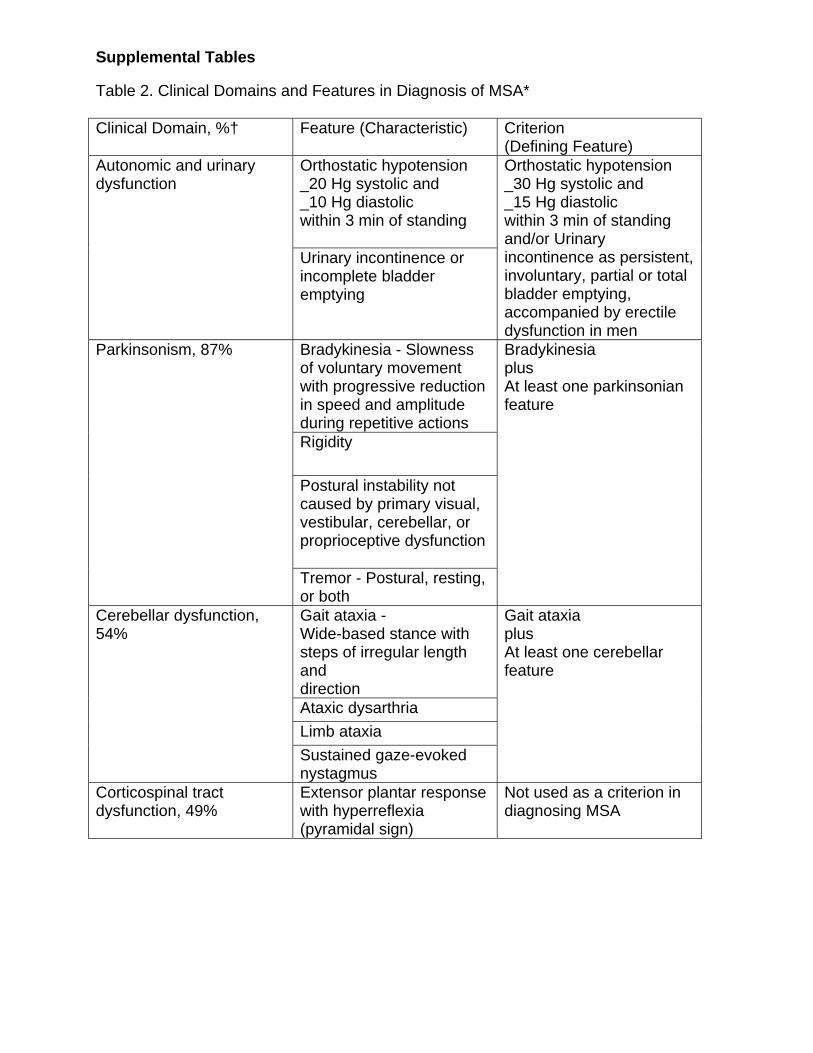
Supplemental Tables Table 2. Clinical Domains and Features in Diagnosis of MSA* Clinical Domain, %† Feature (Characteristic) Criterion
(Defining Feature) Orthostatic hypotension _20 Hg systolic and _10 Hg diastolic within 3 min of standing
Autonomic and urinary dysfunction
Urinary incontinence or incomplete bladder emptying
Orthostatic hypotension _30 Hg systolic and _15 Hg diastolic within 3 min of standing and/or Urinary incontinence as persistent,involuntary, partial or total bladder emptying, accompanied by erectile dysfunction in men
Bradykinesia - Slowness of voluntary movement with progressive reduction in speed and amplitude during repetitive actions Rigidity
Postural instability not caused by primary visual, vestibular, cerebellar, or proprioceptive dysfunction
Parkinsonism, 87%
Tremor - Postural, resting, or both
Bradykinesia plus At least one parkinsonian feature
Gait ataxia - Wide-based stance with steps of irregular length and direction Ataxic dysarthria Limb ataxia
Cerebellar dysfunction, 54%
Sustained gaze-evoked nystagmus
Gait ataxia plus At least one cerebellar feature
Corticospinal tract dysfunction, 49%
Extensor plantar response with hyperreflexia (pyramidal sign)
Not used as a criterion in diagnosing MSA

Supplemental Tables Table 3. Exclusion Criteria for Diagnosis of MSA* History Symptomatic onset younger than age 30 years
Family history of similar disorder Systemic diseases or other identifiable causes for features listed in Table 2 Hallucinations unrelated to medication
Physical Examination Diagnostic and Statistic Manual for Mental Disorders, Fourth Edition criteria for dementia. Prominent slowing of vertical saccades or vertical supranuclear gaze palsy. Evidence of focal cortical dysfunction such as aphasia, alien limb syndrome, and parietal dysfunction.
Laboratory Investigation
Metabolic, molecular genetic, and imaging evidence of alternative cause of features listed in Table 2
Table 4. Diagnostic Categories of MSA* Possible MSA One criterion plus
Two features from separate other domains When criterion is parkinsonism, a poor levodopa response qualifies as one feature (hence only one additional feature required)
Probable MSA Criterion for autonomic failure and urinary dysfunction plus Poorly levodopa-responsive parkinsonism or cerebellar dysfunction
Definitive MSA Pathologically confirmed by presence of high density of GCIs in association with degenerative changes in nigrostriatal and olivopontocerebellar pathways
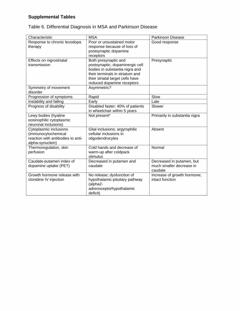
Supplemental Tables Table 6. Differential Diagnosis in MSA and Parkinson Disease Characteristic MSA Parkinson Disease Response to chronic levodopa therapy
Poor or unsustained motor response because of loss of postsynaptic dopamine receptors
Good response
Effects on nigrostriatal transmission
Both presynaptic and postsynaptic; dopaminergic cell bodies in substantia nigra and their terminals in striatum and their striatal target cells have reduced dopamine receptors
Presynaptic
Symmetry of movement disorder
Asymmetric?
Progression of symptoms Rapid Slow Instability and falling Early Late Progress of disability Disabled faster; 40% of patients
in wheelchair within 5 years Slower
Lewy bodies (hyaline eosinophilic cytoplasmic neuronal inclusions)
Not present* Primarily in substantia nigra
Cytoplasmic inclusions (immunocytochemical reaction with antibodies to anti-alpha-synuclein)
Glial inclusions; argyrophilic cellular inclusions in oligodendrocytes
Absent
Thermoregulation, skin perfusion
Cold hands and decrease of warm-up after coldpack stimulus
Normal
Caudate-putamen index of dopamine uptake (PET)
Decreased in putamen and caudate
Decreased in putamen, but much smaller decrease in caudate
Growth hormone release with clonidine IV injection
No release; dysfunction of hypothalamic-pituitary pathway (alpha2-adrenoceptorhypothalamic deficit)
Increase of growth hormone; intact function

Supplemental Tables Table 7. Differential Diagnosis in MSA and Pure Autonomic Failure Characteristic MSA Pure Autonomic Failure CNS involvement Multiple involvement Unaffected Site of lesion Mainly preganglionic, central;
degeneration of intermediolateral cell columns; ganglionic neurons relatively intact
Mainly postganglionic; loss of ganglionic neurons
Progression Fast; median survival 6.5-9.5 y
Slow; some survive more than 10-15 y
Prognosis Poor Good Extrapyramidal involvement Common Not present Cerebellar involvement Common Not present Gastrointestinal symptoms Uncommon Absent, except constipation Plasma supine norepinephrine level
Normal Reduced
Antidiuretic hormone (ADH) response to tilt
Impaired because of catecholaminergic denervation of hypothalamus (but normal ADH response to osmotic stimuli)
Maintained
Adrenocorticotropic hormone and beta-endorphin response to hypoglycemia
Impaired because of central cholinergic dysfunction or dysfunction of adrenergic input to paraventricular nucleus
Normal
Growth hormone release with clonidine IV injection
No release; dysfunction of hypothalamic-pituitary pathway (alpha2- adrenoceptor-hypothalamic deficit)
Increase of growth hormone; intact function
Substance P, catecholamine, 5-HT, and acetylcholine markers in cerebrospinal fluid
Decreased levels
Lewy bodies Mostly absent Present in autonomic neurons BP response to oral water intake
Increased Increased but variable
BP response to ganglionic blockade
Profound decrease Modest decrease

MSA Glossary
Agonist A drug that imitates a neurotransmitter. Dopamine agonists are drugs that imitate the actions of dopamine. Akinesia Inability to move ("freezing") or difficulty in beginning or maintaining a body motion Anticholinergic A drug that blocks the action of acetylcholine, a neurotransmitter in the brain. Anticholinergic drugs are often effective in reducing the tremor of Parkinson's disease Antiparkinsonian medication A medicine used to treat Parkinson's disease. Artane (trihexyphenidyl HCL) An anticholinergic drug that is often effective at reducing parkinsonian tremor. The most common side effects include anxiety, blurry vision, dry mouth, and nausea. It may also cause confusion. Ataxia A mobility-impairment condition marked by loss of balance and decreased coordination Athetosis Slow, repetitive, involuntary movements, especially in the hands Basal ganglia Large clusters of neurons deep within the brain that are responsible for voluntary movements such as walking and movement coordination. Includes the striatum, the subthalamic nucleus, and the substantia nigra Bilateral surgery Surgery performed on both sides of the brain Blood-brain barrier A thin layer of tightly packed cells separating the central nervous system from the body's blood stream. This layer blocks the ability of many substances, including certain drugs, from entering the brain. Bradykinesia The slowing down and loss of spontaneous and voluntary movement Bromocriptine The generic name of a dopamine agonist drug that can alleviate Parkinson's symptoms. The most common brand name is Parlodel.

MSA Glossary Carbidopa A drug often used in conjunction with levodopa—as in the drug Sinemet—to increase levodopa's efficacy by allowing more to reach the brain. Carbidopa also reduces levodopa's unpleasant side effects such as nausea. Central Nervous System (CNS) A term referring to the brain and spinal cord Chorea A general term for nervous disorders characterized by involuntary, random, jerking movements of muscles in the body, face, or extremities CT (CAT) scan Computed tomography, a technique that uses a series of X-rays to create image "slices" of the body from different orientations to create a two-dimensional image of the body. The term CAT scan (computed axial tomography) refers to a specific orientation of images. Cogwheeling A jerky or ratchet-like sensation felt by a physician when a patient's limb is moved around a joint COMT Inhibitor A drug that blocks an enzyme (catchol-O-methyltransferase) that breaks down dopamine. COMT inhibitors include entacapone (Comtan) and tolcapone (Tasmar) DBS (deep brain stimulation) Application of an electrical current to a deep brain target via an implanted electrode connected to a programmable power source inserted in the chest wall (similar to a cardiac pacemaker) Deprenyl The generic name of the drug that inhibits the enzyme monoamine oxidase type B (MAO-B), thereby increasing the level of dopamine in the brain. The most common side effects include nausea, dizziness, insomnia, agitation, and confusion. Diagnosis Identification or naming of a disease by its signs and symptoms Dopamine A neurotransmitter chemical produced in the brain that helps control movement, balance, and walking. Lack of dopamine is the primary cause of Parkinson's symptoms. dx (dx'd) Diagnosed (i.e. dx 5 years ago) Dysarthria Slurred or otherwise impaired speech

MSA Glossary Dysequilibrium Unsteadiness or balance problems Dyskinesias Involuntary, uncontrollable, and often excessive movement. These movements can be lurching, dance-like or jerky, and are distinct from the rhythmic tremor commonly associated with Parkinson's disease. A common side effect of many drugs used to treat Parkinson's disease. Dysphagia Difficulty in swallowing Dystonia Abnormal and awkward posture or sustained movements of a hand, foot, or other part of the body; may be accompanied by rigidity and twisting Eldepryl The brand name for the version of deprenyl made by Somerset Pharmaceuticals Enzyme A protein that catalyzes or speeds up chemical reactions Essential tremor A fast tremor (about eight cycles per second) that is most pronounced when performing an action such as writing or bringing a hand to a target Festination A quickening of steps and shuffling after starting to walk Freezing Abrupt and temporary inability of Parkinson's patients to move that frequently occurs at a boundary such as a door or when exiting a car Genetic Referring to genes, the inherited code ("DNA") for human structure and function. Hereditary Genetic predisposition The inherited genetic pattern that may make some individuals more prone to certain conditions than others with a different genetic makeup Globus Pallidus A structure (group of nerve cells) deep in the brain affecting movement, balance, and walking. It is often used as a target for pallidotomy or DBS, two surgical procedures Heterogeneity The variable appearance of a condition

MSA Glossary Hypokinesia Reduced number of movements Hypomimia Immobile, expressionless face with reduced blinking Lesion An area of cell damage or cell death Levodopa Also called L-dopa, it is the most commonly administered drug to treat Parkinson's symptoms (its brand name is Sinemet in the United States). Levodopa helps restore levels of dopamine, a chemical messenger in the brain responsible for smooth, coordinated movement and other motor and cognitive functions. Lewy bodies Abnormal structures seen in dead or dying dopamine-producing cells of the substantia nigra in Parkinson's disease. They are frequently the most precise way to diagnose Parkinson's. Microelectrodes Thin metallic tubes inserted into the brain and guided by stereotactic methods. They are connected to the operating room computer and used to measure the electrical signal from brain cells during surgical procedures, such as pallidotomy. Micrographia Small, cramped handwriting that is a symptom for many Parkinson's patients Mirapex The brand name of a dopamine agonist, pramipexole, made by Pharmacia, which is often used to treat Parkinson's disease Monoamine oxidase inhibitors (MAO) Drugs that enhance the effect of dopamine by preventing enzymes from breaking them down Movement disorders Refers to several conditions, many of them neurodegenerative, that prevent normal movement. Some are characterized by either lack of movement (bradykinesia, hypokinesia, etc) or excessive movement (chorea, athetosis, dystonia, tremor). Besides Parkinson's, other conditions often defined as movement disorders include essential tremor, multiple system atrophy, progressive supranuclear palsy, Huntington's disease, Tourette's syndrome and cerebral palsy. MRI (Magnetic Resonance Imaging) Three-dimensional images of the brain obtained in a scanner using a powerful magnet

MSA Glossary Multiple System Atrophy (Shy-Drager Syndrome) A degenerative condition characterized by low blood pressure when standing, parkinsonism, rigidity, ataxia, fainting, and incontinence. Neurodegenerative Refers to conditions such as Parkinson's that are characterized by the loss of cells in the central nervous system Neurologist A physician specializing in diseases and disorders of the brain, spinal cord, nerves, and muscles, including stroke, Parkinson's disease, epilepsy, Alzheimer's disease, and muscular dystrophy Neuron A nerve cell used to transmit information within the central nervous system Neurosurgeon A doctor who operates on the brain and central nervous system Neurotransmitter A chemical that carries impulses from one neuron to another On-Off Phenomenon Sudden loss of activity of levodopa lasting minutes to hours after a brief period of effectiveness. The term also sometimes refers to a cyclical response to medication where the patient can function adequately at times but is too stiff and immobile to function at other times. Orthostatic hypotension Sudden drop in blood pressure upon standing. Pallidotomy A surgical procedure in which lesions are produced in the globus pallidus region of the brain in an effort to lessen Parkinson's symptoms such as tremors, rigidity, and Bradykinesia Palsy Antiquated term referring to paralysis or an uncontrollable shaking of the body. Parkinson's disease was originally called the "shaking palsy" Paralysis agitans Antiquated name for Parkinson's disease Parkinsonism Generic term referring to slowness and mobility problems that look like Parkinson's disease. Several conditions, such as multiple system atrophy and progressive supranuclear palsy, and a number of medications produce this appearance.

MSA Glossary Parlodel The brand name for the dopamine agonist bromocriptine that is made by Novartis PD Abbreviation for Parkinson's disease Pergolide The generic name of a dopamine agonist used to treat Parkinson's disease. The brand name is Permax Permax The brand name for the dopamine agonist pergolide that is made by Eli Lilly PET scan An acronym for "positron emission tomography," an imaging technique used to monitor and produce pictures of metabolic or biochemical activity in the brain Pill-rolling One of the characteristic slower tremors in the fingers of Parkinson's patients; the alternating movements of the thumb and forefinger give the appearance of rolling a small object between the fingers Prognosis The expected future course of an illness Progressive Supranuclear Palsy A degenerative disease of unknown cause characterized by problems looking up and down, frequent falls and parkinsonism that is not helped consistently by levodopa Rigidity Abnormal stiffness in a limb or other body part. It is most apparent when an examiner moves a patient's limb -- as in cogwheeling. Shy-Drager Syndrome (Multiple System Atrophy) A degenerative condition characterized by low blood pressure when standing. It may lead to parkinsonism, rigidity, ataxia, fainting, or incontinence. Sinemet The brand name of the most commonly prescribed version of the drug levodopa, made by Du Pont Pharmaceuticals Stereotactic Brain surgery, guided by brain images from CAT or MRI scans, usually involving a metallic frame bolted to a patient's head to prevent any movement Striatum Also known as the corpus striatum, it is the largest component of the basal ganglia in the brain and controls movement, balance, and walking

MSA Glossary Substantia Nigra Literally means "black substance." A part of the basal ganglia, located in the midbrain, that is rich in dopamine-producing nerve cells and the black pigment neuromelanin (hence its name). In Parkinson's the loss of nerve cells from this region leads to a dopamine deficit and subsequently to Parkinson's symptoms. Subthalamic Nucleus (STN) A nerve center near the substantia nigra. The STN may be targeted for deep brain stimulation (DBS) to reduce Parkinson's symptoms Tasmar The brand name of the COMT inhibitor tolcapone, that is made by Roche Laboratories. Thalamotomy A surgical procedure in which cells in the thalamus are destroyed in an effort to eradicate debilitating tremors. Thalamus A mass of gray matter (nerve cells) located deep in the brain that is responsible for motor control and serves as a relay center for sensory signals. Tolcapone A drug in the COMT inhibitor class that is sometimes prescribed in tandem with levodopa. The drug has been known to cause serious liver problems and has been withdrawn from the Canadian and European markets. Tremor Unwanted rhythmic movements (may be fast or slow) that may affect the hands, head, voice or other body parts. Trigger Event An external or environmental factor such as head trauma, exposure to a toxin, or stress that contributes to the development of a condition or disease. Wearing Off Loss of effectiveness of Parkinson's medications between doses. If the effectiveness of a medication does not last until the next dose is due, it "wears off".