Multiple Sclerosis Treatment | Stem Cell Treatment for Multiple Sclerosis
Upload
nguyenduongCategory
view
215download
0
Care at home
GUIDEMultiple Sclerosis & Urology

contents
Foreword ...................................................... 3
Fundamentals................................................ 4
neurogenic bladder dysfunction in Ms ............................................................ 6
neuro-urological diagnosis examinations ............................................... 7 Imaging procedures 8
Functional diagnosis 8
Bladder dysfunction ................................... 10
Intermittent self catheterisation ............... 11
Urinary tract infections ............................. 14 clinical signs 14
other treatment options 16
Incontinence ............................................... 17
Bowel emptying .......................................... 19
sexuality with multiple sclerosis .............. 19
Glossary of main terms .............................. 21
Urine measurement ................................... 23
Homecare urology products ....................... 24
2 contents

Multiple sclerosis is primarily a neurological disease which mainly affects the patient’s capacity for physical activity and can eventually lead to wheelchair dependency.
Patients are frequently reluctant to report bladder and bowel dysfunction, or they may not associate them with their primary neurological disease. Indeed, not all MS patients are seriously impaired by bladder or bowel dysfunction, but if prevalent, they can be very seriously affected. Incontinence further limits quality of life - significantly. The disorder can even lead to serious physical consequences.
MS can also lead to erectile dysfunction. The number of unrecorded cases is also considerable here because many men are embarrassed to talk about it with a physician.
Foreword
The following guide is therefore a source of information for MS patients and their relatives on the association of bladder and bowel problems and erectile dysfunction with the primary disease; it also offers help in talking to a physician to enable optimum cooperation in relieving the problems.
There are many medical interventions available, usually with a successful outcome. Such deterioration can be kept to a minimum through individual therapy.
Dr. Walter MerkleUrologist, DKD Wiesbaden
Foreword 3

External sphincterProstate
PenisUrethra
Ureter
Bladder
Neck of the bladder
Urethral opening
Renal pelvis
External sphincter
Our urinary tract is divided into an upper and lower section. The upper urinary tract consists of the kidneys and ureters. The lower urinary tract includes the bladder, sphincter, urethra and, in men, the prostate gland as well. The kidneys produce urine which is transported by the ureters into the bladder. The lower urinary tract has two functions: to store urine and to empty the bladder.
To do these jobs, not only must the individual organs be intact but their interaction must be controlled and coordinated. The nervous system fulfils these functions.
The nervous system (see fig. right) receives impulses from the bladder (e.g. the current fill status), conveys this information via the nerves in the lesser pelvis to the spinal cord and then the cerebral cortex. All information is collated here and controlled and guided by centres in the cerebrum. The working instructions follow the route in reverse (cerebral cortex – spinal cord – nerves in the lesser pelvis) and cause the desired actions. The centres for voluntary control
(cerebrum) and coordination of signals (cerebral cortex) therefore lie above the spinal cord.
As MS lesions may lie in the brain or the spinal cord, bladder or rectal dysfunction caused by these can vary significantly.
If the MS lesions lie in the cerebrum including the cerebral cortex, bladder impulses may hardly be perceived at all, or voluntary emptying of the bladder on the toilet may not be successfully carried out.
With an MS lesion in the spinal cord the position in relation to the sacral micturition centre is crucial (i.e. this centre receives and sends signals directly to and from the bladder): Although this centre switches over the impulses from the brain, it cannot coordinate them.
If the MS lesion lies below this centre, uncoordinated activity of the bladder and sphincter muscles occurs, and this can sometimes involve incontinence or in- complete emptying of the bladder.
FUndaMentals
UrInary tract In woMen UrInary tract In Men
4 Fundamentals

Path of healthy nerve impulses in the spinal cord
Urinary bladder
If a lesion is above the sacral micturition centre, the position in relation to the nerve tracts which lead to the sympathetic trunk is relevant. If the MS lesion is below, i.e. below the spinal cord segment T8 (8th thoracic vertebra), release and retention of water is autonomic, i.e. independent of brain control, but there is usually little residual urine.
If the lesion lies above T8, however, the disorder is usually more serious: It then includes control of blood pressure and other autonomic organs. The fill quantity of the bladder here leads to an increase in blood pressure (full bladder – high blood pressure; empty bladder – normal blood pressure). This increase in blood pressure cannot be satisfactorily controlled by the medicines normally used to lower blood pressure.
Coordination disorders of the lower urinary tract also occur as a result of other primary diseases and are influenced by these: for example, spinal cord damage (congenital or caused by injury = spina bifida), inter-vertebral disc prolapse with pressure on the spinal cord, diabetes mellitus or MS.
It is also important to know that bowel function is controlled in a similar way to bladder function. Bowel disorders such as diarrhoea and constipation or alternation of the two are also often found with neurogenic bladder dysfunction, especially chronic constipation.
In men, erectile dysfunction can also be disturbed by an MS lesion which has affected the tract of the spinal cord. Treatment depends on the individual, but in principle differs little from the known methods for alleviating erectile dysfunction.
As this is a guide for MS patients, reference will subsequently be made only to the patterns of bladder disease which occur with MS.
This does not mean of course that a female MS patient cannot also get (non-neurogenic) stress incontinence because she has for example given birth to several children, or that a male MS patient cannot have an enlarged, obstructive prostate which can only be adequately treated by an operation.
In terms of the occurrence of these non-neurogenic diseases, the MS patient is no different from non-MS patients. The only difference is that in the treatment of these "everyday" diseases, attention must be paid to the individual MS situation, both in diagnosis and therapy.
Fundamentals 5

Soma/cell body
Myelin (nerve isolation) Axon/nerve fibre
Synapses
Healthy conduction of stimulation
Limited/interrupted conduction of stimulation with MS
Destroyed myelin
neUrone FUnctIon
Bladder dysfunction caused by MS is perceived as involuntary passage of urine, i.e. incontinence.
Occasionally, incontinence is also the first symptom of an otherwise not yet diagnosed MS. However, neurogenic urinary incontinence does not usually occur for years and begins gradually with an in- creasing urge to urinate which is so strong in the end that urine is involuntarily passed.
This passage of urine is usually felt as a strong urge to urinate which cannot be suppressed.
Many MS patients find this embarrassing and do not discuss it with their physician.
However, they can be very easily helped: after correct detailed diagnosis, it is not difficult to provide reliable continence again.
If you are an MS sufferer who is passing urine involuntarily, talk to your physician, who will be happy to refer you to a specialist neurologist.
wHy does Ms caUse UrInary IncontInence? As shown, the ability to hold onto urine is linked with an intact neurological anatomy and functionality. MS lesions can lie anywhere in the brain and spinal cord. Consequently, also in the structures which are
responsible for control of bladder function. This is not the case for all MS patients, so MS does not inevitably mean you will become incontinent.
When an MS lesion lies in the bladder control tract, though, the autonomic communication between the bladder and the brain no longer works.
The brain and its pontine micturition centre (an area in the cerebral cortex; from pons = bridge) is instructed to receive feedback on the fill condition of the bladder. As the bladder is monitored by the brain from early childhood toilet training and is prevented from emptying involuntarily until it is full by instructions from the central nervous system, disorders are bound to occur if an MS lesion occurs in this functional chain.
If an MS lesion like this heals up after cortisone therapy for example, the disrupted chain of function is often reinstated.
By interrupting the passage of stimulation between the brain and the bladder this becomes autonomous again, as in childhood, and empties itself involuntarily once it is filled to a certain quantity.
If this happens without any residual urine, then, although the incontinence is stressful, it is not dangerous.
neUroGenIc Bladder dysFUnctIon In Ms
6 neurogenic bladder dysFunction in ms

Unfortunately, however, neurogenic incontinence caused by MS leads to further disruption of "neuro-communication", to what is known as detrusor sphincter dyssynergia, abbreviated to DSD. With detrusor sphincter dyssynergia, the sphincter muscle does not open in a coordinated manner when the bladder muscle contracts. On the contrary: The sphincter closes more tightly with bladder muscle contraction (i.e. works dyssynergically). This leads to an increase in bladder muscle mass in order to overcome the additional resistance. Unfortunately, complete emptying is not achieved: residual urine remains. This rise in muscle mass increases the loss of urine and also the strength of the urge to urinate.
Residual urine also represents a risk for urinary tract infection. This in turn increases the urge to urinate.
The increasing bladder pressure also leads to the following situation: sooner or later the openings of the ureter into the bladder, which are normally
closed during urination so that urine is not forced up into the kidneys, can no longer resist the massive bladder pressure. Urine is then forced at high pressure up into the kidneys. This is known as reflux and damages the kidneys within a few months, to the extent that they can be destroyed. Then dialysis is imminent.
That is why it is urgent that MS patients immediately report to their physician the symptom of "involuntary loss of urine".
Similar symptoms affect the bowel so that a change-over between constipation and diarrhoea can occur.
Sometimes sexual function is also impaired, such as stiffening of the penis, ejaculation, moistening of the sheath or capacity for orgasm.
A typical initial examination consists of a detailed discussion (medical history), a physical exami-nation, urinalysis, an ultrasound scan of the kidneys and bladder and video urodynamic testing. Whether or not further examinations are necessary will be decided by the results of this "basic diagnosis". A video-urodynamics investigation usually follows (bladder function measuring, see below). In addition, kidney function should be determined using blood and urine tests or by kidney function testing (the renal clearance test used in nuclear medicine). What do these terms mean in detail?
MedIcal HIstory A consultation between neuro-urologist and patient leads to precise determination of which problems with bladder function (e.g. incontinence, urinary
tract infection) have occurred, which medicines are being taken and how satisfied the person is with the current treatment. The effect of the bladder problems on quality of life can be determined with the aid of questionnaires, for example. The form of emptying the bowels and sexual function and any desire to have children should be discussed together.
As many of the points addressed can change be- tween two follow-up examinations, it is extremely important that these consultations take place regularly at each follow-up examination. As there are usually several months or years between follow-up examinations, it is sensible to keep a record at home of the frequency of urinary tract infections and make a note of any questions.
Besides the neurologist, the specialist (neuro-) urologist is the most important partner for someone with multiple sclerosis.
neUro-UroloGIcal dIaGnosIs – exaMInatIons
neurogenic bladder dysFunction in ms | neuro-urological diagnosis 7

Imag
e su
pplie
d by
: SPC
Not
twil
contrast radIoGrapHy oF tHe UretHra (UretHroGrapHy)
The introduction of contrast medium into the male urethra allows the visualisation of constrictions, scars or injuries to the urethra. For women, this examination is only necessary in very rare cases. An allergy to the contrast medium is not a reason to stop this test from being carried out.
FUnctIonal dIaGnosIs
VIdeo UrodynaMIc testInG (radIoGrapHIc or cystoManoMetry)
Video urodynamic testing (measurement of bladder pressure), also known as radiographic cystomanometry, allows examination of bladder function and a check on whether urine is flowing back to the kidneys (reflux). It involves the intro-duction of a catheter into the urethra to measure the pressure and to slowly fill the bladder with sterile contrast medium. To avoid false pressure measurements due to pressure variations in the abdominal space, a soft catheter measures the pressure in the rectum at the same time. Electrodes are also attached to record the muscle activity of the
IMaGInG procedUres
sonoGrapHy (UltrasoUnd)
Ultrasound can be used to assess the position and appearance of kidneys and bladder without exposure to radiation. The technique can detect stones in the urinary tract, bladder outflow obstruction (which prevents urine drainage), scar tissue on the kidney or renal tumours. Stones or tumours can be found when the bladder is full. In addition, sonography is a quick and easy way to measure the urine that remains after the bladder has been emptied (residual urine). Special probes that are inserted into the rectum can also be used if there are particular problems and to determine the size and appearance of the prostate by ultrasound.
Ultrasound can assess the appearance but not the function of the kidneys. This means that another method of measuring kidney function, besides ultrasound, must be used (see below).
Ultrasound image of the bladder with residual urine.
Imag
e su
pplie
d by
: SPC
Not
twil
8 neuro-urological diagnosis

Imag
e su
pplie
d by
: SPC
Not
twil
sphincter. The radiation load is very low with modern X-ray equipment. By means of continuous measurement of the pressure conditions in the bladder during filling and emptying in combination with the test for reflux, this measurement of pressure is the sole procedure that can be used to measure the precise classification of the bladder dysfunction and estimate the risk for kidney function.
Measurement of bladder pressure does not have to be combined with radiography at every assessment. An examination without radiography is known as urodynamic testing or cystomanometry.
Bladder exaMInatIon (cystoscopy)
Cystoscopy is used for direct inspection of the inside of the urethra and urinary bladder. A thin optical device is pushed through the urethra into the bladder. This makes the inner walls of the bladder and urethra visible. Scars, stones, tumours, foci of infection and other pathological changes are identified directly. Changes which cannot be identified by other imaging procedures can be diagnosed early by means of this visual inspection.
renal scIntIGrapHy
This examination is the most accurate procedure used to measure kidney function. A radioactive agent is injected into a vein and the distribution of the radioactivity in the kidneys is subsequently measured. The quantity of radioactivity or radiation administered is extremely low.
Ideally blood tests, urinalysis and scintigraphy are carried out in rotation so that an examination involving radioactive substances is necessary every 2-5 years at the most.
UrInalysIs
Urinalysis can be performed using a test strip or under the microscope. Test strips are more suited to preliminary testing. A detailed examination requires white and red blood cells to be counted under the microscope and a test of whether bacteria are present in the urine. Urine culture is performed to determine any evidence of bacteria. The bacteria are precisely classified in the laboratory and the appropriate antibiotic is tested.
Test strips for urinalysis.
neuro-urological diagnosis 9

Increased residual urine, reduction in bladder muscle strength.
Normal bladder capacity in healthy people with the same voidance rate.
Bladder capacity
treatMent oF Bladder dysFUnctIon
The principle treatment for neurogenic, urinary incontinence caused by MS is with medicines.
Anticholinergics are prescribed which reduce the generation of pressure in the bladder muscle so the occlusive pressure in the urethra is again sufficient to maintain continence, even with an urge to urinate, until the patient can get to a toilet.
If this is achieved, any possible blood pressure dysregulation will also disappear.
However, the problem with anticholinergic therapy is detrusor sphincter dyssynergia. It leads to an increase in residual urine and may even be the cause of residual urine occurring for the first time, because the strength of the bladder muscle is artificially reduced.
However, residual urine is the main cause of urinary tract infections which in their turn can cause MS attacks and in the long term can also damage the kidneys due to rising infection (can also cause prostate inflammation in men).
Occasionally this residual urine can be lowered or eliminated by the additional administration of alpha blockers, although this does not often succeed.
In these cases, patients are taught to intermittently self catheterise.
This eliminates the residual urine, increasing the functional bladder capacity and further promoting continence.
Residual urine is usually emptied 3-5 times a day and more frequently with infections. Finally, a balance must be created between the quantity of fluid drunk and the quantity excreted. If the necessary 1.5 to 2 litres of fluid is drunk, regular emptying of the bladder (also taking account of individual bladder function, as shown in the urodynamics) must take place; with a normal bladder capacity of about 400 ml it is necessary to pass urine at the same rate as a healthy person.
10 treatment oF bladder dysFunction
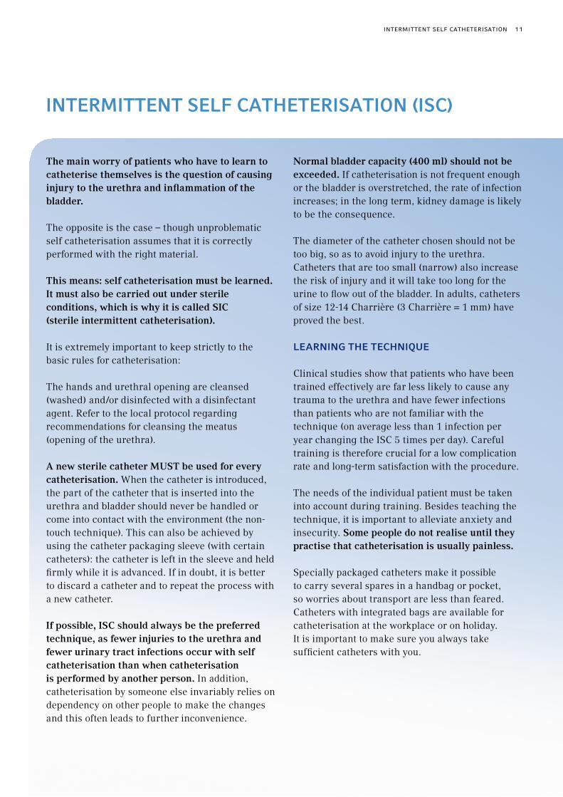
InterMIttent selF catHeterIsatIon (Isc)
The main worry of patients who have to learn to catheterise themselves is the question of causing injury to the urethra and inflammation of the bladder.
The opposite is the case – though unproblematic self catheterisation assumes that it is correctly performed with the right material.
This means: self catheterisation must be learned. It must also be carried out under sterile conditions, which is why it is called SIC (sterile intermittent catheterisation).
It is extremely important to keep strictly to the basic rules for catheterisation:
The hands and urethral opening are cleansed (washed) and/or disinfected with a disinfectant agent. Refer to the local protocol regarding recommendations for cleansing the meatus (opening of the urethra).
A new sterile catheter MUST be used for every catheterisation. When the catheter is introduced, the part of the catheter that is inserted into the urethra and bladder should never be handled or come into contact with the environment (the non-touch technique). This can also be achieved by using the catheter packaging sleeve (with certain catheters): the catheter is left in the sleeve and held firmly while it is advanced. If in doubt, it is better to discard a catheter and to repeat the process with a new catheter.
If possible, ISC should always be the preferred technique, as fewer injuries to the urethra and fewer urinary tract infections occur with self catheterisation than when catheterisation is performed by another person. In addition, catheterisation by someone else invariably relies on dependency on other people to make the changes and this often leads to further inconvenience.
Normal bladder capacity (400 ml) should not be exceeded. If catheterisation is not frequent enough or the bladder is overstretched, the rate of infection increases; in the long term, kidney damage is likely to be the consequence.
The diameter of the catheter chosen should not be too big, so as to avoid injury to the urethra. Catheters that are too small (narrow) also increase the risk of injury and it will take too long for the urine to flow out of the bladder. In adults, catheters of size 12-14 Charrière (3 Charrière = 1 mm) have proved the best.
learnInG tHe tecHnIqUe
Clinical studies show that patients who have been trained effectively are far less likely to cause any trauma to the urethra and have fewer infections than patients who are not familiar with the technique (on average less than 1 infection per year changing the ISC 5 times per day). Careful training is therefore crucial for a low complication rate and long-term satisfaction with the procedure.
The needs of the individual patient must be taken into account during training. Besides teaching the technique, it is important to alleviate anxiety and insecurity. Some people do not realise until they practise that catheterisation is usually painless.
Specially packaged catheters make it possible to carry several spares in a handbag or pocket, so worries about transport are less than feared. Catheters with integrated bags are available for catheterisation at the workplace or on holiday. It is important to make sure you always take sufficient catheters with you.
intermittent selF catheterisation 11

Magnified catheter eye. The example shown here is of an eye that is rounded off internally and externally (SafetyCat®).
Different types of catheter tips:
Ergothan Tip
Nelaton Tip
Tiemann Tip
There are numerous different catheters these days which sometimes differ significantly from each other.
The basic difference is between coated, hydrophilic catheters and catheters which are introduced with the aid of a sterile lubricant gel.
Almost all manufacturers offer hydrophilic catheters today because they have better lubricant properties. They are also easier to handle because no additional lubricant gel is required. Nevertheless, there are certain situations where the use of lubricant gel can have its advantages.
Another basic difference is the length of the catheter. There are shorter catheters for women and longer ones for men. Because of their longer urethra, men need a longer catheter.
There are straight catheter tips and "curved" tips known as Tiemann tips. The latter are more suitable
for men with prostate enlargement, to overcome the curvatures of the male urethra. When using a Tiemann catheter it is important that the curve of the catheter head is always pointing upwards to follow the anatomy of the urethra. Otherwise injury to the urethra can occur.
IC technique is used by those affected several times daily for a long indefinite period. Because of this, the catheters must fulfil certain quality requirements to provide safety even during long-term use.
The openings in the catheter through which the urine runs, known as the eyes of the catheter, must be round and smooth, so as not to cause any injuries.
The coating must not lose its lubrication during use – you have to be able to pull out the catheter as easily as introducing it, even if emptying takes a little longer.
12 intermittent selF catheterisation

While men can hold their penis with one hand so that the other hand can introduce the catheter, it is rather more difficult for women. But they can also learn to carry out self catheterisation without problems.
They must make sure to spread the labia properly so that the urethral opening above the vaginal opening is clear.
In the introductory phase, it can be sensible to use a mirror to see the genital area and the urethral opening better. However, after the learning and practising phase, most women are in a position to find their urethral opening "blind", i.e. by feel, without using a mirror, and carry out the catheterisation correctly.
In the learning phase there is some risk of infection until the process has been hygienically mastered.
We therefore recommend taking Ciprofloxacin 250 mg daily in the first week of sterile self catheterisation. After that taking an antibiotic is unnecessary.
The risk of suffering from a urinary tract infection during correctly carried out sterile self catheterisation has been studied. If catheterisation is carried out 5 times a day there is a statistical risk of 0.7 in- fections a year.
The risk of an infection is therefore very low.
On the other hand, the risk of suffering an infection with an indwelling catheter, which remains in the bladder, is a great deal higher. The bladder is usually colonised by bacteria after 48 to 72 hours if an indwelling catheter is in the bladder. Indwelling catheters are therefore contraindicated for the treatment of residual urine, even in the special form of an abdominal wall catheter (cystostomy).
Indwelling catheter
Suprapubic fistula catheter (SC) (abdominal wall catheter)
Intermittent catheterisation
intermittent selF catheterisation 13

When bacteria (or other microorganisms) multiply in the urinary tract (bladder, urethra, kidney and prostate), this is described as colonisation of germs. As these microorganisms attack the mucosa they induce a defensive reaction on the part of the body. On account of this, white and possibly red blood cells penetrate the urine. As soon as colonisation by germs causes clinical symptoms, it is described as inflammation of the urinary tract. Inflammation of the renal pelvis is particularly dangerous, as inflammation of the kidneys generates serious symptoms with chills and fever and can also leave scarring on the kidney tissue.
Urinary tract infections are more likely to occur in MS patients. Residual urine, inadequately treated spastic bladder and catheterisation represent risk factors for infections. The risk of infection with indwelling catheters is much higher than with IC.
clInIcal sIGns
Not every germ colonisation of the bladder has to be treated. Neither is it useful to test the urine regularly e.g. with test strips if you do not have any symptoms.
Clinical signs of urinary tract infection can be fever without other symptoms, recently occurring involuntary loss of urine, sudden reduction in bladder capacity, pain in the abdomen and the urethra, increased spasticity, generally feeling unwell or loss of capacity. Fever indicates severe inflammation which can progress in extreme cases to blood poisoning or kidney damage and rapid advanced diagnosis is a matter of urgency.
A change in the odour of the urine or cloudy urine can be initial signs of urinary tract infection, but does not require treatment when it is the only symptom, if there is no particularly negative impact on the person concerned.
dIaGnostIcs
If urinary tract infection is suspected, the urine should be tested using test strips or better still by examination under the microscope (urinary sediment). If bacteria and white blood cells are present (the body’s defence cells; increased white blood cells in the urine show that the body is fighting the bacteria. If there is evidence of bacteria without white blood cells, "peaceful coexistence" can be assumed), a urine culture is set up to determine the type of bacteria and the antibiotics to which these pathogens are sensitive.
adVanced dIaGnostIcs
In the case of severe, febrile urinary tract infections or repeatedly occurring (recurrent) infections a physical examination and ultrasound scan of the kidneys, bladder and the prostate in men, if necessary, should be carried out to exclude organ involvement.
treatMent
Treatment of acute urinary tract infection depends on how severe it is. Infections without fever and with only a few symptoms can be treated by drinking more water (> 1.5 litres/day).
Cranberry juice or tablets are a treatment option. Other plant treatments are bearberry leaf tea which should only be drunk for a certain period and at a dose that is not too high (no more than 3 cups/day) and a mixture of nasturtium and horseradish. Homeopathic medicines are also
UrInary tract InFectIons
14 urinary tract inFections
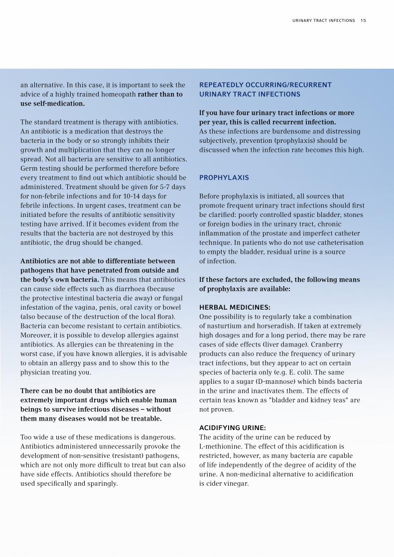
an alternative. In this case, it is important to seek the advice of a highly trained homeopath rather than to use self-medication.
The standard treatment is therapy with antibiotics. An antibiotic is a medication that destroys the bacteria in the body or so strongly inhibits their growth and multiplication that they can no longer spread. Not all bacteria are sensitive to all antibiotics. Germ testing should be performed therefore before every treatment to find out which antibiotic should be administered. Treatment should be given for 5-7 days for non-febrile infections and for 10-14 days for febrile infections. In urgent cases, treatment can be initiated before the results of antibiotic sensitivity testing have arrived. If it becomes evident from the results that the bacteria are not destroyed by this antibiotic, the drug should be changed.
Antibiotics are not able to differentiate between pathogens that have penetrated from outside and the body’s own bacteria. This means that antibiotics can cause side effects such as diarrhoea (because the protective intestinal bacteria die away) or fungal infestation of the vagina, penis, oral cavity or bowel (also because of the destruction of the local flora). Bacteria can become resistant to certain antibiotics. Moreover, it is possible to develop allergies against antibiotics. As allergies can be threatening in the worst case, if you have known allergies, it is advisable to obtain an allergy pass and to show this to the physician treating you.
There can be no doubt that antibiotics are extremely important drugs which enable human beings to survive infectious diseases – without them many diseases would not be treatable.
Too wide a use of these medications is dangerous. Antibiotics administered unnecessarily provoke the development of non-sensitive (resistant) pathogens, which are not only more difficult to treat but can also have side effects. Antibiotics should therefore be used specifically and sparingly.
repeatedly occUrrInG/recUrrent UrInary tract InFectIons
If you have four urinary tract infections or more per year, this is called recurrent infection. As these infections are burdensome and distressing subjectively, prevention (prophylaxis) should be discussed when the infection rate becomes this high.
propHylaxIs
Before prophylaxis is initiated, all sources that promote frequent urinary tract infections should first be clarified: poorly controlled spastic bladder, stones or foreign bodies in the urinary tract, chronic inflammation of the prostate and imperfect catheter technique. In patients who do not use catheterisation to empty the bladder, residual urine is a source of infection.
If these factors are excluded, the following means of prophylaxis are available:
HerBal MedIcInes:One possibility is to regularly take a combination of nasturtium and horseradish. If taken at extremely high dosages and for a long period, there may be rare cases of side effects (liver damage). Cranberry products can also reduce the frequency of urinary tract infections, but they appear to act on certain species of bacteria only (e.g. E. coli). The same applies to a sugar (D-mannose) which binds bacteria in the urine and inactivates them. The effects of certain teas known as "bladder and kidney teas" are not proven.
acIdIFyInG UrIne:The acidity of the urine can be reduced by L-methionine. The effect of this acidification is restricted, however, as many bacteria are capable of life independently of the degree of acidity of the urine. A non-medicinal alternative to acidification is cider vinegar.
urinary tract inFections 15

Injection of botulinum toxin
Imag
e su
pplie
d by
: SPC
Not
twil
Bladder rInsInG:The regular rinsing of the bladder with disinfectant solutions or water is not suitable for avoiding infection if the bladder is emptied by IC.
HoMeopatHIc aGents:Self-medication (from a "homeopathic medicine chest") is not advisable as prophylaxis of urinary tract infections. In general, it is true to say that homeopathic medicines belong in the hands of well trained experts (homeopaths or physicians who are trained in homeopathy), if they are intended to be used against a chronic disease (such as preventing urinary tract infections): no-one would simply order an antibiotic from the internet without specialist knowledge and the same applies to homeopathic medicines.
antIBIotIcs:Prophylaxis with antibiotics should only be carried out for self catheterisation in exceptional cases and on express medical instruction over a short period, e.g. in the first few days of the learning phase. Otherwise antibiotic prophylaxis must be avoided.
otHer treatMent optIons
There are two other treatment options for neurogenic urinary incontinence: one is the injection of a nerve poison (botulinum toxin) into the bladder muscle; the other is the implantation of a bladder pacemaker (neuromodulation).
BotUlInUM toxIn aBotulinum toxin is highly effective and almost al- ways stops the uninhibited bladder contractions which cause loss of urine, though it does have the disadvantage of being difficult to dose. That is why the side effect of (temporary) bladder paralysis often occurs, which makes regular self catheterisation necessary. Also, it must be injected directly into the bladder muscle using a bladder speculum. The intervals are about 3 months, as this is the duration of effectiveness of the product.
neUroModUlatIonNeuromodulation is a procedure which externally resembles a heart pacemaker. Electrodes are implanted next to the nerves of the bladder in the region of the sacrum; these are joined to a control mechanism, the neuromodulator. This can be controlled with a small hand-held device (size of a mobile phone), so retention and release of water can be programmed more or less at the touch of a button. Its disadvantage is that the urge to urinate is no longer sensed, which is why good fluid management is essential.
In addition, the neuromodulator must be implanted by means of an operation under anaesthetic. Depending on the strength of flow, the battery be- comes exhausted after a few years, so it needs to be changed in another operation.
It is also expensive.
Not every MS patient is suitable for this in principle elegant method of neuromodulation. Only patients with stable MS can be considered. With progressive (including slow progression) MS, the procedure is ruled out as it cannot be sufficiently adjusted for this progression.
16 urinary tract inFections

IncontInence Urinary incontinence, an involuntary and uncontrollable loss of urine, is both a medical and a social problem. Incontinence can lead to inflammation of the bladder, skin changes (fungal infestation, inflammation); in addition, the odour nuisance, the need to use incontinence aids and the resulting lack of confidence have a negative effect on quality of life.
Incontinence may occur as the result of an overactive bladder which squeezes the urine through an intact sphincter. This type of incontinence is called urge incontinence. A particular form which affects MS patients is (neurogenic) reflex incontinence. This can be treated by calming the spastic bladder.
Another form is stress incontinence, which is caused by a weak sphincter.
Stress incontinence (loss of urine when coughing, in sport, lifting loads, etc.) can be treated conservatively by a drug which increases sphincter tone (Duloxetine), biofeedback controlled pelvic floor exercises and in many cases surgically.
Videodynamics are usually used with a urethra pressure profile to find out which form of urinary incontinence is present and which therapy is the most sensible. Videodynamics is necessary before any urinary incontinence therapy, to avoid mis-treatment, which can have negative consequences; for example, a sphincter operation should not be carried out for reflex incontinence because otherwise the bladder and later the kidneys will be damaged.
There are several procedures for the methods of operation; in principle they should increase the release resistance if the problem is due to stress incontinence. For women there is also the Burch colposuspension, in which the neck of the bladder is lifted; this is the most widespread method.
Depending on the results of the urethra pressure profile, fascial sling plasty, artificial ligaments or even in exceptional cases an artificial "sphincter" can also be considered.
If an artificial sphincter is used, a plastic cuff is placed around the exit to the bladder, which is connected to a balloon and a pump. All parts are inserted into the body. The pump is placed in the scrotum or in the labia majora so that it can be operated from the outside. The cuff, which is filled with liquid, ensures that no urine loss occurs. When the bladder is voided the cuff is emptied using the pump. After a few minutes, the cuff automatically refills with liquid and the bladder is "tight" again.
The Scott artificial urinary sphincter
Reservoir
Bladder
Sphincter cuff
Balloon pump
incontinence 17

The risks associated with the operation are the typical complications connected with every procedure in which foreign bodies are inserted: a material defect and infection of the device implanted. Approximately 30% of patients need to be operated on for a second time within 5 years.
The Burch procedure, fascia sling plasty and artificial ligaments are carried out via a small skin incision and depending on the method via an incision in the vagina.
When the diagnosis is carried out carefully, all the operative methods have a good success rate.
As both types of incontinence may also occur together, an essential prerequisite of successful treatment is initial testing to ascertain which type of incontinence is present. It is not uncommon for surgery and medical therapy to be combined. The wrong therapy is not just frustrating, it also involves medical risks which may include damage to kidney function.
In forms of reflex incontinence which cannot be treated medically, botulinum toxin injection into the bladder muscle or neuromodulation is considered. Again, a careful urodynamic diagnosis is a prerequisite.
aIds For UrInary IncontInence
These involve urine capture systems. Effective urine capture systems are only available for men and are known as adhesive urinary sheaths. Recently, there have also been special underpants which include a capture system which can be used for a long time without smelling.
Ask your local continence advisor to make sure your are assessed for the home delivery pad service, which is available on prescription at no charge.
Urinary sheath and urine collection bag
18 incontinence

With MS, chronic constipation can also occur because the nerve damage is in many ways similar.
A balanced therapy including the regular intake of roughage is therefore important. Conventional laxatives are on the other hand not suitable for long-term use, but only for individual cases, their use over a long period of time can lead to bowel damage and have the opposite effect from the one intended.
aIds For Bowel IncontInence
If the pelvic floor and sphincter are not entirely paralysed, an attempt can be made to use anal suppositories. These work in much the same way as a tampon. However, it is better to use bulking agents and other materials which affect the bowel. These must be individually tried.
Depending on the detailed findings, there are also surgical measures available which include neuromodulation.
The nerve supply to the genitals uses the same nerves as the supply to the bladder. If paralysis of the bladder is present, sexual dysfunction will also have to be confronted.
Sexual dysfunction includes sensory dysfunction, orgasmic capacity and fertility. In women, vaginal moistening and in men erection and ejaculation may not function normally.
MoIstenInG oF tHe VaGIna
If the vagina cannot be moistened, lubricants (oils such as extra virgin olive oil, Vaseline or water-based lubricants) can be used.
ejacUlatIon
With early ejaculation an improvement can be achieved with medicines or a mild anaesthetic cream on the glans.
If a local anaesthetic cream (e.g. Emla®) is applied an hour beforehand, the effect is reliable. Certain psychopharmaceutical drugs are also effective, although they demonstrate a high rate of side effects. Priligy® can also delay ejaculation. Stop-start-squeeze training can also be helpful, but does need some practice.
Treatment of effusion into the bladder can also be attempted with medicines (e.g. Midodrine). If none of these is successful, sperm can be obtained from the urine, e.g. for fertilisation.
If there is no ejaculate, electrostimulation can be used to obtain sperm. This can be achieved by a type of vibrostimulation; if this fails, electrostimulation can be tried with a probe introduced into the rectum. Both procedures can be carried out independently of sexual intercourse and serve only to obtain sperm for fertilisation; the last procedure in particular can involve significant side effects (hypertensive crisis, massive spasticity, pain) and should only be carried out under medical supervision.
Bowel eMptyInG
sexUalIty wItH MUltIple sclerosIs
bowel emptying | sexuality with multiple sclerosis 19

FeMale FertIlIty
A pregnancy is just as possible for a woman with MS as it is for a healthy woman. Contraception must therefore be used if the patient does not want children. Basically the pill or the coil (IUD) are considered for contraception. A diaphragm (a cap in the vagina) is less reliable.
Some drugs to suppress bladder spasticity are not allowed during pregnancy. Pregnant women with bladder dysfunction requiring medical treatment should therefore contact a urologist as early as possible.
erectIle dysFUnctIon
Erectile dysfunction in MS is usually caused by a nerve dysfunction (although MS patients who smoke can also demonstrate erectile dysfunction of vascular cause). This can usually be well treated with one of the new PDE5 inhibitors, which are available in tablet form and must be taken before sexual intercourse.
As there are contraindications for this, taking such products must be discussed with an experienced physician. The current possibility of ordering these
drugs over the internet for self medication is extremely dangerous. These pills are cheaper than the original products, but we know that many of these pills are counterfeits and do not contain any active substance, and in some cases even contain harmful substances.
If these PDE5 inhibitors are not effective or are contraindicated, there is still the possibility of CCAT, an erection pump or penis prosthesis.
With corpus callosum auto-injection therapy (CCAT) the patient learns to inject a substance causing an erection into his own penis when he wants to have intercourse.
The erection pump creates low pressure and en- courages blood to fill the penis; this can support the CCAT method.
The final option in exceptional cases is a penile prosthesis; it is an implant which may not be accepted by the body and can become infected. This method will be very carefully considered.
aIds For sexUalIty
There are a number of different aids available. Do not be embarrassed to talk to your physician about this or send for appropriate catalogues. The products offered there are significantly cheaper than the relevant "medical" items.
For example, suction pumps can be considered to promote stiffening of the penis (possibly in combination with CCAT) as well as lubricant creams.
20 sexuality with multiple sclerosis

antIcHolInerGIcs:Drugs which affect the bladder muscle so that it does not contract so strongly. They are used for involuntary loss of urine as a result of bladder cramps (urge incontinence). Typical side effects include a dry mouth and occasionally impaired vision. However, other side effects are very rare.
In older people, memory deficits and similar can occasionally occur – depending on the drug used.
BotUlInUM toxIn:Drug which can be used for MS patients if anticholinergics are insufficient for cramplike urine loss. The disadvantage is that Botulinum toxin must be injected into the bladder muscle using a bladder speculum.
detrUsor:The detrusor is the spherical bladder muscle which contracts when the bladder is emptied.
detrUsor spHIncter dyssynerGIa (dsd):This is the lack of cooperation between the bladder muscle and the bladder sphincter. They must function synergically when the bladder is to be emptied. With DSD the sphincter contracts instead of relaxing to allow urine flow when the bladder contracts. DSD is typical in many MS patients with spinal cord lesions.
contInence/IncontInence:Continence is the capacity to retain urine, while incontinence describes the lack of ability to retain urine. There are several forms of urinary incontinence. The form caused by MS lesions is urge incontinence. It can occur both with and without DSD depending on the location of the lesion.
neUroModUlator:So-called "bladder pacemaker". This is an electronic device implanted under the skin, which uses electrodes placed on the nerve roots in the area of the sacrum and which largely takes over control of the bladder. There is seldom an indication for MS patients and it is only provided by neuro-urological centres.
pde5 InHIBItors:Drugs to promote erectile function. They are taken in tablet form about half an hour before sexual intercourse is planned.
There are variations in the duration of effect. A urologist must decide on the choice of drug.
Caution is advised for patients taking heart medication: PDE5 inhibitors are prohibited for men who have to take nitrate or Molsidomine.
penIle prostHesIs: This is implanted into the body of the penis. There are rigid systems which have the effect of permanently thickening the penis. These are "controlled" by bending the inbuilt metal wire as required. They are rarely used.
A flexible inflatable penile prosthesis is usually implanted today. They can be inflated by pumping up a fluid from a reservoir located in the abdominal space, causing an artificial erection.
Glossary oF tHe MaIn terMs
glossary oF the main terms 21

reFlUx: This is the (pathological) backflow of urine into the ureter, possibly as far as the kidneys. It can occur in MS patients if there is bladder dysfunction which leads to high pressure in the bladder, so that the natural reflux protection is destroyed. In the long term, reflux can lead to serious kidney dysfunction.
ccat: This acronym stands for corpus cavernosum auto-injection therapy. For this, a drug is injected by the patient into the corpus cavernosum of the penis (after instruction by a physician), which creates an artificial erection that allows sexual intercourse to take place. It can only be carried out on prescription.
spHIncter:This is the muscle which keeps the bladder closed and is integrated into the urethra; its predominantly ring-shaped muscle fibres voluntarily interrupt urinary flow. It also contains fibres which are "autonomously" controlled, i.e. ensure continence at rest.
sterIle InterMIttent catHerIsatIon (sIc): Sterile self catheterisation
UretHral closInG pressUre: Measuring the pressure is a component of the urodynamic test and is especially important if loss of urine occurs.
UrGe IncontInence: Urge incontinence means that the bladder creates involuntary pressure waves which can be so strong that urine escapes. This form of urinary incontinence is diagnosed by means of urodynamics.
UrodynaMIcs: Examination technique for measuring the pressure in the bladder with a thin measuring probe. From the transfer of the pressure to a time scale we can draw conclusions about the bladder dysfunction and its cause.
22 glossary oF the main terms
© MEDICAL SERVICE 2011
The medical information was kindly supplied by Dr. med. Merkle, DKD Wiesbaden. It is subject to change on the basis of more recent medical findings. This brochure is intended as a guide only and is not a substitute for a visit to the physician or for medical treatment.
Please always ask your physician if you have medical problems. Medical Service cannot accept any liability for the accuracy or completeness of the information given in this brochure.
Concept & Design: DART, www.dartwork.de

MedIcal serVIce GmbH • A Teleflex Company • Luisenstraße 875378 Bad Liebenzell • [email protected] • www.medical-service.de
naMe: date:
You can easily use this table as a master copy to make further copies.
time amount drunk amount of urine wet pad dry pad remarks
total
urine measurement 23
UrIne MeasUreMent

HoMecare UroloGy prodUctsHoMecare UroloGy prodUcts
LIQUICK® PLUSHYDROPHILIC CATHETER SYSTEM
LIQUICK® BASEHYDROPHILIC CATHETER SYSTEM
Liquick® Plus is our most recent development from the range of hydrophilic products. The integrated sachet containing saline solution makes it even easier to use. Once the sachet is burst the coating is activated. The product has an integral collection bag and once filled, may be securely closed and in turn discretely emptied and disposed of.
Liquick® Base is the modern flexible compact product from our range of hydrophilic catheter systems. Ease of handling, particular rapidity of use and reduced packaging were the focus when Liquick® Base was developed. The SafetyCat® safety catheter and the sachet with sterile saline solution are both contained in the outer packaging. The practical blue protective wrapper makes for a non-touch catheterisation.
24 homecare urology products

FLOCATH INTROGELMORE MOBILITY, DISCRETION AND SAFETY
Flocath IntroGel contains an integrated urine collection bag as well as the catheter and aqueous catheter lubricating gel. The catheter is equipped with a urethra insertion aid 15 mm in length. The aid is used to by-pass the region of the urethra with the highest germ colonisation. This prevents germs from being pushed inside and from causing infections when the catheter is inserted into the urethra. Flocath IntroGel is designed for aseptic use without direct contact between catheter and hands.
FLOCATH QUICK 1Never wAs A CATHeTer mOre geNTLe
Our Flocath quick is a catheter system that is as quick to use as Liquick® Base. It contains a nelaton tipped intermittent catheter with hydrophilic coating from the Flocath range with outstanding lubrication and a sachet with sterile saline solution to activate the coating. This catheter may also be considered to be ‘non-touch’ due to the presence of the blue catheter sleeve.
1 FloCath Quick is not for sale in Germany
homecare urology products 25
26 homecare urology products
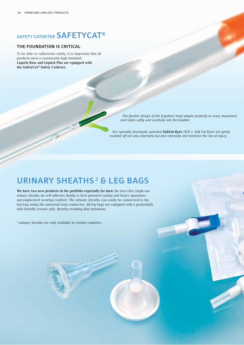
We have two new products in the portfolio especially for men: the latex-free single-use urinary sheaths are self-adhesive thanks to their patented coating and hence guarantee uncomplicated wearing comfort. The urinary sheaths can easily be connected to the leg bag using the universal step connector. All leg bags are equipped with a particularly skin-friendly reverse side, thereby avoiding skin irritations.
2 uninary sheaths are only available in certain countries
URINARY SHEATHS 2 & LEG BAGS
The flexible design of the Ergothan head adapts perfectly to every movement and slides softly and carefully into the bladder.
Our specially developed, patented SoftCat-Eyes (SCE = Soft Cat Eyes) are gently rounded off not only externally but also internally and minimise the risk of injury.
tHe FoUndatIon Is crItIcal
To be able to catheterise safely, it is important that all products meet a consistently high standard.Liquick Base and Liquick Plus are equipped with the SafetyCat® Safety Catheter.
saFety catHeter saFetycat®

homecare urology products 27

940650-000001 · REV A · MC / WM · 11 12 01
yoUr contacts For eUrope, tHe MIddle east and aFrIca (eMea):
teleFlex MedIcal HeadqUarters eMea, Ireland
Teleflex Medical Europe Ltd., IDA Business Park, Athlone, Co Westmeath
Phone +353 (0)9 06 46 08 00 · Fax +353 (0)14 37 07 73
aUstrIa +43 (0)1 402 47 72
BelGIUM +32 (0)2 333 24 60
cZecH repUBlIc +420 (0)495 759 111
France +33 (0)5 62 18 79 40
GerMany +49 (0)7151 406 0
Greece +30 210 67 77 717
Italy +39 0362 58 911
netHerlands +31 (0)88 00 215 00
portUGal +351 22 541 90 85
sloVaK repUBlIc +421 (0)3377 254 28
soUtH aFrIca +27 (0)11 807 4887
spaIn +34 918 300 451
swItZerland +41 (0)31 818 40 90
UnIted KInGdoM +44 (0)1494 53 27 61
For detailed information see www.teleflex.com
FloCath Quick is not for sale in Germany
The products in this catalogue are only available in the EMEA (Europe, Middle East, Africa) region. For further information contact your local representative. All data current at time of printing (11/2012). Subject to technical changes without further notice.