Multiple representations and algorithms for reinforcement learning in the cortico-basal ganglia...
6
Available online at www.sciencedirect.com Multiple representations and algorithms for reinforcement learning in the cortico-basal ganglia circuit Makoto Ito 1 and Kenji Doya 1,2 Accumulating evidence shows that the neural network of the cerebral cortex and the basal ganglia is critically involved in reinforcement learning. Recent studies found functional heterogeneity within the cortico-basal ganglia circuit, especially in its ventromedial to dorsolateral axis. Here we review computational issues in reinforcement learning and propose a working hypothesis on how multiple reinforcement learning algorithms are implemented in the cortico-basal ganglia circuit using different representations of states, values, and actions. Addresses 1 Neural Computation Unit, Okinawa Institute of Science and Technology, Okinawa 904-0412, Japan 2 Computational Neuroscience Laboratories, Advanced Telecommunications Research Institute International, Kyoto 619-0288, Japan Corresponding author: Doya, Kenji ([email protected]) Current Opinion in Neurobiology 2011, 21:368–373 This review comes from a themed issue on Behavioral and cognitive neuroscience Edited by Ann Graybiel and Richard Morris Available online 29th April 2011 0959-4388/$ – see front matter # 2011 Elsevier Ltd. All rights reserved. DOI 10.1016/j.conb.2011.04.001 Introduction The loop network composed by the cerebral cortex and the basal ganglia is now recognized as the major site for decision making and reinforcement learning [1,2]. The theory of reinforcement learning [3] prescribes a number of steps that are required for decision making: 1) recog- nize the present state of the environment by disambig- uating sensory inputs; 2) evaluate the candidate actions in terms of expected future rewards (action values); 3) select an action that is most advantageous; and 4) update the action values based on the discrepancy between the predicted and the actual rewards. Simplistic models of reinforcement learning in the basal ganglia (e.g. [4]) proposed that the cerebral cortex represents the present state and the striatal neurons compute action values [5]. An action is selected in the downstream, the globus pallidus, and the dopamine neurons signal the reward prediction error [6], which enables learning by dopamine- dependent synaptic plasticity in the striatum [7]. Recent studies, however, have shown that the reality may be more complex. Discriminating the environmental state behind noisy observation is in itself a hard problem, known as perceptual decision making [8,9]. Activities related to action values are found not only in the striatum, but also in the pallidum [10,11 ] and the cortex [12 ]. Different parts of the striatum, especially in its ventro- medial to dorsolateral axis, have different roles in goal- directed and habitual behaviors [13]. Action selection may be performed not just in one locus in the brain but by competition and agreement among distributed decision networks [14]. Finally, a subset of midbrain dopamine neurons located in the dorsolateral part signal not only rewarding but also aversive signals [15 ]. Based primarily on primate studies, Samejima and Doya [16] proposed that different cortico-basal ganglia subloops realize decisions in motivational, context-based, spatial, and motor domains. In this article, we consider how different algorithms of decision making, such as model- based and hierarchical reinforcement learning algorithms, can be implemented in the cortico-basal ganglia circuit with a focus on the ventromedial to dorsolateral axis in the rodent striatum. Computational axes in action learning In looking into the computational mechanisms of decision making and reinforcement learning, there are several axes that are useful for sorting out the process. From state recognition through valuation to action selection Where in the brain is the locus of decision making? Exactly how an action is selected may depend on the modality and clarity of sensory evidence, available methods of value estimation, and the preparedness in implementation of the action. From flexible learning to efficient execution Early in learning, actions have to be exploratory and require a high cognitive load. As behavior becomes well learned, actions can be smooth, stereotyped, and needing less cognitive demands. Such a transition can be due to changes in the parameters like the ‘temperature’ for action selection [3], but also the shift in the recruitment of the brain loci implementing different algorithms, most notably model-based, predictive strategies and model- free, retrospective strategies [17]. From whole life to movement details Animal behaviors have hierarchical structure in time and space. To satisfy the fundamental needs of life like Current Opinion in Neurobiology 2011, 21:368–373 www.sciencedirect.com
-
Upload
makoto-ito -
Category
Documents
-
view
214 -
download
1
Transcript of Multiple representations and algorithms for reinforcement learning in the cortico-basal ganglia...

Available online at www.sciencedirect.com
Multiple representations and algorithms for reinforcementlearning in the cortico-basal ganglia circuitMakoto Ito1 and Kenji Doya1,2
Accumulating evidence shows that the neural network of the
cerebral cortex and the basal ganglia is critically involved in
reinforcement learning. Recent studies found functional
heterogeneity within the cortico-basal ganglia circuit, especially
in its ventromedial to dorsolateral axis. Here we review
computational issues in reinforcement learning and propose a
working hypothesis on how multiple reinforcement learning
algorithms are implemented in the cortico-basal ganglia circuit
using different representations of states, values, and actions.
Addresses1 Neural Computation Unit, Okinawa Institute of Science and
Technology, Okinawa 904-0412, Japan2 Computational Neuroscience Laboratories, Advanced
Telecommunications Research Institute International, Kyoto 619-0288,
Japan
Corresponding author: Doya, Kenji ([email protected])
Current Opinion in Neurobiology 2011, 21:368–373
This review comes from a themed issue on
Behavioral and cognitive neuroscience
Edited by Ann Graybiel and Richard Morris
Available online 29th April 2011
0959-4388/$ – see front matter
# 2011 Elsevier Ltd. All rights reserved.
DOI 10.1016/j.conb.2011.04.001
IntroductionThe loop network composed by the cerebral cortex and
the basal ganglia is now recognized as the major site for
decision making and reinforcement learning [1,2]. The
theory of reinforcement learning [3] prescribes a number
of steps that are required for decision making: 1) recog-
nize the present state of the environment by disambig-
uating sensory inputs; 2) evaluate the candidate actions in
terms of expected future rewards (action values); 3) select
an action that is most advantageous; and 4) update the
action values based on the discrepancy between the
predicted and the actual rewards. Simplistic models of
reinforcement learning in the basal ganglia (e.g. [4])
proposed that the cerebral cortex represents the present
state and the striatal neurons compute action values [5].
An action is selected in the downstream, the globus
pallidus, and the dopamine neurons signal the reward
prediction error [6], which enables learning by dopamine-
dependent synaptic plasticity in the striatum [7]. Recent
studies, however, have shown that the reality may be
more complex. Discriminating the environmental state
Current Opinion in Neurobiology 2011, 21:368–373
behind noisy observation is in itself a hard problem,
known as perceptual decision making [8,9]. Activities
related to action values are found not only in the striatum,
but also in the pallidum [10,11�] and the cortex [12��].Different parts of the striatum, especially in its ventro-
medial to dorsolateral axis, have different roles in goal-
directed and habitual behaviors [13]. Action selection
may be performed not just in one locus in the brain
but by competition and agreement among distributed
decision networks [14]. Finally, a subset of midbrain
dopamine neurons located in the dorsolateral part signal
not only rewarding but also aversive signals [15��].
Based primarily on primate studies, Samejima and Doya
[16] proposed that different cortico-basal ganglia subloops
realize decisions in motivational, context-based, spatial,
and motor domains. In this article, we consider how
different algorithms of decision making, such as model-
based and hierarchical reinforcement learning algorithms,
can be implemented in the cortico-basal ganglia circuit
with a focus on the ventromedial to dorsolateral axis in the
rodent striatum.
Computational axes in action learningIn looking into the computational mechanisms of decision
making and reinforcement learning, there are several axes
that are useful for sorting out the process.
From state recognition through valuation to action
selection
Where in the brain is the locus of decision making?
Exactly how an action is selected may depend on the
modality and clarity of sensory evidence, available
methods of value estimation, and the preparedness in
implementation of the action.
From flexible learning to efficient execution
Early in learning, actions have to be exploratory and
require a high cognitive load. As behavior becomes well
learned, actions can be smooth, stereotyped, and needing
less cognitive demands. Such a transition can be due to
changes in the parameters like the ‘temperature’ for
action selection [3], but also the shift in the recruitment
of the brain loci implementing different algorithms, most
notably model-based, predictive strategies and model-
free, retrospective strategies [17].
From whole life to movement details
Animal behaviors have hierarchical structure in time and
space. To satisfy the fundamental needs of life like
www.sciencedirect.com
Multiple representations and algorithms for reinforcement learning in the cortico-basal ganglia circuit Ito and Doya 369
eating, drinking and mating, an animal has to organize a
temporal sequence of exploration, approaching, and
manipulation involving movements of the whole body
to body parts and individual muscles. Dealing with such
temporal and physical hierarchy requires proper mech-
anisms for coordination and credit assignment. Appropri-
ate weighting is needed for the reward attained at the goal
and cost or danger incurred along the way, and also for
immediate and long-term outcomes.
In the following sections, we will first look into the
computational frameworks to deal with such complex-
ities. We will then review experimental works that pro-
vide clues as to how they could be implemented in the
cortico-basal ganglia network. We will finally propose a
working hypothesis on the implementation of hierarchical
reinforcement learning in the ventral-dorsal axis of the
basal ganglia.
Algorithms for reinforcement learningModel-free reinforcement learning algorithms
In the basic theory of reinforcement learning, the learning
agent does not initially know how its actions affect the
environmental state or how much rewards are given in
what state. The action value-based algorithms, including
Q-learning and SARSA, use actual experience of state,
action, and reward to estimate the action value function
Q(state,action), which evaluates how much future reward
is expected by taking a particular action at a given state.
An action can be selected greedily or stochastically by
comparing the action values of the candidate actions.
Another popular algorithm for model-free reinforcement
learning is the actor-critic, in which the critic learns to
predict the future rewards in the form of the state value
function V(state) and the actor improves action policy
P(actionjstate) using the reward prediction error as the
reinforcement signal. A good feature of actor-critic
method is that after sufficient learning, only the actor
part is needed for real-time control with less compu-
tational demand.
Model-based algorithms
In the classical theory of optimal control and decision
making, the knowledge of the environment, i.e. the state
transition probability P(new statejstate,action) is sup-
posed to be available. Such a model of the environmental
dynamics can facilitate decisions and learning in multiple
ways.
The typical way is the search for a good action or a
sequence of actions that gives the largest rewards. This
enables flexible adaptation to a new reward setting.
However, as the numbers of available actions, possible
outcomes, and steps to reach the goal increase, running a
full tree search requires a lot of time and working memory
www.sciencedirect.com
load. The evaluation of intermediate states in the form of
state value function can help truncating a deep search.
In addition to such an on-line use for action selection,
dynamic models can be used for learning value functions
and/or action policies by off-line through simulated
experience either forward or backward in time. Another
important use of environmental dynamic models is for
estimating the state of the environment given past actions
and sensory observations [9].
Hierarchical architectures
Hierarchical reinforcement learning algorithms (e.g.
[18,19]) have been proposed for dividing a large com-
plex problem into small simpler problems and reusing
the solutions for sub tasks to better cope with new
situations. A typical way of dividing a task is for an
‘action’ in a higher level to serve as the context or
activation signal for the lower level [20,21]. A proper
reward signal should be given upon completion of a
specified subtask, even if the entire behavior may not
be rewarded. In some cases, the state value for a
subtask can be seen as the action value for choosing
the subtask by the upper level, which can blur the
distinction between the two.
Model-based analysis of learner’s variablesIn order to describe how an animal’s choices change
dynamically depending on the reward experience, a
straight forward way is to take a Markov model in
which the conditional probability of action choice given
previous state, action, and reward is computed. Such
non-parametric, hypothesis-neutral description is help-
ful in measuring goodness of more elaborate model-
based explanation [11�]. Recent use of normative
models, especially those by reinforcement learning
algorithms, has turned out to be powerful tools for
characterizing subjects’ decision strategies and
parameters, and searching for their neural correlates
[22–25].
One problem in normative model-based behavior
analysis is the choice of the learning algorithm and
the estimation algorithm. In order to describe the
subject’s learning process, most studies assume just
one or a few learning algorithms, such as Q-learning
[5] and its variants [11�,26], or semi-parametric models
[27,28]. Different studies use different algorithms for
estimation of the model parameters, such as maximal
likelihood estimate, Kalman filter [29], and particle
filter [5,11�]. Establishment of standard methods and
tools is strongly in demand.
Another issue is validation of the models. Common criteria
for evaluating model performance are Akaike’s Infor-
mation Criterion (AIC; [28,30,31�,32��]) and Bayesian
information criterion (BIC; [29,31�,33–37]). However, a
Current Opinion in Neurobiology 2011, 21:368–373

370 Behavioral and cognitive neuroscience
care must be taken as the validity of these measures is
under some assumptions, such as incremental complexity.
A more robust comparison can be made by cross validation
[11�,31�,38]. Furthermore, the best fit models to a subject’s
sample behavioral sequence may not reproduce the sub-
ject’s behavior when it is run on its own in the same task. It
is important to check if the model reproduces some stat-
istics of the subject’s behavior, such as the total reward and
learning speed.
Possible implementation in the cortico-basalganglia networkBased on the computational requirements and possible
reinforcement learning algorithms, we now review neural
recording and brain imaging results, many of which
through the model-based analysis described above, that
shed light on how they could be implemented in the
cortico-basal ganglia network.
From state recognition through valuation to action
selection
Information about states, actions, and rewards has been
found in the striatum [11�,31�,39–43]. Furthermore, infor-
mation of past actions and rewards is also represented in
the striatum [11�,31�,42,44,45].
State value
Monkey studies using a free choice task reported that
state-value coding neurons were few in the dorsal stria-
tum (DS) [5,46]. In rat studies, the state value coding
neurons was found in both DS and the vental striatum
(VS), although the proportion was not large [11�,31�].Human imaging studies suggest that VS play a role of
critic in the actor-critic algorithm [47].
Action value
In primates, the action value coding has been reported in
DS [5,10,12��,41,46], the internal pallidum [10], and the
supplemental motor area [12��]. In rodents, however, the
population of action-value coding neurons was reported to
be significant but small in both DS and VS [11�,31�,48].
Action command
While the representation of upcoming action has been
reported in DS [5,10,31�,49], its representation in the
ventral striatum is not consistent, with both positive
[48] and negative reports [11�,31�,44], Action command
is also represented in the cortical areas in the loop circuit
with the striatum, such as the supplementary and pre-
supplementary motor area [50], the prefrontal cortex [49]
and the parietal cortex [51].
Chosen value
The action value for selected action Q(selected action),
named chosen value, is necessary for comparison with the
actually delivered reward for learning. Chosen-value cod-
Current Opinion in Neurobiology 2011, 21:368–373
ing was also found in DS in monkeys [46] and in both DS
and VS in rats [31�].
Reward prediction error (RPE)
Functional MRI studies have reported RPE in VS
[29,32��,34,47,52] and DS [29,47,52]. This is consistent
with the RPE coding of the dopamine neurons projecting
to the striatum because the fMRI signal is known to
respond strongly to the presynaptic inputs [53]. It is
reported in rats that a fraction of the striatal neurons
coded RPE [54]. Recently Dickerson and Delgado [55]
reported in a probabilistic learning task with rats that RPE
signal was observed in not only DS but also hippocampus,
suggesting the involvement of episodic memory system
in decision making. Recently Nomoto et al. [56�] showed
in monkeys using a random dot motion discrimination
task with different rewards for different directions that
the midbrain dopamine neurons show two-phase cue
responses, the early one in proportion to the average
reward and the later one reflecting the deviation from
the average.
While dopamine neurons code RPE, serotonin neurons
have been hypothesized to code aversive prediction error
[57]. However, Miyazaki et al. [58] reported increased
serotonin activity during waiting for delayed rewards, but
not for unexpected reward omission. Aversive prediction
error signal was found instead in the lateral habenula
neurons [59] and a subset of midbrain dopamine neurons
[15��].
From flexible learning to efficient execution
Studies using the devaluation paradigm in rodents
showed the distinct roles of dorsomedial and dorsolateral
striatum in goal-directed and habitual behaviors, respect-
ively [60–64].
Brain imaging studies investigated possible realization of
model-based action and learning strategies, such as a
Kalman filter [29] or a hidden Markov model [34], in
the cortico-basal ganglia system [17]. Recently, a hybrid
model of the model-based and the model-free strategies
was reported to show the higher prediction accuracy than
that of the single model-base or model-free strategy
[32��].
From whole life to movement details
A possible implementation of hierarchical reinforce-
ment learning in the cortico-basal ganglia loop is along
the ventro-dorsal axis within the striatum. The ventral
striatum is connected with the limbic system, which
represent primary reward information and regulates the
affects and motivation of the whole animal. The dorsal
striatum, on the contrary, is connected with the sen-
sory-motor cortices that control detailed body move-
ments required for acquisition of reward and avoidance
of punishment.
www.sciencedirect.com

Multiple representations and algorithms for reinforcement learning in the cortico-basal ganglia circuit Ito and Doya 371
A possible reason why action command and action value
signals have not been found in VS (but [48]) is that the
actions treated in the VS was not like a left and right, but
‘do the task’ or ‘do not’. The nucleus accumbens core in
VS has been thought to be an important site for the
motivation [65,66] (for review, see [67]).
The spiral organization of the connections between the
striatum and the dopamine neurons might be used for
passing reward signal from the higher level learner to the
lower level learner [68]. Recently it was found that some
dopamine neurons located dorsally in the substantia nigra
and VTA respond to both reward and aversive stimuli
[15��]. Although such bidirectional response is generally
regarded as encoding salience, another possibility is that
they represent reinforcing signal for avoidance behaviors.
Hierarchical reinforcement learning in thecortico-basal ganglia loopsAnatomically and neurophysiologically, DS and VS have
the same basic structure and there is no clear boundary
[69], suggesting a possibility that DS and VS work with
the same mechanism. On the contrary, input from the
cortex has a dorsolateral–ventromedial gradient in the
modality: the more dorsolateral striatum receives sensor-
imotor-related information and the more ventromedial
part receives associative and motivational information
[69]. These striatal subdivisions send their output
through the pallidum, the substantia nigra, and the
thalamus to the cortical areas to form parallel but partly
overlapped loops. A possible reason for such gradient in
the input and the output is for implementation of hier-
archical reinforcement learning in the striatum [16,68].
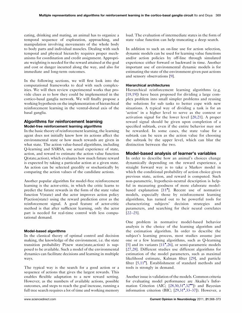
Here, we propose a working hypothesis that the ventral
striatum (VS), the dorsomedial striatum (DMS), and the
Figure 1
a’’=Do a task, Take a rest, …
a’=Turn left, Turn right, Go straight…
a =Control of each limb
Higher actionexecuted in coarse time scale
Lower actionexecuted in finer time scale
Q(a’’,s’’)
Q(a’,s’)
Q(a,s)DLS
DMS
VS
s’’=Context A, Context B, …
s’= Cue A, Cue B, …
s= Sensorimotor feedback
Current Opinion in Neurobiology
A working hypothesis that the dorsolateral (DLS), the dorsomedial
(DMS), and the ventral striatum are parallel and hierarchical Q-learning
modules that are in charge of actions at different physical and temporal
scales.
www.sciencedirect.com
dorsolateral striatum (DLS) are parallel and hierarchical
learning modules that are in charge of actions at different
physical and temporal scales (Figure 1). VS is the coarsest
module in charge of the action of the whole animal, such
as aiming for a goal, avoiding a danger, or just take a rest.
The decision is related to the overall goodness of the
choice at a coarse time scale. DMS is the middle module
in charge of abstract actions, such as turn left or go
straight. DLS is the finest module in charge of physical
actions, such as the control of each limb. The finer-level
behaviors are more distally and conditionally linked with
primary rewards because their sequentially and parallelly
coordinated executions are necessary for achieving a goal
or avoiding a danger. They also have to be memorized
longer term for possible reuse in different contexts.
These modules work in parallel [70�], which is why
bilateral lesions of one among DLS, DMS and VS, do
not impair a performance in a simple instrumental learn-
ing [60,71].
Such differences in the physical and temporal scales may
lead to the differential use of model-free and model-
based algorithms in the ventromedial to dorsolateral axis.
Model-based search algorithms are useful with coarsely
discretized action and state representations in the VS and
DMS connected with the prefrontal cortex. Model-free
algorithms are more appropriate for DLS that has to deal
with finer motor actions in shorter latency.
ConclusionWe reviewed computational issues and possible algor-
ithms for decision making and reinforcement learning
and recent findings on the neural correlates of the
variables in those algorithms. Then we proposed a
working hypothesis: the dorsolateral, the dorsomedial,
and the ventral striatum comprise a parallel and hier-
archical reinforcement learning modules that are in
charge of actions at different physical and temporal
scales. The parallelism of the decision modules has
been suggested also in the prefrontal cortex [17,32��]and the hippocampal system [55,72,73�]. Manipulation
of the different subparts of the parallel networks com-
bined with careful task design and computation model
would be necessary to further clarify their specialization
and coordination.
References and recommended readingPapers of particular interest, published within the annual period ofreview, have been highlighted as:
� of special interest�� of outstanding interest
1. Doya K: Reinforcement learning: computational theory andbiological mechanisms. HFSP J 2007, 1:30-40.
2. Doya K: Modulators of decision making. Nat Neurosci 2008,11:410-416.
3. Sutton RS, Barto AG: Reinforcement Learning. Cambridge, MA:MIT Press; 1998.
Current Opinion in Neurobiology 2011, 21:368–373

372 Behavioral and cognitive neuroscience
4. Doya K: Complementary roles of basal ganglia and cerebellumin learning and motor control. Curr Opin Neurobiol 2000,10:732-739.
5. Samejima K, Ueda Y, Doya K, Kimura M: Representation ofaction-specific reward values in the striatum. Science 2005,310:1337-1340.
6. Schultz W, Dayan P, Montague PR: A neural substrate ofprediction and reward. Science 1997, 275:1593-1599.
7. Reynolds JN, Hyland BI, Wickens JR: A cellular mechanism ofreward-related learning. Nature 2001, 413:67-70.
8. Kiani R, Shadlen MN: Representation of confidence associatedwith a decision by neurons in the parietal cortex. Science 2009,324:759-764.
9. Rao RP: Decision making under uncertainty: a neural modelbased on partially observable markov decision processes.Front Comput Neurosci 2010, 4:146.
10. Pasquereau B, Nadjar A, Arkadir D, Bezard E, Goillandeau M,Bioulac B, Gross CE, Boraud T: Shaping of motor responses byincentive values through the basal ganglia. J Neurosci 2007,27:1176-1183.
11.�
Ito M, Doya K: Validation of decision-making models andanalysis of decision variables in the rat basal ganglia. JNeurosci 2009, 29:9861-9874.
The authors showed that a modified version of Q-learning with a forget-ting term was able to predict rats’ choice behavior with the highestaccuracy among 11 algorithms. They also found that the informationabout action value was coded in the ventral striatum and the ventralpallidum but was less dominant than information about state, action, andreward.
12.��
Wunderlich K, Rangel A, O’Doherty JP: Neural computationsunderlying action-based decision making in the human brain.Proc Natl Acad Sci USA 2009, 106:17199-17204.
Using a special choice task where subjects were required to select anoption by different movements (hand or eye), neuronal activities correlat-ing with the action value were detected for the first time in imagingstudies, in the supplementary motor area, the lateral parietal cortex, theanterior cingulate cortex, and the dorsal putamen.
13. Pennartz CM, Berke JD, Graybiel AM, Ito R, Lansink CS, van derMeer M, Redish AD, Smith KS, Voorn P: Corticostriatalinteractions during learning, memory processing, anddecision making. J Neurosci 2009, 29:12831-12838.
14. Cisek P: Cortical mechanisms of action selection: theaffordance competition hypothesis. Philos Trans R Soc Lond BBiol Sci 2007, 362:1585-1599.
15.��
Matsumoto M, Hikosaka O: Two types of dopamine neurondistinctly convey positive and negative motivational signals.Nature 2009, 459:837-841.
The authors found that a large proportion of dopamine neurons located inthe dorsolateral parts of the substantia nigra and the ventral tegmentalarea were excited by not only reward-predicting stimuli but also punish-ment-predicting stimuli. The signal coded by these neurons might usedfor signaling saliency, or avoidance learning rather than value learning inthe striatum.
16. Samejima K, Doya K: Multiple representations of belief statesand action values in corticobasal ganglia loops. Ann N Y AcadSci 2007, 1104:213-228.
17. Daw ND, Niv Y, Dayan P: Uncertainty-based competitionbetween prefrontal and dorsolateral striatal systems forbehavioral control. Nat Neurosci 2005,8:1704-1711.
18. Sutton RS, Precup D, Singh S: Between MDPs and semi-MDPs:a framework for temporal abstraction in reinforcementlearning. Artif Intell 1999, 112:181-211.
19. Dietterich TG: Hierarchical reinforcement learning with theMAXQ value function decomposition. J Artif Intell Res 1999,13:227-303.
20. Dayan P, Hinton GE: Feudal reinforcement learning. InAdvances in Neural Information Processing Systems. Edited byCowan JD, Tesauro G, Alspector J. Morgan Kaufmann; 1993.
Current Opinion in Neurobiology 2011, 21:368–373
21. Morimoto J, Doya K: Acquisition of stand-up behavior by a realrobot using hierarchical reinforcement learning. Rob AutonSyst 2001, 36:37-51.
22. Daw ND, Doya K: The computational neurobiology of learningand reward. Curr Opin Neurobiol 2006, 16:199-204.
23. Corrado G, Doya K: Understanding neural coding through themodel-based analysis of decision making. J Neurosci 2007,27:8178-8180.
24. O’Doherty JP, Hampton A, Kim H: Model-based fMRI and itsapplication to reward learning and decision making. Ann N YAcad Sci 2007, 1104:35-53.
25. Doya K, Ito M, Samejima K: Model-based analysis of decisionvariables. In Decision Making, Affect, and Learning: Attention andPerformance XXIII. Edited by Delgado MR, Phelps EA, RobbinsTW. Oxford University Press; 2011.
26. Barraclough DJ, Conroy ML, Lee D: Prefrontal cortex anddecision making in a mixed-strategy game. Nat Neurosci 2004,7:404-410.
27. Sugrue LP, Corrado GS, Newsome WT: Matching behavior andthe representation of value in the parietal cortex. Science 2004,304:1782-1787.
28. Lau B, Glimcher PW: Dynamic response-by-response modelsof matching behavior in rhesus monkeys. J Exp Anal Behav2005, 84:555-579.
29. Daw ND, O’Doherty JP, Dayan P, Seymour B, Dolan RJ: Corticalsubstrates for exploratory decisions in humans. Nature 2006,441:876-879.
30. Akaike H: A new look at the statistical model identification.IEEE Trans Autom Control 1974, 19:716-723.
31.�
Kim H, Sul JH, Huh N, Lee D, Jung MW: Role of striatum inupdating values of chosen actions. J Neurosci 2009,29:14701-14712.
Authors recorded neuronal activity from the ventral and dorsal striatum ofrats during a choice task. They demonstrated that in both areas theneuronal signal of chosen action value was increased and persisted afteranimal’s choice. The signals of reward prediction error and updatedaction value were represented after the outcome was revealed.
32.��
lascher J, Daw N, Dayan P, O’Doherty JP: States versus rewards:dissociable neural prediction error signals underlying model-based and model-free reinforcement learning. Neuron 2010,66:585-595.
A hybrid model combining model-based and model-free learners wasused for the first time to explain the choice behavior in a decision task.Authors demonstrated that state prediction error, which is used in themodel-based learner, is encoded by fMRI signals in the interparietalsulcus and lateral prefrontal cortex, while reward prediction error, whichis used for model-free strategy, is encoded in the ventral striatum.
33. Schwarz G: Estimating the dimension of a model. Ann Stat1978, 6:461-464.
34. Hampton AN, Bossaerts P, O’Doherty JP: The role of theventromedial prefrontal cortex in abstract state-basedinference during decision making in humans. J Neurosci 2006,26:8360-8367.
35. Seo H, Lee D: Temporal filtering of reward signals in the dorsalanterior cingulate cortex during a mixed-strategy game. JNeurosci 2007, 27:8366-8377.
36. Seo H, Barraclough DJ, Lee D: Lateral intraparietal cortex andreinforcement learning during a mixed-strategy game. JNeurosci 2009, 29:7278-7289.
37. Rutledge RB, Lazzaro SC, Lau B, Myers CE, Gluck MA,Glimcher PW: Dopaminergic drugs modulate learning ratesand perseveration in Parkinson’s patients in a dynamicforaging task. J Neurosci 2009, 29:15104-15114.
38. Bishop CM: In Pattern recognition and machine learning. Editedby Jordan M, Kleinberg J, Scholkopf B. New York: Springer; 2006.
39. Hollerman JR, Tremblay L, Schultz W: Influence of rewardexpectation on behavior-related neuronal activity in primatestriatum. J Neurophysiol 1998, 80:947-963.
www.sciencedirect.com

Multiple representations and algorithms for reinforcement learning in the cortico-basal ganglia circuit Ito and Doya 373
40. Lau B, Glimcher PW: Action and outcome encoding in theprimate caudate nucleus. J Neurosci 2007, 27:14502-14514.
41. Hori Y, Minamimoto T, Kimura M: Neuronal encoding of rewardvalue and direction of actions in the primate putamen. JNeurophysiol 2009, 102:3530-3543.
42. Kimchi EY, Laubach M: The dorsomedial striatum reflectsresponse bias during learning. J Neurosci 2009,29:14891-14902.
43. Cohen MX, Axmacher N, Lenartz D, Elger CE, Sturm V,Schlaepfer TE: Neuroelectric signatures of reward learning anddecision-making in the human nucleus accumbens.Neuropsychopharmacology 2009, 34:1649-1658.
44. Kim YB, Huh N, Lee H, Baeg EH, Lee D, Jung MW: Encoding ofaction history in the rat ventral striatum. J Neurophysiol 2007,98:3548-3556.
45. Yamada H, Matsumoto N, Kimura M: History- and currentinstruction-based coding of forthcoming behavioraloutcomes in the striatum. J Neurophysiol 2007, 98:3557-3567.
46. Lau B, Glimcher PW: Value representations in the primatestriatum during matching behavior. Neuron 2008, 58:451-463.
47. O’Doherty J, Dayan P, Schultz J, Deichmann R, Friston K,Dolan RJ: Dissociable roles of ventral and dorsal striatum ininstrumental conditioning. Science 2004,304:452-454.
48. Roesch MR, Singh T, Brown PL, Mullins SE, Schoenbaum G:Ventral striatal neurons encode the value of the chosen actionin rats deciding between differently delayed or sized rewards.J Neurosci 2009, 29:13365-13376.
49. Pasupathy A, Miller EK: Different time courses of learning-related activity in the prefrontal cortex and striatum. Nature2005, 433:873-876.
50. Hoshi E, Tanji J: Differential roles of neuronal activity in thesupplementary and presupplementary motor areas: frominformation retrieval to motor planning and execution. JNeurophysiol 2004, 92:3482-3499.
51. Roitman JD, Shadlen MN: Response of neurons in the lateralintraparietal area during a combined visual discriminationreaction time task. J Neurosci 2002, 22:9475-9489.
52. Tanaka SC, Doya K, Okada G, Ueda K, Okamoto Y, Yamawaki S:Prediction of immediate and future rewards differentiallyrecruits cortico-basal ganglia loops. Nat Neurosci 2004,7:887-893.
53. Schonberg T, O’Doherty JP, Joel D, Inzelberg R, Segev Y,Daw ND: Selective impairment of prediction error signaling inhuman dorsolateral but not ventral striatum in Parkinson’sdisease patients: evidence from a model-based fMRI study.Neuroimage 2010, 49:772-781.
54. Oyama K, Hernadi I, Iijima T, Tsutsui K: Reward prediction errorcoding in dorsal striatal neurons. J Neurosci 2010,30:11447-11457.
55. Dickerson KC, Li J, Delgado MR: Parallel contributions ofdistinct human memory systems during probabilistic learning.Neuroimage 2010.
56.�
Nomoto K, Schultz W, Watanabe T, Sakagami M: Temporallyextended dopamine responses to perceptually demandingreward-predictive stimuli. J Neurosci 2010,30:10692-10702.
In this study, midbrain dopamine neurons were recorded during a ran-dom-dot motion detection task. For weakly coherent motions, dopamineneurons showed two-phase responses to the stimuli, the first for anystimulus and the second for a rewarding stimulus, which were consistentwith the time course required for estimation of expected reward value thatparallels the motion discrimination processing.
www.sciencedirect.com
57. Daw ND, Kakade S, Dayan P: Opponent interactions betweenserotonin and dopamine. Neural Netw 2002, 15:603-616.
58. Miyazaki K, Miyazaki KW, Doya K: Activation of dorsal rapheserotonin neurons underlies waiting for delayed rewards. JNeurosci 2011, 31:469-479.
59. Matsumoto M, Hikosaka O: Lateral habenula as a source ofnegative reward signals in dopamine neurons. Nature 2007,447:1111-1115.
60. Yin HH: The sensorimotor striatum is necessary for serial orderlearning. J Neurosci 2010, 30:14719-14723.
61. Yin HH, Knowlton BJ, Balleine BW: Lesions of dorsolateralstriatum preserve outcome expectancy but disrupt habitformation in instrumental learning. Eur J Neurosci 2004,19:181-189.
62. Yin HH, Knowlton BJ, Balleine BW: Blockade of NMDA receptorsin the dorsomedial striatum prevents action-outcome learningin instrumental conditioning. Eur J Neurosci 2005, 22:505-512.
63. Yin HH, Knowlton BJ, Balleine BW: Inactivation of dorsolateralstriatum enhances sensitivity to changes in the action-outcome contingency in instrumental conditioning. BehavBrain Res 2006, 166:189-196.
64. Yin HH, Ostlund SB, Knowlton BJ, Balleine BW: The role of thedorsomedial striatum in instrumental conditioning. Eur JNeurosci 2005, 22:513-523.
65. Shiflett MW, Martini RP, Mauna JC, Foster RL, Peet E, Thiels E:Cue-elicited reward-seeking requires extracellular signal-regulated kinase activation in the nucleus accumbens. JNeurosci 2008, 28:1434-1443.
66. Talmi D, Seymour B, Dayan P, Dolan RJ: Human pavlovian-instrumental transfer. J Neurosci 2008, 28:360-368.
67. Cardinal RN, Parkinson JA, Hall J, Everitt BJ: Emotion andmotivation: the role of the amygdala, ventral striatum, andprefrontal cortex. Neurosci Biobehav Rev 2002, 26:321-352.
68. Haruno M, Kawato M: Heterarchical reinforcement-learningmodel for integration of multiple cortico-striatal loops: fMRIexamination in stimulus-action-reward association learning.Neural Netw 2006, 19:1242-1254.
69. Voorn P, Vanderschuren LJ, Groenewegen HJ, Robbins TW,Pennartz CM: Putting a spin on the dorsal-ventral divide of thestriatum. Trends Neurosci 2004, 27:468-474.
70.�
Thorn CA, Atallah H, Howe M, Graybiel AM: Differential dynamicsof activity changes in dorsolateral and dorsomedial striatalloops during learning. Neuron 2010, 66:781-795.
The functional dissociation between the dorsomedial and the dorsolateralstriatum during learning is an important issue. Authors demonstrate thatthe dorsomedial striatum developed ensemble spike activity mainlyduring the action phase during learning of a T-maze task, while thedorsolateral striatum develops the activity that is heightened at actionboundaries of the task.
71. Cardinal RN, Cheung TH: Nucleus accumbens core lesionsretard instrumental learning and performance with delayedreinforcement in the rat. BMC Neurosci 2005, 6:9.
72. Packard MG, Knowlton BJ: Learning and memory functions ofthe Basal Ganglia. Annu Rev Neurosci 2002, 25:563-593.
73.�
van der Meer MA, Johnson A, Schmitzer-Torbert NC, Redish AD:Triple dissociation of information processing in dorsalstriatum, ventral striatum, and hippocampus on a learnedspatial decision task. Neuron 2010, 67:25-32.
Identification of multiple systems in the decision making is a crucial issue.Authors demonstrated that future paths of rats from the decision pointwere represented in the ventral striatum and the hippocampus but not inthe dorsal striatum. On the contrary, the future reward from the decisionpoint was represented only in the dorsal striatum.
Current Opinion in Neurobiology 2011, 21:368–373