Multiphoton autofluorescence and second-harmonic generation...
6
Multiphoton autofluorescence and second-harmonic generation imaging of the tooth Min-Huey Chen National Taiwan University Hospital Taipei 100, Taiwan and National Taiwan University College of Medicine School of Dentistry Taipei 100, Taiwan Wei-Liang Chen Yen Sun Peter Tramyeon Fwu Chen-Yuan Dong National Taiwan University Department of Physics Taipei 106, Taiwan Abstract. In this study, we used an epi-illuminated multiphoton mi- croscope to image three main components of ex vivo human tooth. In particular, we obtained two-photon excited autofluorescence AF and second-harmonic generation SHG images of the enamel, dentin, and periodontal ligaments PLs and constructed three-dimensional projections of sequentially and axially acquired images. We found that the enamel has a strong two-photon AF signal, clearly revealing the structures of the enamel rods. The dentin, on the other hand, has both AF and SHG signals. The contrast provided by the combination of these two imaging modalities can be used to reveal the structure of peritubular dentin and to distinguish the less mineralized circumpul- pal dentins. The SHG and multiphoton AF imaging also showed the structure of the PL and the distribution of cells around the PL, respec- tively. For comparison, we also obtained scanning electron micros- copy images of the enamel, dentin, circumpulpal dentin, and the PL. Our results demonstrate the effectiveness of using multiphoton mi- croscopy to visualize the major constituents of teeth, including enamel, dentin, and the PL, and the potential of this minimally inva- sive technique for monitoring the morphological developments during tooth regeneration. © 2007 Society of Photo-Optical Instrumentation Engineers. DOI: 10.1117/1.2812710 Keywords: multiphoton autofluorescence; second-harmonic generation SHG; microscopy; dentistry. Paper 06339R received Nov. 20, 2006; revised manuscript received Jun. 10, 2007; accepted for publication Jun. 29, 2007; published online Nov. 28, 2007. 1 Introduction The development of an effective, minimally invasive imaging modality is invaluable for investigating a variety of physi- ological phenomena and pathological conditions in dentistry. Finding an effective and minimally invasive method for im- aging the structures of teeth is invaluable in active areas of current dental research including tooth regeneration. The tra- ditional method of studying calcified sections of tooth in- volved the cutting of the tooth with a diamond saw or carbo- rundum disk. In such methods, the tooth specimens need to be ground and polished until they are sufficiently thin to be viewed with reflected or transmitted light. For years, the structure of enamel has been described and studied with these procedures. For a more detailed view of the structure of teeth, it is necessary to decalcify and stain the tooth specimens, which would likely alter the highly mineralized structures of the tooth. The limited light transmission due to the mineral- ization and tooth thickness decreased the effectiveness of us- ing confocal microscopy to study dental structures. The appli- cation of SEM has increased the understanding of the dental structures. However, the preparation for scanning electron mi- croscopy SEM examination is complicated. In recent years, multiphoton microscopy has become an important research tool in biological research. 1–5 A number of advantages render multiphoton microscopy the preferred imaging modality in many biomedical applications. To begin with, the use of near infrared excitation allows deep sample penetration. In addi- tion, unlike single photon fluorescence microscopy, the non- linear fluorescence and harmonic generation of multiphoton microscopy produce a signal only at the focal point. The pointlike excitation feature has the benefits of causing less sample photodamage and achieving sectional images without the need of a confocal pinhole. Signal collection without a confocal pinhole also improves the detection efficiency be- cause more of the sample scattered light can be detected. The characteristic autofluorescence AF from cellular constituents can provide both morphological and functional information. In addition, many noncentrosymmetric biological structures can be used to produce second-harmonic generation SHG signals. SHG is a coherent, nonlinear process that generates particularly strong signals when the molecules are orderly ar- ranged. Biological structures such as fibril collagen, muscle fibers, and microtubules are known to be effective in generat- ing second-harmonic signals. 6–9 Among tissues capable of generating second-harmonic signals, collagen is a particularly 1083-3668/2007/126/064018/6/$25.00 © 2007 SPIE Address all correspondence to M. H. Chen, National Taiwan University Hospi- tal, College of Medicine, Dept. of Dentistry, Taipei 100, Taiwan; Tel: +886–2– 2312–3456; Fax: +886–2–2383–1346; E-mail: [email protected], or C. Y. Dong, National Taiwan University, Dept. of Physics, Taipei 106, Taiwan; Tel: +8862–3366–5155; Fax: +8862–2363–9984; E-mail: [email protected] Journal of Biomedical Optics 126, 064018 November/December 2007 Journal of Biomedical Optics November/December 2007 Vol. 126 064018-1
Transcript of Multiphoton autofluorescence and second-harmonic generation...
Mg
MNT
NCST
WYPCNDT
1
TmoFacdvrgvspiwtiics
At2Y+
Journal of Biomedical Optics 12�6�, 064018 �November/December 2007�
J
ultiphoton autofluorescence and second-harmoniceneration imaging of the tooth
in-Huey Chenational Taiwan University Hospital
aipei 100, Taiwanand
ational Taiwan Universityollege of Medicinechool of Dentistryaipei 100, Taiwan
ei-Liang Chenen Suneter Tramyeon Fwuhen-Yuan Dongational Taiwan Universityepartment of Physics
aipei 106, Taiwan
Abstract. In this study, we used an epi-illuminated multiphoton mi-croscope to image three main components of ex vivo human tooth. Inparticular, we obtained two-photon excited autofluorescence �AF�and second-harmonic generation �SHG� images of the enamel, dentin,and periodontal ligaments �PLs� and constructed three-dimensionalprojections of sequentially and axially acquired images. We foundthat the enamel has a strong two-photon AF signal, clearly revealingthe structures of the enamel rods. The dentin, on the other hand, hasboth AF and SHG signals. The contrast provided by the combinationof these two imaging modalities can be used to reveal the structure ofperitubular dentin and to distinguish the less mineralized circumpul-pal dentins. The SHG and multiphoton AF imaging also showed thestructure of the PL and the distribution of cells around the PL, respec-tively. For comparison, we also obtained scanning electron micros-copy images of the enamel, dentin, circumpulpal dentin, and the PL.Our results demonstrate the effectiveness of using multiphoton mi-croscopy to visualize the major constituents of teeth, includingenamel, dentin, and the PL, and the potential of this minimally inva-sive technique for monitoring the morphological developments duringtooth regeneration. © 2007 Society of Photo-Optical Instrumentation Engineers.
�DOI: 10.1117/1.2812710�
Keywords: multiphoton autofluorescence; second-harmonic generation �SHG�;microscopy; dentistry.Paper 06339R received Nov. 20, 2006; revised manuscript received Jun. 10, 2007;accepted for publication Jun. 29, 2007; published online Nov. 28, 2007.
Introduction
he development of an effective, minimally invasive imagingodality is invaluable for investigating a variety of physi-
logical phenomena and pathological conditions in dentistry.inding an effective and minimally invasive method for im-ging the structures of teeth is invaluable in active areas ofurrent dental research including tooth regeneration. The tra-itional method of studying calcified sections of tooth in-olved the cutting of the tooth with a diamond saw or carbo-undum disk. In such methods, the tooth specimens need to beround and polished until they are sufficiently thin to beiewed with reflected or transmitted light. For years, thetructure of enamel has been described and studied with theserocedures. For a more detailed view of the structure of teeth,t is necessary to decalcify and stain the tooth specimens,hich would likely alter the highly mineralized structures of
he tooth. The limited light transmission due to the mineral-zation and tooth thickness decreased the effectiveness of us-ng confocal microscopy to study dental structures. The appli-ation of SEM has increased the understanding of the dentaltructures. However, the preparation for scanning electron mi-
ddress all correspondence to M. H. Chen, National Taiwan University Hospi-al, College of Medicine, Dept. of Dentistry, Taipei 100, Taiwan; Tel: +886–2–312–3456; Fax: +886–2–2383–1346; E-mail: [email protected], or C.. Dong, National Taiwan University, Dept. of Physics, Taipei 106, Taiwan; Tel:
8862–3366–5155; Fax: +8862–2363–9984; E-mail: [email protected]ournal of Biomedical Optics 064018-
croscopy �SEM� examination is complicated. In recent years,multiphoton microscopy has become an important researchtool in biological research.1–5 A number of advantages rendermultiphoton microscopy the preferred imaging modality inmany biomedical applications. To begin with, the use of nearinfrared excitation allows deep sample penetration. In addi-tion, unlike single photon fluorescence microscopy, the non-linear fluorescence and harmonic generation of multiphotonmicroscopy produce a signal only at the focal point. Thepointlike excitation feature has the benefits of causing lesssample photodamage and achieving sectional images withoutthe need of a confocal pinhole. Signal collection without aconfocal pinhole also improves the detection efficiency be-cause more of the sample scattered light can be detected. Thecharacteristic autofluorescence �AF� from cellular constituentscan provide both morphological and functional information.In addition, many noncentrosymmetric biological structurescan be used to produce second-harmonic generation �SHG�signals. SHG is a coherent, nonlinear process that generatesparticularly strong signals when the molecules are orderly ar-ranged. Biological structures such as fibril collagen, musclefibers, and microtubules are known to be effective in generat-ing second-harmonic signals.6–9 Among tissues capable ofgenerating second-harmonic signals, collagen is a particularly
1083-3668/2007/12�6�/064018/6/$25.00 © 2007 SPIE
November/December 2007 � Vol. 12�6�1
iv
tnatttptarfdo
fdnfitbc
pcbecsdcg
2T3
Fpa
Chen et al.: Multiphoton autofluorescence and second-harmonic generation imaging…
J
nteresting subject of study due to its wide distribution inarious tissues.
To apply multiphoton microscopy in research areas such asooth development �odontogenesis� and dental tissue engi-eering, it is important to test the ability of multiphoton im-ging in resolving the various structures of a fully grownooth. The main components of teeth include the enamel, den-in, pulp, and the periodontal ligaments �PLs�. The enamel ishe most visible part of a tooth. It is highly mineralized torovide the strength to withstand the force of mastication ando protect the dentin. The dentin consists of mostly collagennd forms a channel structure called a dentinal tubule thatadiates from the pulp to the enamel and cementum. Dentin isormed by the odontoblasts located in the dental pulp close toentin. This process of dentinogenesis occurs throughoutne’s life span.
Obtaining the structural information of teeth is importantor the study of dental development and regeneration. Theental pulp is at the center of teeth where blood vessels,erves, and odontoblasts are located. The PLs are collagenbers that hold a tooth in place by connecting the cementum
o the jaw bone. PLs are produced and destroyed by the fibro-lasts that are located throughout the ligaments and can beategorized according to their orientation.10
Although multiphoton imaging has been previously ap-lied to the study of dental structures including dentalarries,11–14 our work uses multiphoton microscopy to revealoth the two- and three-dimensional microscopic structures ofnamel and dentin as well as that of the PL without the de-alcification treatment of the tooth. We also compare our re-ults with the structures acquired with SEM in an attempt toemonstrate that the images obtained from multiphoton mi-roscopy serve as effective structural models for the investi-ation of teeth.
Materials and Methodshe tooth sample used in this study was obtained from a
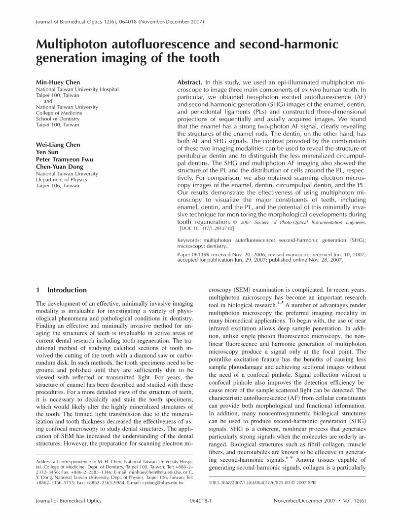
ig. 1 Photo of the longitudinal section of the permanent secondaryremolar used in this study. Structures of the enamel, dentin, pulp, PL,nd alveolar bone attached to the root surface are indicated.
0-year old adult patient who had the upper left secondary
ournal of Biomedical Optics 064018-
Fig. 2 �A� The AF �green� image of enamel. The arrow points to one ofthe enamel rods. �B� Three-dimensional projection of the enamel�depth of 24 �m�, constructed by sequential axial images acquired1 �m apart. �C� Combined AF and SHG image of the enamel withadjacent dentin. The darker area along the dentin-enamel junctionindicates reduced mineralization along that region. �D� SEM image ofthe tooth’s enamel.
November/December 2007 � Vol. 12�6�2

Fmbtriap3tdentinal tubules.
Chen et al.: Multiphoton autofluorescence and second-harmonic generation imaging…
Journal of Biomedical Optics 064018-
premolar extracted for orthodontic treatment. With the pa-tient’s consent and the approval of the Review Committee ofCollege of Medicine at National Taiwan University, we pro-ceeded to prepare the tooth for imaging. First, we cut thetooth along the longitudinal direction with an IsoMet saw�Buehler Ltd., Lake Bluff, Illinois�. The tooth specimen isthen stored in normal saline solution prior to multiphoton im-aging acquisition. In addition to obtaining the multiphotonimaging of the tooth, we also obtained SEM images of thetooth for comparison.
The multiphoton AF and SHG imaging were acquired on ahome-built epi-illuminated laser scanning system similar toone described previously by Sun et al.3 The system consists ofa modified upright microscope �E800, Nikon, Tokyo, Japan�with excitation provided by an 80-MHz femtosecond Ti-sapphire laser system �Millenia X, Tsunami, Spectra Physics,Mountain View, California� tuned to 780 nm. The raster scan-ning of the laser excitation is performed by a pair of scanningmirror systems �Model 6220, Cambridge Technology, Cam-bridge, Massachusetts�. The scanning laser beam is then ex-panded to overfill the back aperture of a water-immersionobjective �S FLUOR 40�, numeric aperture 0.8, Nikon�. Ashort pass dichroic mirror �700DCSPXRUV-3p, Chroma
Fig. 4 �A� Combined multiphoton AF �green� and SHG �blue� imagesof the dentin-pulp junction. The red arrow indicates newly producedcircumpulpal dentin, which is unmineralized and produces only theSHG signal. Further away from the pulp, the dentin is mineralized andemits AF in addition to SHG. �B� Three-dimensional reconstruction ofthe area shown in �A� using a sequence of 30 images taken 1-�mapart. �C� SEM image of the dentin near the pulp demonstrating thecurcumpulpal dentin with less regular arrangement of dentinaltubules.
ig. 3 �A� Multiphoton dentin images showing the combination ofultiphoton AF and SHG. The mineralized dentinal tubules, indicatedy the yellow arrow, emit AF �green� and SHG �blue� signals, whilehe collagen fibers of the dentinal structure, indicated by the red ar-ow, produce mainly a SHG signal. �B� Cross-sectional view of anmage stack over the red dashed line area in �A�. Yellow and redrrows indicate, respectively, the mineralized and unmineralizedarts of dentinal tubules. �C� Three-dimensional projection �depth of5 �m� of the middle dentinal area constructed by sequential images
aken 1-�m apart. �D� SEM image of the dentin showing the aligned
Technology, Rockingham, Vermont� was used to separate theNovember/December 2007 � Vol. 12�6�3
ea��opJtScaabid
3FttbewptsTa
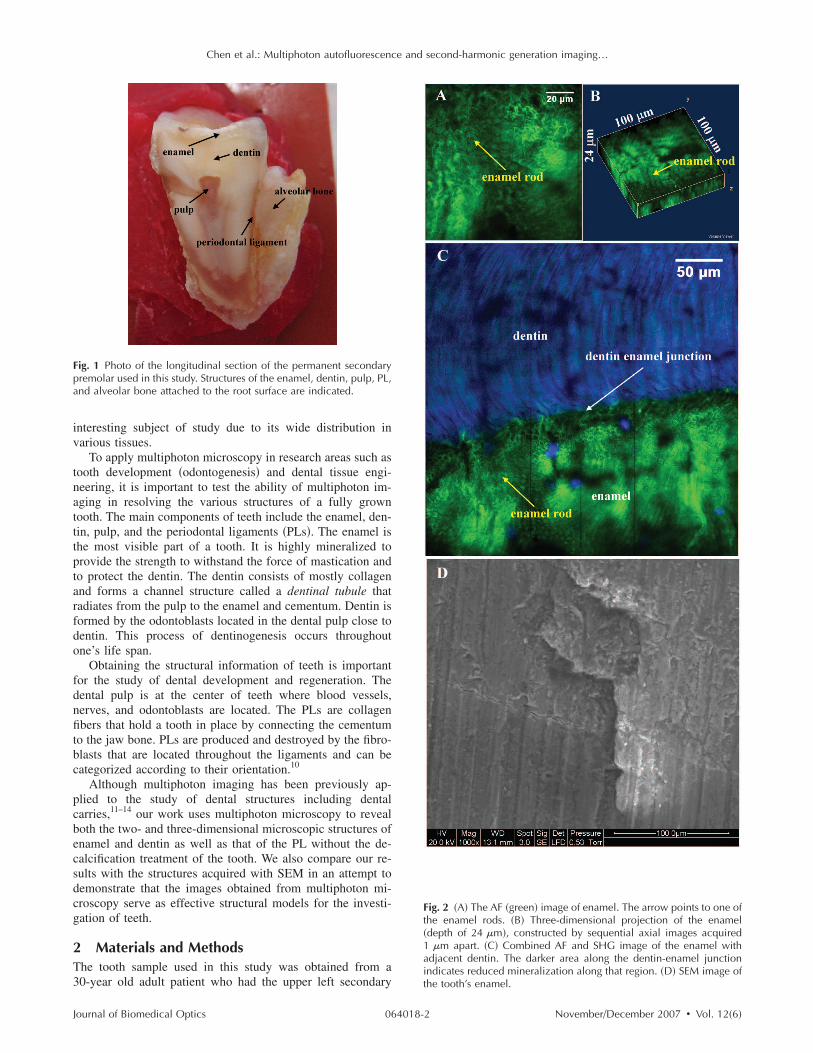
Fs llow arrow points to what is likely to be a fibroblast cell.
Chen et al.: Multiphoton autofluorescence and second-harmonic generation imaging…
J
xcitation and emission. The epi-illuminated multiphoton AFnd SHG signals are separated by a long pass dichroic mirror435DCXR, Chroma Technology� and two bandpass filtersE435LP-E700SP, HQ390/20-2p, Chroma Technology�. Eachf the two signals is detected with a single-photon countinghotomultiplier tube �R7400P, Hamamatsu, Hamamatsu City,apan�, processed by a discriminator, and sent to the computero form images. The motorized sample stage �H101, Priorcientific Instruments, Cambridge, United Kingdom� is alsoontrolled by the computer for automated sequential scanninglong the x, y, and z axes. The sequentially acquired imageslong the x and y directions in the image plane can be com-ined to produce large area images, and sequentially acquiredmages along the axial z axis can be used to construct three-imensional projections of the specimen.
Resultsigure 1 shows the longitudinal section of the tooth used in
his study. The various components of the tooth are labeled inhe figure. In the multiphoton image of the enamel �Fig. 2�,oth the AF and SHG signals were used to characterize thenamel and related structures. In this and subsequent images,e used the green pseudocolor to denote AF and the blueseudocolor to represent SHG signal. Figure 2�A� shows awo-dimensional AF image of the enamel, and Fig. 2�B�hows a three-dimensional projection to a depth of 24 �m.he three-dimensional projection is composed of sequential
ig. 5 �A� Large area multiphoton image around the PL. �B� and �C�hown in �A�. �D� Enlarged view of the selected region in �A�. The ye
are the respective AF and SHG images of the selected area of the PL image
xial images acquired 1 �m apart. These images reveal that
ournal of Biomedical Optics 064018-
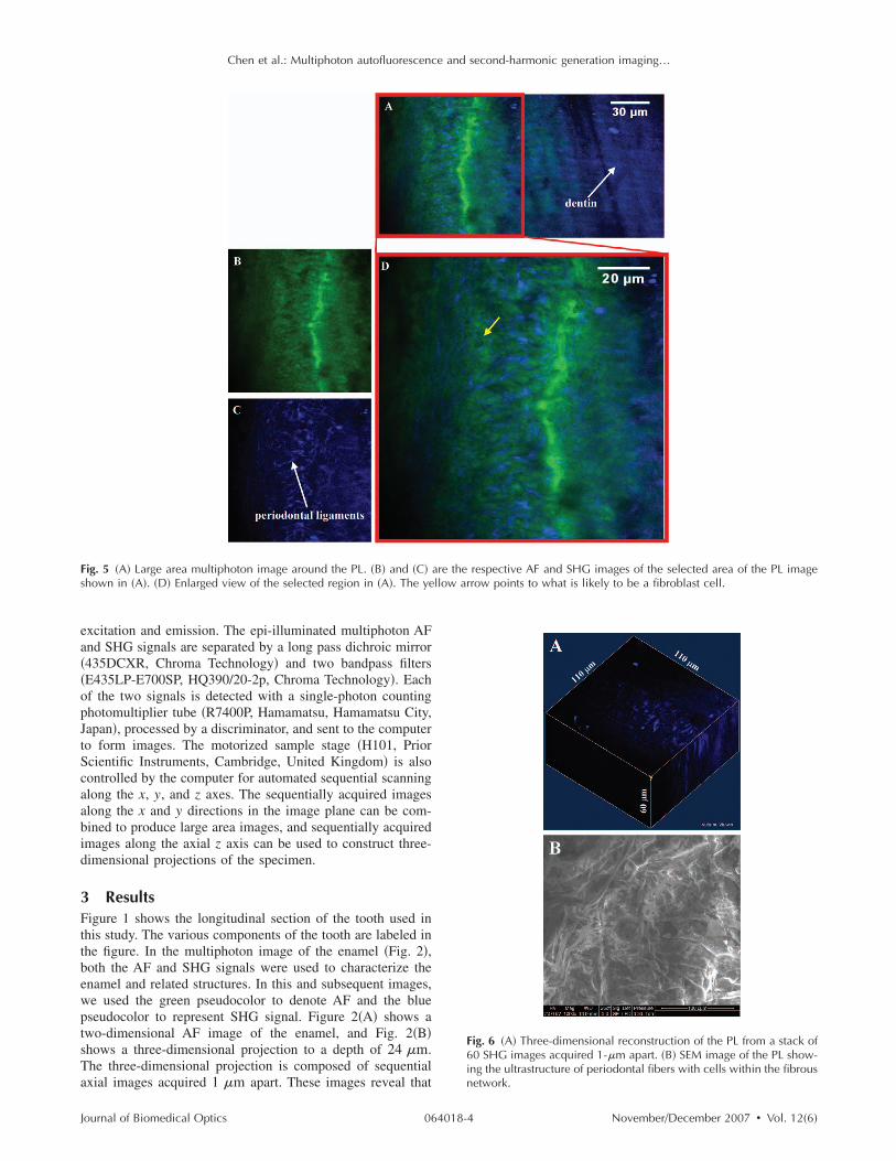
Fig. 6 �A� Three-dimensional reconstruction of the PL from a stack of60 SHG images acquired 1-�m apart. �B� SEM image of the PL show-ing the ultrastructure of periodontal fibers with cells within the fibrous
network.November/December 2007 � Vol. 12�6�4

tl
tcleeictbmsedFTstq3paAalvcat
daanAdFcptsftTd
tlttotilctaSt
Chen et al.: Multiphoton autofluorescence and second-harmonic generation imaging…
J
wo-photon AF imaging is effective in revealing the morpho-ogical features of the mineralized enamel rods.
The mineralized enamel rods are formed during the secre-ory stage by the ameloblasts in the Tomes’s process as theells migrate away from dentin during amelogenesis. The yel-ow arrows in Figs. 2�A� to 2�C� point to examples of thenamel rods, which also show the parallel nature of thenamel rod aggregates. The dentin-enamel junction is shownn Fig. 2�C�, where a darker area approximately 20-�m wideorresponds to reduced mineralization along the junction. Al-hough enamel produces mainly strong AF, dentin generatesoth AF and SHG signals. When compared with the structuralotif observed under SEM �Fig. 2�D��, the morphological
imilarity of the images acquired using the two techniques arevident. The combined two-photon AF and SHG imaging ofentin structure near the enamel-dentin junction is shown inig. 3�A�, where the structure of the dentinal tubule is visible.he dentinal tubule structure can be better visualized in theectional image �Fig. 3�B�� and the three-dimensional projec-ion �Fig. 3�C�� constructed from a stack of sequentially ac-uired axial images acquired 1-�m apart �total thickness of5 �m�. The collagen composition of the dentinal tubulesroduces a strong SHG signal �red arrows�, while the miner-lized part of the dentinal tubules produce both the SHG andF signals �yellow arrows�. Because the mineralized dentin
ppears to surround each dentinal tubule, it is called peritubu-ar dentin.10 By combining the AF and SHG imaging, we canisualize the distribution of the peritubular dentin. As in thease of enamel imaging, comparison shows that multiphotonnd SEM imaging �Fig. 3�D�� reveal similar structural archi-ectures of dentin.
We also applied multiphoton microscopy in imaging theentin region around the pulp area �shown in Fig. 4�A��, andthree-dimensional projection of a 30-�m thick image stack
cquired at the same position is shown in Fig. 4�B�. Theewly produced predentins are located just next to the pulp.s shown in Figs. 4�A� and 4�B�, these unmineralized pre-entins �red arrows� produce only the SHG signal with no AF.urther away from the pulp, the dentins are mineralized andan produce AF in addition to the SHG signal. The circum-ulpal dentins are incrementally deposited dentin that occurshroughout life. The presence of predentin in our tooth samplehows that the deposition of dentin was an ongoing processor the 30-year-old patient. Continual deposition of dentin ishe probable cause for the smaller pulp area shown in Fig. 1.he comparative SEM image of the incrementally depositedentin structure is shown in Fig. 4�C�.
In addition to imaging the enamel and dentin, we also ob-ained multiphoton images of the PL. Figure 5�A� shows aarge area image covering both the dentin and PL. The AF andhe SHG of the selected �red square� region is shown respec-ively in Figs. 5�B� and 5�C�. Figure 5�D� is the enlarged viewf the selected region of Fig. 5�A�. From Fig. 5�D�, we seehat the fiber structures of the PL can be visualized by SHGmaging. In addition, we also found AF structures within theigament �yellow arrow� that, most likely, are the fibroblastells in the ligament. Finally, we acquired and displayed ahree-dimensional projection of the PL from a sequence ofxially acquired SHG images in Fig. 6�A�. Compared to theEM image �Fig. 6�B�� of the PL, one can see that multipho-
on microscopy provides clear three-dimensional structural in-
ournal of Biomedical Optics 064018-
formation of the fiber organization information within thePLs.
In conclusion, we applied and found that multiphoton mi-croscopy can be used to visualize the morphological featuresof the enamel, dentin, and PL, the three main components ofa tooth. We demonstrated that the structure of enamel rods,the mineralized structure of dentinal tubules, and most likely,the cells of the PL can be imaged using two-photon AF sig-nals; whereas the unmineralized structure of dentin and thefiber structure of the PLs can be imaged with the SHG signal.In addition to the ability to obtain morphological informationwith reduced photodamage and deep sample penetration, mul-tiphoton microscopy can also be combined with polarizationresolved methods to obtain structural information below opti-cal resolution. In particular, polarization SHG microscopy hasbeen used to determine the pitch angle of the myosin coil inmuscle fiber.15 Such technique can be applied to determinesubmicron structural information for collagen and other tis-sues that produce SHG signals. This feature and the ability ofmultiphoton microscopy to visualize the major components ofteeth make it a powerful tool for the investigation of dentalphysiological processes such as tooth regeneration and thediagnosis of tooth pathological conditions.
AcknowledgmentsThe authors thank the Core Facility from NTUH Medical Re-search Center, and the Core Facility of the Institute of Cellularand Organismic Biology, Academia Sinica, for assistance inenvironmental SEM and the Optical Molecular Imaging Mi-croscopy Core Facility �A5� of the National Research Pro-gram for Genomic Medicine, National Science Council, Tai-wan for multiphoton image acquisition. We also like to thankNational Taiwan University Hospital �NTUH 95-525� and theNational Science Council of Taiwan �Grant No. NSC 94-3112-B-002-015-Y� for financial support.
References1. W. Denk, J. H. Strickler, and W. W. Webb, “2-Photon laser scanning
fluorescence microscopy,” Science 248�4951�, 73–76 �1990�.2. P. T. C. So, C. Y. Dong, B. R. Masters, and K. M. Berland, “Two-
photon excitation fluorescence microscopy,” Annu. Rev. Biomed.Eng. 2, 399–429 �2000�.
3. Y. Sun, J. W. Su, W. Lo, S. J. Lin, S. H. Jee, and C. Y. Dong,“Multiphoton polarization imaging of the stratum corneum and thedermis in ex-vivo human skin,” Opt. Express 11�25�, 3377–3384�2003�.
4. S. J. Lin, R. J. Wu, H. Y. Tan, W. Lo, W. C. Lin, T. H. Young, C. J.Hsu, J. S. Chen, S. H. Jee, and C. Y. Dong, “Evaluating cutaneousphotoaging by use of multiphoton fluorescence and second-harmonicgeneration microscopy,” Opt. Lett. 30�17�, 2275–2277 �2005�.
5. H. S. Lee, Y. Liu, H. C. Chen, L. L. Chiou, G. T. Huang, W. Lo, andC. Y. Dong, “Optical biopsy of liver fibrosis by use of multiphotonmicroscopy,” Opt. Lett. 29�22�, 2614–2616 �2004�.
6. W. R. Zipfel, R. M. Williams, R. Christie, A. Y. Nikitin, B. T. Hy-man, and W. W. Webb, “Live tissue intrinsic emission microscopyusing multiphoton-excited native fluorescence and second harmonicgeneration,” Proc. Natl. Acad. Sci. U.S.A. 100�12�, 7075–7080�2003�.
7. A. Zoumi, A. Yeh, and B. J. Tromberg, “Imaging cells and extracel-lular matrix in vivo by using second-harmonic generation and two-photon excited fluorescence,” Proc. Natl. Acad. Sci. U.S.A. 99�17�,11014–11019 �2002�.
8. P. J. Campagnola and L. M. Loew, “Second-harmonic imaging mi-croscopy for visualizing biomolecular arrays in cells, tissues and or-ganisms,” Nat. Biotechnol. 21�11�, 1356–1360 �2003�.
9. E. Brown, T. McKee, E. di Tomaso, A. Pluen, B. Seed, Y. Boucher,
November/December 2007 � Vol. 12�6�5

1
1
1
Chen et al.: Multiphoton autofluorescence and second-harmonic generation imaging…
J
and R. K. Jain, “Dynamic imaging of collagen and its modulation intumors in vivo using second-harmonic generation,” Nat. Med. 9�6�,796–800 �2003�.
0. J. K. Avery, P. F. Steele, and N. Avery, Oral Development and His-tology, Thieme Medical Publishers Inc., New York �2001�.
1. J. M. Girkin, A. F. Hall, and S. L. Creanor, “Multi-photon imaging ofintact dental tissue,” in, Proceedings of the 4th Annual Indiana Con-ference, G. K. Stookey, Ed., Indiana University School of Dentistry,Indianapolis, pp. 155–168 �1999�.
2. A. Hall and J. M. Girkin, “A review of potential new diagnosticmodalities for Caries lesions,” IADR, pp. 89–94 �2004�.
ournal of Biomedical Optics 064018-
13. J. C. Chen, T. C. Yang, and F. J. Kao, “Laser scanning microscopy ofenamel and dentin sections with two-photon fluorescence and secondharmonic generation,” Presented at CLEO/Pacific Rim 2003. The 5thPacific Rim Conference on Lasers and Electro-Optics, 2003 �2003�.
14. N. P. Piesco, “Histology of dentin,” in Oral Development and Histol-ogy, pp. 242–261, Thieme Medical Publishers, New York �1994�.
15. S. V. Plotnikov, A. C. Millard, P. J. Campagnola, and W. A. Mohler,“Characterization of the myosin-based source for second-harmonicgeneration from muscle sarcomeres,” Biophys. J. 90�2�, 693–703�2006�.
November/December 2007 � Vol. 12�6�6