Multidetector CT in Renal Tuberculosis - Springer CT in Renal Tuberculosis ... This article gives...
11
ABDOMINAL CT IMAGING (A JOSHI, SECTION EDITOR) Multidetector CT in Renal Tuberculosis Ashwini Sankhe • Anagha R. Joshi Published online: 12 September 2014 Ó Springer Science+Business Media New York 2014 Abstract Tuberculosis (TB) is one of the commonest infectious diseases in the developing countries with rising incidence seen due to HIV and AIDS. Pulmonary tuber- culosis is the commonest form seen. Renal Tuberculosis is the most common extrapulmonary form. There are no specific clinical symptoms associated with renal tubercu- losis. The constitutional symptoms like low grade fever, weight loss, anorexia commonly seen with pulmonary TB are not usually seen with renal TB. Renal TB is also cause of interstitial nephritis which leads to end-stage renal failure. Hence the role of radiologist assumes importance in supporting the timely diagnosis although cultures or his- tologic analysis is required for definitive diagnosis. The individual imaging finding may not be diagnostic as it can be seen with other renal pathologies too, however, presence of multiple imaging findings in a single case is pointer to this disease. Renal TB involves both cortex and collecting system. This article gives review of pathophysiology and computed tomography features of Renal TB. Keywords Extrapulmonary tuberculosis Á CT urography Á Renal abscess Á Papillary necrosis Á Caliectasis Á Putty kidney Introduction Renal tuberculosis is a subset of genitourinary tuberculosis accounting for 20 % of extrapulmonary tuberculosis. Genitourinary tuberculosis is the most common form of extrapulmonary tuberculosis [1]. Globally, tuberculosis is a common disease, with 8–10 million new cases annually and a rising incidence, particularly in regions with a high incidence of HIV infection [2]. Approximately 95 % of cases occur in developing countries. In India, more than 1,000 lives are lost every day due to TB despite the availability of modern diagnostic aids and treatment [3]. Tuberculosis can involve both the renal parenchyma and the collecting system (calyces, renal pelvis, ureter, bladder, and urethra) and results in different clinical presentations and radiographic appearances. This article focuses on the role of computed tomography in evaluation of renal parenchyma, calyces, and renal pelvis. The appearance of renal TB-CT is shown in Flowchart 1. Pathogenesis Renal and urinary tract tuberculosis is generally caused by the human tubercle bacillus, M. tuberculosis, but the bovine tubercle bacillus, M. bovis, can be responsible occasionally. The intravesical instillation of Bacille Cal- mette-Gue ´rin vaccine done for the treatment of superficial bladder cancer can also result in renal lesions as a com- plication [2]. Renal Tuberculosis occurs through hematogenous spread from a primary focus like lung and is usually a bilateral disease [4]. The pathogenesis of this disease begins with the initial localization of the tubercle bacilli in the cortex adjacent to the glomeruli causing mechanical stress which leads to alteration in the cell morphology, This article is part of Topical Collection on Abdominal CT Imaging. A. Sankhe (&) Á A. R. Joshi Lokmanya Tilak Municipal Medical College and General Hosp. Sion, Mumbai 400022, Maharashtra, India e-mail: [email protected] A. R. Joshi e-mail: [email protected] 123 Curr Radiol Rep (2014) 2:69 DOI 10.1007/s40134-014-0069-5
Transcript of Multidetector CT in Renal Tuberculosis - Springer CT in Renal Tuberculosis ... This article gives...
ABDOMINAL CT IMAGING (A JOSHI, SECTION EDITOR)
Multidetector CT in Renal Tuberculosis
Ashwini Sankhe • Anagha R. Joshi
Published online: 12 September 2014
� Springer Science+Business Media New York 2014
Abstract Tuberculosis (TB) is one of the commonest
infectious diseases in the developing countries with rising
incidence seen due to HIV and AIDS. Pulmonary tuber-
culosis is the commonest form seen. Renal Tuberculosis is
the most common extrapulmonary form. There are no
specific clinical symptoms associated with renal tubercu-
losis. The constitutional symptoms like low grade fever,
weight loss, anorexia commonly seen with pulmonary TB
are not usually seen with renal TB. Renal TB is also cause
of interstitial nephritis which leads to end-stage renal
failure. Hence the role of radiologist assumes importance in
supporting the timely diagnosis although cultures or his-
tologic analysis is required for definitive diagnosis. The
individual imaging finding may not be diagnostic as it can
be seen with other renal pathologies too, however, presence
of multiple imaging findings in a single case is pointer to
this disease. Renal TB involves both cortex and collecting
system. This article gives review of pathophysiology and
computed tomography features of Renal TB.
Keywords Extrapulmonary tuberculosis � CT urography �Renal abscess � Papillary necrosis � Caliectasis � Putty
kidney
Introduction
Renal tuberculosis is a subset of genitourinary tuberculosis
accounting for 20 % of extrapulmonary tuberculosis.
Genitourinary tuberculosis is the most common form of
extrapulmonary tuberculosis [1]. Globally, tuberculosis is a
common disease, with 8–10 million new cases annually
and a rising incidence, particularly in regions with a high
incidence of HIV infection [2]. Approximately 95 % of
cases occur in developing countries. In India, more than
1,000 lives are lost every day due to TB despite the
availability of modern diagnostic aids and treatment [3].
Tuberculosis can involve both the renal parenchyma and
the collecting system (calyces, renal pelvis, ureter, bladder,
and urethra) and results in different clinical presentations
and radiographic appearances. This article focuses on the
role of computed tomography in evaluation of renal
parenchyma, calyces, and renal pelvis. The appearance of
renal TB-CT is shown in Flowchart 1.
Pathogenesis
Renal and urinary tract tuberculosis is generally caused by
the human tubercle bacillus, M. tuberculosis, but the
bovine tubercle bacillus, M. bovis, can be responsible
occasionally. The intravesical instillation of Bacille Cal-
mette-Guerin vaccine done for the treatment of superficial
bladder cancer can also result in renal lesions as a com-
plication [2].
Renal Tuberculosis occurs through hematogenous
spread from a primary focus like lung and is usually a
bilateral disease [4]. The pathogenesis of this disease
begins with the initial localization of the tubercle bacilli in
the cortex adjacent to the glomeruli causing mechanical
stress which leads to alteration in the cell morphology,
This article is part of Topical Collection on Abdominal CT Imaging.
A. Sankhe (&) � A. R. Joshi
Lokmanya Tilak Municipal Medical College and General Hosp.
Sion, Mumbai 400022, Maharashtra, India
e-mail: [email protected]
A. R. Joshi
e-mail: [email protected]
123
Curr Radiol Rep (2014) 2:69
DOI 10.1007/s40134-014-0069-5
increased rate of protein synthesis and proliferation of
resident glomerular cell [4]. The cortical granulomas may
remain dormant, asymptomatic, and stable as sequelae of a
primary pulmonary infection from as long ago as
10–15 years (Khan et al. 2004). Renal tuberculosis may
occur as a result of reactivation after this period of dor-
mancy, even when there is no evidence of active pul-
monary tuberculosis, or it may be due to reinfection from
active tuberculosis (Ahmed and Murty 2003) [5]. If the
infection is not controlled in this phase, the organism then
gains access to the tubular fluid by the sloughing of small
glomerular caseating necrotic lesions. While within
tubules, further dissemination of the bacilli to the renal
medulla and pyramidal tissues can take place. This leads to
papillitis that extends into the proximal loop of Henle and
eventually papillary necrosis occurs. Thereafter, the degree
of progression varies from self-limiting process to
advanced destructive lesions of the kidneys. The infection
may remain localized in the renal parenchyma and/or gain
access to the calyceal system causing pyelocalyceal
destruction and subsequently the ureters and urinary blad-
der become involved. This may progress to hydronephrosis
and pyonephrosis secondary to strictures and obstruction
[4]. As in the primary site, granuloma formation, caseous
necrosis, and cavitation within the renal parenchyma are
the hallmark stages of progressive infection [5].
The host’s response induces fibrotic parenchyma which
may lead to renal pelvic traction, calcium deposition with
stone formation within the urinary collection system, and
stricture formation contributing to obstruction and pro-
gressive renal dysfunction. The end-stage result of diffuse
disease is destruction, loss of function, fibrosis, and calci-
fication in a lobular distribution of the entire kidney pro-
ducing autonephrectomy [18]. If granulomas spread to the
collection system, regional spread by either ascension or
descension of the bacilli to the renal pelvis, ureters, urinary
bladder, and accessory genital organs is possible [4, 5].
Clinical Features
There are no early symptoms in renal TB. Presenting
symptoms can be hematuria, nocturia, dysuria, pyuria,
increased frequency, back pain, suprapubic pain. These
symptoms are also seen with bacterial cystitis and in most
cases renal TB is suspected only after usual antibacterial
treatment is ineffective [2]. Constitutional symptoms like
malaise, anorexia, low grade fever, weight loss commonly
seen with pulmonary TB are generally not seen with renal
TB.
Laboratory Diagnosis
Tuberculosis can be diagnosed histopathologically by
biopsy material on routine solid LJ media, microbiologi-
cally by isolation of mycobacterium from urine or by
radiometry. Urine samples of first void are preferred over
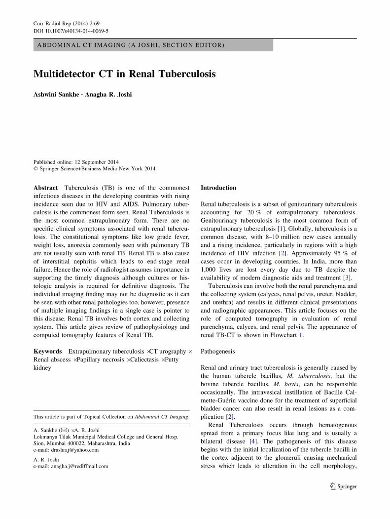
Flowchart 1 Renal TB-CT appearance
69 Page 2 of 11 Curr Radiol Rep (2014) 2:69
123
24-h urine collections as mycobacterial viability decreases
with prolonged exposure to acid urine [5, 6].
Newer rapid detection techniques based on nucleic acid
amplification technique like polymerase chain reaction
(PCR) are being used recently for diagnosing mycobac-
terium tuberculosis and other environmental mycobacte-
ria. PCR is especially efficient in detecting myco-
bacterium from sputum sample. PCR technique is also
specific and sensitive for diagnosing genitourinary TB [2,
7]. PCR is also used in urine samples of disseminated
tuberculosis related to HIV and identifies mycobacterial
DNA where traditional methods are not specific and
sensitive [8]. GeneXpert MTB/RIF is relatively new
technique used for detection of mycobacterium by iden-
tifying DNA sequence specific for TB and rifampicin
resistance by PCR.
Computed Tomography
Diagnosis of renal tuberculosis may be delayed or missed
as the disease may not cause symptoms in early stages or
maybe clinically misdiagnosed for other renal diseases
causing similar symptoms. Hence the radiological investi-
gations play an important role in reaching the diagnosis.
Earlier Intravenous pyelography assumed an important role
in diagnosing the extent of disease however the current
generation multidetector CT scanner with short scanning
time and a minimum slice thickness, achieve high diag-
nostic accuracy with good resolution, excellent sagittal and
coronal reformation, and minimum artifacts.
CT is helpful in determining the extrarenal spread of
tuberculous disease as well as in assessing its severity in
terms of loss of renal function [9, 10•]. It is the most
sensitive modality for identifying renal calcifications which
occur in almost 50 % cases of renal TB [9]. CT is also the
best modality for demonstrating the extent, nature, and
distribution of calcification within the abnormal kidney
[10•]. CT, in general, shows more details of pathologic
anatomy due to the availability of axial images for review
and is superior to retrograde pyelography (RGP), IVU, and
USG in detecting multiple small urothelial lesions [10•].
CT urography is generally performed for evaluation of
renal pathologies.
We perform CT Urography with 75 ml of 300 mg % of
non-ionic contrast medium followed by saline injection.
Generally four phases are taken which includes unen-
hanced scan followed by corticomedullary phase, nephro-
graphic, and pyelographic i.e., excretory phase.
Unenhanced CT is performed from upper pole of kidneys
upto the symphysis pubis. It is used to evaluate calcifica-
tion, fat, and baseline-unenhanced Hounsfield values of
masses so that comparable enhancement of lesions can be
done in contrast-enhanced scans. Corticomedullary phase
is not employed by some groups as arterial phase infor-
mation is generally not required [11•].
CT features of renal TB are varied and depend upon the
stage of the disease. They result from a combination of
papillary necrosis and parenchymal destruction. Typically,
the papillae are involved first and this is followed by cor-
tical damage. Communication with the collecting system
results in thickening, ulceration, and fibrosis, often with
stricture formation [10•, 12, 13] and consequent obstruc-
tion. The most valuable feature of renal TB is the multi-
plicity of abnormal findings [5, 12–14]. A lobar pattern of
caseation, arising from the assimilation of the calyces into
the caseous parenchyma of each destroyed lobe, is virtually
diagnostic of renal TB.
Imaging characteristics can be divided into renal
parenchymal changes and pelvicalyceal system changes.
Renal Tuberculosis can also be classified according to
manifestation as extensive cavitation (open or extensive
forms) or fibrosclerosis (closed forms) [11•]
Renal Parenchymal Changes
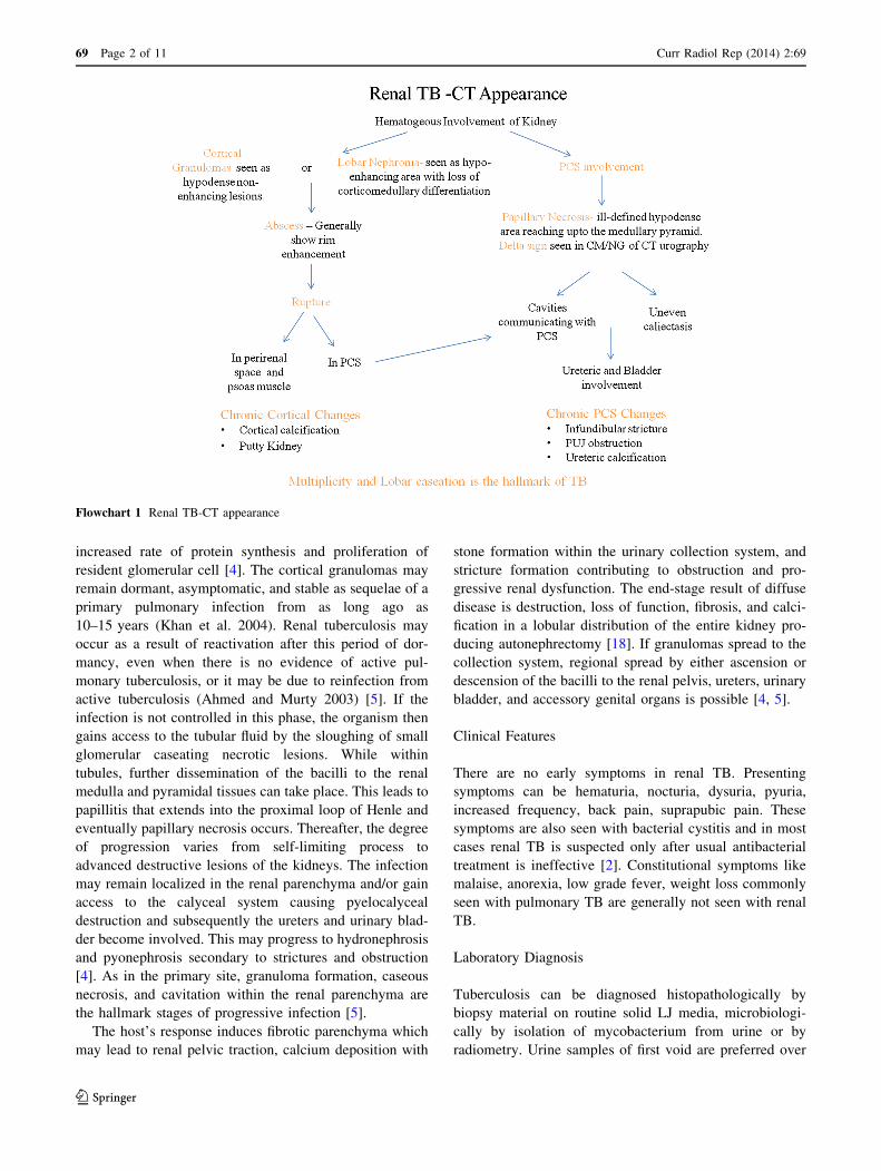
Early changes in renal TB include granulomas of B3 mm
in size and papillary necrosis. Both corticomedullary and
nephrographic phases need to be carefully evaluated for
these. A TB granuloma is seen as a solid mass with little or
minimal enhancement after contrast administration [14]
(Fig. 1) and is usually accompanied by collecting system
changes [14]. In rare cases, there may be single or multiple
parenchymal nodules, without collecting system involve-
ment [15]. The nodules are variable-sized, well-defined
parenchymal lesions on cross-sectional images and may
mimic renal neoplasms, which may lead to unnecessary
Fig. 1 Multiple hypodense lesions in both kidneys, spleen and liver
s/o granulomas
Curr Radiol Rep (2014) 2:69 Page 3 of 11 69
123
surgery; these are therefore labeled as the ‘pseudo-tumoral’
type [15–17].
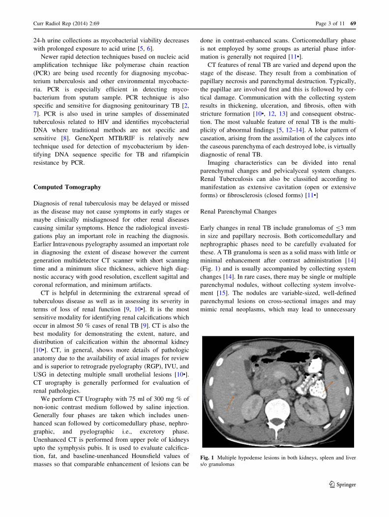
Localized tissue edema and vasoconstriction caused by
active inflammation results in focal hypoperfusion as seen
on nephrographic phase of CT Urography, a finding similar
to that seen in acute pyelonephritis caused by other
organisms [18] (Fig. 2a, b). Coalesced cortical granulomas
containing either caseous or calcified material are readily
identified at CT [9]. The majority reveals evidence of
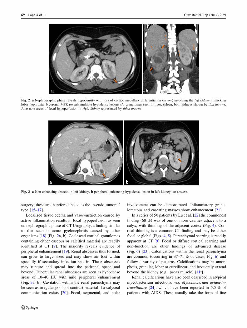
peripheral enhancement [19]. Renal abscesses thus formed,
can grow to large sizes and may show air foci within
specially if secondary infection sets in. These abscesses
may rupture and spread into the perirenal space and
beyond. Tubercular renal abscesses are seen as hypodense
areas of 10–40 HU with mild peripheral enhancement
(Fig. 3a, b). Cavitation within the renal parenchyma may
be seen as irregular pools of contrast material if a calyceal
communication exists [20]. Focal, segmental, and polar
involvement can be demonstrated. Inflammatory granu-
lomatous and caseating masses show enhancement [21].
In a series of 50 patients by Lu et al. [22] the commonest
finding (68 %) was of one or more cavities adjacent to a
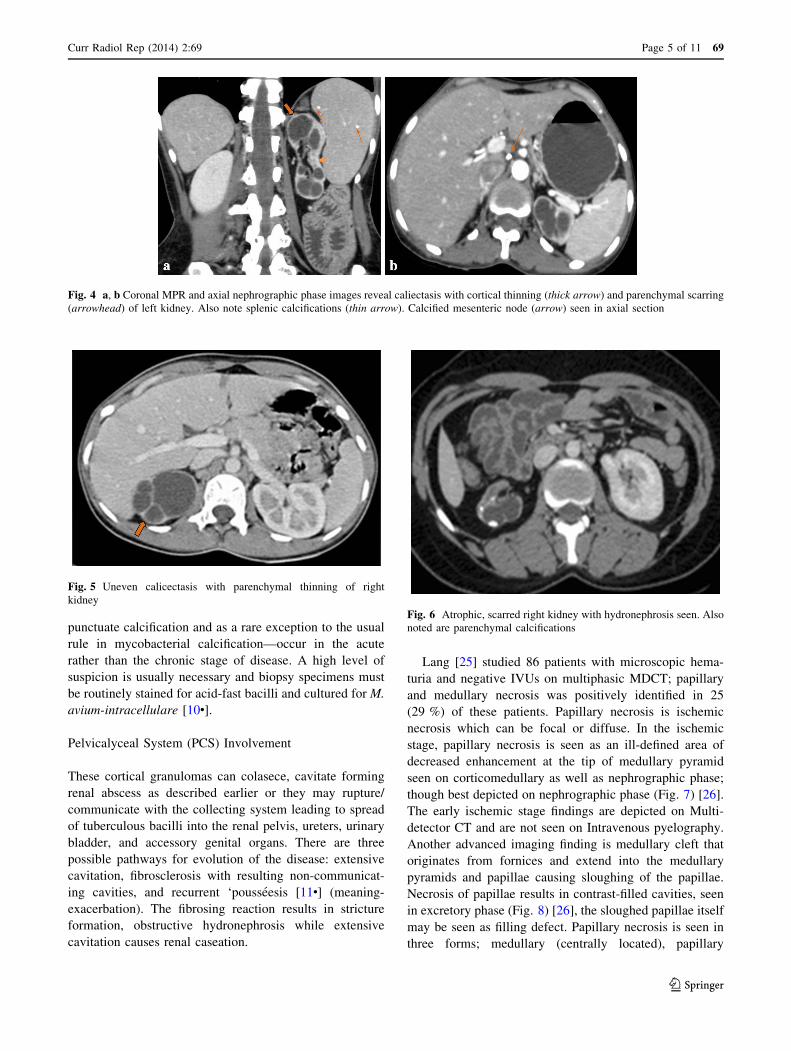
calyx, with thinning of the adjacent cortex (Fig. 4). Cor-
tical thinning is a common CT finding and may be either
focal or global (Figs. 4, 5). Parenchymal scarring is readily
apparent at CT [9]. Focal or diffuse cortical scarring and
non-function are other findings of advanced disease
(Fig. 6) [23]. Calcifications within the renal parenchyma
are common (occurring in 37–71 % of cases; Fig. 6) and
follow a variety of patterns. Calcifications may be amor-
phous, granular, lobar or curvilinear, and frequently extend
beyond the kidney (e.g., psoas muscle) [11•].
Renal calcifications have also been described in atypical
mycobacterium infections, viz, Mycobacterium avium-in-
tracellulare [24], which have been reported in 5.5 % of
patients with AIDS. These usually take the form of fine
Fig. 2 a Nephrographic phase reveals hypodensity with loss of cortico medullary differentiation (arrow) involving the left kidney mimicking
lobar nephronia, b coronal MPR reveals multiple hypodense lesions s/o granulomas seen in liver, spleen, both kidneys shown by thin arrows.
Also note areas of focal hypoperfusion in right kidney represented by thick arrows
Fig. 3 a Non-enhancing abscess in left kidney, b peripheral enhancing hypodense lesion in left kidney s/o abscess
69 Page 4 of 11 Curr Radiol Rep (2014) 2:69
123
punctuate calcification and as a rare exception to the usual
rule in mycobacterial calcification—occur in the acute
rather than the chronic stage of disease. A high level of
suspicion is usually necessary and biopsy specimens must
be routinely stained for acid-fast bacilli and cultured for M.
avium-intracellulare [10•].
Pelvicalyceal System (PCS) Involvement
These cortical granulomas can colasece, cavitate forming
renal abscess as described earlier or they may rupture/
communicate with the collecting system leading to spread
of tuberculous bacilli into the renal pelvis, ureters, urinary
bladder, and accessory genital organs. There are three
possible pathways for evolution of the disease: extensive
cavitation, fibrosclerosis with resulting non-communicat-
ing cavities, and recurrent ‘pousseesis [11•] (meaning-
exacerbation). The fibrosing reaction results in stricture
formation, obstructive hydronephrosis while extensive
cavitation causes renal caseation.
Lang [25] studied 86 patients with microscopic hema-
turia and negative IVUs on multiphasic MDCT; papillary
and medullary necrosis was positively identified in 25
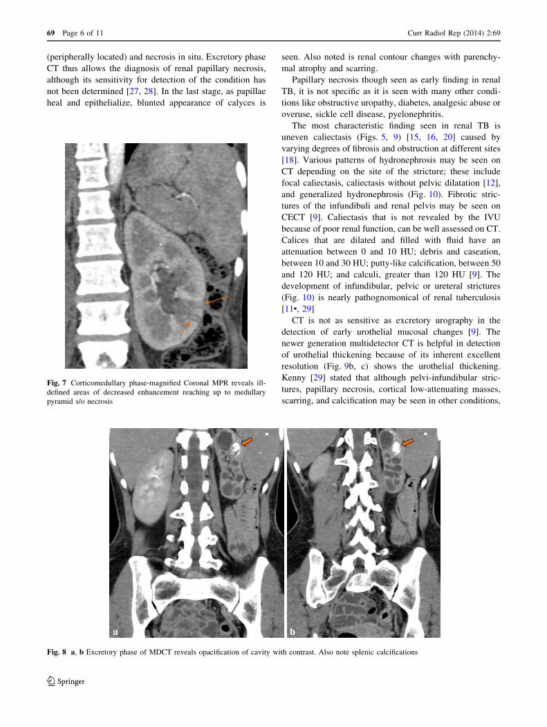
(29 %) of these patients. Papillary necrosis is ischemic
necrosis which can be focal or diffuse. In the ischemic
stage, papillary necrosis is seen as an ill-defined area of
decreased enhancement at the tip of medullary pyramid
seen on corticomedullary as well as nephrographic phase;
though best depicted on nephrographic phase (Fig. 7) [26].
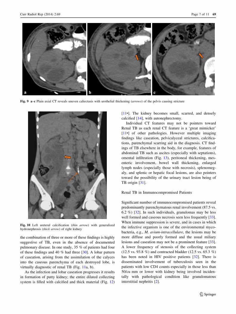
The early ischemic stage findings are depicted on Multi-
detector CT and are not seen on Intravenous pyelography.
Another advanced imaging finding is medullary cleft that
originates from fornices and extend into the medullary
pyramids and papillae causing sloughing of the papillae.
Necrosis of papillae results in contrast-filled cavities, seen
in excretory phase (Fig. 8) [26], the sloughed papillae itself
may be seen as filling defect. Papillary necrosis is seen in
three forms; medullary (centrally located), papillary
Fig. 4 a, b Coronal MPR and axial nephrographic phase images reveal caliectasis with cortical thinning (thick arrow) and parenchymal scarring
(arrowhead) of left kidney. Also note splenic calcifications (thin arrow). Calcified mesenteric node (arrow) seen in axial section
Fig. 5 Uneven calicectasis with parenchymal thinning of right
kidney
Fig. 6 Atrophic, scarred right kidney with hydronephrosis seen. Also
noted are parenchymal calcifications
Curr Radiol Rep (2014) 2:69 Page 5 of 11 69
123
(peripherally located) and necrosis in situ. Excretory phase
CT thus allows the diagnosis of renal papillary necrosis,
although its sensitivity for detection of the condition has
not been determined [27, 28]. In the last stage, as papillae
heal and epithelialize, blunted appearance of calyces is
seen. Also noted is renal contour changes with parenchy-
mal atrophy and scarring.
Papillary necrosis though seen as early finding in renal
TB, it is not specific as it is seen with many other condi-
tions like obstructive uropathy, diabetes, analgesic abuse or
overuse, sickle cell disease, pyelonephritis.
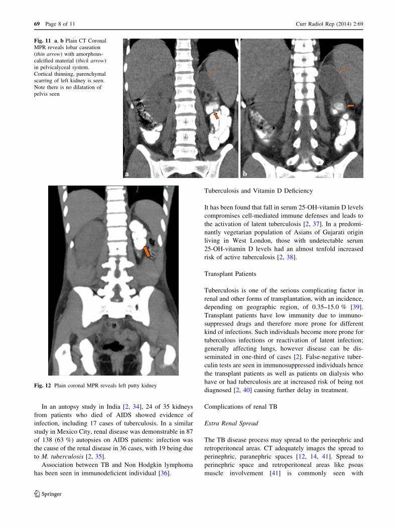
The most characteristic finding seen in renal TB is
uneven caliectasis (Figs. 5, 9) [15, 16, 20] caused by
varying degrees of fibrosis and obstruction at different sites
[18]. Various patterns of hydronephrosis may be seen on
CT depending on the site of the stricture; these include
focal caliectasis, caliectasis without pelvic dilatation [12],
and generalized hydronephrosis (Fig. 10). Fibrotic stric-
tures of the infundibuli and renal pelvis may be seen on
CECT [9]. Caliectasis that is not revealed by the IVU
because of poor renal function, can be well assessed on CT.
Calices that are dilated and filled with fluid have an
attenuation between 0 and 10 HU; debris and caseation,
between 10 and 30 HU; putty-like calcification, between 50
and 120 HU; and calculi, greater than 120 HU [9]. The
development of infundibular, pelvic or ureteral strictures
(Fig. 10) is nearly pathognomonical of renal tuberculosis
[11•, 29]
CT is not as sensitive as excretory urography in the
detection of early urothelial mucosal changes [9]. The
newer generation multidetector CT is helpful in detection
of urothelial thickening because of its inherent excellent
resolution (Fig. 9b, c) shows the urothelial thickening.
Kenny [29] stated that although pelvi-infundibular stric-
tures, papillary necrosis, cortical low-attenuating masses,
scarring, and calcification may be seen in other conditions,
Fig. 7 Corticomedullary phase-magnified Coronal MPR reveals ill-
defined areas of decreased enhancement reaching up to medullary
pyramid s/o necrosis
Fig. 8 a, b Excretory phase of MDCT reveals opacification of cavity with contrast. Also note splenic calcifications
69 Page 6 of 11 Curr Radiol Rep (2014) 2:69
123
the combination of three or more of these findings is highly
suggestive of TB, even in the absence of documented
pulmonary disease. In one study, 35 % of patients had four
of these findings and 40 % had three [30]. A lobar pattern
of caseation, arising from the assimilation of the calyces
into the caseous parenchyma of each destroyed lobe, is
virtually diagnostic of renal TB (Fig. 11a, b).
As the infection and lobar caseation progresses it results
in formation of putty kidney; the entire dilated collecting
system is filled with calcified and thick material (Fig. 12)
[11•]. The kidney becomes small, scarred, and densely
calcified [14], with autonephrectomy.
Individual CT features may not be pointers toward
Renal TB as each renal CT feature is a ‘great mimicker’
[11•] of other pathologies. However multiple imaging
findings like caseation, pelvicalyceal strictures, calcifica-
tions, parenchymal scarring aid in the diagnosis. CT find-
ings of TB elsewhere in the body, for example, features of
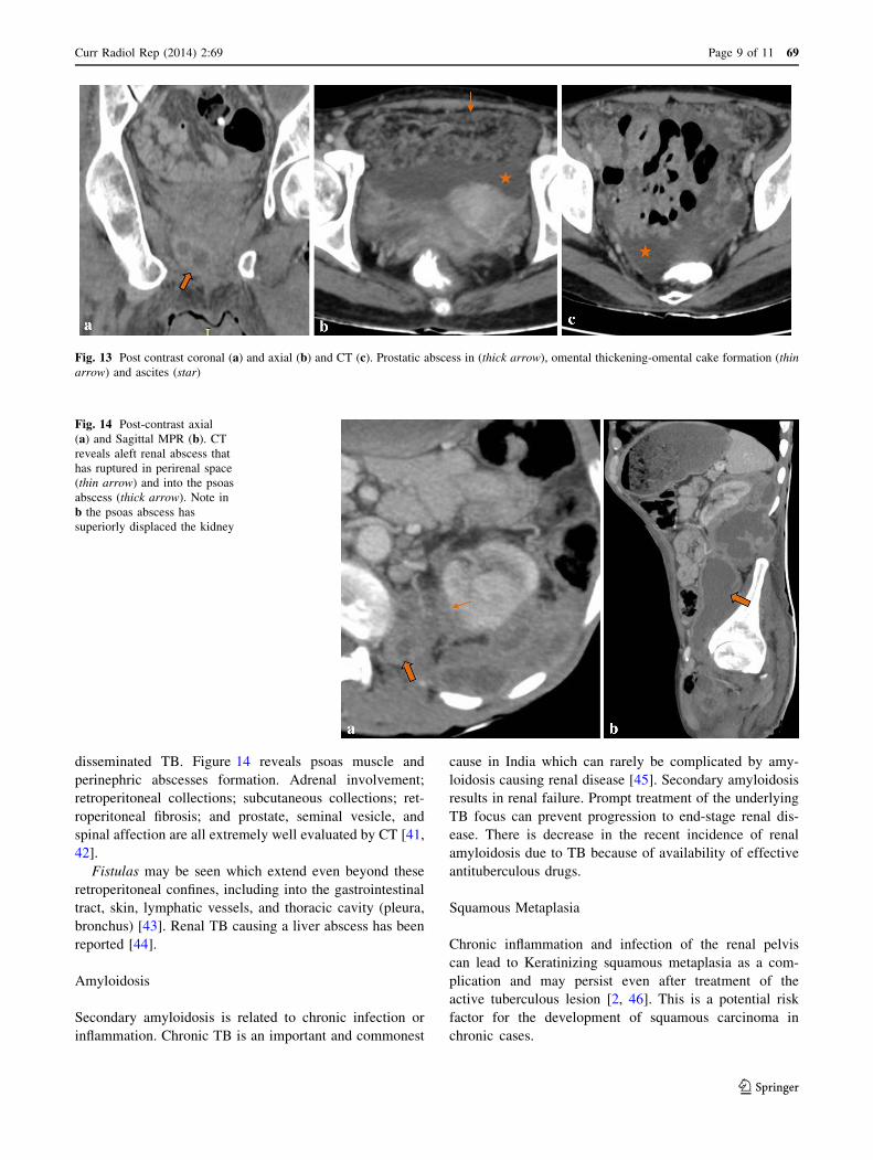
abdominal TB such as ascites (especially with septations),
omental infiltration (Fig. 13), peritoneal thickening, mes-
enteric involvement, bowel wall thickening, enlarged
lymph nodes (especially those with necrosis), splenomeg-
aly, and splenic or hepatic focal lesions, are also pointers
toward the possibility of the urinary tract lesion being of
TB origin [31].
Renal TB in Immunocompromised Patients
Significant number of immunocompromised patients reveal
predominantly parenchymatous renal involvement (87.5 vs.
6.2 %) [32]. In such individuals, granulomas may be less
well formed and caseous necrosis seen less frequently [33].
When immune suppression is severe, and in cases in which
the infective organism is one of the environmental myco-
bacteria, e.g., M. avium-intracellulare, the lesions may be
more diffuse and poorly formed and the usual miliary
lesions and caseation may not be a prominent feature [33].
A lower frequency of stenosis of the collecting system
(12.5 vs. 93.8 %) and contracted bladder (12.5 vs. 65.3 %)
has been noted in HIV positive patients [32]. There is
disseminated involvement of tuberculosis seen in the
patients with low CD4 counts especially in those less than
50/cu mm or lower with kidney being involved inciden-
tally with pathological condition like granulomatous
interstitial nephritis [2].
Fig. 9 a–c Plain axial CT reveals uneven caliectasis with urothelial thickening (arrows) of the pelvis causing stricture
Fig. 10 Left ureteral calcification (thin arrow) with generalized
hydronephrosis (thick arrow) of right kidney
Curr Radiol Rep (2014) 2:69 Page 7 of 11 69
123
In an autopsy study in India [2, 34], 24 of 35 kidneys
from patients who died of AIDS showed evidence of
infection, including 17 cases of tuberculosis. In a similar
study in Mexico City, renal disease was demonstrable in 87
of 138 (63 %) autopsies on AIDS patients: infection was
the cause of the renal disease in 36 cases, with 19 being due
to M. tuberculosis [2, 35].
Association between TB and Non Hodgkin lymphoma
has been seen in immunodeficient individual [36].
Tuberculosis and Vitamin D Deficiency
It has been found that fall in serum 25-OH-vitamin D levels
compromises cell-mediated immune defenses and leads to
the activation of latent tuberculosis [2, 37]. In a predomi-
nantly vegetarian population of Asians of Gujarati origin
living in West London, those with undetectable serum
25-OH-vitamin D levels had an almost tenfold increased
risk of active tuberculosis [2, 38].
Transplant Patients
Tuberculosis is one of the serious complicating factor in
renal and other forms of transplantation, with an incidence,
depending on geographic region, of 0.35–15.0 % [39].
Transplant patients have low immunity due to immuno-
suppressed drugs and therefore more prone for different
kind of infections. Such individuals become more prone for
tuberculous infections or reactivation of latent infection;
generally affecting lungs, however disease can be dis-
seminated in one-third of cases [2]. False-negative tuber-
culin tests are seen in immunosuppressed individuals hence
the transplant patients as well as patients on dialysis who
have or had tuberculosis are at increased risk of being not
diagnosed [2, 40] causing further delay in treatment.
Complications of renal TB
Extra Renal Spread
The TB disease process may spread to the perinephric and
retroperitoneal areas. CT adequately images the spread to
perinephric, paranephric spaces [12, 14, 41]. Spread to
perinephric space and retroperitoneal areas like psoas
muscle involvement [41] is commonly seen with
Fig. 11 a, b Plain CT Coronal
MPR reveals lobar caseation
(thin arrow) with amorphous-
calcified material (thick arrow)
in pelvicalyceal system.
Cortical thinning, parenchymal
scarring of left kidney is seen.
Note there is no dilatation of
pelvis seen
Fig. 12 Plain coronal MPR reveals left putty kidney
69 Page 8 of 11 Curr Radiol Rep (2014) 2:69
123
disseminated TB. Figure 14 reveals psoas muscle and
perinephric abscesses formation. Adrenal involvement;
retroperitoneal collections; subcutaneous collections; ret-
roperitoneal fibrosis; and prostate, seminal vesicle, and
spinal affection are all extremely well evaluated by CT [41,
42].
Fistulas may be seen which extend even beyond these
retroperitoneal confines, including into the gastrointestinal
tract, skin, lymphatic vessels, and thoracic cavity (pleura,
bronchus) [43]. Renal TB causing a liver abscess has been
reported [44].
Amyloidosis
Secondary amyloidosis is related to chronic infection or
inflammation. Chronic TB is an important and commonest
cause in India which can rarely be complicated by amy-
loidosis causing renal disease [45]. Secondary amyloidosis
results in renal failure. Prompt treatment of the underlying
TB focus can prevent progression to end-stage renal dis-
ease. There is decrease in the recent incidence of renal
amyloidosis due to TB because of availability of effective
antituberculous drugs.
Squamous Metaplasia
Chronic inflammation and infection of the renal pelvis
can lead to Keratinizing squamous metaplasia as a com-
plication and may persist even after treatment of the
active tuberculous lesion [2, 46]. This is a potential risk
factor for the development of squamous carcinoma in
chronic cases.
Fig. 13 Post contrast coronal (a) and axial (b) and CT (c). Prostatic abscess in (thick arrow), omental thickening-omental cake formation (thin
arrow) and ascites (star)
Fig. 14 Post-contrast axial
(a) and Sagittal MPR (b). CT
reveals aleft renal abscess that
has ruptured in perirenal space
(thin arrow) and into the psoas
abscess (thick arrow). Note in
b the psoas abscess has
superiorly displaced the kidney
Curr Radiol Rep (2014) 2:69 Page 9 of 11 69
123
End-Stage Renal Disease
End-stage renal disease in renal TB can occur due to
obstructive uropathy as a result of multiple strictures
especially at pelviureteric junction. Secondary amyloidosis
or obliterative endarteritis of intrarenal vessels is the other
cause [47]. Disseminated Tuberculosis seen commonly
with immunocompromised individuals may affect the
kidney resulting in granulomatous interstitial nephritis
eventually leading to renal failure [2].
Certain biochemical abnormalities are seen with renal
TB in end-stage renal disease; hypercalcemia being one of
them. Hypercalcemia is secondary to abnormal/elevated
calcitriol production by the granulomatous tissue [2, 47].
Tuberculosis is an uncommon cause of renal failure
which is treatable and potentially preventable. The overall
incidence of renal failure related to TB, reported in the
literature is 24 % [47].
Acute Interstitial Nephritis as the term suggest involves
renal tubules and/or interstitium sparing the glomerulus. It
is characterized histopathologically by inflammation and
edema of the renal interstitium [48]. Acute interstitial
nephritis (AIN) is due to cellular injury caused by viral,
fungal or bacterial infections or as a hypersensitivity
reaction to drugs or by immunologic conditions. AIN
rapidly and abruptly causes the renal failure [47, 48].
Granulomatous interstitial nephritis (GIN) is an
uncommon form of acute interstitial nephritis [48]; tuber-
culosis being the rare cause. Pathology is granuloma for-
mation with epithelioid giant cells usually found in AIN
secondary to tuberculosis, sarcoidosis or Wegener’s gran-
ulomatosis [48]. AIN can also be caused due to RIFAM-
PICIN drug used in treatment of TB [47]. Renal biopsy is
the definitive test for diagnosis of AIN [48, 49].
As the tubules have regenerative capacity, the renal
function in AIN can be made reversible/or maintained if
the diagnosis is made before complete deterioration of
renal function occurs using a combination of anti-tuber-
cular treatment and corticosteroids [2].
Conclusion
Renal tuberculosis is one of the commonest extrapulmo-
nary form, frequently seen in endemic areas and is not
easily diagnosed clinically due to absence of classical
symptoms. It is also one of the treatable cause of renal
failure. Hence the radiologist and imaging assumes an
important role in reaching the diagnosis.
Computed tomography is highly sensitive for detection of
calcification which is commonly seen with TB. With advent
of newer generation Multidetector scanners, increased res-
olution has resulted in increased sensitivity and detection of
renal lesions. CT urography performed on the current high-
end MDCT scanners have the potential to detect early TB
changes like papillary necrosis that were usually noted on
IVU. Though individual features may not help in diagnosis;
multiple cortical lesions, parenchymal scarring and pelvic-
alyceal system involvement in form of papillary necrosis,
lobar caseation, calcification, uneven caliectasis and stric-
tures are pathogonomic of renal TB. Awareness of these
imaging findings assists in making the diagnosis which is
further confirmed by histopathology. Thus CT imaging will
continue to play an important role in establishing the initial
diagnosis as well as in follow up studies.
Acknowledgements We acknowledge Dr Ashank Bansal for pre-
paring the images and Prof Suleman Merchant, HOD dept of
Radiology, LTMGH and LTMMC, Sion, Mumbai, India.
Compliance with Ethics Guidelines
Conflict of Interest Dr. Ashwini Sankhe declares no potential
conflicts of interest. Dr. Anagha R. Joshi is a section editor for
Current Radiology Reports.
Human and Animal Rights and Informed Consent This article
does not contain any studies with human or animal subjects
performed by any of the authors.
References
Papers of particular interest, published recently, have been
highlighted as:• Of importance•• Of major importance
1. Das P, Ahuja A, Gupta SD. Incidence, etiopathogenesis and
pathological aspects of genitourinary tuberculosis in India: A
journey revisited. Indian J Urol. 2008;24(3):356–61.
2. Eastwood JB, Corbishley CM, Grange JM. Tuberculosis and the
kidney. JASN. 2001;12(6):1307–14.
3. Chauhan LS, Tonsing J. Revised national TB control programme
in India. Tuberculosis (Edinb). 2005;85:271–6.
4. Chijioke A. Current concepts on pathogenesis of renal tubercu-
losis. West Afr J Med. 2001;20(2):107–10.
5. Langemeier Jane. Tuberculosis of the genitourinary system. Urol
Nurs. 2007;27(4):279–84.
6. Pasternak MS, Rubin RH. Urinary tract tuberculosis. In: Schrier
RW, editor. Diseases of the kidney and urinary tract. 7th ed.
Philadelphia: Lippincott Williams & Wilkins; 2001. p. 1017–37.
7. Sechi LA, Pinna MP, Sanna A, Pirina P, Ginesu F, Saba F, Aceti
A, Turrini F, Zanetti S, Fadda G. Detection of Mycobacterium
tuberculosis by PCR analysis of urine and other clinical samples
from AIDS and non-HIV-infected patients. Mol Cell Probes.
1997;11:281–5.
8. Aceti A, Zanetti S, Mura MS, Sechi LA, Turrini F, Saba F,
Babudieri S, Mannu F, Fadda G. Identification of HIV patients
with active pulmonary tuberculosis using urine based polymerase
chain reaction assay. Thorax. 1999;54:145–6.
9. Gibson MS, Puckett ML, Shelly ME. Renal tuberculosis. Ra-
diographics. 2004;24:251–6.
69 Page 10 of 11 Curr Radiol Rep (2014) 2:69
123
10. • Merchant S, Bharati A, Merchant N. Tuberculosis of the gen-
itourinary system-Urinary tract tuberculosis: renal tuberculosis-
Part II. Genitourin Obstet Radiol. 2013; 23(1):64–7. The refer-
ence is latest and gives comprehensive information about geni-
tourinary Koch’s.
11. • Quaia E. Imaging findings in renal tuberculosis on computed
tomography urography. Eur Urol Rev. 2010;5(2):69–72. This
reference article is one of the latest article about CT in Renal
Koch’s and included as it is within 5 years.
12. Wang LI, Wong YC, Chen CJ, Lim KE. CT features of genito-
urinary tuberculosis. J Comput Assist Tomogr. 1997;21:254–8.
13. Hartman DS, Stagg PL. Diagnosis please. Case 3: renal tuber-
culosis. Radiology. 1998;209:69–72.
14. Goldman SM, Fishman EK, Hartman DS, Siegelman SS. Com-
puted tomography of renal tuberculosis and its pathological
correlates. J Comput Assist Tomogr. 1985;9:771–6.
15. Kim SH. Urogenital tuberculosis. In: Pollack HM, McClennan
BL, editors. Clinical urography. 2nd ed. Philadelphia: WB
Saunders Co; 2000. p. 1193–228.
16. Wang LJ, Wu CF, Wong YC, Chuang CK, Chu SH, Chen CJ.
Imaging findings of urinary tuberculosis on excretory urography
and computerized tomography. J Urol. 2003;169:524–8.
17. Goldman SM, Fishman EK. Upper urinary tract infection: the
current role of CT, ultrasound, and MRI. Semin Ultrasound CT
MR. 1991;12:335–60.
18. Engin G, Acuna B, Acuna G, Tunaci M. Imaging of extrapul-
monary tuberculosis. Radiographics. 2000;20:471–88.
19. Li Y, Yang ZG, Guo YK, Min PQ, Yu JQ, Ma ES, et al. Dis-
tribution and characteristics of hematogenous disseminated
tuberculosis within the abdomen on contrast-enhanced CT.
Abdom Imaging. 2007;32:484–8.
20. Harisinghani MG, McLoud TC, Shepard JA, Ko JP, Shroff MM,
Mueller PR. Tuberculosis from head to toe. Radiographics.
2000;20:449–70.
21. Buxi TB, Sud S, Vohra R. CT and MRI in the diagnosis of
tuberculosis. Indian J Pediatr. 2002;69:965–72.
22. Lu P, Li C, Zhou X. Significance of the CT scan in renal
tuberculosis. Zhonghua Jie He He Hu Xi Za Zhi. 2001;24:407–9.
23. Kawashima A, Sandler CM, Ernst RD, Goldman SM, Raval B,
Fishman EK. Renal inflammatory disease: the current role of CT.
Crit Rev Diagn Imaging. 1997;38:369–415.
24. Miller FH, Parikh S, Gore RM, Nemcek AA Jr, Fitzgerald SW,
Vogelzang RL. Renal manifestations of AIDS. Radiographics.
1993;13:587–96.
25. Lang EK, Macchia RJ, Thomas R, Watson RA, Marberger M,
Lechner G, et al. Improved detection of renal pathologic features
on multiphasic helical CT compared with IVU in patients pre-
senting with microscopic hematuria. Urology. 2003;61:528–32.
26. Jung DC, Kim SH, Jung SI, Hwang SI, Kim SH. Renal papillary
necrosis: review and comparison of findings at multi-detector row
CT and intravenous urography. Radiographics. 2006;26:1827–36.
27. Lang EK, Macchia RJ, Thomas R, et al. Detection of medullary
and papillary necrosis at an early stage by multiphasic helical
computerized tomography. J Urol. 2003;170(1):94–8.
28. Bittar EE, Misanik L. Renal necrotizing papillitis. Am J Med.
1963;34:82–7.
29. Kenney PJ. Imaging of chronic renal infections. AJR Am J
Roentgenol. 1990;155:485–94.
30. Okazawa N, Sekiya T, Tada S. Computed tomographic features
of renal tuberculosis. Radiat Med. 1985;3:209–13.
31. Zissin R, Gayer G, Chowers M, Shapiro-Feinberg M, Kots E,
Hertz M. Computerized tomography findings of abdominal
tuberculosis: report of 19 cases. Isr Med Assoc J. 2001;3:414–8.
32. Figueiredo AA, Lucon AM, Junior RF, Ikejiri DS, Nahas WC,
Srougi M. Urogenital tuberculosis in immunocompromised
patients. Int Urol Nephrol. 2009;41:327–33.
33. Eastwood JB, Corbishley CM, Grange JM. Tuberculosis and the
kidney. J Am Soc Nephrol. 2001;12:1307–14.
34. Lanjewar DN, Ansari MA, Shetty CR, Maheshwary MB, Jain P.
Renal lesions associated with AIDS—An autopsy study. Indian J
Pathol Microbiol. 1999;42:63–8.
35. Soriano-Rosas J, Avila-Casado MC, Carrera-Gonzalez E, Cha-
vez-Mercado L, Cruz-Ortiz H, Rojo J. AIDS-associated
nephropathy: 5-year retrospective morphologic analysis of 87
cases. Pathol Res Pract. 1998;194:567–70.
36. Wise GJ. Urinary tuberculosis: modern issues. Curr Urol Rep.
2009;10:313–8.
37. Davies PDO. Vitamin D and tuberculosis. Am Rev Respir Dis.
1989;139:1571.
38. Wilkinson RJ, Llewelym M, Toossi Z, Patel P, Pasvol G, Lalvani
A, Wright D, Latif M, Davidson RN. Influence of vitamin D
deficiency and vitamin D receptor polymorphisms on tuberculosis
among Gujarati Asians in west London: a case-control study.
Lancet. 2000;355:618–21.
39. Singh N, Paterson DL. Mycobacterium tuberculosis infection in
solid-organ transplant recipients: impact and implications for
management. Clin Infect Dis. 1998;27:1266–77.
40. Woeltje KF, Mathew A, Rothstein M, Seiler S, Fraser VJ.
Tuberculosis infection and anergy in hemodialysis patients. Am J
Kidney Dis. 1998;31:848–52.
41. Merchant SA. Tuberculosis of the genitourinary system. Indian J
Radiol Imaging. 1993;3:253–74.
42. Benchekroun A, Lachkar A, Soumana A, Farih MH, Belahnech
Z, Marzouk M, et al. Urogenital tuberculosis. 80 cases. Ann Urol
(Paris) 1998;32:89–94.
43. Jafri SZ, Roberts JL, Berger BD. Fistulas of the genitourinary
tract. In: Pollack HM (ed) Clinical urography. 2nd edn. Saunders,
Philadelphia. 2000. pp. 2992–3011.
44. Shah HN, Jain P, Chibber PJ. Renal tuberculosis simulating
xanthogranulomatous pyelonephritis with contagious hepatic
involvement. Int J Urol. 2006;13:67–8.
45. Chugh KS, Datta BN, Singhal PC, Jain SK, Sakhuja V, Dash SC.
Pattern of renal amyloidosis in Indian patients. Postgrad Med J.
1981;57:31–5.
46. Byrd RB, Viner NA, Omell GH, Trunk G. Leukoplakia associ-
ated with renal tuberculosis in the chemotherapeutic era. Br J
Urol. 1976;48:377–81.
47. Krishnamoorthy S, Gopalakrishnan G. Surg Manag Renal Tub-
erc. Indian J Urol Symp 2008;24(3):369–75.
48. Kodner CM, Kudrimoti A. University of Louisville School of Medi-
cine, Louisville, Kentucky. Diagnosis and Management of Acute
Interstitial Nephritis. Am Fam Physician. 2003;67(12):2527–34.
49. Sampathkumar K, Sooraj YS, Mahaldar AR, Ramakrishnan M,
Rajappannair A et al. Granulomatous interstitial nephritis due to
tuberculosis-a rare presentation. Saudi J Kidney Dis Transplant.
2009;20(5):842–5.
Curr Radiol Rep (2014) 2:69 Page 11 of 11 69
123