Multi-Biochemical Test System for Distinguishing - American Society
7
APuPED MICROBIOLOGY, Sept. 1971, p. 408-414 Vol. 22, No. 3 Copyright © 1971 American Society for Microbiology Printed in U.S.A. Multi-Biochemical Test System for Distinguishing Enteric and Other Gram-Negative Bacilli HARRY R. ELSTON, JUDITH A. BAUDO, JOANN P. STANEK, AND MERCEDES SCHAAB Microbiology Research Laboratory, Veterans Administration Hospital, Omaha, Nebraska 68105 Received for publication 26 April 1971 A multi-biochemical test system consisting of nine tests, entitled Enterotube, was evaluated in parallel with conventional tests to determine its value in the identifica- tion of enteric and certain other gram-negative bacilli. The 242 bacterial strains studied were from a variety of pathological specimens and from our culture collec- tion. When the results with individual tests represented in both test systems were compared, no discrepancies were noted in the indole test, and one discrepancy was recorded for dextrose. In 7 of 242 hydrogen sulfide tests, 3 of 242 phenylalanine tests, 22 of 242 urease tests, 15 of 242 dulcitol tests, 12 of 242 lactose tests, 27 of 217 lysine decarboxylase tests, and 5 of 242 citrate tests, the Enterotube results were contrary to those obtained with conventional methods. The lysine decar- boxylase test in the Enterotube posed a problem of interpretation and readability and is not an acceptable alternative to the conventional methods. Fifteen of the strains studied were incorrectly identified by the Enterotube system and four could not be differentiated from other closely related strains. Salmonella could be iden- tified as to group, whereas Shigella strains were frequently misidentified as Esche- richia. The Enterotube method is simple and convenient, and all media are inocu- lated at once from a single colony. Methods used for differential identification of enteric and related gram-negative bacilli have been dependent mostly on biochemical tests. Many of these tests are time-consuming, and 72 hr may elapse before the organism can be properly identified. Investigators have constantly sought simpler and more rapid procedures to differentiate gram-negative bacilli. Until recently, the vast majority of biochemical tests have been diverse and separate in media and methods. Today a number of rapid biochemical test systems are available to the microbiologist. One such test system is the reagent-impregnated paper strip. Several workers (1, 5, 9) have reported on the use of the paper-impregnated strip as a more rapid substitute for conventional methods. Paper strips impregnated with reagents to test for indole, acetylmethylcarbinol production, urease, cyto- chrome oxidase, phenylalanine deaminase, lysine decarboxylase activity, niacin, and nitrate reduc- tion are available (1, 5, 7, 9, 11). An R/B enteric differential system (Diagnostic Research Inc., Roslyn, N. Y.), which uses two tubes of culture media and various reagents, has been developed for determination of certain biochemical tests. Tube 1 is used to test for phenylalanine deaminase, lactose, glucose, hydrogen sulfide, and lysine decarboxylase activity. Tube 2 is used to test for indole, ornithine activity, and motility. Another newly formulated test system, Enterotube (Roche Diagnostic Div., Hoffman-LaRoche, Inc., Nutley, N.J.), is a compartmental tube containing a number of different types of biochemical test media used for the identification of Entero- bacteriaceae (8). This test system is based on the findings of Edwards and Ewing (4) that eight biochemical reactions can serve to separate members of the enteric family into Shigella-Es- cherichia, Salmonella-Arizona-Citrobacter, Klebsi- ella-Aerobacter-Serratia, and Proteus-Providence divisions. This paper concerns a study which tests the use of a multi-biochemical test system (En- terotube) for distinguishing enteric and certain other gram-negative bacilli and compares the re- sults with conventional tests. MATERIALS AND METHODS Cultures tested. Gram-negative bacilli obtained from various kinds of pathological specimens received in the clinical laboratory formed the material for this study. The strains comprised primary isolates from urine, sputa, wounds, body fluids, stool specimens, and isolates from our culture collection. Initially, each clinical specimen was Gram-stained, transferred to 408 Downloaded from https://journals.asm.org/journal/am on 21 February 2022 by 180.182.225.240.
Transcript of Multi-Biochemical Test System for Distinguishing - American Society
APuPED MICROBIOLOGY, Sept. 1971, p. 408-414 Vol. 22, No. 3Copyright © 1971 American Society for Microbiology Printed in U.S.A.
Multi-Biochemical Test System for DistinguishingEnteric and Other Gram-Negative BacilliHARRY R. ELSTON, JUDITH A. BAUDO, JOANN P. STANEK, AND
MERCEDES SCHAABMicrobiology Research Laboratory, Veterans Administration Hospital, Omaha, Nebraska 68105
Received for publication 26 April 1971
A multi-biochemical test system consisting of nine tests, entitled Enterotube, wasevaluated in parallel with conventional tests to determine its value in the identifica-tion of enteric and certain other gram-negative bacilli. The 242 bacterial strainsstudied were from a variety of pathological specimens and from our culture collec-tion. When the results with individual tests represented in both test systems werecompared, no discrepancies were noted in the indole test, and one discrepancy wasrecorded for dextrose. In 7 of 242 hydrogen sulfide tests, 3 of 242 phenylalaninetests, 22 of 242 urease tests, 15 of 242 dulcitol tests, 12 of 242 lactose tests, 27 of217 lysine decarboxylase tests, and 5 of 242 citrate tests, the Enterotube resultswere contrary to those obtained with conventional methods. The lysine decar-boxylase test in the Enterotube posed a problem of interpretation and readabilityand is not an acceptable alternative to the conventional methods. Fifteen of thestrains studied were incorrectly identified by the Enterotube system and four couldnot be differentiated from other closely related strains. Salmonella could be iden-tified as to group, whereas Shigella strains were frequently misidentified as Esche-richia. The Enterotube method is simple and convenient, and all media are inocu-lated at once from a single colony.
Methods used for differential identification ofenteric and related gram-negative bacilli havebeen dependent mostly on biochemical tests.Many of these tests are time-consuming, and 72 hrmay elapse before the organism can be properlyidentified. Investigators have constantly soughtsimpler and more rapid procedures to differentiategram-negative bacilli. Until recently, the vastmajority of biochemical tests have been diverseand separate in media and methods. Today anumber of rapid biochemical test systems areavailable to the microbiologist. One such testsystem is the reagent-impregnated paper strip.Several workers (1, 5, 9) have reported on theuse of the paper-impregnated strip as a morerapid substitute for conventional methods. Paperstrips impregnated with reagents to test for indole,acetylmethylcarbinol production, urease, cyto-chrome oxidase, phenylalanine deaminase, lysinedecarboxylase activity, niacin, and nitrate reduc-tion are available (1, 5, 7, 9, 11). An R/B entericdifferential system (Diagnostic Research Inc.,Roslyn, N. Y.), which uses two tubes of culturemedia and various reagents, has been developedfor determination of certain biochemical tests.Tube 1 is used to test for phenylalanine deaminase,lactose, glucose, hydrogen sulfide, and lysine
decarboxylase activity. Tube 2 is used to test forindole, ornithine activity, and motility. Anothernewly formulated test system, Enterotube (RocheDiagnostic Div., Hoffman-LaRoche, Inc., Nutley,N.J.), is a compartmental tube containing anumber of different types of biochemical testmedia used for the identification of Entero-bacteriaceae (8). This test system is based on thefindings of Edwards and Ewing (4) that eightbiochemical reactions can serve to separatemembers of the enteric family into Shigella-Es-cherichia, Salmonella-Arizona-Citrobacter, Klebsi-ella-Aerobacter-Serratia, and Proteus-Providencedivisions. This paper concerns a study which teststhe use of a multi-biochemical test system (En-terotube) for distinguishing enteric and certainother gram-negative bacilli and compares the re-sults with conventional tests.
MATERIALS AND METHODS
Cultures tested. Gram-negative bacilli obtained fromvarious kinds of pathological specimens received in theclinical laboratory formed the material for this study.The strains comprised primary isolates from urine,sputa, wounds, body fluids, stool specimens, andisolates from our culture collection. Initially, eachclinical specimen was Gram-stained, transferred to
408
Dow
nloa
ded
from
http
s://j
ourn
als.
asm
.org
/jour
nal/a
m o
n 21
Feb
ruar
y 20
22 b
y 18
0.18
2.22
5.24
0.
MULTI-BIOCHEMICAL TEST SYSTEM
routine culture media consisting of Thioglycollatemedium (BBL), blood (human), and MacConkey(Difco) agar except for stool cultures which wereplanted on MacConkey, S-S (Difco), and BrilliantGreen (Difco) agars. Individual colonies with identicalcolonial morphology were used as inoculum for theEnterotube system and Kligler iron agar (KIA). Con-ventional biochemical media were inoculated fromKIA.
Enterotube biochemical test system. Enterotubes(Hoffman-LaRoche, Inc.) are plastic tubes with oneside round and the adjacent side flat. The flat side iscovered with a thin plastic seal. Each tube has eightcompartments containing a given biochemical testmedium. A single inoculating needle extends length-wise through the center of the media in each com-partment and protrudes at each end of the tube. Oneend of the wire and tube is covered by a blue screwcap, and the other is covered by a white screw cap.The inoculating end of the wire is covered by thewhite cap. Aerobic conditions during incubation areinsured by three small air holes which appear on theside of the tube, and these are covered by a blue stripwhich is removed at the time of inoculation. Eachcompartment is labeled with the appropriate name ofthe media it contains. The Enterotube is inoculatedby removing the white plastic cap, touching the centerof an isolated colony with the straight wire, and thenwithdrawing the wire from the tube. The wire inocu-lates each of the test media in the compartments. Thequestion of sufficient inoculum arises when inoculating(8) test media simultaneously with a single needle froma bacterial colony. With rare exceptions, each of theeight compaitments seemed to be inoculated satis-factorily after withdrawing the inoculating needle fromthe Enterotube. The wire may now be used to inocu-late other test media such as gelatin and motility testmedia which are used routinely in our identificationschema. We have inoculated as many as four sub-strates with success.The type of media in the compartments and the re-
actions were as follows.Dextrose-agar. The fermentation of dextrose
changedthe indicator, phenol red, from pink to yellow.Hydrogen sulfide-indole-agar. Hydrogen sulfide was
demonstrated by the formation of a black precipitatealong the inoculation line, whereas indole productionwas tested by injecting 0.3 ml of Kovac's reagentthrough the plastic film with a needle and syringe ontothe culture growth in the compartment. A red colorindicated the presence of indole.
Phenylalanine deaminase-agar. Deamination wastested by injecting 0.3 ml of a 10% ferric chloridesolution through the plastic film with a needle andsyringe onto the culture growth in the compartment.The formation of a dark green color on the surface ofthe culture slant indicated a positive test. One shouldinject the Kovac's and ferric chloride solutions usedin the indole and phenylalanine deaminase tests withthe Enterotubes in an upright position to prevent theflow of these reagents into other compartments viathe inoculating canal.
Urea-agar. The production of ammonia from ureawas shown by a change in the phenol red indicatorfrom yellow to pink.
Dulcitol- and lactose-agar. The fermentation ofthese carbohydrates was indicated by a change in thephenol red indicator from pink to yellow in theirrespective compartments.
Lysine-lactose-agar. A positive lysine decarboxyl-ase test was noted by a change in the phenol red in-dicator from yellow to pink or orange.Simmons citrate agar. A positive test was shown by
a change in the bromothymol blue indicator fromgreen to blue. All reactions in the compartments wereread after incubation at 37 C for 18 to 24 hr and againat 48 hr.
Conventional biochemical tests were run in con-junction with the Enterotubes. The types of media ormethods used were as follows.
Carbohydrate test media. Seitz-filtered 10% carbo-hydrate solutions of dextrose, lactose, and dulcitolwere added aseptically to cool autoclaved bromo-cresol purple broth basal medium (Difco). The mediawere then dispensed in sterile tubes (125 by 16 mm).
Hydrogen sulfide. Formation of hydrogen sulfidewas determined in Kligler iron agar.
Tryptone broth. Production of indole was testedwith Kovac's reagent after 18 to 24 hr of growth intryptone broth (Difco).
Phenylalanine deaminase agar. Production ofphenylpyruvic acid from phenylalanine was tested bythe method of Ewing, Davis, and Reavis (6) after 24hr of incubation on phenylalanine agar slants (Difco).
Urea-agar. Hydrolysis of urea was tested inChristensen (2) nutrient urea agar.
Lysine media. The two lysine decarboxylase meth-ods used were a modified lysine-lactose-agar tech-nique and the lysine broth method of Moeller (10).The Moeller broth was read daily for 4 days.
Lysine-lactose-agar. The lysine-lactose decarbox-ylase test medium slants consisted of lysine de-carboxylase basal medium (Difco) with 0.5% L-lysine, 1.0% lactose, 1.5% agar, and bromocresolpurple indicator. A control slant without the aminoacid was also used.
Lysine broth. The lysine decarboxylase test brothconsisted of decarboxylase basal medium (Difco)with 0.5% L-lysine and bromocresol purple indi-cator. A control broth without the amino acid wasalso used.Simmons citrate agar. The use of citrate as a sole
carbon source was indicated by the production ofammonia and a change in the color of the medium(Difco) from green to blue. The test reactions wereread after incubation for 24 and 48 hr except whenotherwise stated.
RESULTS
In all, 242 cultures of various groups of entericand other gram-negative bacilli were studied.The organisms were identified by the two testsystems and the results were compared as shownin Table 1. Additional biochemical or serologicaltests were sometimes necessary to confirm theidentity of the organisms.
Fermentation reactions by the Enterotube andconventional methods correlated well with dex-
VOL. 22, 1971 409
Dow
nloa
ded
from
http
s://j
ourn
als.
asm
.org
/jour
nal/a
m o
n 21
Feb
ruar
y 20
22 b
y 18
0.18
2.22
5.24
0.
ELSTON ET AL. APPL. MICROBIOL.
TABLE 1. Comparison of a number of biochemical tests by conventional tests and the Enterotube system
Organism
ShigellaShigella groupA.Shigella group B......Shigella group D.....
Alkalescens-Dispar.....Escherichia.
Klebsiella pneumoniae.Eniterobacter aerogenies.E. cloacae..........E. hafniae..Serratia marcescens.....
Proteus mirabilis-.P. rettgeri.........P. morganii.........P. vulgaris.........Providence group.
SalmonellaS. paratyphi B......S. typhi.........S. gallinarum..Salmonella group E....
Citrobacter group.Arizona arizonae......
Pseudomonas aeruginosa...Herellea....
60
12
2710
4
122
27
205
Dextrose H2S
Ca E C IE
22
260
6064124
27104
3
22
11
2
5
2
2
60
6064124
27104
3
22
11
2
05
00000
00000
2302
0
220
22
0
0
00000
00000
1900
0
220
2
00
Indole
CjE
202
59
20000
0104
3
000020
00
202
59
20000
0104
3
0
0
0
0
2
0
Phenyl- UreasealanineI Dulcitol ILactose ILysine Citrate
C IE C IE C E C IE
00000
00000
2684
3
0
0
0
0
0
0
0 0
0 0
00000
00000
2693
2
0
0
0
0
0
0
0
0
00000
51627
27104
0
0
0
0
0
4
0
a C, Conventional; E, Enterotube.
b' Includes E. coli and atypical Etcherichia (Paracolon coliforme, E. intermedia).
With Escherichia, 23 strains were positive by both methods.
00000
46300
27104
0
0
0
0
0
0
7 5
2 2
000
30
7
0
00
000
2
2
9
0
0
0
0000
26C
60
30
00000
22
90
00
0000
59
606460
00000
000062
00
0000
56
586440
00000
C E C E
00020
3
00
4
00000
0 2
0 2
0 1
0 1
3 3
0 2
0
0
0
23
20054
12000
2
0
32
trose and fairly well with dulcitol and lactose. All
of the organisms examined except Pseudomonas
aeruginosa fermented dextrose both by the con-
ventional method and by the Enterotube. One of
20 P. aeruginosa strains produced acid in the con-
ventional dextrose broth but was acid-negative
in the Enterotube. When retested this strain gave
the same results. There were 15 discrepanciesbetween the two systems among the 242 strains
in the fermentation of dulcitol. Three E. coil, one
Enterobacter cloacae, and one E. hafniae strain
failed to ferment dulcitol by the conventional
method but did so in the Enterotubes. One
Alkalescens-Dispar strain, seven Escherichia in-
cluding two atypical Escherichia (late lactose-
fermenters), one K. pneumoniae, and one Entero-
bacter aerogenes fermented this alcohol in the
conventional tubes but not in the Enterotubes.
With lactose, 230 of 242 tests were in agreement
by both test systems. Three atypical Escherichia
(two Paracolon coliforme; one E. intermedia) , two
K. pneumoniae, two hafniae, three Citrobacter,
and two Arizona produced acid in the conven-
tional broth but not in the Enterotubes. One
atypical Escherichia culture failed to utilize lac-
tose by both methods.
Hydrogen sulfide. Hydrogen sulfide formation
was in accord most of the time on both types of
test media. Four Proteus mirabilis cultures and
two P. morganii strains produced a small amount
of hydrogen sulfide in KIA but not in the Entero-
tubes. It appears that small amounts of hydrogen
sulfide and late production of the gas cannot be
determined in the hydrogen sulfide-indole-agarafter the addition of the indole reagent. A number
of organisms produced a weak brown color in
the Enterotubes which could not be detected after
the addition of Kovac's reagent. The nine Citro-
bacter organisms which failed to produce hydro-
gen sulfide on KIA were C. freundii strains.
Indole. The Enterotube indole reactions of the
strains were identical with those of the conven-
tional tests. One of the E. coli strains was re-
peatedly indole-negative but all other criteria for
Escherichia were met, including a positive methylred and a negative Voges-Proskauer reaction.
410
00002
6064124
207003
2
2
11
2
20
5
00002
6064124
194003
220
112
20
5
Dow
nloa
ded
from
http
s://j
ourn
als.
asm
.org
/jour
nal/a
m o
n 21
Feb
ruar
y 20
22 b
y 18
0.18
2.22
5.24
0.
MULTI-BIOCHEMICAL TEST SYSTEM
Two of 60 K. pneumoniae strains consistently gavea positive indole test, and all other characteristicswere those of Klebsiella including a negativemotility. One of these strains gave a positiveserological reaction in group 2 typing serum.
Phenylalanine deaminase. The results of the twomethods correlated very well. In only threeinstances out of 242 total tests was there a dis-crepancy between the two methods. In one in-stance, the Enterotube was positive with a strainof P. rettgeri, whereas the conventional test wasequivocal. With one P. morganii and one Provi-dence strain, the conventional test was positiveand the Enterotube was negative. One P. mirabilisand one P. rettgeri strain failed to producephenylpyruvic acid by both techniques. Repeatedtesting of these organisms with inocula from thesame source gave similar results.
Urea-agar. There were 60 K. pnewnoniaestrains tested. Of these strains, 51 gave positiveconventional urease tests and 9 were negative,whereas 46 were positive in the Enterotubes. Ofthe 46 positive strains, 10 were positive only after48 hr of incubation. Three of six E. aerogenesorganisms urease-positive by the conventionaltests were negative on Enterotubes. All four E.cloacae strains showed negative Enterotube tests,but two were positive with the conventional tests.There was one positive Serratia strain by bothtest systems. Of the 12 E. hafniae strains tested, 7produced positive tests only by the conventionalmethod, reactions being confined to the cultureslants after overnight incubation. All 42 strains ofProteus produced positive urease tests by both ofthe test methods. Four of 11 Citrobacter strainsshowed positive reactions on Christensen agarslants, whereas none was positive by the Entero-tube test after 48 hr of incubation. However, oneof these strains was positive after 72 hrof growth.All but two of the P. aeruginosa strains gavenegative urease tests in 48 hr. The other urease-positive strains, five on conventional slants andthree on Enterotubes, showed reactions from 48hr to 5 days.
Lysine decarboxylase. There was some difficultyencountered in the interpretation of the decarbox-ylase reaction in the Enterotubes. In a number oftests, the color of the medium changed to purple,the intensity of which deepened after 48 hr ofincubation. This was especially true with some ofthe Proteus and Citrobacter strains. In thesesituations, the color reaction in the conventionallysine-lactose-agar (Table 1) was taken as thedeterminant for the results. Of seven Shigella-Alkalescens-Dispar strains, two were positive andthree were negative by both methods and two gavepositive tests only in the Enterotubes. All 60
Escherichia strains showed negative results by theconventional agar method, but 3 atypical strainswere positive in the Enterotubes. Althoughpositive, the three reactions were weak and notclear-cut. Of 60 K. pneumoniae strains tested, 57were negative by the conventional agar method,whereas 58 were negative by Enterotube testing.Of six E. aerogenes strains, five were negative bythe conventional agar method compared with sixof six by the Enterotube method. All 12 E.hafniae strains were negative by the conventionalagar technique, but five were positive with theEnterotubes. Again, the reactions were weak andnot clear-cut. The seven remaining negative E.hafniae strains were positive in conventionalbroth. Of the five E. hafniae strains that werepositive with the Enterotubes, four were positivewith the conventional broth. The results of theEnterotube tests could not be compared with theconventional broth of Moeller (10), since lactosefermenters usually gave negative results. Forty-sixE. coli, 38 K. pnewnoniae, and 6 of 6 E. aerogenesstrains which were lysine decarboxylase-negativeby the Enterotube were positive by the con-ventional broth test. Two atypical Escherichiastrains (previously designated P. coliforme andE. intermedia) were also positive by the con-ventional broth test. There was agreement betweenthe Enterotube system and conventional agar with30 of 42 (71.4%) Proteus strains which werelysine decarboxylase-negative in the Enterotubes.All P. mirabilis strains were negative by con-ventional tests. Of 15 P. mirabilis strains whosetests were recorded as negative in the Entero-tubes, 7 gave questionable reactions (violet orpurple color) the results of which could not beinterpreted. The 12 remaining strains were posi-tive in the Enterotubes, but 9 of these strains gaveweak (pale orange color in the medium) reactions.The results obtained in the conventional broth ofMoeller (10) with Proteus strains correlatedclosely with that of the conventional agar methodexcept for one P. rettgeri and two P. morganiistrains. These strains were lysine decarboxylase-positive upon testing with the conventional brothmedium but were negative by the other two testsystems. One of three Providence cultures pro-duced a weak reaction in the Enterotubes. Therewere two false-negative Enterotube tests withSalmonella strains comprising one S. typhi andthe one S. gallinarum culture. Tests for lysine de-carboxylase activity of the Citrobacter groupshowed 8 of 11 strains to be negative by both testsystems. One strain that gave a weak reactionwith the Enterotube gave a strong positive by theconventional broth method of Moeller (10).With two different strains, the results could not be
VOL. 22, 1971 411
Dow
nloa
ded
from
http
s://j
ourn
als.
asm
.org
/jour
nal/a
m o
n 21
Feb
ruar
y 20
22 b
y 18
0.18
2.22
5.24
0.
ELSTON ET AL.
interpreted in the Enterotubes and the strainswere considered negative. The two Arizonastrains were positive by both methods.
Citrate-agar. When the results of the citratetests with the two methods were compared (Table1), it was found that the results of 237 of 242species tested were in agreement. A few of thestrains produced a delayed positive reaction withthe Enterotube method. Three P. rettgeri, one P.mirabilis, and the single strain of S. gallinarumwere positive in the conventional test but negativein the Enterotube.A number of Pseudomonas and Herellea
organisms were tested since they are frequentlyencountered in clinical material, and screeningtests used for the Enterobacteriaceae with a fewadditions may be used for these bacteria. Also,colorless colonies produced by some of thesebacteria cannot be delineated from non-lactose-fermenting enteric bacilli on eosin methylene blueor MacConkey agar and would be tested as aroutine procedure.
Possible variation between different lots ofEnterotubes was studied. Five batches of Entero-tubes with different control numbers were ob-tained from the manufacturer. The results ofrepeated tests with E. coli, K. pnewnoniae, E.aerogenes, P. mirabilis, and Citrobacter organismswith each batch were sufficiently constant to pre-clude errors attributed to different lots.
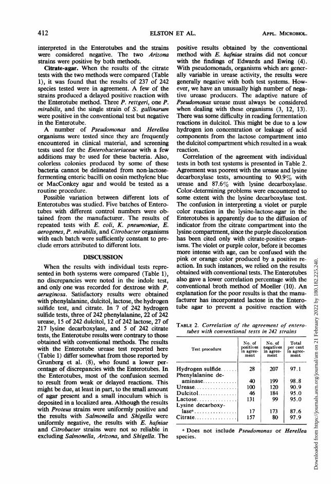
DISCUSSIONWhen the results with individual tests repre-
sented in both systems were compared (Table 1),no discrepancies were noted in the indole test,and only one was recorded for dextrose with P.aeruginosa. Satisfactory results were obtainedwith phenylalanine, dulcitol, lactose, the hydrogensulfide test, and citrate. In 7 of 242 hydrogensulfide tests, three of 242 phenylalanine, 22 of 242urease, 15 of 242 dulcitol, 12 of 242 lactose, 27 of217 lysine decarboxylase, and 5 of 242 citratetests, the Enterotube results were contrary to thoseobtained with conventional methods. The resultswith the Enterotube urease test reported here(Table 1) differ somewhat from those reported byGrunberg et al. (8), who found a lower per-centage of discrepancies with the Enterotubes. Inthe Enterotubes, most of the confusion seemedto result from weak or delayed reactions. Thismight be due, at least in part, to the small amountof agar present and a small inoculum which isdeposited in a localized area. Although the resultswith Proteus strains were uniformly positive andthe results with Salmonella and Shigella wereuniformly negative, the results with E. hafniaeand Citrobacter strains were not so reliable inexcluding Salmonella, Arizona, and Shigella. The
positive results obtained by the conventionalmethod with E. hafniae strains did not concurwith the findings of Edwards and Ewing (4).With pseudomonads, organisms which are gener-ally variable in urease activity, the results weregenerally negative with both test systems. How-ever, we have an unusually high number of nega-tive urease producers. The adaptive nature ofPseudomonas urease must always be consideredwhen dealing with these organisms (3, 12, 13).There was some difficulty in reading fermentationreactions in dulcitol. This might be due to a lowhydrogen ion concentration or leakage of acidcomponents from the lactose compartment intothe dulcitol compartment which resulted in a weakreaction.
Correlation of the agreement with individualtests in both test systems is presented in Table 2.Agreement was poorest with the urease and lysinedecarboxylase tests, amounting to 90.9% withurease and 87.6% with lysine decarboxylase.Color-determining problems were encountered tosome extent with the lysine decarboxylase test.The confusion in interpreting a violet or purplecolor reaction in the lysine-lactose-agar in theEnterotubes is apparently due to the diffusion ofindicator from the citrate compartment into thelysine compartment, since the purple discolorationhas been cited only with citrate-positive organ-isms. The violet or purple color, before it becomesmore intense with age, can be confused with thepink or orange color produced by a positive re-action. In such instances, we relied on the resultsobtained with conventional tests. The Enterotubesalso gave a lower correlation percentage with theconventional broth method of Moeller (10). Anexplanation for the poor results is that the manu-facturer has incorporated lactose in the Entero-tube agar to prevent a positive reaction with
TABLE 2. Correlation of the agreementt of entero-tubes with conventional tests in 242 strains
No. of No. of TotalTest procedure positives negatives per centin agree- in agree- in agree-
ment ment ment
Hydrogen sulfide 28 207 97.1Phenylalanine de-aminase............. 40 199 98.8
Urease................ 100 120 90.9Dulcitol............... 46 184 95.0Lactose............... 131 99 95.0Lysine decarboxy-
lasea ................ 17 173 87.6Citrate................ 157 80 97.9
aDoes not includespecies.
Pseudomonas or Herellea
412 APPL. MICROBIOL.
Dow
nloa
ded
from
http
s://j
ourn
als.
asm
.org
/jour
nal/a
m o
n 21
Feb
ruar
y 20
22 b
y 18
0.18
2.22
5.24
0.
MULTI-BIOCHEMICAL TEST SYSTEM
lactose-fermenting organisms. This is because themethod enhances the identification of certainSalmonella and is helpful in differentiating theseorganisms and Arizona strains from Citrobacter.Nonetheless, the exclusion of lactose from thelysine decarboxylase medium seems desirable toextend its range of usefulness to other membersof the Enterobacteriaceae family isolated fromthose specimens in which salmonellae andshigellae are not detected. This would increase theaccuracy of the test with rapid lactose-positiveArizona strains. Our strains were late lactosefermenters and both were lysine-positive.The results of the Enterotube citrate tests were
consistent when compared with Simmons citrateagar slants. These results disagree with those ofGrunberg et al. (8), who found 27 of 38 (71%)of the Enterotube tests in agreement with theSimmons citrate test. This is considerably lessthan our 97.9% agreement rate (Table 2).The urease test and dulcitol fermentation do
not always produce consistent results with certainbacteria by conventional tests so that negativeresults from these tests must be carefully scruti-nized. The lysine decarboxylase tests in theEnterotube system cannot be recommended asthey are at present constituted because of a con-siderable number of discrepancies that occurredbetween the Enterotubes and those obtained byconventional methods. The violet-to-purple colorthat appeared with some of our organisms makesinterpretation of the results difficult, if not impos-sible, to read.From Table 1 it may be seen that a number of
discrepancies exist between the Enterotube systemand conventional methods. Quite often theEnterotube identifies strains as to genus ratherthan species. Occasionally aberrant forms arerecognized which do not fit into the schema, butusually these are readily identified by furthertesting. It is obvious, of course, that the Entero-tube test is not useful for nonfermentative gram-negative rods which do not belong to the entericfamily. The addition of motility and gelatinstudies when using Enterotubes would be mosthelpful in the differentiation of Salmonella,Shigella, Klebsiella, Enterobacter, and Serratia.Additional fermentation studies in arabinose,raffinose, xylose, and sodium malonate may benecessary to separate Serratia organisms from E.liquefaciens. The anaerogenic nature of theseorganisms is also a differential point. We do notagree with Grunberg et al. (8) that rapid lactose-positive members of the Klebsiella-Aerobacter-Serratia groups can be classified as Klebsiella on
the basis of their reaction with lysine, dulcitol, andurease in the Enterotubes. Without other tests,
motility, gelatin, etc., these organisms probablyshould be identified only as to group. AlthoughP. aeruginosa and Herellea strains were tested,the Enterotube system was not designed for theidentification of these organisms.Without additional tests some of the members
of the Enterobacteriaceae family may be mis-identified by the Enterotube test system. Fromthe results obtained from the Enterotubes, one oftwo Shigella group A strains, the two group Bstrains, and the two Alkalescens-Dispar organ-isms were classified as atypical Escherichia. TheShigella group A strains gave negative lysinedecarboxylase tests by the conventional method,but one gave a false-positive in the Enterotube.Acid with no gas production in fermentativecarbohydrates, no reactions in lactose andsucrose, lysine decarboxylase, motility, and gelatintests were conventional procedures, along withserological studies that were used to identify thesestrains. The two indole-positive Klebsiella werefalsely identified as atypical Escherichia. Addi-tional testing revealed their true identity. Threeof four E. cloacae strains could not be differ-
TABLE 3. Frequenicy of error in identificationt ofstrains by the Enterotube system
No. ofOrganisms No. of errors in Per cent
strains identi- of errorfication
Shigella group A ....... 2 1 50.0Shigella group B ....... 2 2 100Shigella group D0......IAlkalescens-Dispar.... 2 2 100Escherichiaa........... 60 0 0
Klebsiella pneumoniae 60 2 3.3Enterobacter aero-genes................ 6 0 0
E. cloacae............. 4 3 75.0E. hafniae............. 12 0 0Serratia marcescens.. 4 4 100
Proteus mirabilis....... 27 0 0P. rettgeri............. 10 3 30.0P. morganii............ 4 0 0P. vulgaris............. 1 0 0Providence group...... 3 0 0
Salmoniella paratyphiBb................... 2 0 0
S. typhi............... 2 1 50.0S. gallinarum.......... 1 1I 100Salmonella group E... 1 0 0Citrobacter group 11 0 0Arizona arizonae....... 2 0 0
a Includes E. coli and atypical Escherichia.b Identified as to genus.
VOL. 22, 1971 413
Dow
nloa
ded
from
http
s://j
ourn
als.
asm
.org
/jour
nal/a
m o
n 21
Feb
ruar
y 20
22 b
y 18
0.18
2.22
5.24
0.
414 ELSTOI
entiated from E. aerogenes. A negative lysinedecarboxylase test, motility, and gelatinaseactivity were criteria used to identify E. cloacae.Without further study, the four nonchromogenicSerratia strains were incorrectly identified as E.hafniae in the Enterotubes. The Serratia strainswere separated by rapid liquefaction of gelatin,inability to attack sodium malonate, and ferment-ative characteristics in carbohydrates with littleor no gas production. Three of seven P. rettgeristrains could not be differentiated from P. morganiiin the Enterotubes because of their failure toutilize citrate and were called P. morganii.Fermentation tests with mannitol, inositol, andsucrose and the ornithine decarboxylase reactionwere conventional tests used to separate thesetwo organisms. Of the two Salmonella lysinedecarboxylase-negative strains, S. typhi was mis-identified as a Citrobacter organism, whereas S.gallinarum could not be differentiated. Usually,without serological study and a more elaborateset of biochemical tests the Salmonella organismscould be identified only as to group in the Entero-tubes as indicated by Grunberg et al. (8). Theseresults are complied in Table 3 to show thefrequency of error in identification of the abovestrains by the Enterotube system. This indicatesthat the use of the Enterotube test system mayresult in frequent misidentification of entericgram-negative bacilli.
According to our studies, there are obviousdeficiencies in the system. If these deficiencieswere amended by the manufacturer, then the useof a combined chemical unit such as offered bythe Enterotube is of value in rapid and simplebiochemical testing. Such a method would haveapplication in the busy diagnostic laboratory
ET AL. APPL. MICROBIOL.
which does not prepare or stock differentialmedia and reagents.
LITERATURE CITED
1. Amsterdam, D., and M. W. Wolfe. 1968. Comparison ofreagent-impregnated paper strips and conventional tests fordistinguishing Escherichia from Aerobacter: correlationwith colonial morphology. Appl. Microbiol. 16:1460-1464.
2. Christensen, W. B. 1946. Urea decomposition as a means ofdifferentiating Proteus and paracolon cultures from eachother and from Salmonella and Shigella types. J. Bacteriol.52:461-466.
3. DeTurk, W. 1955. The adaptive formation of urease by washedsuspension of Pseudomonas aeruginosa. J. Bacteriol. 70:187-191.
4. Edwards, P. R., and W. H. Ewing. 1964. Identification ofEnterobacteriaceae, 2nd ed. Burgess Publishing Co.,Minneapolis.
5. Elston, H. R. 1969. Comparative study of the paper striptechnique with the micro-spot plate and liquid mediummethods for niacin. Amer. Rev. Resp. Dis. 100:729-731.
6. Ewing, H. W., B. R. Davis, and R. W. Reavis. 1957. Phenyl-alanine and malonate medium and their uses in entericbacteriology. Pub. Health Lab. 15:153.
7. Gandelman, A. L., and P. H. Mann. 1965. An evaluation ofreagent-impregnated paper strips for use in the process ofidentifying certain species of clinically important bacteria.Curr. Ther. Res. 7:130-138.
8. Grunberg, E., E. Titsworth, G. Beskid, R. Cleeland, Jr., andW. F. Delorenzo. 1969. Efficiency of a multitest system(Enterotube) for rapid identification of Eniterobacteriaceae.Appl. Microbiol. 18:207-213.
9. Matsen, J. M. and J. C. Sherris. 1969. Comparative study ofseven paper-reagent strips and conventional biochemicaltests in identifying gram-negative organisms. Appl. Micro-biol. 18:452-457.
10. Moeller, V. 1955. Simplified tests for some amino acid de-carboxylases and for the arginine dihydrolase system. ActaPathol. Microbiol. Scand. 36:158-172.
11. Quigley, H. J., Jr., and H. R. Elston. 1970. Nitrite test stripfor detection of nitrate reduction by Mycobacteria. Amer.J. Clin. Pathol. 53:663-665.
12. Rhodes, M. D. 1959. The characterization of Pseudomonasfluorescens. J. Gen. Microbiol. 21:221-263.
13. Stewart, D. J. 1965. Urease activity of fluorescent pseudo-monads. J. Gen. Microbiol. 41:169-174.
N
Dow
nloa
ded
from
http
s://j
ourn
als.
asm
.org
/jour
nal/a
m o
n 21
Feb
ruar
y 20
22 b
y 18
0.18
2.22
5.24
0.