Síndromes Coronaris Sense Lesions Coronaries ... · Lesions Coronaries Significatives (SLCS,

Oral Lichenoid LesionsDifferences in expression of TLR4 and TLR9 in Oral Lichen Planus and amalgam induced Oral Lichenoid Lesions
Mariken BrecheisenJulia PerssonHandledare:Liv Kroona, Oral patologi Gunnar Warfvinge, Oral patologi
Examensarbete (30 hp) Malmö högskolaTandläkarprogrammet Odontologiska fakulteten2014-02-14 205 06 Malmö

140214
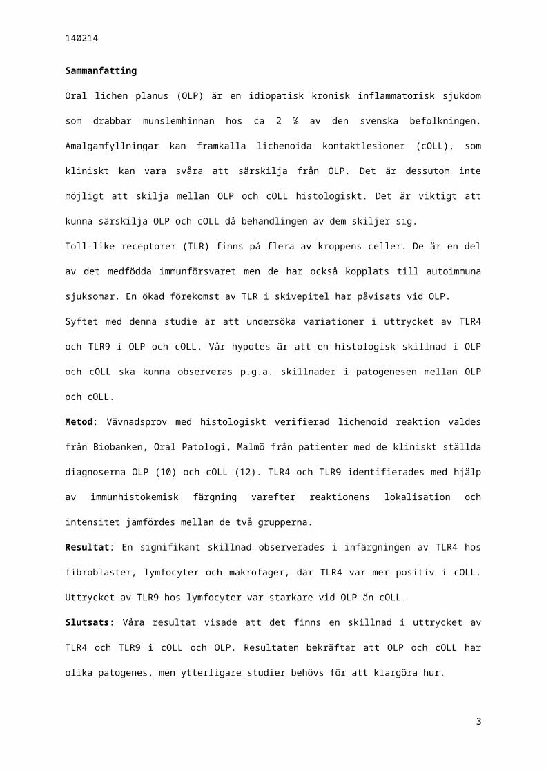
Sammanfatting
Oral lichen planus (OLP) är en idiopatisk kronisk inflammatorisk sjukdom som drabbar munslemhinnan
hos ca 2 % av den svenska befolkningen. Amalgamfyllningar kan framkalla lichenoida kontaktlesioner
(cOLL), som kliniskt kan vara svåra att särskilja från OLP. Det är dessutom inte möjligt att skilja mellan
OLP och cOLL histologiskt. Det är viktigt att kunna särskilja OLP och cOLL då behandlingen av dem
skiljer sig.
Toll-like receptorer (TLR) finns på flera av kroppens celler. De är en del av det medfödda
immunförsvaret men de har också kopplats till autoimmuna sjuksomar. En ökad förekomst av TLR i
skivepitel har påvisats vid OLP.
Syftet med denna studie är att undersöka variationer i uttrycket av TLR4 och TLR9 i OLP och cOLL.
Vår hypotes är att en histologisk skillnad i OLP och cOLL ska kunna observeras p.g.a. skillnader i
patogenesen mellan OLP och cOLL.
Metod: Vävnadsprov med histologiskt verifierad lichenoid reaktion valdes från Biobanken, Oral
Patologi, Malmö från patienter med de kliniskt ställda diagnoserna OLP (10) och cOLL (12). TLR4 och
TLR9 identifierades med hjälp av immunhistokemisk färgning varefter reaktionens lokalisation och
intensitet jämfördes mellan de två grupperna.
Resultat: En signifikant skillnad observerades i infärgningen av TLR4 hos fibroblaster, lymfocyter och
makrofager, där TLR4 var mer positiv i cOLL. Uttrycket av TLR9 hos lymfocyter var starkare vid OLP
än cOLL.
Slutsats: Våra resultat visade att det finns en skillnad i uttrycket av TLR4 och TLR9 i cOLL och OLP.
Resultaten bekräftar att OLP och cOLL har olika patogenes, men ytterligare studier behövs för att
klargöra hur.
2
140214
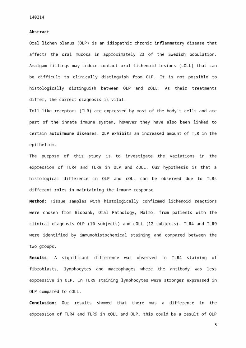
Abstract
Oral lichen planus (OLP) is an idiopathic chronic inflammatory disease that affects the oral mucosa in
approximately 2% of the Swedish population. Amalgam fillings may induce contact oral lichenoid
lesions (cOLL) that can be difficult to clinically distinguish from OLP. It is not possible to histologically
distinguish between OLP and cOLL. As their treatments differ, the correct diagnosis is vital.
Toll-like receptors (TLR) are expressed by most of the body's cells and are part of the innate immune
system, however they have also been linked to certain autoimmune diseases. OLP exhibits an
increased amount of TLR in the epithelium.
The purpose of this study is to investigate the variations in the expression of TLR4 and TLR9 in OLP
and cOLL. Our hypothesis is that a histological difference in OLP and cOLL can be observed due to
TLRs different roles in maintaining the immune response.
Method: Tissue samples with histologically confirmed lichenoid reactions were chosen from Biobank,
Oral Pathology, Malmö, from patients with the clinical diagnosis OLP (10 subjects) and cOLL (12
subjects). TLR4 and TLR9 were identified by immunohistochemical staining and compared between
the two groups.
Results: A significant difference was observed in TLR4 staining of fibroblasts, lymphocytes and
macrophages where the antibody was less expressive in OLP. In TLR9 staining lymphocytes were
stronger expressed in OLP compared to cOLL.
Conclusion: Our results showed that there was a difference in the expression of TLR4 and TLR9 in
cOLL and OLP, this could be a result of OLP being an autoimmune disorder. Further studies on this
subject are recommended on this subject.
MeSH: "Dental Amalgam", "Dermatitis, Allergic Contact", "Immunohistochemistry", "Lichen Planus,
Oral", "Toll-Like Receptor 4", "Toll-Like Receptor 9"
3

140214
Introduction
Oral lichen planus and oral lichenoid reaction lesions
Oral Lichen Planus (OLP) is a chronic inflammatory disorder, occurring in 1.85% of the adult
population in Sweden, and is most common among middle-aged women (1). The clinical and
histological features of OLP are well-defined lesions generally located in the buccal mucosa and may
also occur on the gingiva, tongue, floor of the mouth and lips (1-3). Symptoms can vary from simply a
feeling of tissue “roughness” to severe pain (4). Clinically there are six types of OLP; papular, reticular,
plaque, atrophic, bullous and erosive (1). The reticular type is the most common form characterized by
white striations often localized bilaterally in the buccal mucosa (5, 6). The less common ulcerative
type, usually located on the tongue and gingiva, is characterized as red lesions and often with
concurrent symptoms (2). OLP is believed to be an autoimmune disease but the pathogenesis is still
not fully known (7).
Oral lichenoid lesions (OLL) are lesions that are similar to OLP but either have a different etiology or
an atypical clinical appearance. Oral contact allergy may appear as an OLL and is often associated
with dental materials (8-10). It is currently not possible to differentiate between OLL and OLP by
merely examining their histological features (3). Histologically, OLP and OLL are characterized by
band like lymphocytic infiltrate below the basal layer, hyperkeratosis and saw-tooth ridged epithelium
(5). The lesions typically exhibit degeneration of the basal cell layer (11). Civatte bodies are dead
keratinocytes and commonly seen in the epithelium and are most common in thinner epithelium. (12)
Amalgam
Amalgam fillings may induce contact allergy with an appearance of OLL (cOLL). These lesions are
usually limited to juxtaposed mucosa (13). Dental amalgam contains 50% mercury mixed with other
metals. Mercury may form haptens that initiate a delayed hypersensitivity reaction. Clinically, the
reaction manifests as an amalgam-associated OLL. Studies show that in patients with cOLL, 27% to
70% had a positive reaction to mercury in patch-tests. Consequently, sensitization to mercury does not
necessarily predict which patients will develop cOLL (14). Individuals with cOLL have significantly
higher lymphocyte reactivity to inorganic mercury due to chronic low grade exposure that causes a
systemic sensitization (15). Removal of amalgam fillings adjacent to cOLL results in an improvement
4
140214
to the mucosa in 92% to 98% of the patients. Histological differentiation between OLP and cOLL has a
low sensitivity and specificity of 40% and 32%, respectively (14).
Toll-like receptors (TLR)
Toll-like receptors (TLR) are glycoproteins located on cell membranes that are expressed by
numerous different cells, including antigen presenting cells (APC) and other immune cells (16-18).
TLR is a key sensor for countering invading bacteria, virus and allergens and binds to components
such as lipoproteins, peptidoglycans, LPS, single- or double stranded RNA and viral DNA (19, 20).
Oral epithelial cells express more TLR compared to other human cells (21). Bound TLR can initiate
APC and generate a T-cells response by upregulation of cytokines and an enhanced expression of the
major histocompatibility complex II (MHC-II) (17, 22). This shows that TLR plays a big part in the auto
reactivity of T-cells and thereby also for the innate and adaptive immune response (21, 23, 24).
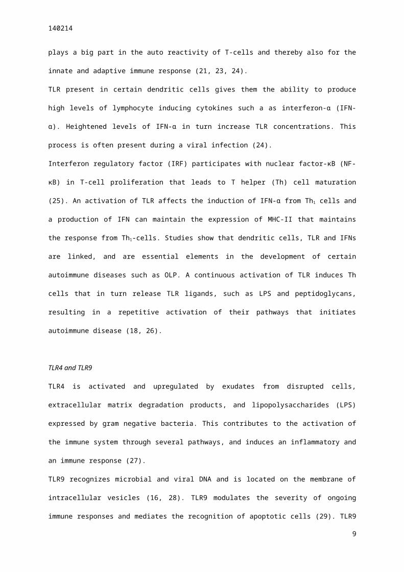
TLR present in certain dendritic cells gives them the ability to produce high levels of lymphocyte
inducing cytokines such a as interferon-α (IFN-α). Heightened levels of IFN-α in turn increase TLR
concentrations. This process is often present during a viral infection (24).
Interferon regulatory factor (IRF) participates with nuclear factor-κB (NF-κB) in T-cell proliferation that
leads to T helper (Th) cell maturation (25). An activation of TLR affects the induction of IFN-α from Th1
cells and a production of IFN can maintain the expression of MHC-II that maintains the response from
Th1-cells. Studies show that dendritic cells, TLR and IFNs are linked, and are essential elements in the
development of certain autoimmune diseases such as OLP. A continuous activation of TLR induces
Th cells that in turn release TLR ligands, such as LPS and peptidoglycans, resulting in a repetitive
activation of their pathways that initiates autoimmune disease (18, 26).
TLR4 and TLR9
TLR4 is activated and upregulated by exudates from disrupted cells, extracellular matrix degradation
products, and lipopolysaccharides (LPS) expressed by gram negative bacteria. This contributes to the
activation of the immune system through several pathways, and induces an inflammatory and an
immune response (27).
TLR9 recognizes microbial and viral DNA and is located on the membrane of intracellular vesicles (16,
28). TLR9 modulates the severity of ongoing immune responses and mediates the recognition of
5

140214
apoptotic cells (29). TLR9 activates dendritic cells, macrophages, B-cells and also starts a Th1-cell
response (28).
The MyD88 pathway is a cellular signal system important for all TLR to meditate the production of
cytokines (26). When activated, both TLR4 and TLR9 signal via the adaptor protein MyD88, however,
the consequences of the initiation differ (30). Signals through MyD88 activate transcription factor NF-
κB and lead to an induced expression of pro-inflammatory cytokines (20).
TLR4, activated by LPS, signals via two pathways: early MyD88 dependent reaction, and late TIR-
domain-containing adapter-inducing interferon-β (TRIF) reaction (20, 31). For TLR4 to create a
cytokine response, it needs to activate both the MyD88 and TRIF, while TLR9 only requires a single
pathway to induce a cytokine response (28).
Stimulation of TLR4 leads to an activation of transcription factor interferon regulatory factor 3 (IRF-3)
which will lead to an IFN-β production that maintains the inflammation (19, 32). TRIF is responsible for
the delayed activation of NF-κB that activates the maturation of dendritic cells (26, 31). (Fig. 1)
The amount of expressed NF- κB correlates to the amount of expressed TLR4, and TLR4 may be the
basis for maintaining the inflammatory response (23).
Li et.al found that there is an increased expression of TLR9 in OLP compared to normal mucuous
membrane. TLR9 is also seen in dendritic cells present in auto immune skin diseases, but not in
healthy controls (24).
Sensitization and TLR
Studies have shown that patients with cOLL, exhibiting hypersensitivity towards components in
amalgam, often are hypersensitive towards nickel (33). Nickel has been shown to directly activate
TLR4 signaling by crosslinking of histidine residues on the TLR. Just like LPS, the activation of TLR4
by nickel generates a proinflammatory gene expression that can generate a co-stimulatory signal
during T-cell activation. Thus, contact allergy to nickel can be promoted via TLR4 (34). Cobalt and
palladium has been shown to react in a similar way as nickel (35), but it is still not known whether
mercury has a role in TLR-activation.
6

140214
Keratinocytes are required cells for developing contact dermatitis, as they respond to TLR4-triggering
haptens, by secreting IL-1β and IL-18, as well as TNF. These cytokines activate dendritic cells and
promotes cell migration to the lymph nodes (20).
Purpose of this study
The current consensus of studies is that it histologically is not possible to differentiate between OLP
and cOLL (36). The clinical differences between the lesions are also not clear cut (10). It is important
to distinguish between OLP and cOLL since the treatment of the two differs considerably. OLP often
requires extensive corticosteroid treatment, whereas cOLL can resolve by simply removing the
adjacent amalgam filling (10, 14). Due to the different causative agents, the immunologic reaction
could differ, leaving discernable traces. Hypothetically, these traces could be identified by detecting
deviances in the reactions.
The purpose of this study is to immunohistochemically differentiate between OLP and cOLL by
examining variations in the expression of TLR4 and TLR9 with immunohistochemical staining.
Our hypothesis is that there is a difference in TLR-expression between OLP and cOLL that can be
observed due to TLRs separate roles in maintaining the immune response depending on the present
TLR-ligands.
This study could give the clinician an alternate tool for diagnosing and distinguishing OLP from cOLL.
7

140214
Material and method
Study population
The biopsies were retrieved from the patient data at Malmö University’s PAD database from the
Biobank.
The subjects were chosen from patients with diagnoses of either OLP or cOLL. The OLP group
required bilateral lesions while the cOLL lesions were required to be solely located by amalgam
fillings. Subjects with Candida albicans infection or with known medication were excluded. The
subjects in the cOLL group were selected from an earlier study when the diagnosis had been
confirmed.
The subjects were divided into two groups comprising healthy, non-smoking, non-medicated patients:
the first group with idiopathic OLP, the second group with amalgam induced cOLL.
The OLP biopsies were from 2012 and 2013 where the average subject age was 55 years old with a
standard deviation of 18 years. The cOLL biopsies were from 1987 and 1988, where the average age
was 38 years old with a standard deviation of 9 years. There were 4 men and 6 women in the OLP
group and 5 men and 7 women in the cOLL group. A power of 80% was considered significant.
Immunohistochemical staining
Polyclonal rabbit antibodies to human TLR4 and monoclonal mouse antibodies to human TLR9 were
purchased from Abcam, United Kingdom.
The dilutions were set after evaluating background staining to 1:50 for the TLR4 antibody and 1:100
for the TLR9 antibody. Three µm sections on Thermo Scientific+ glasses were dried at 57°C for 1
hour. Paraffin was removed in graded ethanol. The sections were microwaved in TEG-buffer at 98°C
for 24 min to expose antigen epitopes in the fixed material. The sections were incubated for 30 min
with Bovine Serum Albumin (BSA) and then with primary antibody for 60 min. Excess antibodies were
rinsed off with TRIS-buffer. The sections were then incubated with hydrogen peroxide blocking
solution for 10 min followed by Envision for 20 min. The sections were rinsed with TRIS-buffer and
then incubated with HRP-substrate (DAB) for 12 min. Afterwards, the sections were counterstained
with Hematoxylin for 1 min and mounted.
Biopsies from both healthy mucosa and lymph nodes were used as positive controls to verify the
validity of the method and to reveal potential discrepancies between older and newer biopsies that
8
140214
would affect the results. A negative control was performed using an irrelevant non-binding antibody as
well as a control without a primary antibody.
Sample processing
The results of the TLR staining were divided into three categories; negative, weakly positive and
strongly positive. Negative was defined as the absence of staining, strongly positive as intense
staining of the majority of the specific cells, while weakly positive was characterized with less intense
staining.
Histological features such as epithelial form, thickness and degree of keratinization, present
inflammatory and non-inflammatory cells and their locations were recorded.
Calibration between the two observers was achieved by following a protocol during individual and
collective recording of the results and calibration with our supervisor.
The differences between OLP and cOLL sections stained with TLR4 and TLR9 respectively were
statistically evaluated with Mann-Whitney U-test. P ≤ 0.05 was considered to be statistically significant.
9

140214
Results
Hematoxylin and eosin stained sections
Histological differences included a slightly thicker basal cell layer in OLP compared to cOLL. Civatte
bodies were common in OLP and rarely found in cOLL. Plasma cells were present in half of the OLP
cases and half of the cOLL cases, however the location differed. In OLP the plasma cells were located
generally located in the submucosa, while plasma cells found in cOLL were located in the lamina
propria adjacent to the basal cell layer. It was common for both OLP and cOLL to exhibit only a few
lymphocytes in the epithelial layer. The thickness of the inflammatory infiltrate was similar in both
lesions where the band of infiltrating cells varied between broad and patchy. Macrophages were
present in the infiltrate in both lesions though the amount of present macrophages differed.
Immunohistochemical staining
TLR4 and TLR9 expression in the two test groups are summarized in figure 2 and 3.
Inflammatory cells. In OLP stained with TLR4, only 1 of 10 slides expressed positive lymphocytes
while in cOLL, all of the slides expressed positive lymphocytes. The difference was significant (p<
0.001).
In OLP stained with TLR9, 8 of 10 slides were strongly positive in lymphocytes and 1 of 10 were
weakly positive, compared to cOLL where 3 of 12 slides were strongly positive and 6 of 12 were
weakly positive, with a significant difference (p=0.036).
The majority of the plasma cells were negative in all groups.
Macrophages were less positive for TLR4 in OLP compared to cOLL. In OLP only 2 of 10 slides had
macrophages expressing TLR4, while in cOLL 9 of 10 slides had macrophages expressing TLR4
which was a significant difference (p=0.007). In TLR9, some macrophages were positive with slight
variations between the groups but there was no significant difference.
The majority of mast cells were strongly positive for TLR4 and TLR9 in all OLP and cOLL slides.
Non-inflammatory cells. TLR4 expression in fibroblasts showed a significant difference between OLP
and cOLL (p=0.011). In OLP, 6 of 10 slides showed a weakly positive expression while the expression
in cOLL slides showed 6 strongly positive and 3 weakly positive slides out of 10. Fibroblast in OLP and
cOLL, both showed a strong expression of TLR9 fibroblasts.
10

140214
Endothelial cells in OLP stained with TLR4 showed that half of the slides were strongly positive and 1
slide weakly positive. Similar results were seen in cOLL where half of the slides were strongly positive
and 2 were weakly positive for TLR4. Endothelial cells in OLP and cOLL expressed TLR9 with varying
intensity.
Non-specific staining. Non-specific staining was defined as diffuse staining not limited to individual
cells or certain structures, but as a general staining present in the whole slide. In the epithelial layer
non-specific staining was common with TLR4 antibodies where two 2 of the OLP slides and 4 of the
cOLL slides had strong non-specific staining. With TLR9 antibodies 5 of the OLP slides and 5 of the
cOLL slides had weak non-specific staining. In cOLL the connective tissue had strong non-specific
staining in 1 slide and weak non-specific staining in 3 slides. Only 1 of the TLR9 slides showed a weak
non-specific staining of the connective tissue in cOLL. None of the OLP slides stained with TLR9
showed non-specific staining.
Due to excessive non-specific staining 2 samples from the cOLL group stained with TLR4 were
excluded.
11
140214
Discussion
Our results implies that despite only minor histological differences between OLP and cOLL there is a
possibility to differentiate the two based on differences in TLR expression.
The thicker basal cell layer present in OLP might be affected by the lesion’s current state at the time
the biopsy was taken. The histological appearance of OLP in an initial stage may differ from OLP in an
advanced stage, as a decrease in epithelial thickness is linked to the more advanced stages (12).
Therefore our recording of a thicker basal cell layer in OLP compared to cOLL is not necessarily a
reliable marker to histologically differentiate OLP and cOLL.
The presence of Civatte bodies was seen more often in the OLP slides compared to the cOLL slides.
Earlier studies have discussed the correlation between the amount of lymphocyte infiltration in the
epithelium and the amount of Civatte bodies in the basal cell layer (37). In our findings, we saw the
same amount of lymphocyte infiltrate in the epithelium in both OLP and cOLL. The amount of Civatte
bodies differed between the two groups. This may therefore be the result of basal keratinocyte
destruction (12) attributed to a higher cell activity that results in Civatte bodies. The difference in the
amount of present Civatte bodies could be due to the fact that cOLL and OLP have different etiologies,
where cOLL are induced by the presence of Hg haptens that diffuse in to the tissue while OLP’s
etiology is still unknown.
The higher presence of plasma cells observed in cOLL correlates to findings made by Thornhill et al
where plasma cells in the connective tissue were seen (36). This may be attributable to cOLL being a
reaction caused by a specific hapten and triggering a greater amount of plasma cells compared to the
amount present in OLP (20).
Macrophages and plasma cells were not always possible to differentiate. This problem could have
been alleviated by a double staining with cell specific antibodies.
Two of the cOLL stained with TLR4 were excluded due to a very strong non-specific staining. It would
have been ideal to redo the excluded slides to avoid reducing the number of participating subjects.
This was not possible in the scope of this study. The strong non-specific staining is attributable to the
12
140214
fact that the IHC protocol was performed manually allowing varying degrees of staining to have
occurred. More standardized results could have been achieved utilizing an IHC-machine.
Although TLR4 is a membrane receptor, it was expressed by the cytoplasm. This anomaly is
consistent with the results of earlier studies which gave similar results (38). A possible explanation for
this is that TLR4 is a membrane receptor also located in intracellular vesicles (29).
We observed a positive expression of TLR4 in the basal cell layer that correlates to studies by
Janardhanam et al where they observed an up-regulated expression of TLR4 in OLP compared to a
healthy oral epithelium (21).
Our results showed a negative expression of TLR9 in the epithelium. However, in some of the cases
we could see a weak staining but not enough to grade. Our negative results are contradictory to
previous results by Siponen et.al who observed positive staining of TLR9 in the outer epithelium (16).
This can be explained by the use of different antibodies, protocol and interpretation of the sections by
different operators. (Fig. 4)
In cOLL there was a high amount of subepithelial lymphocytes expressing TLR4. In OLP where very
few subepithelial lymphocytes expressed TLR4 and this corroborates with studies by Siponen et.al
that also showed a weak TLR4 expression in the subepithelial inflammatory infiltrate though they
compared this to the TLR4 expression in the basal cell layer of OLP (16). TLR4 staining could
therefore be a valid method to histopathologically differentiate OLP from cOLL. However, it is possible
that the positive staining in cOLL is attributed to the older age of the biopsies as they generally had a
stronger staining. (Fig 4)
Slides stained with TLR9 showed a significant difference in the amount of strongly positive and weakly
positive lymphocytes that were more intense in OLP. The increased expression could be a result of
OLP being an autoimmune disorder affected by dendritic cells expressing TLR9 (24).
Further studies on this subject are recommended to substantiate the conclusions of this study. To
correctly identify which cells express TLR4 and TLR9, cell specific staining could be performed in
combination with TLR4 and TLR9 staining.
The results imply that, by differentiating OLP and cOLL via examining TLR4, may help the clinician
confirm the correct diagnosis. The resulting diagnosis can be used as a basis for choosing the
13

140214
following treatment, such as removal of current filling materials. For example, patients with cOLL that
previously haven’t been correctly diagnosed could avoid prolonged treatment with corticosteroids. This
could have a clinical and economic relevance for patients and healthcare givers.
14

140214
Conclusions
Our results, combined with earlier studies, show that there are detectable histopathologic differences
between OLP and cOLL. However, the characteristics typical for the one lesion can still be present in
the other lesions and therefore they might not be sufficient for histologic differentiation. A difference in
the expression of TLR4 in lymphocytes, macrophages and fibroblasts was observed between the
groups, as well as a difference in the expression of TLR9 in the lymphocytes. These results confirm
the consensus that OLP and cOLL have different pathogenesis. Further research is needed to
corroborate these results.
15
140214
Acknowledgements
Ethical approval
No ethical approval was needed to perform this study.
Source of funding
This project was founded by the Pathology Department of Malmö University.
Acknowledgments
Special thanks to the Pathology Department of Malmö University for all the help and support during
this project.
References
1. Axell T. A prevalence study of oral mucosal lesions in an adult Swedish population. Odontol Revy Suppl. 1976; 36: 1-103.
2. Scully C, Beyli M, Ferreiro MC, Ficarra G, Gill Y, Griffiths M et al. Update on oral lichen planus: etiopathogenesis and management. Crit Rev Oral Biol Med. 1998; 9: 86-122.
3. Wright J. Diagnosis and management of oral lichenoid reactions. J Calif Dent Assoc. 2007; 35: 412-416.
4. Scully C, Carrozzo M. Oral mucosal disease: Lichen planus. Br J Oral Maxillofac Surg. 2008; 46: 15-21.
5. Thornhill MH. Immune mechanisms in oral lichen planus. Acta Odontol Scand. 2001; 59: 174-177.
6. Lu R, Zhou G, Du G, Xu X, Yang J, Hu J. Expression of T-bet and GATA-3 in peripheral blood mononuclear cells of patients with oral lichen planus. Arch Oral Biol. 2011; 56: 499-505.
7. Charazinska-Carewicz K, Ganowicz E, Krol M, Gorska R. A: Assessment of the peripheral immunocompetent cells in patients with reticular and atrophic-erosive lichen planus. Oral Surg Oral Med Oral Pathol Oral Radiol Endod. 2008; 105: 202-205.
8. Mega H, Jiang WW, Takagi M. Immunohistochemical study of oral lichen planus associated with hepatitis C virus infection, oral lichenoid contact sensitivity reaction and idiopathic oral lichen planus. Oral Dis. 2001; 7: 296-305.
9. Cortes-Ramirez DA, Rodriguez-Tojo MJ, Gainza-Cirauqui ML, Martinez-Conde R, Aguirre-Urizar JM. Overexpression of cyclooxygenase-2 as a biomarker in different subtypes of the oral lichenoid disease. Oral Surg Oral Med Oral Pathol Oral Radiol Endod. 2010; 110: 738-743.
10. van der Waal I. Oral lichen planus and oral lichenoid lesions; a critical appraisal with emphasis on the diagnostic aspects. Med Oral Patol Oral Cir Bucal. 2009; 14: E310-4.
11. Huber MA. White oral lesions, actinic cheilitis, and leukoplakia: confusions in terminology and definition: facts and controversies. Clin Dermatol. 2010; 28: 262-268.
12. Brant JM, Vasconcelos AC, Rodrigues LV. Role of apoptosis in erosive and reticular oral lichen planus exhibiting variable epithelial thickness. Braz Dent J. 2008; 19: 179-185.
13. Little MC, Griffiths CE, Watson RE, Pemberton MN, Thornhill MH. Oral mucosal keratinocytes express RANTES and ICAM-1, but not interleukin-8, in oral lichen planus and oral lichenoid reactions induced by amalgam fillings. Clin Exp Dermatol. 2003; 28: 64-69.
14. McCullough MJ, Tyas MJ. Local adverse effects of amalgam restorations. Int Dent J. 2008; 58: 3-9.
15. Stejskal VD, Forsbeck M, Cederbrant KE, Asteman O. Mercury-specific lymphocytes: an indication of mercury allergy in man. J Clin Immunol. 1996; 16: 31-40.
16

140214
16. Siponen M, Kauppila JH, Soini Y, Salo T. TLR4 and TLR9 are induced in oral lichen planus. J Oral Pathol Med. 2012.
17. Abdelsadik A, Trad A. Toll-like receptors on the fork roads between innate and adaptive immunity. Hum Immunol. 2011; 72: 1188-1193.
18. Trucci VM, Salum FG, Figueiredo MA, Cherubini K. Interrelationship of dendritic cells, type 1 interferon system, regulatory T cells and toll-like receptors and their role in lichen planus and lupus erythematosus -- a literature review. Arch Oral Biol. 2013; 58: 1532-1540.
19. Takeda K, Akira S. Toll-like receptors in innate immunity. Int Immunol. 2005; 17: 1-14.
20. Kaplan DH, Igyarto BZ, Gaspari AA. Early immune events in the induction of allergic contact dermatitis. Nat Rev Immunol. 2012; 12: 114-124.
21. Janardhanam SB, Prakasam S, Swaminathan VT, Kodumudi KN, Zunt SL, Srinivasan M. Differential expression of TLR-2 and TLR-4 in the epithelial cells in oral lichen planus. Arch Oral Biol. 2012; 57: 495-502.
22. Trompette A, Divanovic S, Visintin A, Blanchard C, Hegde RS, Madan R et al. Allergenicity resulting from functional mimicry of a Toll-like receptor complex protein. Nature. 2009; 457: 585-588.
23. Ge Y, Xu Y, Sun W, Man Z, Zhu L, Xia X et al. The molecular mechanisms of the effect of Dexamethasone and Cyclosporin A on TLR4 /NF-kappaB signaling pathway activation in oral lichen planus. Gene. 2012; 508: 157-164.
24. Li J, Chen J, Tan Z, Liu H, Liu Z. Expression of TLR9 and its mRNA in the lesions of lichen planus. J Huazhong Univ Sci Technolog Med Sci. 2007; 27: 203-205.
25. Meyer O. Interferons and autoimmune disorders. Joint Bone Spine. 2009; 76: 464-473.
26. Akira S, Takeda K. Toll-like receptor signalling. Nat Rev Immunol. 2004; 4: 499-511.
27. Garate I, Garcia-Bueno B, Madrigal JL, Caso JR, Alou L, Gomez-Lus ML et al. Stress-Induced Neuroinflammation: Role of the Toll-like Receptor-4 Pathway. Biol Psychiatry. 2012.
28. Kawai T, Akira S. The role of pattern-recognition receptors in innate immunity: update on Toll-like receptors. Nat Immunol. 2010; 11: 373-384.
29. Miles K, Heaney J, Sibinska Z, Salter D, Savill J, Gray D et al. A tolerogenic role for Toll-like receptor 9 is revealed by B-cell interaction with DNA complexes expressed on apoptotic cells. Proc Natl Acad Sci U S A. 2012; 109: 887-892.
30. Chuang TH, Lee J, Kline L, Mathison JC, Ulevitch RJ. Toll-like receptor 9 mediates CpG-DNA signaling. J Leukoc Biol. 2002; 71: 538-544.
31. Palsson-McDermott EM, O'Neill LA. Signal transduction by the lipopolysaccharide receptor, Toll-like receptor-4. Immunology. 2004; 113: 153-162.
32. Doyle S, Vaidya S, O'Connell R, Dadgostar H, Dempsey P, Wu T et al. IRF3 mediates a TLR3/TLR4-specific antiviral gene program. Immunity. 2002; 17: 251-263.
33. Ahlgren C, Axell T, Moller H, Isaksson M, Liedholm R, Bruze M. Contact allergies to potential allergens in patients with oral lichen lesions. Clin Oral Investig. 2013.
34. Schmidt M, Raghavan B, Muller V, Vogl T, Fejer G, Tchaptchet S et al. Crucial role for human Toll-like receptor 4 in the development of contact allergy to nickel. Nat Immunol. 2010; 11: 814-819.
35. Rachmawati D, Bontkes HJ, Verstege MI, Muris J, von Blomberg BM, Scheper RJ et al. Transition metal sensing by Toll-like receptor-4: next to nickel, cobalt and palladium are potent human dendritic cell stimulators. Contact Dermatitis. 2013; 68: 331-338.
36. Thornhill MH, Sankar V, Xu XJ, Barrett AW, High AS, Odell EW et al. The role of histopathological characteristics in distinguishing amalgam-associated oral lichenoid reactions and oral lichen planus. J Oral Pathol Med. 2006; 35: 233-240.
37. Bloor BK, Malik FK, Odell EW, Morgan PR. Quantitative assessment of apoptosis in oral lichen planus. Oral Surg Oral Med Oral Pathol Oral Radiol Endod. 1999; 88: 187-195.
17

140214
38. Uronen-Hansson H, Allen J, Osman M, Squires G, Klein N, Callard RE. Toll-like receptor 2 (TLR2) and TLR4 are present inside human dendritic cells, associated with microtubules and the Golgi apparatus but are not detectable on the cell surface: integrity of microtubules is required for interleukin-12 production in response to internalized bacteria. Immunology. 2004; 111: 173-178.
18

140214
Fig.1: TLR4 and TLR9 signaling pathways. LPS and viral DNA binds to TLR4 and TLR9. This will
lead to an activation of the TIR domain-containing adaptor, MyD88, that will bind to the receptor by
ligand binding. This will result in a translocation of NF-κB which induces expression of inflammatory
cytokines. TRIF is essential for the MyD88-independent pathway where there is an activation of IRF-3
and induction of IFN-β.
19
140214
Fig.2: Staining with TLR4. The OLP (a) has no expression of lymphocytes compared to the
lymphocytes in the cOLL (b), where is shows a positive expression. The arrow shows a positive
expression of endothelial cells.
20

140214
Fig. 3: Results of TLR expression. The tables show the amount of expressed TLR4 and TLR9 in
OLP compared to cOLL. There was a significant difference in fibroblasts, lymphocytes and epithelium
stained with TLR4 and lymphocytes stained with TLR9.
* marks the significant differences in the figure.
21

140214
Fig. 4: Stained OLP and cOLL. Picture a) shows OLP expressing TLR4 and picture b) shows OLP
expressing TLR9. Picture c) shows cOLL expressing TLR4 and picture d) shows cOLL expressing
TLR9. In both slides TLR4 is more expressive in the epithelium. cOLL has a thicker basal cell layer
compared to OLP.
22