Mucopolysaccharidosis VI: Pathophysiology, diagnosis and ...
Mucopolysaccharidosis Type I SubtypesPresence of Immunologically Cross-reactive Materialand In Vitro Enhancement of the Residual a-L-lduronidase Activities
Edward H. Schuchman and Robert J. DesnickDivision of Medical Genetics, Mount Sinai School of Medicine, New York, New York 10029
Abstract
The enzymatic and immunologic properties of the defectiveresidual a-L-iduronidase activities were investigated in fibro-blast extracts from the three subtypes of mucopolysacchari-dosis type I, Hurler (MPS IH), Scheie (MPS IS), and Hurler-Scheie (MPS IH-S) diseases. Using 4-methylumbelliferyl-a-L-iduronide (4MU-a-Id), the activities in fibroblast extractsfrom all three subtypes were less than 0.1% of normal. Rocketimmunoelectrophoresis with monospecific rabbit anti-humana-L-iduronidase polyclonal antibodies, as well as immunoblotsusing a monoclonal antibody, revealed the presence of cross-reactive immunologic material (CRIM) in extracts preparedfrom each subtype. When the samples were equalized for 8-hexosaminidase A activity, 38-105% of normal enzyme proteinwas detected. The sequential addition of cystamine, MgCl2 andpyridoxal phosphate increased the residual 4MU-a-Id activi-ties in subtype extracts up to about 35%of normal mean fibro-blast activity. Cystamine, MgC2or pyridoxal phosphate aloneenhanced the residual activities two- to fourfold, whereas thesequential addition of all three compounds was required formaximal effect. Of the six B6 vitamers evaluated, only thenegatively charged forms, pyridoxamine (PLN), pyridoxaminephosphate (PNP), and pyridoxal phosphate (PLP), stimulatedthe residual activities. The addition of dermatan sulfate or he-paran sulfate to the subtype extracts, followed by treatmentwith the effector compounds, similarly inhibited both the nor-mal and enhanced MPSI activities. Heat inactivation experi-ments confirmed the fact that the mutant iduronidase activitywas reconstituted and that the observed increase in enzymaticactivity was not an artifact of the fluorogenic assay. Theseresults suggest that the presence of certain thiol reducingagents, divalent cations and negatively charged B6 vitamers canalter the conformation of the mutant ac-L-iduronidase in vitrosuch that the hydrolysis of 4MU-a-Id is enhanced into theheterozygote range.
Introduction
The type I mucopolysaccharidoses represent three clinicallydelineated, autosomal recessive disorders (Hurler disease,MPSIH; Scheie disease, MPSIS; and Hurler-Scheie disease,
Address reprint requests to Dr. Schuchman, Division of Medical Ge-netics, Mount Sinai School of Medicine, Fifth Avenue and 100thStreet, NewYork, NY 10029.
Received for publication 31 October 1986 and in revised form 4June 1987.
MPSIH-S)1 that result from the deficient activity of the lyso-somal hydrolase, a-L-iduronidase (a-L-iduronide iduronohy-drolase, E.C. 3.2.1.76) (1, 2) and the subsequent lysosomalaccumulation of the glycosaminoglycans (GAGs), dermatansulfate (DS), and heparan sulfate (HS) (3). MPSIH is charac-terized by severe dysostosis multiplex, a characteristic facialdysmorphia, corneal clouding, hepatosplenomegaly, and se-vere mental retardation with demise usually before the seconddecade of life. In contrast, affected individuals with MPSIShave mild dysostosis multiplex, mild corneal clouding, aorticvalvular involvement, normal mentation, and a normal life-span. Patients with MPSIH-S present with an intermediatephenotype (4). Although it had been proposed that MPSIH-Swas the genetic compound of MPSIH and MPSIS alleles (5),reports of patients with consanguineous parents indicated thatdifferent mutant alleles at the a-L-iduronidase locus were re-sponsible for the intermediate phenotypic expression of thedisease (6).
Only limited information is available on the nature of theenzymatic defects in the different MPSI subtypes. Character-ization of the physicokinetic properties of the defective en-zymes in fibroblast and leukocyte extracts has been difficultdue to the low levels of residual activity; estimates have rangedfrom nondetectable to - 1% of normal levels depending onthe substrate and assay conditions used (7-10). However,Hopwood and Muller (7) reported that the apparent Kmvaluesof the residual activity in MPS IH fibroblast extracts wereelevated 4- to 10-fold toward a radiolabeled disaccharide sub-strate derived from heparan sulfate, whereas the apparent Kmvalue for the MPSIS and MPSIH-S residual activities were inthe normal range. These investigators also found the pH op-tima, inhibition by sulfate and chloride ions and thermostabil-ity properties of the MPSIH and MPSIS fibroblast enzymes tobe essentially normal. Using a radiolabeled disaccharide de-rived from dermatan sulfate, no activity was detected in a MPSIH fibroblast extract, whereas a MPSIS extract had residualactivity with a normal apparent Km (10). To date, the onlyreported immunologic study of a-L-iduronidase enzyme pro-tein was in a MPSIH fibroblast extract and no enzyme proteinwas detected (1 1).
Recently, we purified to homogeneity the high and lowuptake forms of human a-L-iduronidase (12), determined thephysicokinetic and immunologic properties of each enzymeform (13) and localized the gene to the chromosomal region,22pter -. ql 1 (14). Since the purified enzyme was relatively
1. Abbreviations used in this paper: CRIM, cross-reactive immunologicmaterial; DS, dermatan sulfate; GAGs, glycosaminoglycans; HS, he-paran sulfate; MPS, mucopolysaccharidosis; MPSIH, Hurler disease;MPSIH-S, Hurler-Scheie disease; MPSIS, Scheie disease; 4MU-a-Id,4-methylumbelliferyl a-L-iduronide; PLN, pyridoxamine; PLP, pyri-doxal phosphate; PNP, pyridoxamine phosphate.
98 E. H. Schuchman and R. J. Desnick
J. Clin. Invest.©The American Society for Clinical Investigation, Inc.0021-9738/88/01/0098/08 $2.00Volume 81, January 1988, 98-105
unstable, the effects of various sulfhydryl reactive reagents anddivalent metal cations on enzyme stability and activity wereevaluated. Sulfhydryl reducing reagents and the metals, Mg2'and Ca2", were found to stabilize the purified activity (13). Inaddition, the effects of various vitamin cofactors were assessedand it was observed that PLP reactivated partially inactivatednormal enzyme. Therefore, an investigation was undertakento evaluate the effects of these "effector" compounds on theresidual activities in fibroblast extracts from the MPSI sub-types. The availability of monospecific polyclonal and mono-clonal antibodies raised against the purified enzyme (12) per-mitted the analysis of the enzyme protein present in subtypeextracts using sensitive rocket immunoelectrophoretic and im-munoblotting tehniques. This manuscript reports the presenceof cross-reactive immunologic material (CRIM) in individualsfrom each of the three MPSI subtypes and the identification ofeffector compounds that enhance the residual activities insubtype extracts into the heterozygote range.
Methods
Materials. 4MU-a-Id was purchased from Calbiochem-Behring Co.,San Diego, CA. Cystamine-di-HCI, dithiothreitol (DTT), iodoacet-amide, methyl-a-D-mannopyranoside and all B6 vitamers were fromSigma Chemical Co., St. Louis, MO. MgCI2 and CaCl2 were fromAldrich Chemical Co., Inc., Milwaukee, WI. Concanavalin A Sepha-rose was from Pharmacia Fine Chemicals, Piscataway, NJ. Seakemagarose was obtained from the FMCCorporation, Rockland, ME.RPMI 1640 tissue culture media and fetal calf serum were from Gibco,Grand Island, NY. Fluorescamine was obtained from Pierce ChemicalCo., Rockford, IL. High purity acetonitrile was purchased from Bur-dick and Jackson Laboratories, Muskegon, MI. A model 250/2.5power supply was obtained from Bio-Rad Laboratories, Richmond,CA. MPS I mutant cell lines were obtained from the Mutant CellRepository, Camden, NJ. Purified DS and HS were supplied by Dr.M. B. Matthews and J. A. Cifonelli, who prepared these standardsunder a contract (No. 1 AM-52205) from the National Institutes ofHealth.
Enzyme and protein assays. a-L-Iduronidase activity was deter-mined using 4MU-a-Id as substrate (1.0 mMfinal concentration) (15).Protein concentrations were measured by a modified fluorescamineassay (16). ,B-Hexosaminidase A enzyme assays were performed aspreviously described (17).
Preparation ofpolyclonal and monoclonal antibodies and immuno-logic studies. Monospecific polyclonal antibodies were produced inNew Zealand white rabbits against purified a-L-iduronidase as pre-viously described (12). For quantitative rocket immunoelectrophore-sis, the a-L-iduronidase activity in cell extracts was concentrated about10-fold by chromatography on Con A-Sepharose and immunoelectro-phoresis was performed in 6.5 X 8.0-cm agarose gels as described (14).Each gel contained 10.0 ul of the antibody preparation dissolved in 4.5ml of 1% agarose. After electrophoresis, the rockets were incubatedovernight in a moist chamber, and precipitin bands were either ob-served directly or visualized by a peroxidase-conjugated second anti-body staining system (18). Equal amounts of extract protein from theMPSI subtype and control fibroblasts were electrophoresed.
In order to further characterize the enzyme protein present in eachof the MPSI subtypes, a monoclonal anti-a-Id antibody was raised inmice by previously described methods (19). For immunoblotting ex-periments, fibroblast extracts were prepared from normal and MPSIindividuals and electrophoresed through a native polyacrylamide gel ina fl-alanine/acetate buffer, pH 4.0, as described (20). For quantitativeexperiments, equal amounts of f-hexosaminidase A activity were ap-plied for each sample. Immunoblotting was performed according tothe manufacturer's instructions using a Bio-Rad trans-blot apparatus.
In vitro enhancement assay. Cultured fibroblasts from MPSI sub-type and normal fibroblasts were harvested from 75 mm2culture flaskswith a rubber policeman, centrifuged at 5,000 g for 10 min and thenwashed twice with a total of 10 ml of Dulbecco's phosphate-bufferedsaline. The cells were resuspended in 100 Ml of 0.4 Msodium formatebuffer, pH 3.5, containing 0.2% Triton X-100, and then were sonicatedfor 90 s (three bursts, 30 s each) at 4VC with a cup sonicator (model200; Branson Sonic Power Co., Danbury, CT). The sonicates werecentrifuged at 20,000 g for 30 min and the supernatants (fibroblastextracts) were either used immediately or frozen at -20'C for subse-quent use.
For the standard enhancement assay, 25 1l of fibroblast extract waspreincubated sequentially with 25 1l of 200 mMcystamine, 100 mMMgCl2, and 1.0 mMPLP. The effector compounds were dissolved indistilled water and each was preincubated for 5 min at 370C with thefibroblast extract before addition of the next effector. After the 1 5-minpreincubation period, 25 1M of 4MU-a-Id (1.0 mMfinal concentration)was added and the reaction mixture was incubated for 1 h at 37°C. Thefluorescence was quantitated as previously described (15). For eachfibroblast line, the stimulation assay was performed in duplicate oncells harvested at midconfluency from three different passages. Sub-strate blanks were prepared by sequentially preincubating each effectorcompound with the fibroblast extracts, incubating the mixture for 1 hat 37°C, and then adding substrate after the reaction was stopped.Enzyme blanks were prepared similarly with the fibroblast extractadded after the reaction was stopped.
Kinetic studies. Kmdeterminations were made for the 4MU-a-Idactivity present in fibroblast extracts from normal and MPSI individ-uals after treatment with the effector compounds. Enzyme assays wereperformed at six different substrate concentrations and Kmvalues werecalculated from Lineweaver-Burk plots. Inhibition experiments wereperformed at two different 4MU-a-Id concentrations using purifiedDSand HS; Ki values were determined from Dixon plots.
Heat inactivation studies. For the heat inactivation experiments,fibroblast extracts from normal and MPSI subtypes were incubated at68°C for 120 min. Aliquots were removed every 15 min and wereeither assayed directly or were incubated with the effector compoundsfor 15 min before assay.
Results
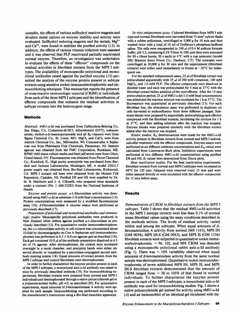
Demonstration of CRIM in fibroblast extracts from the MPSIsubtypes. Table I shows that the residual 4MU-a-Id activitiesin the MPSI subtype extracts were less than 0.1% of normalmean fibroblast values using the assay conditions described inthe methods section. The levels of residual activity variedwithin and among the subtypes. When equal amounts of f3-hexosaminidase A activity from normal (MS 1163), MPSIH(GM 0034), MPSIH-S (GM 0963), and MPSIS (GM 1256)fibroblast extracts were subjected to quantitative rocket immu-noelectrophoresis, - 96, 102, and 98% CRIM was detectedusing a monospecific polyclonal rabbit anti-a-Id antibody(Fig. 1). There was 10% variability observed when equalamounts of ,B-hexosaminidase activity from the same normalsample was electrophoresed. Quantitative rocket immunoelec-trophoresis of seven additional MPSIH, MPSIS, and MPSIH-S fibroblast extracts demonstrated that the amount ofCRIM ranged from - 38 to 105% of that found in normalindividuals. To further characterize the enzyme proteinpresent in each of the MPSI subtypes, a monoclonal anti-a-Idantibody was used for immunoblotting studies. Fig. 2 shows anative polyacrylamide gel stained for activity using 4MU-a-Id(A) and an immunoblot of an identical gel incubated with the
Enzyme Enhancement in the Mucopolysaccharidosis I Subtypes 99
Figure 1. Rocket immunoelec-trophoresis of fibroblast ex-tracts from MPSI subtypes.Rocket immunoelectrophore-
1 2 3 4 5 6 7 sis was performed as describedin the text. Lanes I and 5: nor-
mal fibroblast extracts (MS 1163) containing SUof -hexosamini-dase A activity. Lanes 2, 6, and 7: SUof ft-hexosaminidase A activityfrom cell lines GM0034 (MPS IH), GM0963 (MPS IH-S) and GM1256 (MPS IS), respectively. Lane 3: 2U of fl-hexosaminidase A ac-tivity from cell line GM0034. Lane 4: normal fibroblast extract (MS1163) containing 15U of fl-hexosaminidase A activity.
monoclonal anti-a-Id antibody (B). Although no enzymaticactivity was detected in each of the MPSI subtypes (A), aboutnormal amounts of enzyme protein were identified (B).
In vitro enhancement of the residual activity in the MPSIsubtypefibroblast extracts. Preincubation of the sulfihydryl re-ducing compounds, DTT, and cystamine for 5 min at 370Cbefore the addition of 4MU-a-Id, increased the mean residualactivities in fibroblast extracts from all three subtypes 2.6- to14.7-fold (Table I). The mean residual activities in MPSIH,MPSIH-S, and one MPSIS extract, were increased aboutthreefold by either DTTor cystamine. The residual activity inextracts from the other MPSIS individual was increased - 9-and 15-fold by DTTand cystamine, respectively. For compari-son, the normal mean fibroblast activity was stimulated 1.3-and 1.6-fold by DTTand cystamine, respectively. In contrast,iodoacetamide, which alkylates reduced sulfhydryl groups, hadlittle, if any, effect on the mean residual activities in the sub-type extracts and inhibited the mean activity in normal fibro-blasts by - 50%. The divalent metal cations, Mg2" and Ca2+,also enhanced the residual activities in the subtype extracts.Mg2" and Ca2+ increased the mean residual activities about
A B Figure 2. Immunoblotting of- _-- fibroblast extracts from MPSI
subtypes. Native polyacryl-12 3 4 2 3 4 amide gel electrophoresis and
immunoblotting was per-formed as described in the text. A shows a native polyacrylamide gelstained for 4MU-a-Id activity; lanes 1-4: extracts from fibroblast celllines GM0034 (MPS IH), GM0963 (MPS IH-S), GM1256 (MPSIS), and MS1163 (normal), respectively. B shows an immunoblot ofan identical native polyacrylamide gel reacted with a monoclonalanti-a-Id antibody. Each lane contained 20 U of -hexosaminidase Aactivity.
two- to eightfold and about two- to threefold, respectively,whereas the normal enzyme was essentially unaffected by ei-ther cation (Table I).
Investigation of the effects of various vitamin cofactors onthe residual activities in subtype extracts revealed that onlycertain B6 vitamers enhanced the residual activities, as shownin Table II. PLN, PNP, and PLP each stimulated the meanresidual activities from 2.5- to 16-fold, while pyridoxal andpyridoxine essentially had no effect. None of the B6 vitamersenhanced the normal fibroblast enzymatic activity. Fig. 3shows that the enhancement effect of cystamine, Mg9l2, andPLP on the residual activities in MPSI extracts was concen-tration dependent, with the optimal concentrations about 5.0mMcystamine, 25 mMMgCl2, and 0.25 mMPLP.
Next, the effect of the sequential addition of the effectorcompounds was evaluated. The sequence in which the effectorcompounds were added in the stimulation assay proved criti-cal, with optimal results obtained when the extracts were se-quentially preincubated with cystamine, MgCl2, and PLP.When the subtype extracts were preincubated in the optimaleffector concentrations and sequence, the residual activities
Table I. Effect of Sulzhydryl Reactive Reagents and Divalent Cations on the Residual a-L-IduronidaseActivities in MPSI Subtype Fibroblast Extracts
Enzymatic activity*
Normal controls MPSIH MPSffi-5 MPSISAddition (n = 5) (n = 5) (n = 3) (n = 2)
nmol/h/mg nmol/h/mg nmol/h/mg nmol/h/mg
None Mean 2,840 1.76 1.44Range 885-4,440 0.83-3.97 0.26-2.65 0.33; 3.62
DTT Mean 3,610 4.65 4.02Range 1,970-5,430 1.65-23.0 0.85-11.5 0.92; 14.5
Cystamine Mean 4,550 4.83 5.58Range 3,010-6,720 1.14-13.5 3.96-18.0 2.95; 12.5
lodoacetamide Mean 1,240 1.72 1.37Range 723-2,000 0.67-3.65 0.32-2.97 0.44; 3.08
MgC12 Mean 2,670 3.35 4.46Range 1,010-5,060 1.98-16.5 3.01-14.0 2.66; 17.2
CaCG2 Mean 3,150 3.08 3.92Range 1,160-5,720 1.99-4.66 1.05-5.52 1.06; 6.67
* a-L-Iduronidase
100 E. H. Schuchman and R. J. Desnick
Fibroblast extracts were preincubated with the effector compound for 5 min at 370C before the addition of 4MU-a-Id.assays were performed as described in Methods.
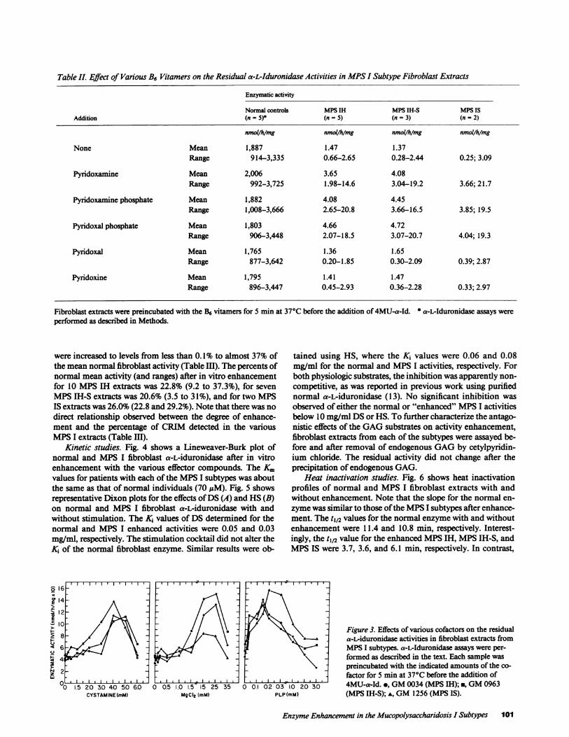
Table II. Effect of Various B6 Vitamers on the Residual a-L-Iduronidase Activities in MPSI Subtype Fibroblast Extracts
Enzymatic activity
Normal controls MPSIH MPSIH-S MPSISAddition (n = 5)* (n = 5) (n = 3) (n = 2)
nmol/h/mg nmol/h/mg nmol/h/mg nmol/h/mg
None Mean 1,887 1.47 1.37Range 914-3,335 0.66-2.65 0.28-2.44 0.25; 3.09
Pyridoxamine Mean 2,006 3.65 4.08Range 992-3,725 1.98-14.6 3.04-19.2 3.66; 21.7
Pyridoxamine phosphate Mean 1,882 4.08 4.45Range 1,008-3,666 2.65-20.8 3.66-16.5 3.85; 19.5
Pyridoxal phosphate Mean 1,803 4.66 4.72Range 906-3,448 2.07-18.5 3.07-20.7 4.04; 19.3
Pyridoxal Mean 1,765 1.36 1.65Range 877-3,642 0.20-1.85 0.30-2.09 0.39; 2.87
Pyridoxine Mean 1,795 1.41 1.47Range 896-3,447 0.45-2.93 0.36-2.28 0.33; 2.97
Fibroblast extracts were preincubated with the B6 vitamers for 5 min at 370C before the addition of 4MU-a-Id. * a-L-Iduronidase assays wereperformed as described in Methods.
were increased to levels from less than 0.1% to almost 37% ofthe mean normal fibroblast activity (Table III). The percents ofnormal mean activity (and ranges) after in vitro enhancementfor 10 MPSIH extracts was 22.8% (9.2 to 37.3%), for sevenMPSIH-S extracts was 20.6% (3.5 to 3 1%), and for two MPSIS extracts was 26.0% (22.8 and 29.2%). Note that there was nodirect relationship observed between the degree of enhance-ment and the percentage of CRIM detected in the variousMPSI extracts (Table III).
Kinetic studies. Fig. 4 shows a Lineweaver-Burk plot ofnormal and MPS I fibroblast a-L-iduronidase after in vitroenhancement with the various effector compounds. The Kmvalues for patients with each of the MPSI subtypes was aboutthe same as that of normal individuals (70 ,M). Fig. 5 showsrepresentative Dixon plots for the effects of DS (A) and HS(B)on normal and MPS I fibroblast a-L-iduronidase with andwithout stimulation. The Ki values of DS determined for thenormal and MPS I enhanced activities were 0.05 and 0.03mg/ml, respectively. The stimulation cocktail did not alter theKi of the normal fibroblast enzyme. Similar results were ob-
E '4 -~ ~
12-
lo
z442w
'0 1.5 2.0 3.0 40 5.0 6.0 0 0.5CYSTAMINE(mM)
I.0 1.5 15 25 35MgC12 (mM) PLP(i
tained using HS, where the Ki values were 0.06 and 0.08mg/ml for the normal and MPSI activities, respectively. Forboth physiologic substrates, the inhibition was apparently non-competitive, as was reported in previous work using purifiednormal a-L-iduronidase (13). No significant inhibition wasobserved of either the normal or "enhanced" MPSI activitiesbelow 10 mg/ml DSor HS. To further characterize the antago-nistic effects of the GAGsubstrates on activity enhancement,fibroblast extracts from each of the subtypes were assayed be-fore and after removal of endogenous GAGby cetylpyridin-ium chloride. The residual activity did not change after theprecipitation of endogenous GAG.
Heat inactivation studies. Fig. 6 shows heat inactivationprofiles of normal and MPSI fibroblast extracts with andwithout enhancement. Note that the slope for the normal en-zyme was similar to those of the MPSI subtypes after enhance-ment. The tj2 values for the normal enzyme with and withoutenhancement were 11.4 and 10.8 min, respectively. Interest-ingly, the t1,2 value for the enhanced MPSIH, MPSIH-S, andMPSIS were 3.7, 3.6, and 6.1 min, respectively. In contrast,
Figure 3. Effects of various cofactors on the residual9 \ ^ * + a-L-iduronidase activities in fibroblast extracts from
MPSI subtypes. a-L-Iduronidase assays were per-formed as described in the text. Each sample was
preincubated with the indicated amounts of the co-factor for 5 min at 370C before the addition of
1.0 20 3.0 4MU-a-Id. *, GM0034 (MPS IH); n, GM0963mM) (MPS IH-S); *, GM1256 (MPS IS).
Enzyme Enhancement in the Mucopolysaccharidosis I Subtypes 101
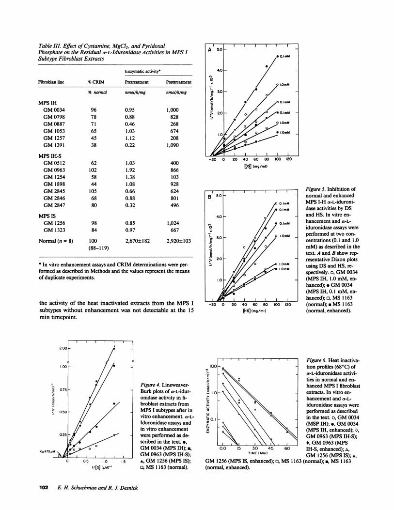
Table III. Effect of Cystamine, MgCl2, and PyridoxalPhosphate on the Residual a-L-Iduronidase Activities in MPSISubtype Fibroblast Extracts
Enzymatic activity*
Fibroblast line %CRIM Pretreatment Posttreatment
% normal nmol/h/mg nmol/h/mg
MPSIHGM0034 96 0.95 1,000GM0798 78 0.88 828GM0887 71 0.46 268GM1053 65 1.03 674GM1257 45 1.12 208GM1391 38 0.22 1,090
MPSIH-SGM0512 62 1.03 400GM0963 102 1.92 866GM1254 58 1.38 103GM1898 44 1.08 928GM2845 105 0.66 624GM2846 68 0.88 801GM2847 80 0.32 496
MPSISGM1256 98 0.85 1,024GM1323 84 0.97 667
Normal (n = 8) 100 2,670±182 2,920+103(88-119)
* In vitro enhancement assays and CRIM determinations were per-formed as described in Methods and the values represent the meansof duplicate experiments.
the activity of the heat inactivated extracts from the MPSIsubtypes without enhancement was not detectable at the 15min timepoint.
[DS] (mg/ml)
-20 0 20 40 60 80 100 120CHS] (mg/ml)
Figure 5. Inhibition ofnormal and enhancedMPSI-H a-L-iduroni-dase activities by DSand HS. In vitro en-hancement and a-L-iduronidase assays wereperformed at two con-centrations (0.1 and 1.0mM)as described in thetext. A and B show rep-resentative Dixon plotsusing DSand HS, re-spectively. o, GM0034(MPS IH, 1.0 mM, en-hanced); * GM0034(MPS IH, 0.1 mM, en-hanced); o, MS1163(normal); * MS1163(normal, enhanced).
Figure 4. Lineweaver-Burk plots of a-L-idur-onidase activity in fi-broblast extracts fromMPSI subtypes after invitro enhancement. a-L-Iduronidase assays andin vitro enhancementwere performed as de-scribed in the text. e,GM0034 (MPS IH);*,GM0963 (MPS IH-S);A, GM1256 (MPS IS);0, MS1163 (normal).
lll Figure6. Heat inactiva-IG.O _ _ tion profiles (680C) of
£ a-L-iduronidase activi-ties in normal and en-hanced MPSI fibroblastE
'5 1.0 \ extracts. In vitro en-, ~ \\ i\khancement and a-L-
iduronidase assays were4 \ \ \ % performed as described
in the text. o, GM0034Xas \\ \ w (MSP IH); *, GM0034z S \ \\\(MPSIH, enhanced); 0,
ffi \. I GM0963 (MPS IH-S);,.,,,,\ ,\ *, GM0963 (MPS
0.0 15 30 45 60 IH-S, enhanced); A,TIME (Min) GM1256 (MPS IS); A,
GM1256 (MPS IS, enhanced); o, MS1163 (normal); ., MS1163(normal, enhanced).
E
Km370MM
1.0
102 E. H. Schuchman and R. J. Desnick

Discussion
Although the immunologic and physicokinetic properties ofnormal human purified a-L-iduronidase from several sourceshave been well documented (1 1-13, 21-30), limited informa-tion is available on the nature of the defective enzyme in theMPS I subtypes. In this communication, fibroblast extractsfrom each subtype were evaluated for the presence of CRIMusing monospecific polyclonal and monoclonal anti-humana-L-iduronidase antibodies. When equal amounts of f3-hexos-aminidase A activity were electrophoresed in a quantitativerocket immunoelectrophoretic system, the subtype extractswere found to have from 38 to 105% of normal a-L-iduroni-dase enzyme protein using the anti-a-Id polyclonal antibodies.It should be noted that these extracts had < 0.1% of normalenzymatic activity. The demonstration of CRIM by immuno-blotting (Fig. 2) using an anti-a-Id monoclonal antibody cor-roborated the results obtained with rocket immunoelectropho-resis. These findings indicated that the different mutations inthe MPSI subtypes examined did not dramatically alter thetranscriptional activity of the a-L-iduronidase gene or the sta-bility of the mutant enzyme proteins, despite the fact that thecatalytic properties of the mutant proteins were severely im-paired. It was notable that CRIM-negative mutations were notdetected in any of the MPSI lines examined. However, the oneMPSIH line previously analyzed for an a-L-iduronidase matu-ration study appeared to be CRIM negative (1 1). This may beexplained by differences in antibody preparations used in thetwo studies, as well as differences in the immunologic tech-niques. Based on the results presented here, it is likely that themajority of MPSI mutations are CRIM positive.
Since sulfhydryl reducing reagents and divalent cationswere required for optimal activity of the normal purified en-zyme (12, 13, 30), and since it had been determined that en-zyme proteins were present in fibroblast extracts from theMPSI subtypes, studies were conducted to determine the ef-fects of these effector molecules on the residual activities in theMPSI subtypes. The finding that the sequential addition ofcystamine, MgCl2, and PLP enhanced the residual activities infibroblast extracts up to - 35% of normal was unexpected assimilar observations had not been reported previously forother lysosomal enzymes, with one notable exception. Theresidual arylsulfatase B activity in leukocyte extracts from fe-line mucopolysaccharidosis VI was enhanced over 10-fold bythe addition of DTTor cysteamine (31). Studies indicated thatthe purified mutant feline enzyme was a monomer that di-merized in the presence of thiol-reducing agents, thereby re-gaining partial enzymatic activity toward both artificial andnatural substrates (31, 32). It is unlikely that thiol-inducedsubunit association was responsible for the activity enhance-ment in the MPSIH subtypes since normal human a-L-idur-onidase is a monomer (12-14, 30).
Although the specific mechanism responsible for enhance-ment of the residual activities is not known, several possibili-ties can be considered. The fact that removal of the GAGsubstrates by cetylpyridinium chloride precipitation from sub-type extracts did not enhance the residual activity toward4MU-a-Id suggests that the mutant enzymes were not inhib-ited by bound excess natural substrate. Therefore, the effectormolecules may have a direct effect on the mutant proteins asopposed to interacting with the natural substrates present in
the extracts. Inhibition studies of a-L-iduronidase from nor-mal and "enhanced" MPSI fibroblast extracts using the natu-ral substrates, DSand HS, have revealed that: (a) these physio-logic substrates inhibit the 4MU-a-Id activity in fibroblast ex-tracts in a manner similar to that previously observed withpurified human enzyme (13). Dixon plots at two differentsubstrate concentrations indicated that the inhibition was ap-parently noncompetitive, as was observed in the previous workmentioned above, and (b) these substrates inhibit the activityof the "enhanced" enzymatic activity in the MPSI extracts ina manner similar to that observed in the normal samples.Thus, the use of 4MU-a-Id as a substrate accurately reflectedthe enhanced activity of the mutant enzymes toward theirnatural substrates and was a reliable measurement of reacti-vated enzyme. Finally, heat inactivation studies confirmed thefact that the enhancement effect was due to the presence ofactive mutant enzyme and was not an artifact of the fluoro-genic assay. Although the t1/2 of the enhanced activities fromthe MPSI subtypes was less (3.5-6.1 min) than that observedfor the normal activities (10.8-11.4 min), the linear inhibitioncurves for normal and mutant enzymes were similar, indicat-ing that the enhancement effect was dependent on the amountof non-heat denatured mutant enzyme protein.
It is likely that the mutant enzyme proteins in each of thesubtypes have altered conformations and are modified by theeffector molecules such that their normal integrity is partiallyrestored and residual activity toward 4MU-a-Id is markedlyincreased. This hypothesis assumes that different mutations inthe structural gene for a-L-iduronidase each result in enzymeproteins which are relatively stable, but have markedly re-duced activities toward the natural and artificial substrates,i.e., presumably, each mutant allele encodes a monomeric en-zyme in which the protein conformation is altered at or nearthe active site of the enzyme. This concept is supported by thefollowing two findings: immunologic demonstration of CRIMin each of the MPSI subtypes, and enhancement of the activi-ties in normal and subtype fibroblast extracts by thiol reducingreagents alone (Table I), indicating that reduction of crucialsulfhydryl groups at or near the active site may be importantfor obtaining optimal activity and involved in maintaining theactive conformation. Wepreviously reported that thiol reduc-ing reagents stimulated the 4MU-a-Id activities of purifieda-L-iduronidase, whereas p-chloromercurobenzoate and ma-leate were potent inhibitors (13). Similar findings for the lowuptake kidney enzyme also have been reported using a differ-ent artificial substrate (23). Furthermore, Clements et al. havefound that DTThelped prevent a gradual loss of enzyme activ-ity during the purification of a-L-iduronidase from humanliver and kidney (30).
The fact that Mg2+ and Ca2+ cations stimulated the residualenzymatic activity in the MPSI subtypes may relate directly tothis concept, as well as to their mechanistic role as effectors ofenzyme enhancement. The metal cations may bind the re-duced sulfhydryl groups in the enzyme and protect these criti-cal residues at or near the active site from oxidation. Such amechanism, involving reduced sulfhydryl groups that aremaintained by zinc cations and PLP, has been suggested toprevent inactivation of 5-aminolevulinate dehydratase(33-36). Although a B6 vitamer is not known to be a require-ment for normal a-L-iduronidase activity, clearly, PLP, PLN,or PNPenhanced the residual activities in the MPSI extracts
Enzyme Enhancement in the jMucopolysaccharidosis I Subtypes 103
(Table II). The pyridoxal moiety may covalently bind the mu-tant protein through a Schiff base mechanism and alter theactive site conformation, making it more accessible to the4MU-a-Id substrate. Alternatively, and more likely, PLP maybind to the metal cations, further preventing the oxidation ofcrucial sulfhydryl groups and maintaining the active confor-mation. It should be noted that B6 vitamers are strong che-laters of divalent metal cations (35) and that only negativelycharged vitamers stimulated the residual activities.
In summary, the data presented here clearly demonstratethe presence of mutant enzyme protein in each of the MPSIsubtypes and, although these studies were performed using anartificial substrate, 4MU-a-Id, the results suggest that it maybe possible to enhance the residual enzymatic activity in pa-tients with MPSI by further investigation of methods to ma-nipulate the conformation and substrate binding of the mutantenzyme. However, caution should be exercised in the extrapo-lation of in vitro findings to in vivo expectations. For example,several investigators have shown that normal a-mannosidaseA and B were stimulated by zinc ions in vitro (37, 38). Zincalso was found to enhance the residual acidic a-mannosidaseactivity in various sources from patients with mannosidosis(37-40). However, a clinical trial of oral zinc supplementationdid not alter the level of residual activity in plasma, leukocytes,or tears from the treated patients (41). Thus, in vivo trials ofa-L-iduronidase enhancement should be performed in animalanalogues (42, 43) before human experimentation, particu-larly since the residual activity in feline MPSI can be signifi-cantly enhanced (Schuchman, E. H., M. E. Haskins, and R. J.Desnick, unpublished results).
Acknowledgments
This work was supported in part by a grant (1-535) from the March ofDimes Birth Defects Foundation and a grant (DK-25759) from theNational Institutes of Health. Dr. Schuchman was the recipient of apostdoctoral fellowship in medical genetics (T32 HDO7105). Dr.Schuchman is the recipient of a Basil O'Conner Award (5-640) fromthe March of Dimes Birth Defects Foundation.
References
1. Matalon, R., J. A. Cifonelli, and A. Dorfman. 1972. Hurler'ssyndrome, an a-L-iduronidase deficiency. Biochem. Biophys. Res.Commun. 47:959-964.
2. Bach, G., R. Friedman, B. Weissmann, and E. F. Neufeld. 1972.The defect in Hurler and Scheie syndromes. Deficiency of a-L-idur-onidase. Proc. Nat!. Acad. Sci. USA. 69:2048-205 1.
3. Roden, L. 1980. Structure and metabolism of connective tissueproteoglycans. In The Biochemistry of Glycoproteins and Proteogly-cans. W. G. Lennarz, editor. Plenum Press, NewYork. 267-37 1.
4. McKusick, V. A., and E. F. Neufeld. 1983. The mucopolysac-charide storage diseases. In The Metabolic Basis of Inherited Disease.5th ed. J. B. Stanbury, J. B. Wyngaarden, D. S. Fredrickson, J. L.Goldstein, and M. S. Brown, editors. McGraw-Hill, New York.751-777.
5. McKusick, V. A. Heritable Disorders of Connective Tissue. 4thed. C. V. Mosby and Co., St. Louis. 521-556.
6. Kaibara, H., M. Eguchi, K. Shibata, and K. Takagishi. 1979.Hurler-Scheie phenotype. A report of two pairs of inbred sibs. Hum.Genet. 53:37-43.
7. Hopwood, J. J., and V. Muller. 1979. Biochemical discrimina-tion of Hurler and Scheie syndromes. Clin. Sci. 57:265-272.
8. Hall, C. W., I. Liebaers, P. DiNatale, and E. F. Neufeld. 1978.Enzymic diagnosis of the genetic mucopolysaccharide storage dis-orders. Methods Enzymol. 50:539-543.
9. Hopwood, J. J., V. Muller, A. Smithson, and N. Baggett. 1979. Afluorometric assay using 4-methylumbelliferyl a-L-iduronide for theestimation of a-L-iduronidase activity and the detection of Hurler andScheie syndromes. Clin. Chim. Acta. 92:257-265.
10. Muller, V., and J. J. Hopwood. 1984. a-L-Iduronidase defi-ciency in mucopolysaccharidosis type 1 against a radiolabelled sulfateddisaccharide substrate derived from dermatan sulfate. Clin. Genet.26:414-421.
11. Myerowitz, R., and E. F. Neufeld. 1981. Maturation of a-L-iduronidase in cultured human fibroblasts. J. Biol. Chem. 256:3044-3048.
12. Schuchman, E. H., N. A. Guzman, and R. J. Desnick. 1984.Human a-L-iduronidase. I. Purification and characterization of thehigh uptake (higher molecular weight) and the low uptake (processed)forms. J. Biol. Chem. 259:3132-3140.
13. Schuchman, E. H., N. A. Guzman, and R. J. Desnick. 1984.Humana-L-iduronidase. II. Comparative biochemical and immuno-logic properties of the purified low and high uptake forms. Enzyme.31:166-175.
14. Schuchman, E. H., K. H. Astrin, P. Aula, and R. J. Desnick.1984. Regional assignment of the structural gene for human a-L-idur-onidase. Proc. NatL. Acad. Sci. USA. 81:1169-1173.
15. Weissmann, B. 1978. Synthetic substrates for a-L-iduronidase.Methods Enzymol. 50:141-150.
16. Bishop, D. F., and R. J. Desnick. 1981. Affinity purification ofa-galactosidase A from human spleen, placenta and plasma with elimi-nation of pyrogen contamination. Properties of the purified splenicenzyme and comparison to other forms. J. Biol. Chem. 255:1307-1316.
17. Srivastava, S. K., and E. Beutler. 1974. Studies on human3-D-N-acetylhexosaminidases. Purification and properties. J. Biol.
Chem. 249:2043-2048.18. Beratis, N. G., G. U. LaBadie, and K Hirschhorn. 1983. Ge-
netic heterogeneity in acid a-glucosidase deficiency. Am. J. Hum.Genet. 35:21-33.
19. Kennett, R. H., T. J. McKearn, and K. B. Bechtol. 1980.Monoclonal Antibodies. Plenum Press, NewYork.
20. McGovern, M. M., D. T. Vine, M. E. Haskins, and R. J.Desnick. 1982. Purification and properties of feline and human aryl-sulfatase B isozymes. Evidence for feline homodimeric and humanmonomeric structures. J. Biol. Chem. 257:12605-12610.
21. Hickman, S., L. J. Shapiro, and E. F. Neufeld. 1974. A recogni-tion marker required for uptake of a lysosomal enzyme by culturedfibroblasts. Biochem. Biophys. Res. Commun. 57:55-61.
22. Sando, G. N., and E. F. Neufeld. 1977. Recognition and recep-tor-mediated uptake of a lysosomal enzyme, a-L-iduronidase, by cul-tured human fibroblasts. Cell. 12:519-527.
23. Rome, L. H., A. J. Garvin, and E. F. Neufeld. 1978. Humankidney a-L-iduronidase; purification and characterization. Arch. Bio-chem. Biophys. 189:344-353.
24. Rome, L. H., B. Weissmann, and E. F. Neufeld. 1979. Directdemonstration of binding of a lysosomal enzyme, a-L-iduronidase, toreceptors on cultured fibroblasts. Proc. Natl. Acad. Sci. USA.76:2331-2334.
25. Rome, L. H., A. J. Garvin, M. M. Allietta, and E. F. Neufeld.1979. Two species of lysosomal organelles in cultured human fibro-blasts. Cell. 173:143-153.
26. Karson, E. M., E. F. Neufeld, and G. N. Sando. 1980. P-Iso-thiocyanotophenyl-6-phospho-a-D-mannopyranoside coupled to al-bumin. A model compound recognized by the fibroblast lysosomaluptake system. II. Biological properties. Biochemistry. 19:3856-3860.
27. Rome, L. H., and J. Miller. 1980. Butadione treatment reducesreceptor binding of lysosomal enzymes to cells and membranes. Bio-chem. Biophys. Res. Commun. 92:986-993.
104 E. H. Schuchman and R. J. Desnick

28. Steiner, A. W., and L. H. Rome. 1982. Assay and purificationof a solubilized membrane receptor that binds the lysosomal enzyme,a-L-iduronidase. Arch. Biochem. Biophys. 214:681-687.
29. Clements, P. R., D. A. Brooks, G. T. P. Saccone, and J. J.Hopwood. 1985. Humana-L-iduronidase. 1. Purification, monoclonalantibody production, native subunit molecular mass. Eur. J. Biochem.152:21-28.
30. Clements, P. R., V. Muller, and J. J. Hopwood. 1985. Humana-L-iduronidase. 2. Catalytic properties. Eur. J. Biochem. 152:29-34.
31. Vine, D. T., M. M. McGovern, E. H. Schuchman, M. E. Has-kins, and R. J. Desnick. 1982. Enhancement of residual arylsulfatase Bactivity in feline mucopolysaccharidosis VI by thiol-induced subunitassociation. J. Clin. Invest. 69:294-302.
32. McGovern, M. M., N. Mandel, M. E. Haskins, and R. J. Des-nick. 1985. Animal model studies of allelism: characterization of aryl-sulfatase B mutations in homoallelic and heteroallelic (genetic com-pound) homozygotes with feline mucopolysaccharidosis VI. Genetics.110:733-749.
33. Anderson, P. M., and R. J. Desnick. 1979. Purification andproperties of -aminolevulinate dehydratase from human erythrocytes.J. Biol. Chem. 254:6924-6930.
34. Barnard, G. F., R. Itoh, L. H. Hohberger, and D. Shemin. 1977.Mechanism of porphobilinogen synthase. Possible role of essentialthiol groups. J. Biod. Chem. 252:8965-8974.
35. Jaffe, E. K., S. P. Salowe, N. T. Chen, and P. A. DeHaven.1984. Porphobilinogen synthase modification with methyl methane-thiosulfonate. J. Biol. Chem. 259:5032-5036.
36. Gibbs, P. N. B., M. G. Gore, and P. M. Jordan. 1985. Investi-gation of the effect of metal ions on the reactivity of thiol groups inhuman 5-amino-laevulinate dehydratase. Biochem. J. 225:573-580.
37. Desnick, R. J., H. L. Sharp, G. A. Grabowski, R. D. Brunning,P. G. Quie, J. H. Sung, R. S. Gorlin, and J. U. Ikonne. 1976. Manno-sidosis: Clinical, morphologic, immunologic and biochemical studies.Pediatr. Res. 10:985-992.
38. Hultberg, B., and P. K. Masson. 1975. Activation of residualacidic a-mannosidase activity in mannosidosis tissues by metal ions.Biochem. Biophys. Res. Commun. 67:1473-1480.
39. Burton, B. K., and H. L. Nadler. 1978. Mannosidosis: Separa-tion and characterization of two acid a-mannosidase forms in mutantfibroblasts. Enzyme. 23:29-33.
40. Bach, G., G. Kohn, E. E. Lasch, M. El Massri, A. Ornoy, E.Sckeles, C. Legum, and M. M. Cohen. 1978. A new variant of manno-sidosis with increased residual enzymatic activity and mild clinicalmanifestations. Pediatr. Res. 12:1010-1016.
41. Grabowski, G. A., L. Walling, and R. J. Desnick. 1980. Humanmannosidosis: In vitro and in vivo studies of co-factor supplementa-tion. In Enzyme Therapy in Genetic Diseases: 2. R. J. Desnick, editor.Alan R. Liss, Inc., NewYork. 319-334.
42. Haskins, M. E., P. F. Jezyk, R. J. Desnick, and D. F. Patterson.1981. Animal model for human disease. Mucopolysaccharidoses I,Hurler, Scheie and Hurler/Scheie syndrome. Comp. Pathol. Bull.8:3-4.
43. Haskins, M. E., G. D. Aguirre, P. F. Jezyk, R. J. Desnick, andD. F. Patterson. 1983. The pathology of the feline model of muco-polysaccharidosis I. Am. J. Pathol. 112:27-36.
Enzyme Enhancement in the Mucopolysaccharidosis I Subtypes 105


![RESEARCHARTICLE Rugby-SpecificSmall-SidedGamesTraining ... · muscle fibre,andmusclefibre area[12].However, todate, therehasbeennopublishedre- searchdirectly investigating theeffects](https://static.fdocuments.net/doc/165x107/5f5dfc9299447d03974b381b/researcharticle-rugby-specificsmall-sidedgamestraining-muscle-fibreandmusclefibre.jpg)