Mucoadhesive Drug Delivery System: A Review Article · Mucoadhesive Drug Delivery System: A Review...
17
T. Dey. et. al./ 8(2) pp- 944- 960 June-2020 International Journal of Pharmacy and Engineering Page 944 International Journal of Pharmacy and Engineering Page 944 1 School of Pharmacy (SOP),Techno India University (TIU),SaltLake,EM-4/1,Sector-V, Kolkata-700091,WestBengal,India, E-mail:[email protected] 2 School of Pharmacy (SOP), Techno India University (TIU), SaltLake,EM-4/1,Sector-V, Kolkata-700091, West Bengal, India, E-mail:[email protected] 3 School of Pharmacy (SOP), Techno India University (TIU), SaltLake,EM-4/1,Sector-V, Kolkata-700091, West Bengal, India,E-mail:[email protected] 4 School of Pharmacy (SOP), Techno India University (TIU), SaltLake,EM-4/1,Sector-V, Kolkata-700091, West Bengal, India, E-mail:[email protected] Mucoadhesive Drug Delivery System: A Review Article Tushar Dey 1 , Dr.Khokan Bera 2* , Bappaditya Samanta 3 and Dr.Beduin Mahanti 4 Received: May 20 th , 2020, Revised: May 25 th , 2020, Accepted: May 30 th , 2020, Licensee Abhipublications Open. This is an open access article licensed under the terms of the Creative Commons Attribution Non- Commercial License (http://www.abhipublications.org/ijpe ) which permits unrestricted, non- commercial use, distribution and reproduction in any medium, provided the work is properly cited. Corresponding Author:*Dr. Khokan Bera, School of Pharmacy, Techno India University, Salt Lake,EM-4/1,Sector-V, Kolkata-700091,West Bengal, India .E-mail: [email protected] Phone No: +91-9432897061
Transcript of Mucoadhesive Drug Delivery System: A Review Article · Mucoadhesive Drug Delivery System: A Review...
T. Dey. et. al./ 8(2) pp- 944- 960 June-2020 International Journal of Pharmacy and Engineering Page 944
International Journal of Pharmacy and Engineering Page 944
1School of Pharmacy (SOP),Techno India University (TIU),SaltLake,EM-4/1,Sector-V, Kolkata-700091,WestBengal,India, E-mail:[email protected]
2 School of Pharmacy (SOP), Techno India University (TIU), SaltLake,EM-4/1,Sector-V, Kolkata-700091, West Bengal, India, E-mail:[email protected]
3 School of Pharmacy (SOP), Techno India University (TIU), SaltLake,EM-4/1,Sector-V, Kolkata-700091, West Bengal, India,E-mail:[email protected]
4 School of Pharmacy (SOP), Techno India University (TIU), SaltLake,EM-4/1,Sector-V, Kolkata-700091, West Bengal, India, E-mail:[email protected]
Mucoadhesive Drug Delivery System: A Review Article
Tushar Dey1, Dr.Khokan Bera2*, Bappaditya Samanta3 and Dr.Beduin Mahanti4
Received: May 20th , 2020, Revised: May 25th , 2020, Accepted: May 30th , 2020, Licensee Abhipublications Open. This is an open access article licensed under the terms of the Creative Commons Attribution Non-Commercial License (http://www.abhipublications.org/ijpe) which permits unrestricted, non-commercial use, distribution and reproduction in any medium, provided the work is properly cited. Corresponding Author:*Dr. Khokan Bera, School of Pharmacy, Techno India University, Salt
Lake,EM-4/1,Sector-V, Kolkata-700091,West Bengal, India .E-mail: [email protected]
Phone No: +91-9432897061
T. Dey. et. al./ 8(2) pp- 944- 960 June-2020 International Journal of Pharmacy and Engineering Page 945
International Journal of Pharmacy and Engineering Page 945
Key words: oral route, mucous layer, gastric emptying rate, first pass metabolism, mucoadhesive strength.
Oral delivery has so far been the most common and preferred route of administration for most of the therapeutic agents[1]. The popularity of the oral route has been attributed to the patient acceptance, ease of administration, accurate dosing, cost effective manufacturing method, least sterility constraints, flexible design of dosage forms and generally improved shelf-life of the product[2]. Mucoadhesive drug delivery has been a topic of interest in the design of drug delivery systems to lengthen the residence time of the dosage form at the site of application or absorption and to facilitate intimate contact of the formulation with the underlying absorption surface, so as to improve and enhance the bioavailability of drug[3]. Mucoadhesive controlled drug delivery systems are beneficial, since they give a controlled drug release over a period of time and can also be utilised for localising the drug to a specific site in the body[4]. Mucoadhesive substances could also be used as therapeutic agents in their own right, to coat and protect and soothe the injured tissues (gastric ulcers or lesions of the oral mucosa) or as lubricants (in the oral cavity, eye and vagina)[5].
Abstract
Abstract The mucoadhesive strength of a dosage form is dependent upon a variety of factors, including the nature of the mucosal tissue, nature of polymer, cohesive strength of coating polymer and the physicochemical properties of the polymeric formulation. This review article aims to provide an overview of the various aspects of mucoadhesion, mucoadhesive materials, factors affecting mucoadhesion etc. The mucosa of the oral cavity presents a barrier to drug penetration and one method of optimizing drug delivery is by the use of adhesive dosage forms and the mucosa has a rich blood supply and it is relatively permeable which provides entering the dosage form in the systemic circulation . It has been noted that buccal mucosa is very suitable for a bioadhesion system due to smooth and relatively immobile surface and accessibility. Bioadhesionon the surface of mucoadhesive tissue can be achieved by using mucoadhesive polymers. Such type of delivery systems is advantageous in enhancing the drug plasma concentrations and also therapeutic activity. In this connection, this review covers the areas of mechanisms and theories of mucoadhesion, factors influencing the mucoadhesive devices and also various mucoadhesive dosage forms.In this review we discussed about potential applications of mucoadhesion and mucoadhesive polymers in drug delivery along with the mechanism of mucoadhesion and the methods for evaluation of this drug delivery systems. Oral route also have some disadvantages such as hepatic first pass metabolism and enzymatic degradation within the GI tract, which inhibit oral administration of certain classes of drugs especially peptides and proteins.
Introduction
T. Dey. et. al./ 8(2) pp- 944- 960 June-2020 International Journal of Pharmacy and Engineering Page 946
International Journal of Pharmacy and Engineering Page 946
Mechanism of Mucoadhesion Mucoadhesion is a complex process involving wetting, adsorption and interpenetration of polymer chains. Mucoadhesion is established in the following stages: Contact stage: Intimate physical con-tact between a bioadhesive/Mucoadhesive material and a
membrane (wetting or swell-ing phenomenon). Consolidation stage: Penetration of the bioadhesive/Mucoadhesive into underlying the tissue or
into the surface of the mucous membrane (interpenetration)[6]. Mucous Membrane Mucous membranes (mucosae) are the moist surfaces, lining the walls of various body cavities such as the gas-trointestinal and respiratory tracts. They consist of a connective tissue layer (the lamina propria) above which is an epithelial layer, the upper part of which is made moist usually due to the presence of a mucus layer. The epithelia may be either single layered (e.g. the stomach, small and large intestine and bronchi) or multilayered/ stratified (e.g. in the oesophagus, vagina and cornea)[7]. The former contain goblet cells which secrete mucus directly onto the epithelial surfaces, the latter contain, or are adjacent to tissues containing, specialised glands such as salivary glands that secrete mucus onto the epithelial surface. Mucus is present as either a gel layer sticking to the mucosal surface or as a soluble or suspended luminal entity. The primary components of all mucus gels are mucin glycoproteins, lipids, inorganic salts and water, the latter comprising more than 95% of its weight, making it a highly hydrated system[8]. The mucin glycoproteins are the most important structure-forming component of the mucus gel, which provide the mucous with its characteristic gel-like, cohesive and adhesive properties. The thickness of this mucus layer varies at different mucosal surfaces, from 50 to 450 μm in the stomach, to less than 1 μm in the oral cavity. The major functions of mucus include protection and lubrication (anti adherents)[9]. Composition of mucus layer Mucus is translucent and viscous secretion which forms a thin, continuous gel layer sticking to the mucosal epithelial surface. Mucus glycoproteins are high molecular weight proteins possessing attached oligosaccharide units containing, L-fucose, D-galactose, N-acetyl-D-glu-cosamine, N-acetyl-D-galactosamine and Sialic acid. Functions of mucous layer Mucous layer is protective because of its hydrophobicity. It influences the bioavailability of drugs as it acts as a barrier in tissue absorption of drugs and
other substrates.[8] It strongly bonds with the epithelial cell surface as a continuous gel layer. It plays a major role in the lubrication of the mucosal membrane and maintenance of its
moisture.[9]
Advantages of Mucoadhesive Drug Delivery System Mucoadhesive delivery system offers several advantages over conventional drug delivery systems which are as follows:
T. Dey. et. al./ 8(2) pp- 944- 960 June-2020 International Journal of Pharmacy and Engineering Page 947
International Journal of Pharmacy and Engineering Page 947
Prolongs the residence time of the dosage form at the site of absorption, hence increases the bioavailability.
Excellent accessibility, rapid onset of action possible. Rapid absorption because of enormous blood supply and good perfusion rate [6] An alternative to oral route, whereby the drug is protected from degradation in the acidic
environment of the GIT. Better patient compliance. Moreover, rapid cellular recovery and healing of the local site.[10] Reduced dosing frequency. Shorter treatment period. Increased safety margin of high potency drugs due to better control of plasma levels. Maximum utilisation of drug enabling reduction in total amount of drug administered.[11]
Disadvantages Drugs, which irritate the oral mucosa, have a bitter or unpleasant taste, odour, cannot be
administered by this route. Drugs, which are unstable at buccal pH, cannot be administered by this route. Only drugs with small dose requirements can be administered. [6] Drugs may be swallowed along with the saliva and lose the advantages of buccal route. Only those drugs, which are absorbed by passive diffusion, can be administered by this route. Eating and drinking may become restricted. In case of vaginal drug delivery, the drug has to be stable in the acidic vaginal pH. The vaginal formulation may interfere with sexual intercourse.[10] The vaginal formulation may leak and cause messiness. The vaginal formulation may be contraindicated in case of pregnancy. In case of ocular formulations, the formulation may cause uneasiness and blurring. In case of nasal formulations, the presence of the formulation may stimulate sneezing and
subsequent dislodgement of the formulation. Over hydration may lead to the formation of slip-pery surface and structural integrity of the
formula-tion may get disrupted by the swelling and hydration of the bioadhesive polymers. [11] Theories of Mucoadhesion Various theories exist to explain at least some of the experimental observations made during the bioadhesion process. Unfortunately, each theoretical model can only explain a limited number of the diverse range of interactions that constitute the bioadhesive bond.[12] However, four main theories can be distinguished. Wetting theory The wetting theory applies to liquid systems which present affinity to the surface in order to spread over it. This affinity can be found by using measuring techniques such as the contact angle. The general rule states that the lower the contact angle, the greater is the affinity [Figure1]. The contact angle should be equal or close to zero to provide adequate spreadability. The spreadability coefficient, SAB, can be calculated from the difference between the surface energies γB and γA and the interfacial energy γAB, as indicated in the equation given below.[13]This theory explains the
T. Dey. et. al./ 8(2) pp- 944- 960 June-2020 International Journal of Pharmacy and Engineering Page 948
International Journal of Pharmacy and Engineering Page 948
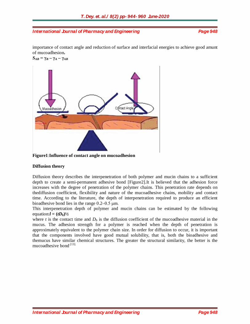
importance of contact angle and reduction of surface and interfacial energies to achieve good amunt of mucoadhesion. SAB = γB – γA – γAB
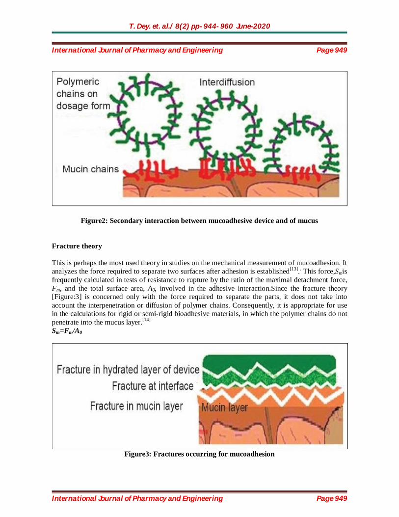
Figure1:Influence of contact angle on mucoadhesion Diffusion theory Diffusion theory describes the interpenetration of both polymer and mucin chains to a sufficient depth to create a semi-permanent adhesive bond [Figure2].It is believed that the adhesion force increases with the degree of penetration of the polymer chains. This penetration rate depends on thediffusion coefficient, flexibility and nature of the mucoadhesive chains, mobility and contact time. According to the literature, the depth of interpenetration required to produce an efficient bioadhesive bond lies in the range 0.2–0.5 μm. This interpenetration depth of polymer and mucin chains can be estimated by the following equation:l = (tDb)½ where t is the contact time and Db is the diffusion coefficient of the mucoadhesive material in the mucus. The adhesion strength for a polymer is reached when the depth of penetration is approximately equivalent to the polymer chain size. In order for diffusion to occur, it is important that the components involved have good mutual solubility, that is, both the bioadhesive and themucus have similar chemical structures. The greater the structural similarity, the better is the mucoadhesive bond.[13].
T. Dey. et. al./ 8(2) pp- 944- 960 June-2020 International Journal of Pharmacy and Engineering Page 949
International Journal of Pharmacy and Engineering Page 949
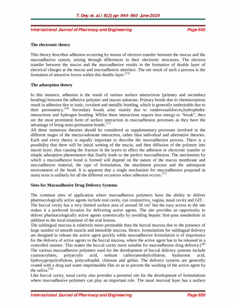
Figure2: Secondary interaction between mucoadhesive device and of mucus Fracture theory This is perhaps the most used theory in studies on the mechanical measurement of mucoadhesion. It analyzes the force required to separate two surfaces after adhesion is established[13].. This force,Smis frequently calculated in tests of resistance to rupture by the ratio of the maximal detachment force, Fm, and the total surface area, A0, involved in the adhesive interaction.Since the fracture theory [Figure:3] is concerned only with the force required to separate the parts, it does not take into account the interpenetration or diffusion of polymer chains. Consequently, it is appropriate for use in the calculations for rigid or semi-rigid bioadhesive materials, in which the polymer chains do not penetrate into the mucus layer.[14] Sm=Fm/A0
Figure3: Fractures occurring for mucoadhesion
T. Dey. et. al./ 8(2) pp- 944- 960 June-2020 International Journal of Pharmacy and Engineering Page 950
International Journal of Pharmacy and Engineering Page 950
The electronic theory This theory describes adhesion occurring by means of electron transfer between the mucus and the mucoadhesive system, arising through differences in their electronic structures. The electron transfer between the mucus and the mucoadhesive results in the formation of double layer of electrical charges at the mucus and mucoadhesive interface. The net result of such a process is the formation of attractive forces within this double layer.[15] The adsorption theory In this instance, adhesion is the result of various surface interactions (primary and secondary bonding) between the adhesive polymer and mucus substrate. Primary bonds due to chemisorptions result in adhesion due to ionic, covalent and metallic bonding, which is generally undesirable due to their permanency.[16] Secondary bonds arise mainly due to vanderwaalsforces,hydrophobic interactions and hydrogen bonding. Whilst these interactions require less energy to “break”, they are the most prominent form of surface interaction in mucoadhesion processes as they have the advantage of being semi-permanent bonds.[17] All these numerous theories should be considered as supplementary processes involved in the different stages of the mucus/substrate interaction, rather than individual and alternative theories. Each and every theory is equally important to describe the mucoadhesion process. There is a possibility that there will be initial wetting of the mucin, and then diffusion of the polymer into mucin layer, thus causing the fracture in the layers to effect the adhesion or electronic transfer or simple adsorption phenomenon that finally leads to the perfect mucoadhesion. The mechanism by which a mucoadhesive bond is formed will depend on the nature of the mucus membrane and mucoadhesive material, the type of formulation, the attachment process and the subsequent environment of the bond. It is apparent that a single mechanism for mucoadhesion proposed in many texts is unlikely for all the different occasions when adhesion occurs.[17] Sites for Mucoadhesive Drug Delivery Systems The common sites of application where mucoadhesive polymers have the ability to deliver pharmacologically active agents include oral cavity, eye conjunctiva, vagina, nasal cavity and GIT. The buccal cavity has a very limited surface area of around 50 cm2 but the easy access to the site makes it a preferred location for delivering active agents. The site provides an opportunity to deliver pharmacologically active agents systemically by avoiding hepatic first-pass metabolism in addition to the local treatment of the oral lesions. The sublingual mucosa is relatively more permeable than the buccal mucosa due to the presence of large number of smooth muscle and immobile mucosa. Hence, formulations for sublingual delivery are designed to release the active agent quickly while mucoadhesive formulation is of importance for the delivery of active agents to the buccal mucosa, where the active agent has to be released in a controlled manner. This makes the buccal cavity more suitable for mucoadhesive drug delivery.[18] The various mucoadhesive polymers used for the development of buccal delivery systems include cyanoacrylates, polyacrylic acid, sodium carboxymethylcellulose, hyaluronic acid, hydroxypropylcellulose, polycarbophil, chitosan and gellan. The delivery systems are generally coated with a drug and water impermeable film so as to prevent the washing of the active agent by the saliva.[19] Like buccal cavity, nasal cavity also provides a potential site for the development of formulations where mucoadhesive polymers can play an important role. The nasal mucosal layer has a surface
T. Dey. et. al./ 8(2) pp- 944- 960 June-2020 International Journal of Pharmacy and Engineering Page 951
International Journal of Pharmacy and Engineering Page 951
area of around 150–200 cm2. The residence time of a particulate matter in the nasal mucosa varies between 15 and 30 min, which has been attributed to the increased activity of the mucociliary layer in the presence of foreign particulate matter[20]. The polymers used in the development of formulations for the development of nasal delivery system include copolymer of methyl vinyl ether, hydroxypropylmethylcellulose (HPMC), sodium carboxymethylcellulose, carbopol-934P and Eudragit RL-100.[21] Due to the continuous formation of tears and blinking of eye lids, there is a rapid removal of the active medicament from the ocular cavity, which results in the poor bioavailability of the active agents. This can be minimized by delivering the drugs using ocular insert or patches[22]. The mucoadhesive polymers used for the ocular delivery include thiolated poly(acrylic acid), poloxamer, celluloseacetophthalate, methyl cellulose, hydroxy ethyl cellulose, poly(amidoamine) dendrimers, poly(dimethyl siloxane) and poly(vinyl pyrrolidone).[23] The vaginal and the rectal lumen have also been explored for the delivery of the active agents both systemically and locally. The active agents meant for the systemic delivery by this route of administration bypass the hepatic first-pass metabolism[24]. Quite often, the delivery systems suffer from migration within the vaginal/rectal lumen, which might affect the delivery of the active agent to the specific location. The use of mucoadhesive polymers for the development of delivery system helps in reducing the migration of the same, thereby promoting better therapeutic efficacy[25]. The polymers used in the development of vaginal and rectal delivery systems include mucin, gelatin, polycarbophil and poloxamer.[26] GIT is also a potential site which has been explored for a long time for the development of mucoadhesive based formulations. The modulation of the transit time of the delivery systems in a particular location of the gastrointestinal system by using mucoadhesive polymers has generated much interest among researchers around the world. The various mucoadhesive polymers which have been used for the development of oral delivery systems include chitosan, poly(acrylic acid), alginate, poly(methacrylic acid) and sodium carboxymethyl cellulose.[27] Mucoadhesive Dosage Forms Tablets Tablets are small, flat, and oval, with a diameter of approximately 5–8 mm.[28] Unlike the conventional tablets, mucoadhesive tablets allow for drinking and speaking without major discomfort. They soften, adhere to the mucosa, and are retained in position until dissolution and/or release is complete. Mucoadhesive tablets, in general, have the potential to be used for controlled release drug delivery, but coupling of mucoadhesive properties to tablet has additional advantages, for example, it offers efficient absorption and enhanced bioavailability of the drugs due to a high surface to volume ratio and facilitates a much more intimate contact with the mucus layer.[29]Mucoadhesive tablets can be tailored to adhere to any mucosal tissue including those found in stomach, thus offering the possibilities of localized as well as systemic controlled release of drugs. The application of mucoadhesive tablets to the mucosal tissues of gastric epithelium is used for administration of drugs for localized action[30]. Mucoadhesive tablets are widely used because they release the drug for a prolonged period, reduce frequency of drug administration and improve the patient compliance. The major drawback of mucoadhesive tablets is their lack of physical flexibility, leading to poor patient compliance for long-term and repeated use.[31] Films
T. Dey. et. al./ 8(2) pp- 944- 960 June-2020 International Journal of Pharmacy and Engineering Page 952
International Journal of Pharmacy and Engineering Page 952
Mucoadhesive films may be preferred over adhesive tablets in terms of flexibility and comfort. In addition, they can circumvent the relatively short residence time of oral gels on the mucosa, which are easily washed away and removed by saliva. Moreover, in the case of local delivery for oral diseases, the films also help protect the wound surface, thus helping to reduce pain, and treat the disease more effectively. An ideal film should be flexible, elastic, and soft, yet adequately strong to withstand breakage due to stress from mouth movements. It must also possess good mucoadhesive strength in order to be retained in the mouth for the desired duration of action. Swelling film, if it occurs, should not be too extensive in order to prevent discomfort.[32] Patches Patches are laminates consisting of an impermeable backing layer, a drug-containing reservoir layer from which the drug is released in a controlled manner, and a mucoadhesive surface for mucosal attachment. Patch systems are similar to those used in transdermal drug delivery. Two methods used to prepare adhesive patches include solvent casting and direct milling. In the solvent casting method, the intermediate sheet from which patches are punched is prepared by casting the solution of the drug and polymer(s) onto a backing layer sheet, and subsequently allowing the solvent(s) to evaporate. In the direct milling method, formulation constituents are homogeneously mixed and compressed to the desired thickness, and patches of predetermined size and shape are then cut or punched out[33]. An impermeable backing layer may also be applied to control the direction of drug release, prevent drug loss, and minimise deformation and disintegration of the device during the application period.[34] Gels and ointments Semisolid dosage forms, such as gels and ointments, have the advantage of easy dispersion throughout the oral mucosa. However, drug dosing from semisolid dosage forms may not be as accurate as from tablets, patches, or films. Poor retention of the gels at the site of application has been overcome by using mucoadhesive formulations. Certain mucoadhesive polymers, for example, sodium carboxymethylcellulose, carbopol, hyaluronic acid,and xanthan gum, undergo a phase change from liquid to semisolid. This change enhances the viscosity, which results in sustained and controlled release of drugs. Hydrogels are also a promising dosage form for buccal drug delivery. They are formed from polymers that are hydrated in an aqueous environment and physically entrap drug molecules for subsequent slow release by diffusion or erosion[35].The application of mucoadhesive gels provides an extended retention time in the oral cavity, adequate drug penetration, as well as high efficacy and patient acceptability. A major application of adhesive gels is the local delivery of medicinal agents for the treatment of periodontitis, which is an inflammatory and infectious disease that causes formation of pockets between the gum and the tooth, and can eventually cause loss of teeth. It has been suggested that mucoadhesive polymers might be useful for periodontitis therapy when incorporated in antimicrobial-containing formulations that are easily introduced into the periodontal pocket with a syringe[36]. Characteristics of an ideal mucoadhesive polymer The polymer and its degradation products should be nontoxic and should be non absorbable
from the GI tract. It should be nonirritant to the mucus membrane.
T. Dey. et. al./ 8(2) pp- 944- 960 June-2020 International Journal of Pharmacy and Engineering Page 953
International Journal of Pharmacy and Engineering Page 953
It should preferably form a strong non covalent bond with the mucin–epithelial cell surfaces. It should adhere quickly to most tissue and should possess some site specificity. It should allow easy incorporation of the drug and should offer no hindrance to its release. The polymers must not decompose on storage or during the shelf life of the dosage form. The cost of polymer should not be high so that the prepared dosage form remains competitive. Evaluation of Mucoadhesive Drug Delivery System In vitro/ex vivo tests Methods determining tensile strength Methods determining shear stress Adhesion weight method Fluorescent probe method Flow channel method Mechanical spectroscopic method Falling liquid film method Viscometer method Thumb method Adhesion number Electrical conductance Swelling properties In vitro drug release studies Mucoretentability studies In vivo methods Use of radioisotopes Use of gamma scintigraphy Use of pharmacoscintigraphy Use of electron paramagnetic resonance (EPR) oximetry X - ray studies Isolated loop technique [37] In vitro methods Methods determining tensile strength In tensile and shear experiments, the stress is uniformly distributed over the adhesive joint, whereas in the peel strength stress is focused at the edge of the joint. Thus tensile and shear measure the mechanical properties of the system, whereas peel strength measures the peeling force.Texture profile analyser is a commercial instrument which is used to measure the force required to remove bioadhesive films from excised tissue in vitro. For this test, a piece of animal mucous membrane was taken and tested for the force required to take away the formulation from a model membrane which consists of disc composed of mucin. The texture analyser, operating in tensile test mode and
T. Dey. et. al./ 8(2) pp- 944- 960 June-2020 International Journal of Pharmacy and Engineering Page 954
International Journal of Pharmacy and Engineering Page 954
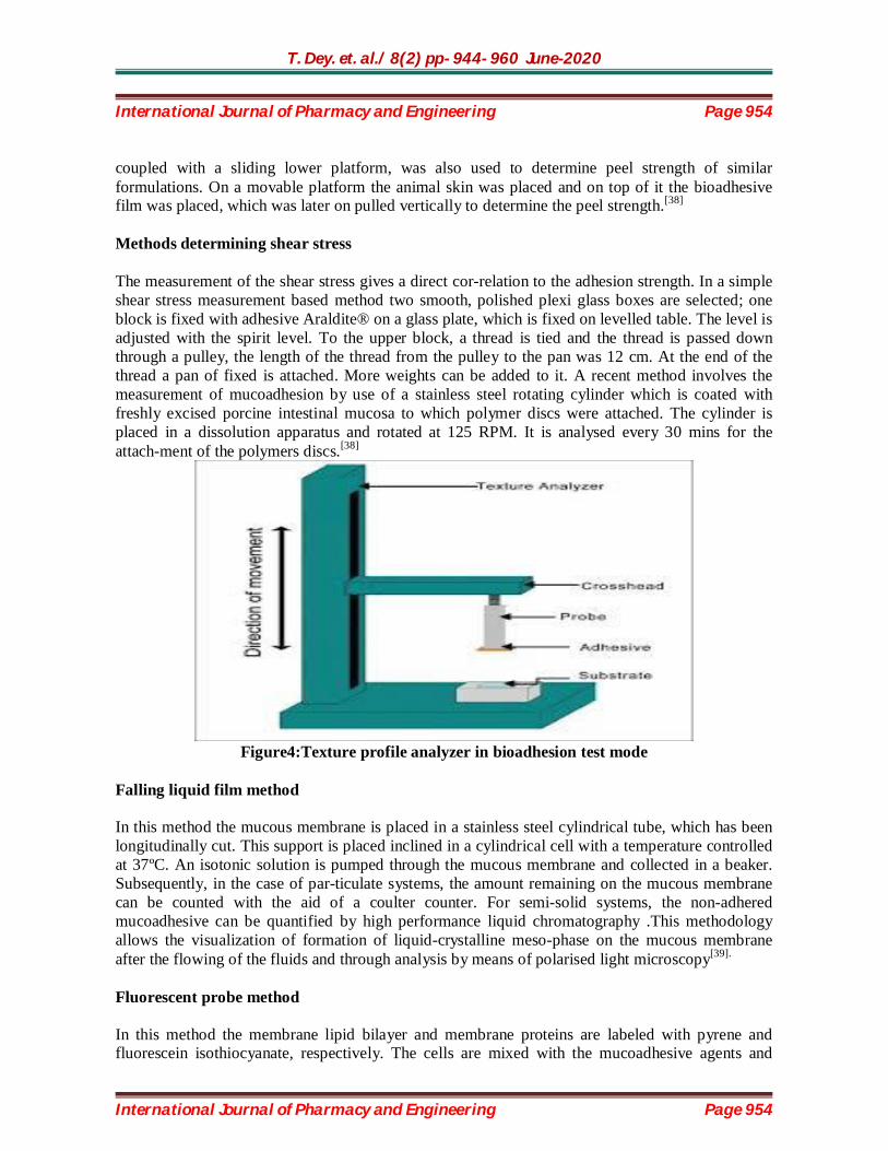
coupled with a sliding lower platform, was also used to determine peel strength of similar formulations. On a movable platform the animal skin was placed and on top of it the bioadhesive film was placed, which was later on pulled vertically to determine the peel strength.[38] Methods determining shear stress The measurement of the shear stress gives a direct cor-relation to the adhesion strength. In a simple shear stress measurement based method two smooth, polished plexi glass boxes are selected; one block is fixed with adhesive Araldite® on a glass plate, which is fixed on levelled table. The level is adjusted with the spirit level. To the upper block, a thread is tied and the thread is passed down through a pulley, the length of the thread from the pulley to the pan was 12 cm. At the end of the thread a pan of fixed is attached. More weights can be added to it. A recent method involves the measurement of mucoadhesion by use of a stainless steel rotating cylinder which is coated with freshly excised porcine intestinal mucosa to which polymer discs were attached. The cylinder is placed in a dissolution apparatus and rotated at 125 RPM. It is analysed every 30 mins for the attach-ment of the polymers discs.[38]
Figure4:Texture profile analyzer in bioadhesion test mode
Falling liquid film method In this method the mucous membrane is placed in a stainless steel cylindrical tube, which has been longitudinally cut. This support is placed inclined in a cylindrical cell with a temperature controlled at 37ºC. An isotonic solution is pumped through the mucous membrane and collected in a beaker. Subsequently, in the case of par-ticulate systems, the amount remaining on the mucous membrane can be counted with the aid of a coulter counter. For semi-solid systems, the non-adhered mucoadhesive can be quantified by high performance liquid chromatography .This methodology allows the visualization of formation of liquid-crystalline meso-phase on the mucous membrane after the flowing of the fluids and through analysis by means of polarised light microscopy[39]. Fluorescent probe method In this method the membrane lipid bilayer and membrane proteins are labeled with pyrene and fluorescein isothiocyanate, respectively. The cells are mixed with the mucoadhesive agents and
T. Dey. et. al./ 8(2) pp- 944- 960 June-2020 International Journal of Pharmacy and Engineering Page 955
International Journal of Pharmacy and Engineering Page 955
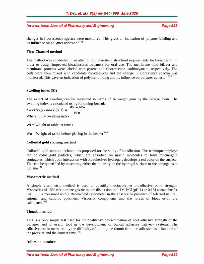
changes in fluorescence spectra were monitored. This gives an indication of polymer binding and its influence on polymer adhesion.[39] Flow Channel method The method was conducted in an attempt to under-stand structural requirements for bioadhesion in order to design improved bioadhesives polymers for oral use. The membrane lipid bilayer and membrane proteins were labeled with pyrene and fluorescence isothiocyanate, respectively. The cells were then mixed with candidate bioadhesives and the change in florescence spectra was monitored. This gave an indication of polymer binding and its influence on polymer adhesion.[39] Swelling index (SI) The extent of swelling can be measured in terms of % weight gain by the dosage form. The swelling index is calculated using following formula.:
Where, S.I = Swelling index Wt = Weight of tablet at time t Wo = Weight of tablet before placing in the beaker [40] Colloidal gold staining method Colloidal gold staining technique is proposed for the study of bioadhesion. The technique employs red colloidal gold particles, which are adsorbed on mucin molecules to form mucin–gold conjugates, which upon interaction with bioadhesives hydrogels develops a red color on the surface. This can be quantified by measuring either the intensity on the hydrogel surface or the conjugates at 525 nm.[41] Viscometric method A simple viscometric method is used to quantify mucinpolymer bioadhesive bond strength. Viscosities of 15% w/v porcine gastric mucin dispersion in 0.1M HCl (pH 1) or 0.1M acetate buffer (pH 5.5) is measured with a Brook-field viscometer in the absence or presence of selected neutral, anionic, and cationic polymers. Viscosity components and the forces of bioadhesion are calculated.[41] Thumb method This is a very simple test used for the qualitative deter-mination of peel adhesive strength of the polymer and is useful tool in the development of buccal adhesive delivery systems. The adhesiveness is measured by the difficulty of pulling the thumb from the adhesive as a function of the pressure and the contact time.[41] Adhesion number:
T. Dey. et. al./ 8(2) pp- 944- 960 June-2020 International Journal of Pharmacy and Engineering Page 956
International Journal of Pharmacy and Engineering Page 956
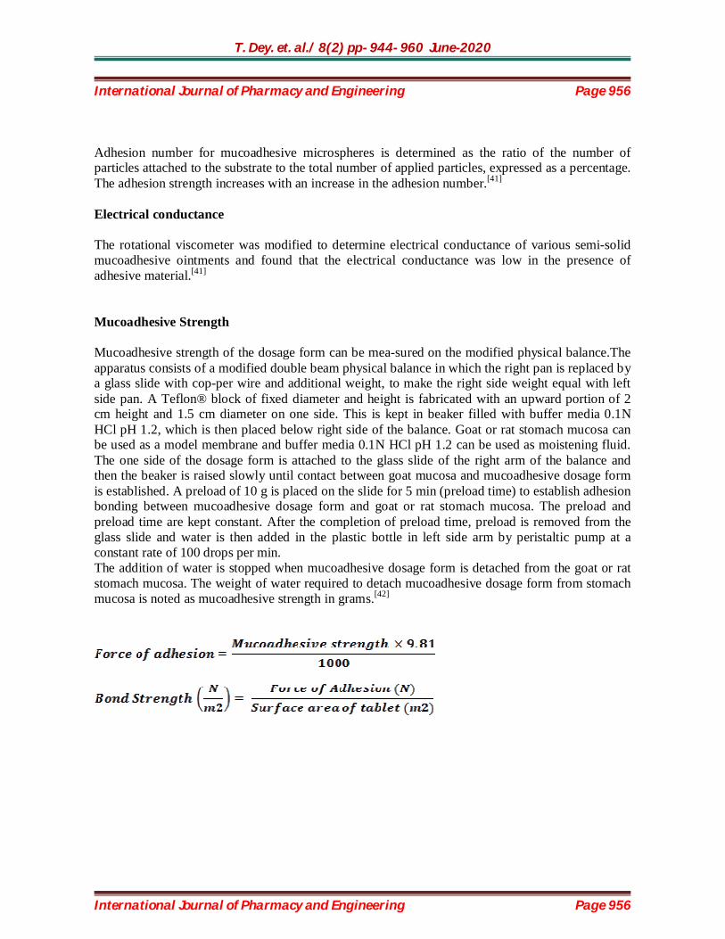
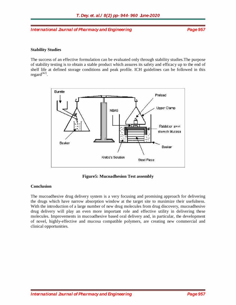
Adhesion number for mucoadhesive microspheres is determined as the ratio of the number of particles attached to the substrate to the total number of applied particles, expressed as a percentage. The adhesion strength increases with an increase in the adhesion number.[41] Electrical conductance The rotational viscometer was modified to determine electrical conductance of various semi-solid mucoadhesive ointments and found that the electrical conductance was low in the presence of adhesive material.[41] Mucoadhesive Strength Mucoadhesive strength of the dosage form can be mea-sured on the modified physical balance.The apparatus consists of a modified double beam physical balance in which the right pan is replaced by a glass slide with cop-per wire and additional weight, to make the right side weight equal with left side pan. A Teflon® block of fixed diameter and height is fabricated with an upward portion of 2 cm height and 1.5 cm diameter on one side. This is kept in beaker filled with buffer media 0.1N HCl pH 1.2, which is then placed below right side of the balance. Goat or rat stomach mucosa can be used as a model membrane and buffer media 0.1N HCl pH 1.2 can be used as moistening fluid. The one side of the dosage form is attached to the glass slide of the right arm of the balance and then the beaker is raised slowly until contact between goat mucosa and mucoadhesive dosage form is established. A preload of 10 g is placed on the slide for 5 min (preload time) to establish adhesion bonding between mucoadhesive dosage form and goat or rat stomach mucosa. The preload and preload time are kept constant. After the completion of preload time, preload is removed from the glass slide and water is then added in the plastic bottle in left side arm by peristaltic pump at a constant rate of 100 drops per min. The addition of water is stopped when mucoadhesive dosage form is detached from the goat or rat stomach mucosa. The weight of water required to detach mucoadhesive dosage form from stomach mucosa is noted as mucoadhesive strength in grams.[42]
T. Dey. et. al./ 8(2) pp- 944- 960 June-2020 International Journal of Pharmacy and Engineering Page 957
International Journal of Pharmacy and Engineering Page 957
Stability Studies The success of an effective formulation can be evaluated only through stability studies.The purpose of stability testing is to obtain a stable product which assures its safety and efficacy up to the end of shelf life at defined storage conditions and peak profile. ICH guidelines can be followed in this regard[42].
Figure5: Mucoadhesion Test assembly Conclusion The mucoadhesive drug delivery system is a very focusing and promising approach for delivering the drugs which have narrow absorption window at the target site to maximize their usefulness. With the introduction of a large number of new drug molecules from drug discovery, mucoadhesive drug delivery will play an even more important role and effective utility in delivering these molecules. Improvements in mucoadhesive based oral delivery and, in particular, the development of novel, highly-effective and mucosa compatible polymers, are creating new commercial and clinical opportunities.
T. Dey. et. al./ 8(2) pp- 944- 960 June-2020 International Journal of Pharmacy and Engineering Page 958
International Journal of Pharmacy and Engineering Page 958
References 1. Verma S, Kaul M, Rawat A, Saini S. An overview on buccal drug delivery system. Int J Pharm
Sci. 2(6): 1303-21 (2011) 2. Rao MBM, Shyam PSS, Shelke N. Preparation and characterisation of solid self micro
emulsifying drug delivery system by adsorbent technique to improve dissolution profile of poorly aqueous soluble drug ramipril. Int J Pharm and Pharm Sci. 2(6): 85-90 (2011)
3. Khairnar GA, Sayyad FJ. Development of buccal drug delivery system based on mucoadhesive polymers. Int J Pharm Tech. 2(1): 719-35 (2010)
4. Das R, Kazi AA, Bandyopadhyay AK. Mucoadhesion and mucoadhesive tablets-a review. Int J Pharma Sci. 6(1): 64-115. (2011)
5. Smart SJD. The basics and underlying mechanisms of mucoadhesion. Adv Drug Deliv Rev. 57(11): 1556–68 (2005)
6. Alexander A, Ajazuddin S, Tripathi DK, Verma T, Maurya J, Patel S. Mechanism responsible for mucoadhesion of mucoadhesive drug delivery system: a review. Int J App Bio and Pharm Tech. 2(1): 434-45 (2011)
7. Allen A, Cunliffe WJ, Pearson JP, Venables CW. The Adherant Gastric Mucus Gel Barrier in Man and Changes in Peptic Ulceration. J. Intern. Med. Res. 228(s732): 83–90 (1990)
8. Vinod KR, Rohit RT, Sandhya S, David B, Venkatram RB. Critical review on mucoadhesive drug delivery systems. Hygeia. J. D. 4(1): 7-28 (2012)
9. Venkatalakshmi R, Sudhakar Y. Buccal drug delivery using adhesive polymeric patches. IJPSR.3(1): 35-41(2012)
10. Madhav NVS, Ojha A, Tyagi Y, Negi M. Mucoadhesion: a novelistic platform for drug delivery system. Int J Pharm. 2(9): 246-58 (2014)
11. Patel AR, Patel DA, Chaudhry SV. Mucoadhesive buccal drug delivery system. Int J of Pharm and Life Sci. 2(6): 848-56 (2011)
12. Longer MA, Robinson JR. Fundamental aspects of bioadhesion. Pharmacy Int. 7:114–7 (1986) 13. Smart JD. The basics and underlying mechanisms of mucoadhesion. Adv Drug Deliv Rev.
57:1556–68 (2005) 14. Hägerström H, Edsman K, Strømme M. Low-frequency dielectric spectroscopy as a tool for
studying the compatibility between pharmaceutical gels and mucus tissue. J Pharm Sci. 92:1869–81 (2003)
15. Dodou D, Breedveld P, Wieringa P. Mucoadhesives in the gastrointestinal tract: Revisiting the literature for novel applications. Eur J Pharm Biopharm. 60:1–16 (2005)
16. Kinloch AJ. The science of adhesion. J Mater Sci. 15:2141–66 (1980) 17. Jiménez-Castellanos MR, Zia H, Rhodes CT. Mucoadhe-sive drug delivery systems. Drug Dev
Ind Pharm. 19:143–94 (1993) 18. Shojaei AH. Buccal mucosa as a route for systemic drug delivery: A review. J Pharm Pharm
Sci. 1:15–30 (1998) 19. Remuñán-López C, Portero A, Vila-Jato JL, Alonso MJ. Design and evaluation of
chitosan/ethylcellulose mucoadhesive bilayered devices for buccal drug delivery. J Control Release. 55:143–52 (1998)
20. Semalty M, Semalty A, Kumar G. Formulation and characterization of mucoadhesive buccal films of glipizide. Indian J Pharm Sci. 70:43–8 (2008)
T. Dey. et. al./ 8(2) pp- 944- 960 June-2020 International Journal of Pharmacy and Engineering Page 959
International Journal of Pharmacy and Engineering Page 959
21. Hornof M, Weyenberg W, Ludwig A, Bernkop SA. Mucoadhesive ocular insert based on thiolated poly (acrylic acid): Development and in vivo evaluation in humans. J Control Release. 89:419–28 (2003)
22. Sultana Y, Aqil M, Ali A. Ocular inserts for controlled delivery of pefloxacin mesylate: Preparation and evaluation. Acta Pharm. 55:305–14 (2005)
23. Wagh VD, Inamdar B, Samanta MK. Polymers used in ocular dosage form and drug delivery systems. Asian J Pharmaceutics. 2:12–7 (2008)
24. Elhadi SS, Mortada ND, Awad GA, Zaki NM, Taha RA. Development of in situ gelling and mucoadhesive mebeverine hydrochloride solution for rectal administration. Saudi Pharm J. 11:150–71 (2003)
25. Neves JD, Amaral MH, Bahia MF. Vaginal drug delivery. In: Gad SC, editor. Pharmaceutical Manufacturing Handbook. NJ: John Willey and Sons Inc; pp. 809–78 (2007)
26. Choi HG, Kim CK. In situ gelling and mucoadhesive liquid suppository containing acetaminophen: Enhanced bioavailability. Int J Pharm.165:23–32 (1998)
27. Asane GS. Mucoadhesive gastro intestinal drug delivery system: An overview. Pharmainfo.net. 5(2007)
28. Schnürch AB. Mucoadhesive systems in oral drug delivery. Drug Discov Today Technol.2:83–7 (2005)
29. Rathbone MJ, Drummond BK, Tucker G. The oral cavity as a site for systemic drug delivery. Adv Drug Deliv Rev.13:1–22(1994).
30. Rajput GC, Majmudar FD, Patel JK, Patel KN, Thakor RS, Patel BP, et al. Stomach specific mucoadhesive tablets as controlled drug delivery system: A review work. Int J Pharm Biol Res.1:30–41(2010)
31. Remeth D, Sfurti S, Kailas M. In-vitro absorption studies of mucoadhesive tablets of acyclovir. Indian J Pharm Educ Res.44:183–8 (2010)
32. Shah D, Gaud RS, Misra AN, Parikh R. Formulation of a water soluble mucoadhesive film of lycopene for treatment of leukoplakia. Int J Pharm Sci Rev Res.12:6–11.(2010)
33. Biswajit B, Kevin G, Thimmasetty J. Formulation and evaluation of pimozide buccal mucoadhesive patches. Int J Pharm Sci Nanotechnol.2:32–41(2010)
34. Wong CF, Yuen KH, Peh KK. Formulation and evaluation of controlled release Eudragit buccal patches. Int J Pharm.178:11–22(1999)
35. Kumar S, Haglund BO, Himmelstein KJ. In situ-forming gels for ophthalmic drug delivery. J Ocul Pharmacol.10:47–56(1994)
36. Jones DS, Woolfson AD, Brown AF, Coulter WA, McClelland C, Irwin CR. Design, characterisation and preliminary clinical evaluation of a novel mucoadhesive topical formulation containing tetracycline for the treatment of periodontal disease. J Control Release.67:357–68(2000)
37. Nielsen LS, Schubert L, Hansen J. Bioadhesive drug delivery system: Characterisation of mucoadhesive properties of systems based on glyceryl mono-oleate and glyceryl monolinoleate.
Eur. J. Pharm. Sci. 3(1): 231- 39 (1998). 38. Rao RKV, Buri PA. Novel in situ method to test polymers and coated micro particles for
bioadhesion. Int. J. Pharm.52(3): 265-70 (1989). 39. Chowdary CPR, Rao YS. Mucoadhesive microspheres for controlled drug delivery. Biol Pharm
Bull.27(11): 1717-24(2004). 40. Patel VM, Prajapati G, Patel M. Mucoadhesive bilayer tablets of propranolol Hydrochloride.
AAPS Pharmasci tech.8(3): 234-42.(2007)
T. Dey. et. al./ 8(2) pp- 944- 960 June-2020 International Journal of Pharmacy and Engineering Page 960
International Journal of Pharmacy and Engineering Page 960
41. Suresh P, Manasa K, Satish BS, Brahmaiah B, Khalilullah S, Sreekanth N. Bioadhesive drug delivery system-a review. Asian J. Pharm. Res.3(1): 30-7(2013).
42. Tangri P. Mucoadhesive drug delivery system: mechanism and methods of evaluation. Int J Pharm Bio Sci. 2(1): 458-67(2011).