MRI in neonatal hypoxic- ischaemic encephalopathy...
56
MRI in neonatal hypoxic- ischaemic encephalopathy: predicting outcome and assessing interventions. M A Rutherford Centre for the Developing Brain Centre for the Developing Brain
Transcript of MRI in neonatal hypoxic- ischaemic encephalopathy...

MRI in neonatal hypoxic-ischaemic encephalopathy:
predicting outcome and assessing interventions. M A Rutherford
Centre for the Developing Brain
Centrefor theDevelopingBrain

The role of neonatal MR imaging
Confirm a normally developed brain Assess severity and pattern of any injuryPredict outcome form pattern of injury and clinical details Assess/ monitor the effect of any intervention
Even with all diagnostic criteria• The spectrum of injury may be wide• The evolution of lesions variable
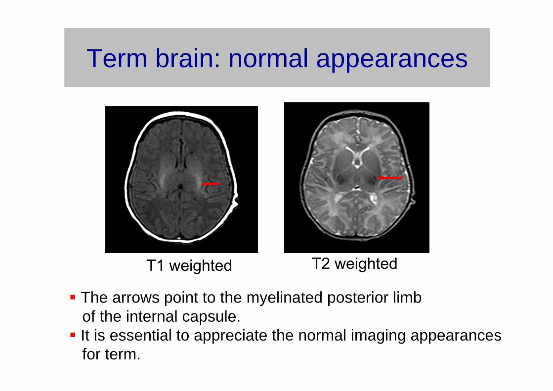
Term brain: normal appearances
T1 weighted T2 weighted
The arrows point to the myelinated posterior limb of the internal capsule.It is essential to appreciate the normal imaging appearances for term.
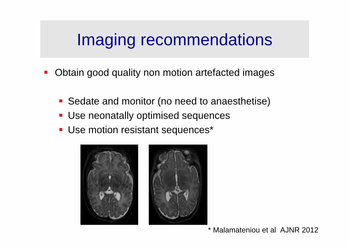
Imaging recommendations
Obtain good quality non motion artefacted images
Sedate and monitor (no need to anaesthetise)Use neonatally optimised sequencesUse motion resistant sequences*
* Malamateniou et al AJNR 2012
Imaging recommendations
Image between 5 and 14 days from delivery
T1 and T2 weighted sequences in axial planeT1 weighted in sagittal plane thinner slicesDiffusion weighted or tensor imaging axial plane• Generate ADC map. MR Venogram to exclude sinus venous thrombosisMR proton spectroscopy. Measure Lactate/Naa ratio

Diffusion tensor imaging
This sequence exploits random motion of water within a tissue . Alterations in signal intensity relate to freedom of motion termed
Diffusivity, measured as a an Apparent Diffusion Coefficient (ADC)
ADC is reduced in acute infarction. Diffusion is the most sensitive sequence to detect early ischaemic lesions

Hypoxic- ischaemic encephalopathy:diagnostsiccriteria
Term born neonate >37 weeks gestationEvidence of fetal distress (abnormal CTG,MSL)Low Apgar ScoresLow umbilical cord pH <7.1Necessity for resuscitationNeurological signs
NB: Always consider dual pathology
Exclude metabolic,infective disorderscongenital malformations

Alternative aetiology in HIE
Pontocerebellar hypoplasia. Diagnosis made on MRI. Normal OFC- this neonate fulfilled all criteria for a cooling trial

Spectrum of injury: antenatal history
Decreased fetal movements• Common, 4-16% pregnancies• Over-represented in neonates with HIE, 15-25%
Imaging findings* 24 out of 70 neonates with HIE referred over 15 month period
• Basal ganglia and thalamic injury in 12%• White matter and cortex injury in 75%
• Consistent with more prolonged insult
Personal data

T1 T2 FLAIR DWI ADC
Case: Decreased fetal movements for 48 hours. Born at 37+3 weeks GA . Unreactive CTG. EMCS performed. Seizures. Imaged day 2
Diffusion imaging excellent for early detection of WM injury-Note abnormal high signal throughout the white matter on DWI andcorresponding low signal in the ADC map
Decreased fetal movements associated with WM injury

Sentinel events
Acute severe hypoxic-ischaemic insult may occur with a sentinel event e.g. uterine rupture, cord prolapse, placental abruption
However only a minority (10%*) of HIE cases have a sentinel event
* Okereafor et al 2008

Acute hypoxic-ischaemic insultsites of abnormality
basal ganglia and thalamiinternal capsulecortexsubcortical white mattermedial temporal lobebrainstem
Increased metabolic rateActively myelinatingIncreased glutamate receptors
These sites are susceptible because they have:

Day 5diffusionDay 11
Day 11
Day 11Day 11T1w
Sites of injury associated with an acute hypoxic ischaemic event
Lesions seen as low signal intensity on the early ADC map and abnormal high signal intensity on the later T1 weighted imagesare predominantly in grey matter

Acute hypoxic-ischaemic insult:sites of abnormality
basal ganglia and thalami (BGT)
BGT lesions give rise to cerebral palsyBGT lesions can be graded as mild, moderate and severeThe severity of neonatal BGT lesion dictates severity of impairment
Moderate; multifocal equivocal or abnormal PLIC
Mild;Focal with normal PLIC
Severe; widespread with abnormal PLIC

Acute hypoxic-ischaemic insultsites of abnormality
basal ganglia and thalamiposterior limb of the internal capsule (PLIC)
Abnormal signal intensity within the PLIC (arrow) predicts abnormal motor outcomeSensitivity= 0.9 Specificity = 1.0 *
* Rutherford et al Pediatrics 1998
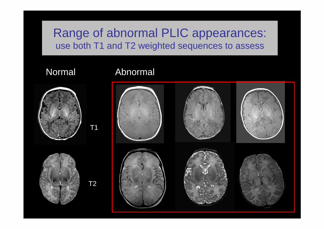
Range of abnormal PLIC appearances: use both T1 and T2 weighted sequences to assess
Normal Abnormal
T1
T2

Injury patterns
Determined by• Nature of insult; chronic, acute
Imaging appearances are influenced by• Sequences used• Time of imaging from injury

Optimal timing conventional imaging
Between 7 and 21 days to ascertain maximum extent of lesions
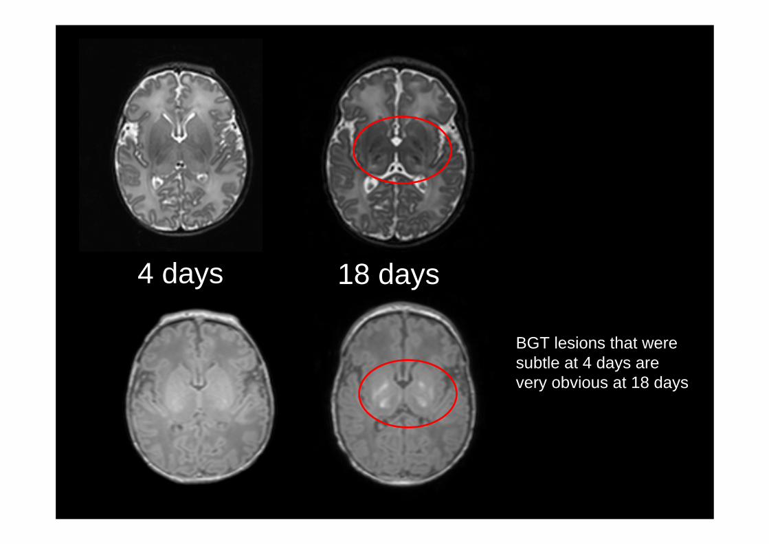
4 days 18 days
BGT lesions that were subtle at 4 days are very obvious at 18 days

T1 and T2 weighted sequences aged 14 days
T1 weighted 6 weeks
Late scanning underestimates severity

Early diffusion imaging
Early conventional imaging may underestimate extent of injuryNeed to use diffusion imagingExcellent for white matter infarctionLess predictable in serial early imaging of BGT injury

Evolution of diffusion changes
day 5
day 11
Low signal intensity regions on these ADC maps are consistent with hypoxic-ischaemic injury. By day 11 the reduced ADC has normalised everywhere. However the low signal intensity in the globus pallidus has become more obvious.

Day 3
Day 22
The day 3 low signal intensity on the ADC map is less marked than the eventual injury on T2W image at day 22.
Diffusion imaging at one time point may underestimate BGT injury
ADC map
T1 T2
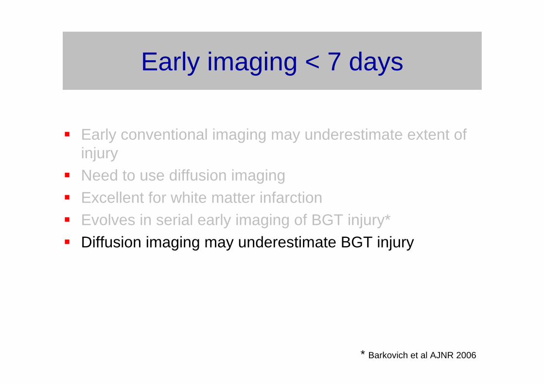
Early imaging < 7 days
Early conventional imaging may underestimate extent of injuryNeed to use diffusion imagingExcellent for white matter infarctionEvolves in serial early imaging of BGT injury* Diffusion imaging may underestimate BGT injury
* Barkovich et al AJNR 2006

Pattern of injury in neonatal HIE
Pattern of brain injury dictated by nature of insult. Take a careful historyExclude infection Exclude metabolic and congenital abnormalities

Predicting Outcome
Pattern of injury dictates neurodevelopmentaloutcome Basal ganglia and thalamic (BGT) lesions associated with cerebral palsyAbnormal PLIC associated with abnormal motor outcome

Patients555 infants included in our 1993-2007
neonatal encephalopathy database
175 infants included
186 infants did not have BGT injury
64 infants had metabolic disorders, congenital
malformations or infections
41 infants were cooled
10 infants were lost to follow-up
20 infants were < 35 wks
59 infants had an MRI scan after 6 weeks
Martinez Biarge Neurology 2011
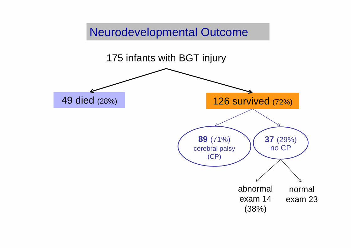
Neurodevelopmental Outcome
175 infants with BGT injury
49 died (28%) 126 survived (72%)
abnormalexam 14
(38%)
normal exam 23
89 (71%)cerebral palsy
(CP)
37 (29%)no CP

No CP / mild CP (level* I)
ModerateCP (levels* II & III)
Severe CP (levels* IV & V)
Mild BGT Moderate BGT & equivocal PLIC
Moderate BGT & abnormal PLIC
Severe BGT
Spearman´s correlation coefficient = 0.773
MRI and motor outcome at 2 years
* Gross motor function score

All children with normal signal intensity in the posterior limb of the internal capsule (PLIC) were walking independently by 2 years
PLIC and ability to walk at age 2 years
PLIC Not walking Walking
Abnormal 73 10
Normal 0 23
Sensitivity = 1.0 Specificity = 0.70
PPV = 0.88
NPV = 1.0
Martinez Biarge Neurology 2011

Acute hypoxic-ischaemic insultsites of abnormality
basal ganglia and thalamiinternal capsule
brainstem
swollen ponsloss of corticospinal tractsponto subicular necrosis

Brainstem lesions andoutcome in HIE (n=175)
No brainstem injury (32%)
Moderate brainstem injury (23%)
Severe brainstem injury (45%)
No deaths 25% died 49% died
T1 T1 T1
Martinez Biarge Neurology 2011

Brain stem lesions in HIE
* Martinez Biarge Neurology 2011
In surviving infants with BGT lesions:
mesencephalic injury was associated with prolonged feeding difficulties (p<0.001)
pontine injury was associated with gastrostomy (p<0.001).

Isolated white matter injury
Uncommon in HIEMore common if history of decreased fetalmovementsMore common if infection Associated with hypoglycaemia
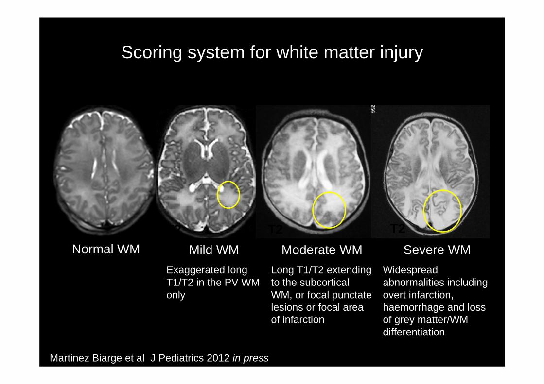
Scoring system for white matter injury
Normal WM Mild WMExaggerated long T1/T2 in the PV WM only
Moderate WMLong T1/T2 extending to the subcorticalWM, or focal punctate lesions or focal area of infarction
Severe WMWidespread abnormalities including overt infarction, haemorrhage and loss of grey matter/WM differentiation
T2 T2 T2 T2
Martinez Biarge et al J Pediatrics 2012 in press

Normal and mild WMn = 22
Moderate WMn = 28
Severe WMn =18
P
Total DQ 112 104.7 85.4 <0.001
Motor 108.4 107.3 91.4 0.15
Social 114.3 108.5 94.6 0.018
Language 111.7 106 79.2 <0.001
Eye and Hand Coordination 109.4 99.3 82.3 <0.001
Performance 115.6 103.5 81.1 <0.001
67 children were evaluated with Griffiths at a median age of 29 months (range 12-56)
Outcomes in isolated WM injury in HIE

The role of MR imaging in HIE
Confirm normally developed brain Assess severity and pattern of injury
Influenced by nature of insult Predict outcome
Associated with pattern of injury
Assess/ monitor effect of intervention

Conventional imaging Visual analysis of lesions with grading
Diffusion tensor imaging Tract Based Spatial Statistics (TBSS)
MRI to assess the effect of interventions in HIE.

Visual analysis: Hypothermia
Does hypothermia alter pattern of lesions?Does it decrease the number of lesions?Does it delay the onset or evolution of lesions?Does it impair the ability of MR to predict outcome?

TOBY trialMulti centre 42 hospitals between 2002-2006
Term neonates >36 weeks GAFetal distressEncephalopathyAbnormal aEEG
Recruit prior to 6 hoursModerate total body hypothermia 33-34° C for 72 hours
Outcome at 18 months
Imaging between 1-3 weeks T1 and T2 weighted sequences in transverse and sagittal planes
Azzopardi et al NEJM 2009
TOBY trial
151 infants of 325 underwent MR imaging
131 scans suitable for analysisGood qualityNo consistent diffusion imaging

Patterns of injury: Haemorrhage
47/131 infants had signs of haemorrhage
39 had subdural , 10 moderate and 29 mild10 infants had haemorrhage in other sites• 3 IVH• 1 caudate head• 1 cerebellum• 5 parenchyma
2 associated with venous sinus thrombosis

Haemorrhage in TOBY trial neonates
The majority of haemorrhages detected were small and not considered to be significant for long term outcome
There was no increase in haemorrhagic lesions associated with cooling 25 cooled v 22 non-cooled (p=0.46)
Haemorrhage

Sinus thrombosis
3/131 infants imaged had signs consistent with sinus thrombosis 2 non cooled and one cooled.

Hypothermia
Does hypothermia alter pattern of lesions? Does it decrease the number of lesionsDoes it delay the evolution of lesions?Does it impair the ability of MR to predict outcome?
NO

Hypothermia reduces tissue injury *
Therapeutic hypothermia was associated with a reduction in:
• Basal ganglia or thalamus lesions (P=0.02)• White matter lesions (P=0.01) • Abnormal posterior limb of the internal capsule (P=0.02).
Cooled infants: • had fewer scans predictive of later neuromotor abnormalities (P=0.03) • were more likely to have normal scans ( P=0.03).
* Rutherford et al Lancet Neurol 2010

Hypothermia
Does hypothermia alter pattern of lesions? Does it decrease the number of lesions?Does it alter the evolution of lesions?Does it impair the ability of MR to predict outcome?
NOYES

Evolution of lesions
Does hypothermia effect evolution of lesions? On conventional imaging• Needs looking at systematically- no obvious effect in TOBY
infants or non Trial infants.
On diffusion imaging• Evolution of diffusion in BGT is prolonged and patterns very
different even without hypothermia• Needs looking at systematically with hypothermia. Suggestion
that hypothermia may prolong diffusion abnormalities*
• However any abnormality in BGT on diffusion clinically significant as one scan likely to underestimate
* Bednarek, et al. Neurology 2012

Hypothermia
Does hypothermia alter pattern of lesions? Does it decrease the number of lesionsDoes it alter the evolution of lesions? Does it impair the ability of MR to predict outcome?
NOYESUNCLEAR
Prediction of outcome
MRI performed at median of 8 days in both cooled and noncooledinfantsThe accuracy of prediction by MRI of death or disability to 18 months of age was 0.84, 95% CI, 0.74-0.94 in the cooled and 0.81, 95% CI, 0.71-0.91 in the non cooled groups.
* Rutherford et al Lancet Neurol 2010

Hypothermia (n=131)
Does hypothermia alter pattern of lesions? Does it decrease the number of lesionsDoes it delay the evolution of lesions? UNCLEARDoes it impair the ability of MR to predict outcome?
NOYES
NO
All these questions need to be asked as new interventions administered e.g. Xenon

Summary1: MRI in HIE
Pattern of brain injury determined by• Nature of insult
Imaging appearances influenced by• Sequences used• Time from injury
Pattern of injury dictates outcome• Sentinel events , acute injury – BGT lesions• prolonged injury, infection , hypoglycaemia- WM lesions

Summary 2: MRI in HIE
Neonatal MR imaging provides excellent surrogate outcome
Hypothermia decreases lesionsNot associated with atypical injuryDoes not alter ability to predict outcome

Recommendations in HIE
Image between 5 and 14 days
T1 and T2 weighted sequences in axial planeT1 weighted in sagittal plane thinner slicesDiffusion weighted imaging axial plane
• Generate ADC map.
MR VenogramMR proton spectroscopy Lactate/Naa ratio
MOTION renders image data uninterpretable.*
* Malamateniou et al AJNR 2012

AcknowledgementsMany thanks to all the staff in the Robert Steiner MR Unit and on the neonatal units of Hammersmith and Queen CharlottesHospitals
Jo HajnalSerena CounsellJoanna AllsopAmy McGuinnessAkudo OkereaforMiriam Martinez BiargeChristina Malamateniou
Frances CowanDenis AzzopardiDavid Edwards
Dulcie Rodrigues
Centrefor theDevelopingBrain