MR of Venous Sinus Thrombosis: A Case Report
3
713 MR of Venous Sinus Thrombosis: A Case Report Wolfgang M. Bauer,' Karl Einhaupl, Sylvia H. Heywang, Thomas Vogl, Manfred Seiderer, and Dimitrios Clados The frequency of aseptic venous sinus thrombosis (VST) is underestimated according to many authors [1-4] . One of the reason s is that until now the correct diagnosis of VST could only be established by cerebral angiography. As pathologic changes can be demonstrated by MR in a patient with angio- graphically proven VST, MR may become an important non- invasive method for its primary diagnosis. To our knowledge, this is the first report of the diagnosis of VST by MR. Materials and Methods MR images were obtained with a 1.5-T superconductive magnet (Magnetom, Siemens AG , Erlangen), which is now operated at 0.35 T. A multislice T2-weighted double-echo spin-echo (SE) technique (TR/TE = 1600/35,70 and 1600/60,120) was used as well as a more T1-weighted SE sequence (TRITE = 400/35,70 and 400/60,120). The spin-echo images were obtained in transverse, coronal, and sagittal planes with a slice thickness of 10 mm. Bilateral carotid angiography was performed using a trans femoral catheter technique in frontal and lateral projections with films taken every second up to 15 sec. Case Report A 44-year-old woman with a 1 O-year history of migraine, who had taken contraceptives for 15 years and had a history of labile hyper- tension , suffered from an increasingly severe headache for several days after a common cold. Five days after the onset of the symptoms a grand mal seizure precipitated hospital admission, where she presented with mild disorientation and right occipital headache. EEG EEG indicated a focal lesion in the right occiput as well as discon- tinuous delta waves of low amplitude. The CSF was xanthochromic with 3300 red blood cells/mm 3 but with no other abnormalities. Radiologic Studies CT on the 10th day showed a small hyperdense area in the left parietal region without enhancement, which was thought to be a Received March 28 , 1985; accepted after revision July 2, 1985. hypertensive hemorrhage. A second CT study on the 11 th day showed narrow ventricles; the basal cisterns were not well defined but the gyri were still di scernible. After enhancement, the empty delta sign was seen (Fig . 1 A) , as was enhancement of the right transverse sinus. Carotid angiography showed thrombosis of the superior sagittal sinus. The ascending cerebral veins were congested and without connection to the superior sagittal sinus (Fi gs. 1 Band 1 C). On MR, the superior sagittal , right transverse , and sigmOid sinu ses displayed high signal intensities on th e T1 - and T2-weighted SE images (Figs. 10-1 G) . Additionally , an area of high signal intensity could be seen in the right parasagittal region, which was interpreted as a hemorrhagically infarcted area of cortex (Fig . 1 G) . Discussion Indirect signs, which are mostly nonspecific, can be ob- tained by CT. Consequently, CT is an important noninvasive method in the early diagnostic work-up of VST. The only specific CT sign is the empty delta sign [5-8] first reported by Buonanno et al. [5]. The sinus rectus sign [9, 10], the falx sign [11] , and the gyral or tentorial enhancement [5 , 7, 12- 14] are found not only with VST. Other signs, such as intracerebral hemorrhage [5 , 7, 15-18], hypodense areas [5, 9, 18], or increased cerebral volume [19], are more frequently seen in other cerebral diseases and are less indicative of VST [20]. Although digital subtraction angiography has been able to show signs of sinus occlusion in some cases [15] , its reliability for the majority of cases is not yet proven . MR signal intensity and contrast not only depend on T1- and T2-values and proton density, but also on flow velocity. Thus, information about blood flow in vessels can be obtained. This is true not only for big arteries but also for the large cerebral venous sinuses such as the superior sagittal sinus, the transverse sinus, and the sigmoid sinus, which can be well assessed, if adequately examined. In a group of 60 patients comprising healthy persons as well as patients with nonvascular cerebral di seases, the su- perior sagittal sinus could be seen without exception as a 1 All authors: Departments of Radiology and Neurology, Klinikum Grosshadern , Uni versity of Munich . Marchionini strass e 15. 8000 Munchen 70. Germany. Address reprint reques ts to W. M. Bauer. AJNR 8:713-715, July/August 1987 01 95-6 108/87/0804-071 3 © Ameri can Society of Neuroradiology
-
Upload
phungkhanh -
Category
Documents
-
view
225 -
download
0
Transcript of MR of Venous Sinus Thrombosis: A Case Report
713
MR of Venous Sinus Thrombosis: A Case Report Wolfgang M. Bauer,' Karl Einhaupl , Sylvia H. Heywang , Thomas Vogl , Manfred Seiderer, and Dimitrios Clados
The frequency of aseptic venous sinus thrombosis (VST) is underestimated according to many authors [1-4] . One of the reasons is that until now the correct diagnosis of VST could only be established by cerebral angiography. As pathologic changes can be demonstrated by MR in a patient with angiographically proven VST, MR may become an important noninvasive method for its primary diagnosis. To our knowledge, this is the first report of the diagnosis of VST by MR.
Materials and Methods
MR images were obtained with a 1.5-T superconductive magnet (Magnetom, Siemens AG, Erlangen), which is now operated at 0.35 T. A multislice T2-weighted double-echo spin-echo (SE) technique (TR/TE = 1600/35,70 and 1600/60,120) was used as well as a more T1-weighted SE sequence (TRITE = 400/35,70 and 400/60,120). The spin-echo images were obtained in transverse, coronal , and sagittal planes with a slice thickness of 10 mm.
Bilateral carotid angiography was performed using a trans femoral catheter technique in frontal and lateral projections with films taken every second up to 15 sec.
Case Report
A 44-year-old woman with a 1 O-year history of migraine, who had taken contraceptives for 15 years and had a history of labile hypertension , suffered from an increasingly severe headache for several days after a common cold . Five days after the onset of the symptoms a grand mal seizure precipitated hospital admission, where she presented with mild disorientation and right occipital headache.
EEG
EEG indicated a focal lesion in the right occiput as well as discontinuous delta waves of low amplitude. The CSF was xanthochromic with 3300 red blood cells/mm3 but with no other abnormalities.
Radiologic Studies
CT on the 10th day showed a small hyperdense area in the left parietal region without enhancement, which was thought to be a
Received March 28, 1985; accepted after revision July 2, 1985.
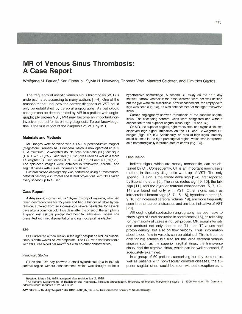
hypertensive hemorrhage. A second CT study on the 11 th day showed narrow ventricles ; the basal cisterns were not well defined but the gyri were still discernible. After enhancement, the empty delta sign was seen (Fig . 1 A), as was enhancement of the right transverse sinus.
Carotid angiography showed thrombosis of the superior sagittal sinus. The ascending cerebral veins were congested and without connection to the superior sagittal sinus (Figs. 1 Band 1 C).
On MR, the superior sagittal , right transverse , and sigmOid sinuses displayed high signal intensities on the T1 - and T2-weighted SE images (Figs. 10-1 G). Additionally , an area of high signal intensity could be seen in the right parasagittal region , which was interpreted as a hemorrhagically infarcted area of cortex (Fig . 1 G) .
Discussion
Indirect signs, which are mostly nonspecific, can be obtained by CT. Consequently, CT is an important noninvasive method in the early diagnostic work-up of VST. The only specific CT sign is the empty delta sign [5-8] first reported by Buonanno et al. [5] . The sinus rectus sign [9 , 10], the falx sign [11] , and the gyral or tentorial enhancement [5 , 7, 12-14] are found not only with VST. Other signs, such as intracerebral hemorrhage [5 , 7, 15-18], hypodense areas [5 , 9, 18], or increased cerebral volume [19] , are more frequently seen in other cerebral diseases and are less indicative of VST [20] .
Although digital subtraction angiography has been able to show signs of sinus occlusion in some cases [15] , its reliability for the majority of cases is not yet proven . MR signal intensity and contrast not only depend on T1- and T2-values and proton density, but also on flow velocity . Thus, information about blood flow in vessels can be obtained. This is true not only for big arteries but also for the large cerebral venous sinuses such as the superior sagittal sinus , the transverse sinus , and the sigmoid sinus , which can be well assessed, if adequately examined .
In a group of 60 patients comprising healthy persons as well as patients with nonvascular cerebral diseases, the superior sagittal sinus could be seen without exception as a
1 All authors: Departments of Radiology and Neurology, Klinikum Grosshadern , Universi ty of Munich . Marchioninistrasse 15. 8000 Munchen 70. Germany. Address reprint requests to W. M. Bauer.
AJNR 8:713-715, July/August 1987 01 95-6108/87/0804-071 3 © American Society of Neuroradiology
714
A
G
BAUER ET AL. AJNR :8, Julyl August 1987
B c
E F
Fig. 1.-A , Enhanced CT scan with empty delta sign (arrow) and gyral enhancement on right. B, Right carotid angiogram, frontal projection, late venous phase: superior sagittal sinus not opacified,
drainage via sphenoparietal sinus and cavernous sinus through base of skull to pterygoid plexus. C, Right carotid angiogram, lateral projection, late venous phase: superior sagittal sinus not opacified,
enlarged tortuous ascending veins, draining inferiorly toward sylvian group of veins. Outflow via sphenoparietal sinus and cavernous sinus through base of skull to pterygoid plexus. Occlusion of anterior part of inferior sagittal sinus. Increased circulation time. Left carotid angiogram showed similar abnormalities.
D, Transverse spin-echo image (TRI TE = 1600/35) with high signal intensity of superior sagittal sinus. E, Transverse spin -echo image (TRI TE = 1600 /35) with high signal intensity of right transverse and
sigmoid sinuses, irregular dark areas next to vessel wall may be indicative of incomplete thrombotic obstruction (arrows) .
F, Slightly parasagittal spin-echo image (TRI TE = 1600/ 70) delineating thrombotic superior sagittal sinus.
G, Coronal spin-echo image (TRI TE = 4001120) shows thrombotic right sigmoid sinus and superior sagittal sinus. Area of high intensity parasagittally on right may correspond to a hemorrhagically infarcted area of cortex.
AJNR :8, July/August 1987 MR OF VENOUS SINUS THROMBOSIS 715
dark area of low signal intensity in T1 - and T2-weighted spinecho images. In the case described above, in which VST had been angiographically confirmed , the area in question displayed high signal intensity (Figs. 10-1 G). As mentioned by Mills et al. [21], these MR findings are characteristic of stationary blood or thrombosis, although very slowly flowing blood may also show increased signal intensity due to "paradoxic" enhancement.
According to Mills et al. [21] , vascular occlusion results in a high signal intensity on the first echo and decreased signal intensity on the second echo, whereas in conditions of slowly flowing blood the second echo-delay signal may be equal to or even higher than the first. In our patient the measurement of signal intensities in the superior sagittal sinus of the first (35,60 msec) and second (70,120 msec) echoes showed significant decrease of signal intensity between the first and second echoes . The results demonstrate the capabilities of MR . This new method provides the possibility of differentiating between flowing and stationary blood without being invasive and without application of contrast material. This may finally lead to a change in diagnostic procedure.
MR may contribute to an earlier diagnosis of VST, as it provides an indication for carotid angiography in suspected or uncertain cases. And while it is not yet possible to eliminate carotid angiography in the primary diagnosis of VST, total substitution of carotid angiography by MR in patients with VST in the future does seem possible. However, more experience and further investigations are necessary.
REFERENCES
1. Kalbag RM , Woolf AL. Cerebral venous thrombosis. London: Oxford University Press, 1967
2. KrayenbOhl HA. Cerebral venous and sinus thrombosis . Clin Neurosurg 1967;14 : 1- 24
3. Towbin A. The syndrome of latent cerebral venous thrombosis . Stroke
1973;4:419- 430 4. Vines FS, Davis DO. Clinical-radiological correlation in cerebral venous
occlusive disease. Radiology 1971;98 :9- 22 5. Buonanno FS, Moody OM , Ball MR, Laster ow. Computed cranial tomo
graphic findings in cerebral sinovenous occlusion . J Comput Assist Tomogr 1978;82 :281-291
6. Brant-ZawadzkiM , Chang GY, McCarty E. Computed tomography in dural sinus thrombosis . Arch Neuro/ 1982;39 :446-447
7. Rao KCVG , Knipi H, Wagner E. Computed tomographic findings in cerebral sinus and venous thrombosis. Radiology 1981 ;140 :391 - 398
8. Zilkha A, Daiz AS. Computed tomography in the diagnosis of superior sagittal sinus thrombosis . J Comput Assist Tomogr 1980;4 : 124- 126
9. Eick JJ , Miller KD, Bell KA, Tutton RH . Computed tomography of deep cerebral venous thrombosis in children . Radiology 1981 ;140 :399-402
10. Wendling LR . Intracranial venous sinus thrombosis . Diagnosis suggested by computed tomography. AJR 1978;130 :978-980
11 . Osborn AG , Anderson RE, Wing SO. The false falx sign. Radiology 1980;134 :421 - 425
12. Banna M, Groves FT. Deep vascular congestion in dural venous thrombosis. J Comput Assist Tomogr 1979;3 :539-541
13. Ingstrup HM, Jorgensen PS . Tentorial changes in sigmoid sinus thrombosis. J Comput Assist Tomogr 1981 ;5 :760-762
14. Naidich TP, Leeds NE, Kricheff II , Pudlowski RM, Naidich JB, Zimmermann RD. The tentorium in axial section. I. Normal CT appearance and nonneoplastic pathology. Radiology 1977;123:631-638
15. Barnes BD, Brant-Zawadzki M, Mentzner W. Digital subtraction angiography in the diagnosis of superior sagittal sinus thrombosis. Neurology 1983;33 :508-510
16. Beal MF, Wechsler LR , Davis KR . Cerebral vein thrombosis and multiple intracranial hemorrhage by computed tomography . Arch Neurol 1982;39 :437-438
17. Brismar J. Computed tomography in superior sagittal sinus thrombosis. Acta Radio/1980 ;21 :321-326
18. Kingsley OPE, Kendall BE , Moseley IF. Superior sagittal sinus thrombosis. An evaluation of the changes demonstrated on computed tomography. J Neurol Neurosurg Psychiatry 1978;41 : 1 065-1 068
19. Munderloh S, Schnapf 0 , Coker S, Citrin C. Changing ventricular size in dural sinus thrombosis . J Comput Tomogr 1981 ;5 :11-16
20. McCormick WF, Rosenfield DB. Massive brain hemorrhage. A review of 144 cases and an examination of their causes. Stroke 1973;4 : 946- 954
21 . Mills CM, Brant-Zawadzki M, Crooks LE , et al. Nuclear magnetic resonance: Principles of blood flow imaging. AJNR 1983;4: 11 61 - 1166, AJR 1984;142 : 165-170