MR Neurography of Neuromas Related to Nerve … · Injury and Entrapment with Surgical Correlation...
6
REVIEW ARTICLE MR Neurography of Neuromas Related to Nerve Injury and Entrapment with Surgical Correlation A. Chhabra E.H. Williams K.C. Wang A.L. Dellon J.A. Carrino SUMMARY: MR imaging of peripheral nerves has been described in relation to abnormalities such as nerve injury, entrapment, and neoplasm. Neuroma formation is a known response to peripheral nerve injury, and here we correlate the MRN appearance of postinjury neuroma formation with intraoperative findings. We also present the MR imaging features of surgical treatment with a synthetic nerve tube and nerve wrap on postoperative follow-up imaging. ABBREVIATIONS: MRN MR neurography; NIC neuroma in continuity; SPACE sampling perfection with application optimized contrasts by using different flip angle evolutions; SPAIR spectral adiabatic inversion recovery; STIR short tau inversion recovery P eripheral nerve injuries and entrapments may lead to for- mation of NIC, neuroma in completely severed nerves, and amputation neuroma. These lesions also demonstrate unique MRN appearances. This article presents MRN and sur- gical correlations of various posttraumatic neuromas with rel- evant case examples. Anatomy and Pathophysiology The basic structure of a peripheral nerve is a neuronal axon enveloped in a myelin sheath composed of Schwann cells and loose connective tissue referred to as the “endoneurium.” Multiple axons are arranged together as bundles called “fasci- cles,” the fundamental neural unit imaged with current high- resolution MRN techniques (Fig 1). Each fascicle is enveloped by a connective tissue layer called the “perineurium.” Groups of fascicles are arranged in yet another connective tissue layer, called the “epineurium,” which serves as the outer sheath of the peripheral nerve. 1,2 In traumatic or entrapment neuropa- thies, the nerves can go through a spectrum of injuries span- ning neurapraxia (focal damage to the myelin sheath without axonal disruption), axonotmesis (disruption of multiple ax- onal fibers without myelin or connective tissue disruption), and neurotmesis (complete loss of axonal continuity along with partial or complete discontinuity of the myelin sheath and supporting connective tissue fibers). A neurapraxia by definition resolves very quickly (within weeks to a few months) because the axons are not damaged and, therefore, do not undergo wallerian degeneration, the process of axonal degradation distal to a site of nerve injury. These injuries are treated conservatively and will fully recover. However, injuries associated with an axonotmesis or neurot- mesis do experience wallerian degeneration; therefore, the nerve has to regenerate from the point of injury to its distal target. The advantage for the patient with an axonotmesis is that the neural track and Schwann cell tract are still intact so that regeneration to the distal targets is more accurate and, therefore, will usually recover good function if treated conser- vatively. In neurotmesis, little recovery can be expected with- out surgical intervention because the nerve is physically di- vided and the orientation of the nerve is disrupted. Surgical exploration is performed to reorient the nerve fiber and repair the nerve. 3-5 Pathologically, any nerve that is lacerated, avulsed, or trau- matized may form a neuroma. These neuromas can be classi- fied into 2 basic types: NIC or an end-bulb neuroma. NIC usually involves all degrees of nerve injury, from normal to neurotmesis, coexisting within a scarred nerve. With time, the proximal injured nerve fascicles sprout in an attempt to unite. However, due to surrounding lattice disruption, disorganized regeneration, hypertrophy of nerve fascicles, and associated fibrosis, the proximal and distal nerve fibers at the site of in- jury may fail to appose and thereby lead to NIC formation. NICs may be of 2 types pathologically: spindle neuromas with intact perineurium or lateral neuromas that occur after partial disruption of the perineurium and after nerve repairs. 6-8 End-bulb neuromas occur anywhere a nerve is completely divided and is unopposed by another neural tissue. These are subcategorized as a neuroma in a completely severed nerve and as an amputation neuroma. Most commonly these occur in lacerations in which the nerve is not repaired in a timely fashion, amputation stumps, and in postoperative patients in whom sensory nerves in the skin may have been divided unknowingly. Clinical Presentation and Management Clinically, NIC may lead to a dysfunctional nerve, disabling pain, alterations in the patient’s lifestyle, and possible progres- sion to chronic pain syndromes. The Tinel sign (local tender- ness over the injured nerve with distally radiating tingling), denervation atrophy of the muscles, and sensory or trophic changes are evident on clinical examination. Treatment of NIC is challenging and is targeted to alleviating the pain and restoring the functional loss caused by the nerve injury. 7 Recent microneurosurgical techniques are a considerable advancement in repairing these lesions. After failure of con- servative treatment, if the patient continues to have severe From the Russell H. Morgan Department of Radiology and Radiological Science (A.C., K.C.W., J.A.C.) and Departments of Plastic Surgery (E.H.W., A.L.D.) and Neurosurgery (A.L.D.), Johns Hopkins Hospital, Baltimore, Maryland; and Dellon Institute for Peripheral Nerve Surgery (E.H.W., A.L.D.), Towson, Maryland. K.C.W. gratefully acknowledges the support of the Radiological Society of North America (RSNA) Research and Education Foundation Fellowship Training grant #FT0904, as well as that of the Walter and Mary Ciceric Research Award. Please address correspondence to Avneesh Chhabra, MD, Johns Hopkins University Hospital, MSK Radiology Section, 601 N Caroline St, JHOC, Ste 3262, Baltimore, MD 21287; e-mail: [email protected] functional deficit. Indicates open access to non-subscribers at www.ajnr.org DOI 10.3174/ajnr.A2002 REVIEW ARTICLE AJNR Am J Neuroradiol 31:1363– 68 Sep 2010 www.ajnr.org 1363
Transcript of MR Neurography of Neuromas Related to Nerve … · Injury and Entrapment with Surgical Correlation...
REVIEW ARTICLE
MR Neurography of Neuromas Related to NerveInjury and Entrapment with Surgical Correlation
A. ChhabraE.H. Williams
K.C. WangA.L. Dellon
J.A. Carrino
SUMMARY: MR imaging of peripheral nerves has been described in relation to abnormalities such asnerve injury, entrapment, and neoplasm. Neuroma formation is a known response to peripheral nerveinjury, and here we correlate the MRN appearance of postinjury neuroma formation with intraoperativefindings. We also present the MR imaging features of surgical treatment with a synthetic nerve tubeand nerve wrap on postoperative follow-up imaging.
ABBREVIATIONS: MRN � MR neurography; NIC � neuroma in continuity; SPACE � samplingperfection with application optimized contrasts by using different flip angle evolutions; SPAIR �spectral adiabatic inversion recovery; STIR � short tau inversion recovery
Peripheral nerve injuries and entrapments may lead to for-mation of NIC, neuroma in completely severed nerves,
and amputation neuroma. These lesions also demonstrateunique MRN appearances. This article presents MRN and sur-gical correlations of various posttraumatic neuromas with rel-evant case examples.
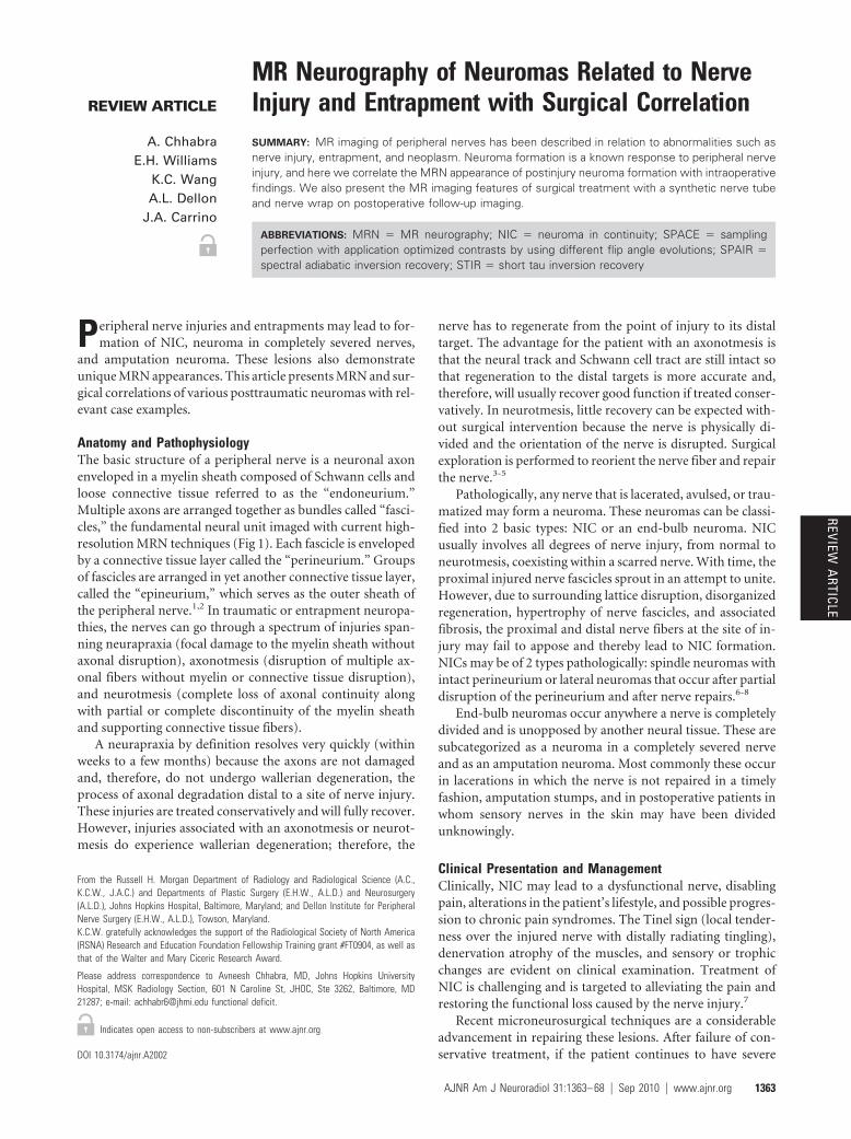
Anatomy and PathophysiologyThe basic structure of a peripheral nerve is a neuronal axonenveloped in a myelin sheath composed of Schwann cells andloose connective tissue referred to as the “endoneurium.”Multiple axons are arranged together as bundles called “fasci-cles,” the fundamental neural unit imaged with current high-resolution MRN techniques (Fig 1). Each fascicle is envelopedby a connective tissue layer called the “perineurium.” Groupsof fascicles are arranged in yet another connective tissue layer,called the “epineurium,” which serves as the outer sheath ofthe peripheral nerve.1,2 In traumatic or entrapment neuropa-thies, the nerves can go through a spectrum of injuries span-ning neurapraxia (focal damage to the myelin sheath withoutaxonal disruption), axonotmesis (disruption of multiple ax-onal fibers without myelin or connective tissue disruption),and neurotmesis (complete loss of axonal continuity alongwith partial or complete discontinuity of the myelin sheathand supporting connective tissue fibers).
A neurapraxia by definition resolves very quickly (withinweeks to a few months) because the axons are not damagedand, therefore, do not undergo wallerian degeneration, theprocess of axonal degradation distal to a site of nerve injury.These injuries are treated conservatively and will fully recover.However, injuries associated with an axonotmesis or neurot-mesis do experience wallerian degeneration; therefore, the
nerve has to regenerate from the point of injury to its distaltarget. The advantage for the patient with an axonotmesis isthat the neural track and Schwann cell tract are still intact sothat regeneration to the distal targets is more accurate and,therefore, will usually recover good function if treated conser-vatively. In neurotmesis, little recovery can be expected with-out surgical intervention because the nerve is physically di-vided and the orientation of the nerve is disrupted. Surgicalexploration is performed to reorient the nerve fiber and repairthe nerve.3-5
Pathologically, any nerve that is lacerated, avulsed, or trau-matized may form a neuroma. These neuromas can be classi-fied into 2 basic types: NIC or an end-bulb neuroma. NICusually involves all degrees of nerve injury, from normal toneurotmesis, coexisting within a scarred nerve. With time, theproximal injured nerve fascicles sprout in an attempt to unite.However, due to surrounding lattice disruption, disorganizedregeneration, hypertrophy of nerve fascicles, and associatedfibrosis, the proximal and distal nerve fibers at the site of in-jury may fail to appose and thereby lead to NIC formation.NICs may be of 2 types pathologically: spindle neuromas withintact perineurium or lateral neuromas that occur after partialdisruption of the perineurium and after nerve repairs.6-8
End-bulb neuromas occur anywhere a nerve is completelydivided and is unopposed by another neural tissue. These aresubcategorized as a neuroma in a completely severed nerveand as an amputation neuroma. Most commonly these occurin lacerations in which the nerve is not repaired in a timelyfashion, amputation stumps, and in postoperative patients inwhom sensory nerves in the skin may have been dividedunknowingly.
Clinical Presentation and ManagementClinically, NIC may lead to a dysfunctional nerve, disablingpain, alterations in the patient’s lifestyle, and possible progres-sion to chronic pain syndromes. The Tinel sign (local tender-ness over the injured nerve with distally radiating tingling),denervation atrophy of the muscles, and sensory or trophicchanges are evident on clinical examination. Treatment ofNIC is challenging and is targeted to alleviating the pain andrestoring the functional loss caused by the nerve injury.7
Recent microneurosurgical techniques are a considerableadvancement in repairing these lesions. After failure of con-servative treatment, if the patient continues to have severe
From the Russell H. Morgan Department of Radiology and Radiological Science (A.C.,K.C.W., J.A.C.) and Departments of Plastic Surgery (E.H.W., A.L.D.) and Neurosurgery(A.L.D.), Johns Hopkins Hospital, Baltimore, Maryland; and Dellon Institute for PeripheralNerve Surgery (E.H.W., A.L.D.), Towson, Maryland.K.C.W. gratefully acknowledges the support of the Radiological Society of North America(RSNA) Research and Education Foundation Fellowship Training grant #FT0904, as well asthat of the Walter and Mary Ciceric Research Award.
Please address correspondence to Avneesh Chhabra, MD, Johns Hopkins UniversityHospital, MSK Radiology Section, 601 N Caroline St, JHOC, Ste 3262, Baltimore, MD21287; e-mail: [email protected] functional deficit.
Indicates open access to non-subscribers at www.ajnr.org
DOI 10.3174/ajnr.A2002
REVIEWA
RTICLE
AJNR Am J Neuroradiol 31:1363– 68 � Sep 2010 � www.ajnr.org 1363

pain or loss of sensory or motor function, nerve exploration isperformed. On exploration, an aggressive neurolysis is per-formed to free the nerve from adhesions and to remove scartissue in and around the nerve itself. If the patient has a totalloss of function and exploration reveals a very badly injurednerve and no function can be restored with intraoperativenerve stimulation, nerve resection and reconstruction may berequired. However, if the patient has partial function of theaffected nerve, often a neurolysis will be sufficient to allowregeneration. Surgeons have tried for decades to prevent re-current scarring around the released nerve to help preventtraction neuritis. There is controversy over how best to do this.Some prefer to start active and passive range of motion early to
allow the nerve to “glide,” preventing adhesions. Others haveadvocated wrapping the nerve with substances designed tolimit scarring. These substances have ranged from siliconetubes, umbilical veins, autologous veins, and now highly en-gineered collagen-based nerve tubes and nerve wrap (Fig 2D).The collagen wrap generally resorbs in a few months’ time,depending on the construct.9
As mentioned, in all end-bulb neuromas or in severe caseswith complete loss of function associated with a NIC, the neu-romatous area is completely excised and the nerve is recon-structed. There are a number of ways to reconstruct nerveinjuries. The traditional method is usually with nerve graftingif the 2 cut ends of the nerve cannot come together without
Fig 1. 3D STIR SPACE in an oblique coronal reconstruction(A ) and axial T2 SPAIR (B ) images demonstrate normalfascicular appearance of the sciatic nerves (arrows).
Fig 2. A 46-year-old woman with a history of attempted righttarsal tunnel release presented with persistent foot andankle pain with numbness in the medial plantar distribution.A and B, Sequential sagittal T2 SPACE images demonstratea spindle-shaped NIC involving the distal tibial nerve (longarrows). Notice the attenuated appearance of the medial(short arrows) and lateral (wavy arrow) plantar nerves. C,Intraoperative photo confirmed the NIC (long arrow) andsmall proximal medial and lateral plantar nerves entrapped inscarring (short arrow). D, Extensive neurolysis was performedand a nerve wrap was placed. Follow-up sagittal T2 SPACEMR image shows the wrap as a hypointense covering aroundthe tibial and proximal medial plantar nerve (curved arrow),with residual hyperintensity of the nerves.
1364 Chhabra � AJNR 31 � Sep 2010 � www.ajnr.org

tension. However, other options now exist. A variety of nerveconduits now allow the surgeon to suture the conduit to theproximal and distal end of the injured nerve instead of har-vesting a nerve graft. Such conduits allow the proximal axonsto regenerate distally in a protected environment to reach thedistal stump and have been shown to be as effective or more socompared with nerve grafts for nerve defects up to 3 cm inlength.10-15
Neuromatous noncritical sensory nerves are often success-fully treated with excision of the neuroma and implantation of
the proximal cut end into a muscle to help prevent recurrentneuroma formation.16
MRNMRN imaging of peripheral nerves depends in part on fat-suppressed fluid-sensitive sequences for detection of nerve ab-normalities. Together with T1-weighted imaging, these tech-niques may be used to demonstrate changes in nerve size,signal intensity, and course. Current high-field systems, used
Fig 4. A 42-old-woman with tingling and numbness in theulnar side of the right hand. Axial T1 (A ) and T2 SPAIR (B )images show a hyperintense ulnar nerve (long arrows) en-trapped at the cubital tunnel due to focal fibrosis (shortarrows) related to previous injury. No denervation atrophywas seen at the time of imaging, and clinical as well aselectromyography findings were in keeping with neurapraxia.
Fig 3. A 48-year-old woman with a history of attempted lefttarsal tunnel release and midfoot fusion presented withsevere pain in the bottom of the foot with toe flexionweakness. A and C, Sagittal T2 SPACE (A ) and axial T2SPAIR images (C ) demonstrate a spindle-shaped NIC involv-ing the tibial nerve (arrow), medial plantar nerve (shortarrow), and relatively less involved lateral plantar nerve(wavy arrow) entrapped in surrounding scarring. Extensivedenervation edema and atrophy of plantar muscles were alsoidentified (not shown). B, The findings correlate well withintraoperative photography. D, Collagen-based nerve wrapsare placed around the neurolysed segments of medial andlateral plantar nerves (curved arrows), with successfulrecovery.
AJNR Am J Neuroradiol 31:1363– 68 � Sep 2010 � www.ajnr.org 1365

with evolving methods of fat suppression, promise to advancethe diagnostic capability of MRN.
Peripheral nerve MR imaging is performed at our institu-tion by using 3T imaging systems (Magnetom Trio, Magne-tom Verio; Siemens, Erlangen, Germany) in combination withthin-section planar (ie, 2- to 3-mm section thickness) and iso-tropic 3D SPACE sequences (eg voxel dimensions from0.8 mm to 1.0 mm). 3D acquisition with isotropic voxel reso-lution allows arbitrary multiplanar postprocessing, such as theoblique coronal plane used to demonstrate the sciatic and tib-ial nerves (Figs 1A, 2, and 3). Fat suppression is achieved withSTIR (Fig 1A) and T2 SPAIR techniques, the latter providing
improved insensitivity to field heterogeneity compared withtraditional chemical fat suppression while also allowing a bet-ter signal intensity–to-noise ratio compared with STIR se-quences (Fig 1B). Isotropic 3D pre- and postgadolinium T1-weighted imaging is also selectively performed, by usingvolumetric interpolated breath-hold sequences.
In neurapraxia and axonotmesis, MRN usually demon-strates enlarged and T2 hyperintense nerves due to varioushypothesized mechanisms, such as, proximally, due to ob-structed axoplasmic flow (Fig 4) and, distally, due to walleriandegeneration. The increasing abnormal T2 hyperintensity ofthe nerve fascicles correlates with the severity of the nerve
Fig 5. Benign peripheral nerve sheath tumor of the sciatic nerve shows the typical split fat sign (arrow) on coronal T1 (A ), target sign (short arrow) on coronal STIR (B ), and nodularenhancement (curved arrow) on a postcontrast coronal T1 fat-saturated image (C ).
Fig 6. A 49-year-old woman with a history of previous carpal tunnel surgery and median nerve injury. A, The median nerve was re-explored in the hand followed by neurolysis and nervetube placement (short arrows). The patient did not recover nerve function following surgery. B and C, MR imaging examination 5 months after surgery shows no significant nerveregeneration and empty fluid-filled nerve tubes (curved arrows). D, Re-exploration demonstrated end-bulb neuromas (wavy arrow), and nerve grafting was performed.
1366 Chhabra � AJNR 31 � Sep 2010 � www.ajnr.org

injury. Also, a return to normal size and signal intensity withinthe nerve correlates with functional recovery. Distal denerva-tion muscle atrophy serves as a useful secondary sign of nerveinjury on MR imaging.17-19
MRN can also depict true discontinuity in the nerve incases of neurotmesis, though hemorrhage in acute stages canobscure the findings. MRN demonstrates the NIC as a base-ball-shaped mass with nerve continuity on either side (Figs 2and 3). To our knowledge, NIC has not been described previ-ously in the radiology literature. The MRN appearance of NICis somewhat similar to that of neurogenic tumors such asschwannomas and neurofibromas. However, in our experi-ence, NIC may often be distinguished from neurogenic tu-mors (Fig 5) by the presence of surrounding scarring, lack of asplit fat or target sign, and absence of abnormal enhancement.A neuroma in a completely severed nerve (end-bulb neuroma)demonstrates a T2 hyperintense nerve terminating in a base-ball-shaped mass resembling a balloon on a string or a greenonion appearance (MRN not shown; see surgical photograph,
Fig 6D). Amputation neuroma (ie, end-bulb neuroma), as thename suggests, arises in an amputated limb and otherwisedemonstrates an appearance similar to neuroma in a com-pletely severed nerve (Fig 7).
In cases of surgical neuroma resection, peripheral nervereconstruction may be performed with either nerve grafts ornerve conduits, as described above, and conduits have beenfound to allow native axonal regeneration. We have foundnerve wraps and conduits to exhibit curvilinear T1 and T2signal-intensity hypointensity on postsurgical follow-upMRN, as shown in Figs 2D, 6, and 8.
MRN techniques may also be helpful in postsurgical fol-low-up after nerve reconstruction. In the setting of peripheralnerve reconstruction with synthetic conduits, postoperativefollow-up MRN has the potential to demonstrate nerve regen-eration. Small areas of nonenhancing soft-tissue intensitieswithin the conduit (Fig 8) are hypothesized to represent earlyregenerating nerve sprouts. However, a controlled study iswarranted to further evaluate the postoperative MRN appear-
Fig 7. A 50-year-old man with previous left leg amputation for epitheloid sarcoma presents with multiple bony metastases. A and B, Notice an enlarged nonenhancing hyperintense sciaticnerve with an amputation end-bulb neuroma on coronal STIR (arrow, A ) and postcontrast T1 3D gradient recalled-echo (curved arrow, B ) images.
Fig 8. A 48-year-old woman presented with a claw hand following injury to the ulnar nerve during ganglion cyst removal from Guyon canal. A and B, During repeat surgery, the severednerve is sutured to the ends of a neurotube (arrows). C�E, MRN examination was performed 1 month after the surgery. Axial T2 SPAIR image (C ) shows an enlarged and hyperintenseulnar nerve proximally related to long-standing obstruction of axoplasmic flow (curved arrow) and postoperative changes. At the level of the hook of the hamate, minimal filling of theneural tube with nonenhancing tissue (arrowhead on the axial T2 SPAIR image in D, and wavy arrow in the postcontrast 3D T1 fat-saturated image in E ) is hypothesized to represent earlynerve sprouts.
AJNR Am J Neuroradiol 31:1363– 68 � Sep 2010 � www.ajnr.org 1367

ance of nerve regeneration and to correlate these findings withclinical and electrophysiologic studies. A better understandingof the postoperative appearance of nerve regenerationthrough conduits could impact management in cases of failedresponse. Nerve transfers may represent a salvage treatmentoption in such cases (Fig 6D).
ConclusionsPeripheral nerve MRN is a useful technique for the preopera-tive diagnosis, localization, and characterization of nerve ab-normalities, including posttraumatic neuroma formation. Inaddition, these techniques promise to play a role in postsurgi-cal evaluation after nerve reconstruction. Radiologists shouldbe aware of the MRN appearance of injury-related neuromasfor appropriate diagnosis and avoid misinterpretation as trueneoplasms.
References1. Bencardino JT, Rosenbert ZS. Entrapment neuropathies of the upper extrem-
ity. In: Stoller DW, ed. Magnetic Resonance Imaging in Orthopaedics and SportsMedicine. Baltimore: Lippincott Williams & Wilkins; 2006
2. Maravilla KR, Bowen BC. Imaging of the peripheral nervous system: evalua-tion of peripheral neuropathy and plexopathy. AJNR Am J Neuroradiol1998;19:1011–23
3. Chen ZL, Yu WM, Strickland S. Peripheral regeneration. Annu Rev Neurosci2007;30:209 –33
4. Aagaard BD, Maravilla KR, Kliot M. Magnetic resonance neurography: mag-netic resonance imaging of peripheral nerves. Neuroimaging Clin N Am2001;11:131– 46
5. Ide C. Peripheral nerve regeneration. Neurosci Res 1996;25:101–216. Mavrogenis AF, Pavlakis K, Stamatoukou A, et al. Current treatment concepts
for neuromas-in-continuity. Injury 2008;39(suppl 3):S43– 48. Epub 2008 Aug19
7. Herndon JH, Hess AV. Neuromas. In: Gelberman RH, ed. Operative NerveRepair and Reconstruction. Philadelphia: JB Lippincott; 1991:1525– 40
8. Midha R, Kline DG. Evaluation of neuroma in continuity. In: Omer G, SpinnerM, Van Beek A, eds. Management of Peripheral Nerve Problems. 2nd ed.Philadelphia: WB Saunders; 1998:319 –27
9. Williams EH, Dellon AL. Overview of nerve repair. In: Slutsky DJ, ed. MasterSkills in Nerve Repair: Tips and Techniques. Chicago: American Society for Sur-gery of the Hand; 2008
10. Murray JA, Willins M, Mountain RE. A comparison of glue and a tube as ananastomotic agent to repair the divided buccal branch of the rat facial nerve.Clin Otolaryngol Allied Sci 1994;19:190 –92
11. Weber RA, Breidenbach WC, Brown RE, et al. A randomized prospective studyof polyglycolic acid conduits for digital nerve reconstruction in humans. PlastReconstr Surg 2000;106:1036 – 45
12. Lundborg G. A randomized prospective study of polyglycolic acid conduitsfor digital nerve reconstruction in humans by Weber, et al. Plast Reconstr Surg2000;106:1046–48. Available at http://journals.lww.com/plasreconsurg/Citation/2000/10000/A_Randomized_Prospective_Study_of_Polyglycolic.14.aspx. Ac-cessed January 7, 2010.
13. Ishikawa N, Suzuki Y, Ohta M, et al. Peripheral nerve regeneration through thespace formed by a chitosan gel sponge. J Biomed Mater Res A 2007;83:33– 40
14. Rosson GD, Williams EH, Dellon AL. Motor nerve regeneration across a con-duit. Microsurgery 2009;29:107–14
15. Al-Qattan MM. Prevention and treatment of painful neuromas of the super-ficial radial nerve by the end-to-side nerve repair concept: an experimentalstudy and preliminary clinical experience. Microsurgery 2000;20:99 –104
16. Wolfort SF, Dellon AL. Treatment of recurrent neuroma of the interdigitalnerve by implantation of the proximal nerve into muscle in the arch of thefoot. J Foot Ankle Surg 2001;40:404 –10
17. Yamabe E, Nakamura T, Oshio K, et al. Peripheral nerve injury: diagnosis withMR imaging of denervated skeletal muscle: experimental study in rats. Radi-ology 2008;247:409 –17
18. Titelbaum DS, Frazier JL, Grossman RI, et al. Wallerian degeneration and in-flammation in rat peripheral nerve detected by in vivo MR imaging. AJNRAm J Neuroradiol 1989;10:741– 46
19. Cudlip SA, Howe FA, Griffiths JR, et al. Magnetic resonance neurography ofperipheral nerve following experimental crush injury, and correlation withfunctional deficit. J Neurosurg 2002;96:755–59
1368 Chhabra � AJNR 31 � Sep 2010 � www.ajnr.org