MPN UPDATE 2017 -...
54
MPN UPDATE 2017 DAVID S. SNYDER, M.D. MARCH 16 2017 MARCH 16, 2017
Transcript of MPN UPDATE 2017 -...

MPN UPDATE 2017
DAVID S. SNYDER, M.D.
MARCH 16 2017MARCH 16, 2017
Click to edit Master Presentation Date

DISCLOSURES
I am a consultant for Ariad, BMS, Gilead, Incyte and Novartis.Gilead, Incyte and Novartis.

The Classic MPN Subtypes: An OverviewThe Classic MPN Subtypes: An Overview
• Polycythemia vera (PV)O d i f d bl d ll (RBC ) f i h i d– Overproduction of red blood cells (RBCs), often with increased white blood cells (WBCs) and platelets
– Variable risk of vascular events and MPN‐related symptoms (e.g., enlarged spleen constitutional symptoms)enlarged spleen, constitutional symptoms)
• Essential thrombocythemia (ET)– Overproduction of platelets– Variable risk of vascular events and MPN‐related symptoms
• Myelofibrosis (MF)– May arise de novo (primary MF [PMF]) or following PV or ET (post‐May arise de novo (primary MF [PMF]) or following PV or ET (post
PV MF or post‐ET MF)– Variable clinical features (i.e., splenomegaly, cytopenias,
constitutional symptoms)– Significantly reduces life expectancy

Driver Mutations in MPNs: Driver Mutations in MPNs: Activating the JAKActivating the JAK‐‐STAT Signaling PathwaySTAT Signaling Pathway
• In 2005, JAK2 gene mutation (JAK2V617F) was found in a high percentage of ti t ith MPN
gg g g yg g y
patients with MPNs• A variety of other gene mutations (e.g., MPL, CALR) are present in ET and PMF
patients with non‐mutated JAK2
14%
ET
7%
PMF2%
1% PV
JAK2 ex 12
Unknown
Unknown Unknown
CALR20%
14%
3%
CALR20%
JAK2 V617FSH2B3 JAK2 V617F
SH2B3 (LNK)
60%
3%
3%
60%
5%
CBL5%
97%
MPL ex 10
SH2B3 (LNK)
MPL ex 10
Pasquier F, et al. Clin Lymphoma Myeloma Leuk. 2014 14S:S23‐S35.
MPL ex 10

Role of cytokine receptors in the oncogenic properties of JAK2V617F and CALR mutants.
William Vainchenker, and Robert Kralovics Blood 2017;129:667-679
©2017 by American Society of Hematology

Frequency and distribution of mutations.
Keyur P. Patel et al. Blood 2015;126:790-797
©2015 by American Society of Hematology
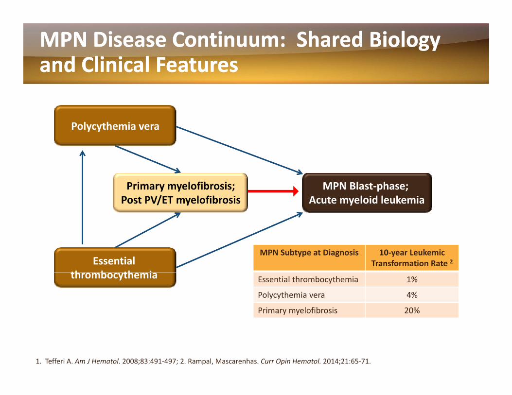
MPN Disease Continuum: Shared Biology MPN Disease Continuum: Shared Biology and Clinical Featuresand Clinical Features
P l h iPolycythemia vera
Primary myelofibrosis;Post PV/ET myelofibrosis
MPN Blast‐phase; Acute myeloid leukemia
Essential th b th i
MPN Subtype at Diagnosis 10‐year Leukemic Transformation Rate 2
thrombocythemia Essential thrombocythemia 1%
Polycythemia vera 4%
Primary myelofibrosis 20%
1. Tefferi A. Am J Hematol. 2008;83:491‐497; 2. Rampal, Mascarenhas. Curr Opin Hematol. 2014;21:65‐71.

NCCN Guidelines Version 1.2017 Myeloproliferative Neoplasms
NCCN Guidelines IndexTable of Contents
Discussion
WORKUP DIAGNOSIShWORKUP• H&P, including spleen size by palpation, evaluation of thrombotic/
hemorrhagic events and cardiovascular risk factors• CBC with differential• Comprehensive metabolic panel with uric acid, lactate dehydrogenase
(LDH), and liver function tests (LFTs)• FISH or RT-PCR for BCR-ABL1 to exclude the diagnosis of CML; if
BCR-ABL1-positive, See NCCN Guidelines for Chronic Myelogenous
• Primary myelofibrosis(PMF)a,i
• Post-PV or Post-ET MFi,j
Low-riskk (MPN-2)
Intermediate-risk 1 (INT-1)k(MPN-3)
DIAGNOSISh
p y gLeukemia
Suspicion of myeloproliferative neoplasms (MPN)
Polycythemia vera (PV)b,i
• Examination of blood smear• Bone marrow aspirate and biopsy with trichrome and reticulin staina,b
• Bone marrow cytogenetics (karyotype ± FISH)a,b
• Molecular testing for JAK2 V617F mutations; if negative, test for CALR and MPL mutations (for patients with ET and MF) and JAK2 Exon 12 mutations (for patients with PV)
• Assessment of symptom burden using MPN Symptom Assessment
Intermediate-risk 2 (INT-2) andHigh-risk (MPN-4)k
Essential thrombocythemia (ET)b,i
• Assessment of symptom burden using MPN Symptom Assessmentform (MPN-SAF)c
• Documentation of transfusion/medication history• Human leukocyte antigen (HLA) testing, if considering allogeneic
hematopoietic cell transplant (HCT)• Serum erythropoietin (EPO) level• Serum iron studies• Coagulation tests to evaluate for acquired von Willebrand disease
d,e
aSee 2016 WHO Diagnostic Criteria for Primary Myelofibrosis (PMF). See (MPN-A).bSee 2016 WHO Diagnostic Criteria for PV and ET. See (MPN-B).cAssessment of symptoms (in provider's office) at baseline using MPN Symptom Assessment
form (MPN SAF 20 items) is recommended for all patients See Assessment of Symptom Burden
fPatients undergoing high-risk surgical procedures and those with elevated platelet count and/or splenomegaly or unexplained bleeding.
gAn expanded panel including von Willebrand factor (VWF) antigen, Factor VIII activity, and VWFmultimers may be useful under certain circumstances
(VWD) and/or other coagulopathies in selected patientsf
¢ Prothrombin time (PT), partial thromboplastin time (PTT), Fibrinogen¢ P l a s m a von Willebrand Factor Antigen (VWFA) measurement¢ Von Willebrand Ristocetin Cofactor (VWF:RCo) activityg
form (MPN-SAF-20 items) is recommended for all patients. See Assessment of Symptom Burden(MPN-C 1 of 3).
dPrognostic models incorporating other mutations have been proposed to identify patients who may beat risk of leukemic transformation. However, the role of next-generation sequencing (NGS) to identifyhigh-risk mutations and the use of the Molecular International Prognostic Scoring System (MIPSS) isless well-established.
eEvaluation for allogeneic HCT is recommended for all patients with intermediate-2-risk (INT-2) and high-risk disease and for patients with intermediate-1-risk (INT-1) disease with low platelet counts and complex cytogenetics. Identification of “higher-risk” mutations may be helpful in the decision-making
multimers may be useful under certain circumstances.hThe diagnosis of MPN is based on the 2016 WHO Criteria and requires a combination of clinical,
laboratory, cytogenetics, and molecular testings.iReferral to specialized centers with expertise in the managment of MPN is strongly recommended for
all patients diagnosed with MF, PV, or ET. These guidelines pertain to the treatment of myelofibrosis.Guidelines are forthcoming for PV and ET.
jDiagnostic criteria for Post-ET or Post-PV MF. See (MPN-E).kDynamic International Prognostic Scoring System (DIPSS)-Plus is preferred for the risk stratification of
myelofibrosis; however, IPSS should be used at diagnosis. DIPSS can be used for risk stratification, if
Note: All recommendations are category 2A unless otherwise indicated.Clinical Trials: NCCN believes that the best management of any patient with cancer is in a clinical trial. Participation in clinical trials is especially encouraged.
p y g g y p gregarding allogeneic HCT. See Prognostic Significance of Mutations in MPN (MPN-D). karyotyping is not available. See Risk Stratification for Patients with Myelofibrosis (MPN-F).
MPN-1Version 1.2017, 09/26/16 © National Comprehensive Cancer Network, Inc. 2016, All rights reserved. The NCCN Guidelines® and this illustration may not be reproduced in any form without the express written permission of NCCN®.

POLYCYTHEMIAPOLYCYTHEMIA VERAVERAPOLYCYTHEMIA POLYCYTHEMIA VERAVERA
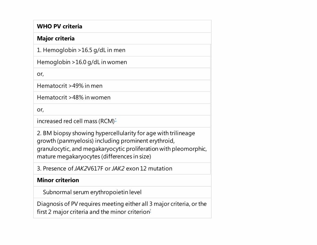
WHO PV criteria
Major criteria
1. Hemoglobin >16.5 g/dL in men
Hemoglobin >16.0 g/dL in women
or, ,
Hematocrit >49% in men
Hematocrit >48% in women
or,
increased red cell mass (RCM)*
2. BM biopsy showing hypercellularity for age with trilineage growth (panmyelosis) including prominent erythroid, granulocytic, and megakaryocytic proliferation with pleomorphic, mature megakaryocytes (differences in size)
3 Presence of JAK2V617F or JAK2 exon 12 mutation3. Presence of JAK2V617F or JAK2 exon 12 mutation
Minor criterion
Subnormal serum erythropoietin level
Diagnosis of PV requires meeting either all 3 major criteria, or the first 2 major criteria and the minor criterion†

Clinical Features of Polycythemia Vera (PV)Clinical Features of Polycythemia Vera (PV)
• Overproduction of red blood cells (erythrocytosis)(erythrocytosis)
– Often with increased white blood cells and platelets
– Patients may have splenomegaly
• Cardiovascular complications due to thrombosis or hemorrhage─ Thrombotic events can be arterial (CVA, ACS),
venous (DVT PE as well as affecting unusualvenous (DVT, PE, as well as affecting unusual locations such as splanchnic circulation), and microcirculatory
• Symptoms ─ May include pruritus, fatigue, shortness of
breath, dizziness, and symptoms due to splenomegaly
• Potential to progress to post‐PV MF or AMLPotential to progress to post‐PV MF or AML
1. Vardiman JW, et al. Blood. 2009;114:937‐951; 2. Vannucchi AM, et al. CA Cancer J Clin. 2009;59:171‐191; 3. Stein BL, et al. Ann Hematol. 2014;93:1965‐76.

Risk Classification for Patients with PVRisk Classification for Patients with PV
• Historically, cardiovascular complications are the most common cause of mortality in patients with PV1
• Conventional PV risk factors are prognostic for thrombosis, and not based on survival2not based on survival2
Risk Category Conventional Risk Factors
Low• Age < 60 years
• No thrombosis history
High
• Age ≥ 60 years
and/or
• Thrombosis history
1. Stein BL, et al. Thrombosis. 2011;2011:874146; 2. Barbui T et al. Clin Oncol. 2011;29(6):761‐770.

RiskRisk‐‐Adapted Management of Patients Adapted Management of Patients with PVwith PV 11
• Hematocrit (HCT) control is a key therapeutic goal– Maintaining HCT < 45% significantly decreases the risk of cardiovascular
death and major thrombotic events2
Conventional Risk Variables TherapyRisk Category
py
Low• Age < 60 years • No thrombosis
• Phlebotomy, and• Correction of CV risk factors, and
history • Aspirin
• Age ≥ 60 years and/or
• Cytoreduction*, and• Correction of CV risk factors, and
*First‐line cytoreductive therapy is hydroxyurea or interferon alfa; busulfan reserved for age >75 years.
High and/or• Thrombosis
history
Correction of CV risk factors, and• Aspirin, and• Phlebotomy
1. Barbui T et al. J Clin Oncol. 2011;29(6):761‐770; 2. Marchioli R, et al. N Engl J Med. 2013;368(1):22‐33.
y py y y ; g y

Phlebotomy and Aspirin in PV ManagementPhlebotomy and Aspirin in PV Management
PhlebotomyR d h t it (HCT) (h i it ) l i HCT 45%1• Reduces hematocrit (HCT) (hyperviscosity); goal is HCT <45%1
• Does not control systemic symptoms or progressive symptomatic splenomegaly well 2
• Iron deficiency is common with repeated phlebotomies2y p p– Associated with fatigue, cognitive impairment, increased pulmonary artery
pressure
Low‐dose aspirin• Persistently enhanced platelet activation contributes to the higher risk of
thrombosis in patients with PV3,4
• Placebo‐controlled ECLAP trial (N= 518): low‐dose aspirin can safely prevent thrombotic complications in patients with PV who have no contraindications tothrombotic complications in patients with PV who have no contraindications to aspirin4
• Screening for acquired von Willebrand syndrome is recommended before administrating aspirin in the presence of extreme thrombocytosis5
1. Marchioli R, et al. N Engl J Med. 2013;368(1):22‐33; 2. Mascarenhas J et al. Haematologica2014;99(6):945‐49; 2. Patrono C, et al. Blood. 2013;121(10):1701‐11; 3. Landolfi R, et al. N Engl J Med 2004;350: 114‐24; 5. Tefferi A, Barbui T. Am. J. Hematol. 2015;90:163‐73.

HydroxyureaHydroxyurea (HU) in PV Management(HU) in PV Management
• HU is often used as a first‐line cytoreductive treatment yfor patients with PV who are at high‐risk for vascular complications1,2
• Clinical activities2
– Controls myeloproliferation– Reduces splenomegaly– May reduce risk of major thrombosis (limited evidence in PV)3
2• Side effects2
– Myelosuppression, leg ulcers, hyperpigmentation, fever, alopecia increased risk of squamous cell carcinomaalopecia, increased risk of squamous cell carcinoma
1. Sever M et al. Leuk Lymphoma. 2014;55(12):2685‐90; 2. Mascarenhas J et al. Haematologica.2014;99(6):945‐49; 3. Fruchtman SM, et al. Semin Hematol. 1997;34(1):17‐23.

Pegylated InterferonPegylated Interferon‐‐αα2a is an Acceptable 2a is an Acceptable Alternative to HU in PV Alternative to HU in PV
Phase II studies: Treatment with PEG‐IFN‐ α2a or α2b resulted in high rates of complete hematologic and molecular response, and low rates of thrombosis.1‐3
IFN α2a (n=40)2 *
7080
100
nders (%)
CompletePartial
100
80
64nt Allele
No. Patients 40 32 32 29 27 23 24 12 10 6 10
40
60
on of R
espo
n
No response 60
40
64
53
41
29
2V617fMutan
1020
0
20
PV
Prop
ortio 20
00 6 12 18 24 30 36 42 48 54 60
19 1316
4.5
179 5
% JA
K2
Time (months)
• Ongoing global randomized phase III studies:─ PEG‐IFN‐α2a vs HU in high‐risk PV and ET (NCT01259856)
*Complete response included absence of thrombosisPV ( )
Polycythemia vera
1. Kiladjian JJ, et al. Blood 2008;112:3065‐72; 2. Quintas‐Cardama A et al. J Clin Oncol. 2009;27:5418‐24; 3. Quintas‐Cardama A et al. Blood 2013;122:893‐901.
PEG IFN α2a vs HU in high risk PV and ET (NCT01259856)─ Novel monopegylated IFN‐α2b in high‐risk PV and ET (NCT01949805)
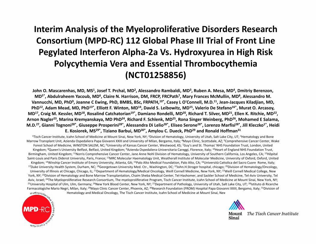
Interim Analysis of the Myeloproliferative Disorders Research Consortium (MPD‐RC) 112 Global Phase III Trial of Front Line ( )Pegylated Interferon Alpha‐2a Vs. Hydroxyurea in High Risk
Polycythemia Vera and Essential Thrombocythemia(NCT01258856)(NCT01258856)
John O. Mascarenhas, MD, MS1, Josef T. Prchal, MD2, Alessandro Rambaldi, MD3, Ruben A. Mesa, MD4, Dmitriy Berenzon, MD5*, Abdulraheem Yacoub, MD6, Claire N. Harrison, DM, FRCP, FRCPath7, Mary Frances McMullin, MD8, Alessandro M.
Vannucchi, MD, PhD9, Joanne C Ewing, PhD, BMBS, BSc, FRPATH,10*, Casey L O'Connell, M.D.11, Jean‐Jacques Kiladjian, MD, PhD12 Ad M d MD PhD13* Elli tt F Wi t MD14 D id S L ib it MD15 V l i D St f 16* M t O APhD12, Adam Mead, MD, PhD13*, Elliott F. Winton, MD14, David S. Leibowitz, MD15, Valerio De Stefano16*, Murat O. Arcasoy,
MD17, Craig M. Kessler, MD18, Rosalind Catchatorian19*, Damiano Rondelli, MD20, Richard T. Silver, MD21, Ellen K. Ritchie, MD22, Arnon Nagler23, Marina Kremyanskaya, MD PhD24, Richard F. Schlenk, MD25, Rona Singer Weinberg, PhD26, Mohamed E Salama, M.D.27, Gianni Tognoni28*, Giuseppe Prosperini28*, Alessandra Di Lelio28*, Eliseo Serone28*, Lorenzo Marfisi28*, Jill Kleczko1*, Heidi
E. Kosiorek, MS29*, Tiziano Barbui, MD30*, Amylou C. Dueck, PhD29 and Ronald Hoffman311Tisch Cancer Institute, Icahn School of Medicine at Mount Sinai, New York, NY; 2Division of Hematology, University of Utah, Salt Lake City, UT; 3Hematology and BoneTisch Cancer Institute, Icahn School of Medicine at Mount Sinai, New York, NY; Division of Hematology, University of Utah, Salt Lake City, UT; Hematology and Bone
Marrow Transplant Unit, Azienda Ospedaliera Papa Giovanni XXIII and University of Milan, Bergamo, Italy; 4Mayo Clinic, Scottsdale, AZ; 5Comprehensive Cancer Center, Wake Forest School of Medicine, WINSTON SALEM, NC; 6University of Kansas Cancer Center, Westwood, KS; 7Guy's and St. Thomas' NHS Foundation Trust, London, United Kingdom; 8Queen's University Belfast, Belfast, United Kingdom; 9Azienda Ospedaliera‐Universitaria Careggi, Florence, Italy; 10Heart of England NHS Foundation Trust,
Birmingham, United Kingdom; 11Norris Comprehensive Cancer Center, Jane Anne Nohl Division of Hematology, University of Southern California, Los Angeles, CA; 12Hôpital Saint‐Louis and Paris Diderot University, Paris, France; 13MRC Molecular Haematology Unit, Weatherall Institute of Molecular Medicine, University of Oxford, Oxford, United Kingdom; 14Winship Cancer Institute of Emory University, Atlanta, GA; 15Palo Alto Medical Foundation, Palo Alto, CA; 16Università Cattolica del Sacro Cuore Rome, Italy;
17Duke University Health System, Durham, NC; 18Georgetown University Med. Ctr., Washington, DC; 19John.H.Stroger hospital, chicago; 20Division of Hematology/Oncology, y y , , ; g y , g , ; g p , g ; gy/ gy,University of Illinois at Chicago, Chicago, IL; 21Department of Hematology/Medical Oncology, Weill Cornell Medicine, New York, NY; 22Weill Cornell Medical College, New
York, NY; 23Division of Hematology and Bone Marrow Transplantation, Chaim Sheba Medical Center, Tel‐Hashomer, and Sackler School of Medicine, Tel‐Aviv University, Tel Aviv, Israel; 24The Myeloproliferative Research Consortium, The myeloproliferative Program, Tisch Cancer Institute, Icahn School of Medicine at Mount Sinai, New York, NY; 25University Hospital of Ulm, Ulm, Germany; 26New York Blood Center, New York, NY; 27Department of Pathology, University of Utah, Salt Lake City, UT; 28Istituto di RicercheFarmacologiche Mario Negri, Milan, Italy; 29Mayo Clinic Cancer Center, Phoenix, AZ; 30Research Foundation (FROM) Hospital Papa Giovanni XXIII, Bergamo, Italy; 31Division of
Hematology and Medical Oncology, The Tisch Cancer Institute, Icahn School of Medicine at Mount Sinai, New York, NY

ConclusionsConclusions• No difference in hematologic CR between the two arms at 12
monthsh l f• BM pathologic responses are more frequent in HU arm at 12
months • No difference in molecular response rates between the arms at 12
monthsmonths • Toxicity is not a major reason for discontinuation in either arm at 12
months • Comparative analysis of QOL and symptom burden also presentedComparative analysis of QOL and symptom burden also presented
at this meeting Mesa et al ASH 2016• Longer term follow up of the entire study population may show
meaningful differences in response and toxicity between these two agents over time
• Final analysis of MPD‐RC 112 will establish optimal first line therapy for high risk ET/PV
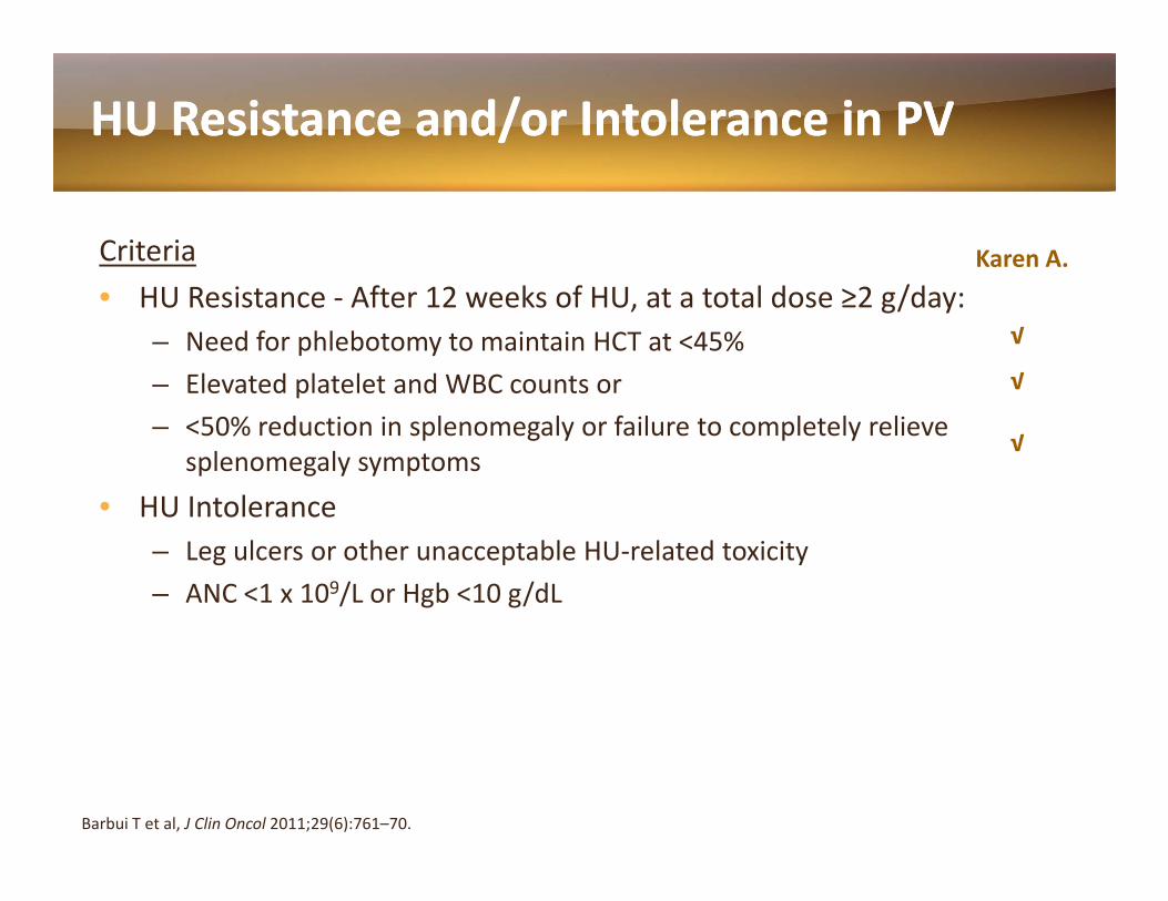
HU Resistance and/or Intolerance in PVHU Resistance and/or Intolerance in PV
Criteria Karen A.• HU Resistance ‐ After 12 weeks of HU, at a total dose ≥2 g/day:
– Need for phlebotomy to maintain HCT at <45%– Elevated platelet and WBC counts or
√
√p– <50% reduction in splenomegaly or failure to completely relieve
splenomegaly symptoms• HU Intolerance
√
• HU Intolerance– Leg ulcers or other unacceptable HU‐related toxicity– ANC <1 x 10⁹/L or Hgb <10 g/dL
Barbui T et al, J Clin Oncol 2011;29(6):761–70.
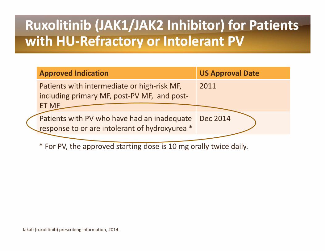
RuxolitinibRuxolitinib (JAK1/JAK2 Inhibitor) for Patients (JAK1/JAK2 Inhibitor) for Patients with HUwith HU‐‐Refractory or Intolerant PVRefractory or Intolerant PVyy
Approved Indication US Approval DatePatients with intermediate or high‐risk MF, including primary MF, post‐PV MF, and post‐ET MF
2011
Patients with PV who have had an inadequate response to or are intolerant of hydroxyurea *
Dec 2014
* For PV, the approved starting dose is 10 mg orally twice daily.
Jakafi (ruxolitinib) prescribing information, 2014.

Treatment SummaryTreatment Summary
• Contemporary treatment for patients with PV combines:p y p– Modification of CV risk factors– Phlebotomy (HCT target <45%)– Antiplatelet/anticoagulation therapy – First‐line cytoreductive therapy: HU or IFN‐alfa– Second‐line: Ruxolitinib for patients resistant to or intolerant of HU
• Other options may include PEG‐IFN or busulfanOther options may include PEG IFN or busulfan
ESSENTIALESSENTIAL THROMBOCYTHEMIATHROMBOCYTHEMIAESSENTIAL ESSENTIAL THROMBOCYTHEMIATHROMBOCYTHEMIA
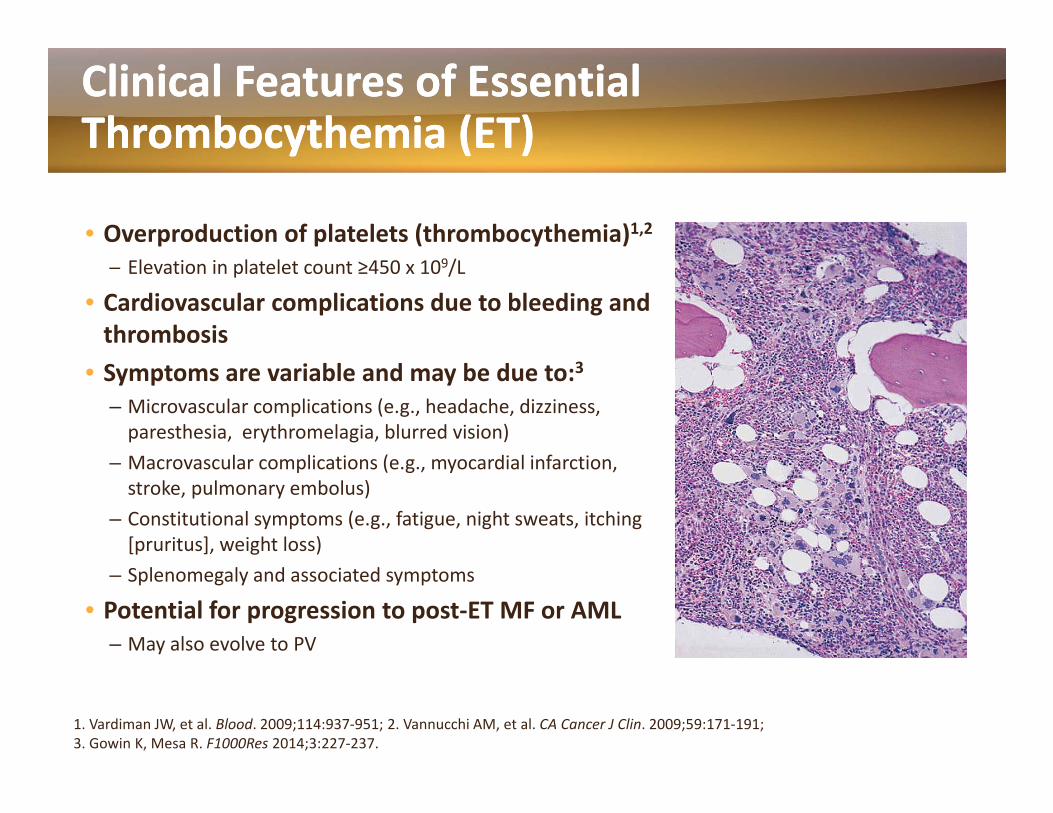
Clinical Features of Essential Clinical Features of Essential Thrombocythemia (ET)Thrombocythemia (ET)y ( )y ( )
• Overproduction of platelets (thrombocythemia)1,2─ Elevation in platelet count ≥450 x 109/L
• Cardiovascular complications due to bleeding and thrombosis
• Symptoms are variable and may be due to:3– Microvascular complications (e.g., headache, dizziness, paresthesia, erythromelagia, blurred vision)M l li ti ( di l i f ti– Macrovascular complications (e.g., myocardial infarction, stroke, pulmonary embolus)
– Constitutional symptoms (e.g., fatigue, night sweats, itching [pruritus], weight loss)
– Splenomegaly and associated symptoms
• Potential for progression to post‐ET MF or AML– May also evolve to PV
1. Vardiman JW, et al. Blood. 2009;114:937‐951; 2. Vannucchi AM, et al. CA Cancer J Clin. 2009;59:171‐191; 3. Gowin K, Mesa R. F1000Res 2014;3:227‐237.

WHO criteria for ET
WHO ET criteria
Major criteria
1. Platelet count ≥450 × 109/L
2. BM biopsy showing proliferation mainly of the megakaryocyte lineage with increased numbers of enlarged, mature megakaryocytes withhyperlobulatednuclei No significant increase or left shift inwith hyperlobulated nuclei. No significant increase or left shift in neutrophil granulopoiesis or erythropoiesis and very rarely minor (grade 1) increase in reticulin fibers
3. Not meeting WHO criteria forBCR‐ABL1+ CML, PV, PMF, myelodysplastic syndromes, or other myeloid neoplasms
4. Presence of JAK2, CALR, orMPLmutation
Minor criterion
Presence of a clonal marker or absence of evidence for reactive thrombocytosis
Diagnosis of ET requires meeting all 4 major criteria or the first 3 major criteriaand the minorcriterioncriteria and the minor criterion
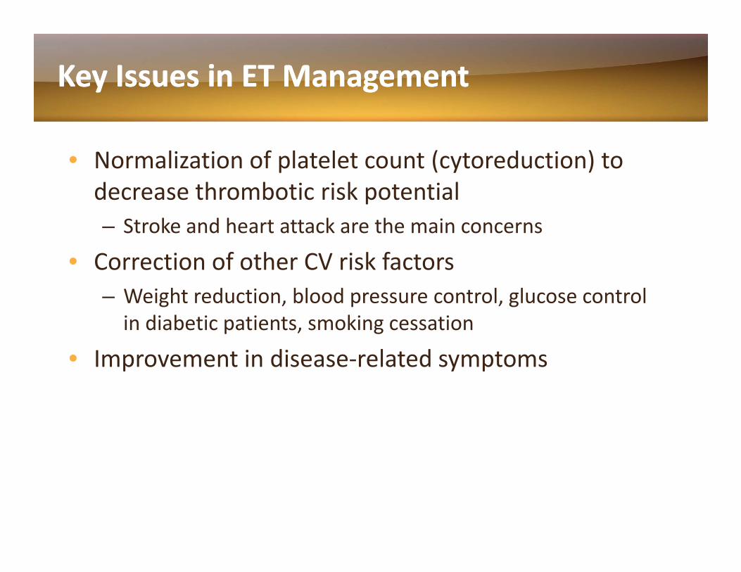
Key Issues in ET Management Key Issues in ET Management
• Normalization of platelet count (cytoreduction) to decrease thrombotic risk potential– Stroke and heart attack are the main concerns
• Correction of other CV risk factors– Weight reduction, blood pressure control, glucose control in diabetic patients smoking cessationin diabetic patients, smoking cessation
• Improvement in disease‐related symptoms
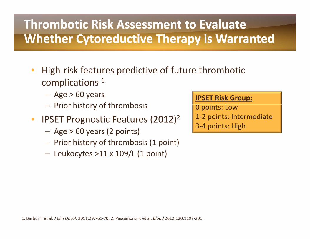
Thrombotic Risk Assessment to Evaluate Thrombotic Risk Assessment to Evaluate Whether Whether CytoreductiveCytoreductive Therapy is Warranted Therapy is Warranted yy pypy
• High‐risk features predictive of future thrombotic g pcomplications 1– Age > 60 years
Prior history of thrombosisIPSET Risk Group:
– Prior history of thrombosis
• IPSET Prognostic Features (2012)2– Age > 60 years (2 points)
0 points: Low1‐2 points: Intermediate3‐4 points: High
g y ( p )– Prior history of thrombosis (1 point)– Leukocytes >11 x 109/L (1 point)
1. Barbui T, et al. J Clin Oncol. 2011;29:761‐70; 2. Passamonti F, et al. Blood 2012;120:1197‐201.

CALR and JAK2 mutations represent 2 disease spectrums in essential thrombocythemiawhereby cases with mutated CALR are characterized by higher platelet levels, lower hemoglobin and leukocyte counts, and lower thrombosis risk compared with JAK2-
mutated patiemutated patie...
Chao M P , and Gotlib J Blood 2014;123:1438-1440

Systemic Therapy for ETSystemic Therapy for ET
• In low‐risk asymptomatic ET, observation is appropriate • High‐risk ET:
– Daily low‐dose aspirin is standard practice for most patients, if not contraindicated*
• Due to high risk of bleeding in patients with platelet counts >1500 x 109/L, cytoreduction may be considered prior to aspirin initiation
– Cytoreductive therapy: y py• Hydroxyurea (HU) is the first‐line treatment of choice• Anagrelide is generally considered 2nd‐line therapy if resistant or intolerant to HUI lf i d f i i h• IFN‐alfa is used for young patients, pregnant women, or patients who are refractory/intolerant to HU
• Consider clinical trials for patients who are intolerant to or have progressed on all 3 approved agentsprogressed on all 3 approved agents
Gowin K, Mesa R. F1000Res 2014;3:227‐237.
*Aspirin is not universally recommended ‐ typically for those with JAK2 positive ET, CV risk factors, or microvascular symptoms.

MYELOFIBROSISMYELOFIBROSISMYELOFIBROSISMYELOFIBROSISMay arise de novo (primary MF) or following PV or ET (post PV MF or post ET MF)(post‐PV MF or post‐ET MF)

WHO criteria for overt PMF
WHO overt PMF criteria
Major criteriaMajor criteria
1. Presence of megakaryocytic proliferation and atypia, accompanied by either reticulin and/or collagen fibrosis grades 2 or 3*
2. NotmeetingWHOcriteria forET, PV,BCR‐ABL1+ CML, 2. Not meeting WHO criteria for ET, PV,BCR ABL1 CML, myelodysplastic syndromes, or other myeloid neoplasms
3. Presence of JAK2, CALR, orMPLmutation or in the absence of these mutations, presence of another clonal marker,† or absence of reactivemyelofibrosis‡reactive myelofibrosis
Minor criteria
Presence of at least 1 of the following, confirmed in 2 consecutive determinations:determinations:
a. Anemia not attributed to a comorbid condition
b. Leukocytosis ≥11 × 109/L
P l bl l l c. Palpable splenomegaly
d. LDH increased to above upper normal limit of institutional reference range
e Leukoerythroblastosis e. Leukoerythroblastosis
Diagnosis of overt PMF requires meeting all 3 major criteria, and at least 1 minor criterion

Clinical Features of Clinical Features of MyelofibrosisMyelofibrosis (MF)(MF)
• Bone marrow fibrosis• Splenomegaly
– Splenomegaly‐associated symptoms include abdominal pain/discomfort, earlyinclude abdominal pain/discomfort, early satiety
• CytopeniasA i th b t i– Anemia, thrombocytopenia
• Constitutional symptoms – Include fatigue, night sweats, pruritus g , g , p
(itching), bone aches, weight loss

MKs play a central role in MPN pathogenesis.
William Vainchenker, and Robert Kralovics Blood 2017;129:667-679
©2017 by American Society of Hematology

Prognostic Models for MyelofibrosisPrognostic Models for Myelofibrosis
At diagnosis of primary MF:• International Prognostic Scoring System (IPSS)1
– Factors associated with a worse prognosis: Age >65 yrs, constitutional symptoms, Hgb <10 g/dL, WBC count > 25 x 109/L, blood blasts ≥1%
Valid at diagnosis and at any point in the course of disease: Valid at diagnosis and at any point in the course of disease:• Dynamic International Prognostic Scoring System (DIPSS)2
– Gives more prognostic weight to the presence of disease‐related anemiaanemia
• DIPSS‐plus3– Incorporates abnormal karyotype as an independent negative
prognostic factor for overall and leukemia‐free survivalprognostic factor for overall and leukemia free survival
1. Cervantes, et al. Blood. 2009;113(13):2895‐2901. 2. Passamonti, et al. Blood. 2010;115(9):1703‐1708. 3. Gangat N et al. J Clin Oncol. 2011;29:29:392‐397.

NCCN Guidelines Version 1.2017 Myeloproliferative Neoplasms
NCCN Guidelines IndexTable of Contents
Discussion
MYELOPROLIFERATIVE NEOPLASM SYMPTOM ASSESSMENT FORMMYELOPROLIFERATIVE NEOPLASM SYMPTOM ASSESSMENT FORMTOTAL SYMPTOM SCORE (MPN-SAF TSS-10 ITEMS)2
(Recommended for monitoring symptoms during the course of treatment)
Symptom 1 to 10 (0 if absent) ranking1 is most favorable and 10 least favorable
Pl t f ti ( i (N F ti ) 0 1 2 3 4 5 6 7 8 9 10 (W t I i bl )
Circle the one number that describes how, during the past week how muchdifficulty you have had with each of the following symptoms
Please rate your fatigue (weariness, tiredness) by circling the one number that best describes your WORST level of fatigue during past24 hours*
(No Fatigue) 0 1 2 3 4 5 6 7 8 9 10 (Worst Imaginable)
difficulty you have had with each of the following symptoms
Filling up quickly when you eat (early satiety)
(Absent) 0 1 2 3 4 5 6 7 8 9 10 (Worst Imaginable)
Abdominal discomfort (Absent) 0 1 2 3 4 5 6 7 8 9 10 (Worst Imaginable)Inactivity (Absent) 0 1 2 3 4 5 6 7 8 9 10 (Worst Imaginable)Problems with (Absent) 0 1 2 3 4 5 6 7 8 9 10 (Worst Imaginable)concentration- comparedto prior to my MPD
( ) ( g )
Numbness/Tingling (in myhands and feet)
(Absent) 0 1 2 3 4 5 6 7 8 9 10 (Worst Imaginable)
Night sweats (Absent) 0 1 2 3 4 5 6 7 8 9 10 (Worst Imaginable)Itching (pruritus) (Absent) 0 1 2 3 4 5 6 7 8 9 10 (Worst Imaginable)
2Emanuel RM Dueck AC Geyer HL et al Myeloproliferative neoplasm (MPN) symptom assessment form total symptom score: prospective international
Bone pain (diffuse not joint pain orarthritis)
(Absent) 0 1 2 3 4 5 6 7 8 9 10 (Worst Imaginable)
Fever (>100 F) (Absent) 0 1 2 3 4 5 6 7 8 9 10 (Daily)Unintentional weight loss last 6 months (Absent) 0 1 2 3 4 5 6 7 8 9 10 (Worst Imaginable)
Note: All recommendations are category 2A unless otherwise indicated.Clinical Trials: NCCN believes that the best management of any patient with cancer is in a clinical trial. Participation in clinical trials is especially encouraged.
(3 OF 3)Version 1.2017, 09/26/16 © National Comprehensive Cancer Network, Inc. 2016, All rights reserved. The NCCN Guidelines® and this illustration may not be reproduced in any form without the express written permission of NCCN®.
MPN-C
2Emanuel RM, Dueck AC, Geyer HL, et al. Myeloproliferative neoplasm (MPN) symptom assessment form total symptom score: prospective international assessment of an abbreviated symptom burden scoring system among patients with MPNs. J Clin Oncol 2012;30:4098-4103.

Kaplan-Meier analysis of survival of PMF patients stratified according to the risk categories defined by a clinical-molecular prognostic model.
Elisa Rumi et al. Blood 2014;124:1062-1069
©2014 by American Society of Hematology

Kaplan-Meier analysis of survival of PMF patients stratified according to their driver mutation.
Elisa Rumi et al. Blood 2014;124:1062-1069
©2014 by American Society of Hematology

RiskRisk‐‐Adapted Treatment Adapted Treatment of of MyelofibrosisMyelofibrosis
Low RiskMinimally symptomatic Observation or InterferonM JAK2 i hibiLow Risk
Intermediate‐1
Many symptoms JAK2 inhibitor
JAK2 inhibitor or allogeneic HSCT or
Intermediate‐2
allogeneic HSCT or anemia treatment
JAK2 inhibitor or ll i HSCT
High Risk
allogeneic HSCT or anemia treatment
Allogeneic HSCT or High Risk JAK2 inhibitor or
anemia treatment
1. Mesa RA. Leuk Lymphoma. 2013;54:242‐51; 2. Geyer HL, Mesa RA. Hematol.2014 277‐86.
HSCT, hematopoietic stem cell transplantation.Anemia treatment may include: IMID, androgens, erythropoietin; clinical trial, splenectomy

Ruxolitinib is the Only JAK Inhibitor Ruxolitinib is the Only JAK Inhibitor Approved for Treatment of Myelofibrosis Approved for Treatment of Myelofibrosis pp ypp y
COMFORT‐I (N = 309)1( l b) ( l b ) h dRuxolitinib vs. placebo in pts
with intermediate‐ or high‐risk MF
• 41.9% (ruxolitinib) vs 0.7% (placebo) had ≥35% reduction in spleen volumea at week 24 (P < 0.001)
COMFORT‐II (N = 219) 2
Ruxolitinib vs. best available h ( ) i i h
• 32% (ruxolitinib) vs 0% (BAT) had ≥ 35% reduction in spleen volumea at week 24therapy (BAT) in pts with
intermediate‐ or high‐risk MF
reduction in spleen volume at week 24 (P < 0.001)
a As assessed by MRIy
1. Verstovsek S, et al. N Engl J Med. 2012;366:799‐807; 2. Harrison C, et al. N Engl J Med. 2012;366:787‐798.

Summary: Summary: RuxolitinibRuxolitinib in Patients with MF in Patients with MF (including PMF, post(including PMF, post‐‐PV MF, and postPV MF, and post‐‐ET MF)ET MF)( g , p( g , p , p, p ))
• COMFORT‐I and COMFORT‐II phase III trials:p– Efficacy
• Spleen size reduction, significant improvement in symptoms, quality of life, performance status
• Not selective for JAK2V617F (i.e., benefits patients with and without JAK2 mutation))
• Possible prolongation of life in patients with advanced disease– Safety
• Myelosuppression (not a cause for stopping therapy)

Kaplan-Meier analysis of time to event outcomes.
Keyur P. Patel et al. Blood 2015;126:790-797
©2015 by American Society of Hematology

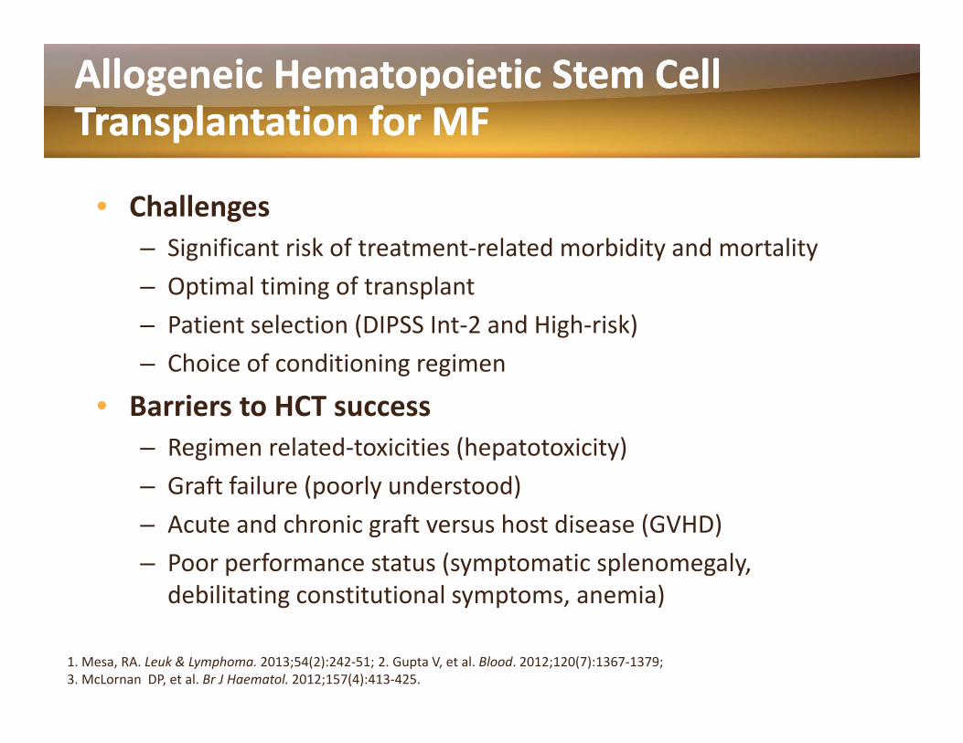
Allogeneic Hematopoietic Stem Cell Allogeneic Hematopoietic Stem Cell Transplantation for MFTransplantation for MFpp
HSCT is a reasonable option for otherwise healthy patients with intermediate ‐2 or high‐risk PMF
• HSCT is the only potentially curative treatment approach • Young otherwise fit patients may be candidates
• Therapeutic efficacy is mediated via: – Antineoplastic effect of pre‐transplant conditioning regimen
• Young, otherwise fit patients may be candidates
– Antineoplastic effect of pre‐transplant conditioning regimen – Alloimmune graft versus leukemia effect
• Reduced intensity conditioning may be considered for: – Older patients– Patients with comorbidities that preclude them from myeloablative
conditioning regimens
1. Mesa, RA. Leuk & Lymphoma. 2013;54(2):242‐51; 2. Gupta V, et al. Blood. 2012;120(7):1367‐1379; 3. McLornan DP, et al. Br J Haematol. 2012;157(4):413‐425.
Allogeneic Hematopoietic Stem Cell Allogeneic Hematopoietic Stem Cell Transplantation for MFTransplantation for MFpp
• Challenges– Significant risk of treatment‐related morbidity and mortality– Optimal timing of transplant
Patient selection (DIPSS Int 2 and High risk)– Patient selection (DIPSS Int‐2 and High‐risk)– Choice of conditioning regimen
• Barriers to HCT success – Regimen related‐toxicities (hepatotoxicity)– Graft failure (poorly understood)– Acute and chronic graft versus host disease (GVHD) – Poor performance status (symptomatic splenomegaly,
debilitating constitutional symptoms, anemia)g y p , )
1. Mesa, RA. Leuk & Lymphoma. 2013;54(2):242‐51; 2. Gupta V, et al. Blood. 2012;120(7):1367‐1379; 3. McLornan DP, et al. Br J Haematol. 2012;157(4):413‐425.

MPN ConclusionsMPN Conclusions
• The MPNs (PV, ET, MF) are progressive hematopoietic di i h h d bi l d li i l fdiseases with shared biology and clinical features– JAK‐STAT pathway activation is hallmark of these diseases– MPNs are associated with significant complications, including g p , g
thrombosis, splenomegaly, cytopenias, constitutional symptoms– Patients can have high symptom burden regardless of the subtype– Risk of leukemic transformation especially for MFRisk of leukemic transformation, especially for MF
• Treatment strategies can vary depending on the individual’s risk status and management needs
• JAK2 inhibitor therapy (ruxolitinib) is improving the outlook for many patients with MF or HU‐resistant/intolerant PV, but it does not cure these diseasesit does not cure these diseases
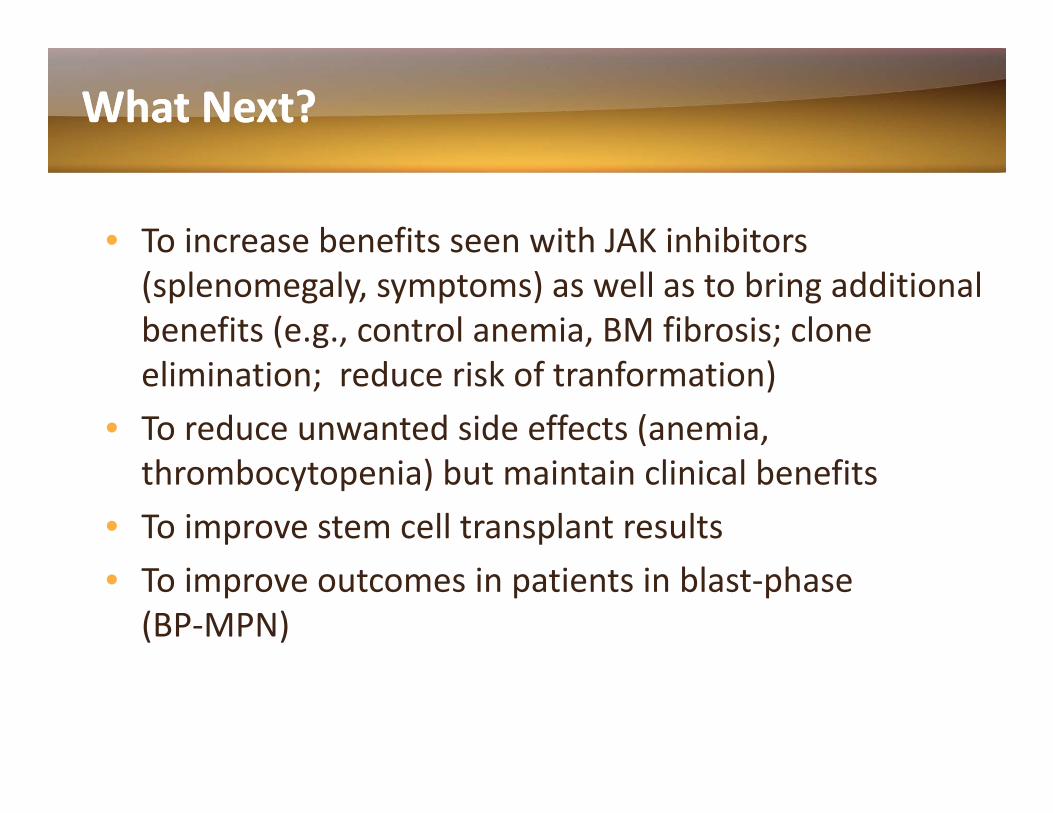
What Next?What Next?
• To increase benefits seen with JAK inhibitors (splenomegaly, symptoms) as well as to bring additional benefits (e.g., control anemia, BM fibrosis; clone elimination; reduce risk of tranformation)
• To reduce unwanted side effects (anemia, thrombocytopenia) but maintain clinical benefits
• To improve stem cell transplant results• To improve outcomes in patients in blast‐phase (BP‐MPN)

PostPost‐‐MPN AML demonstrates limited response to MPN AML demonstrates limited response to conventional AML therapyconventional AML therapy
Mesa R A et al. Blood 2005;105:973-977

Safety and efficacy of combined Ruxolitinib and Decitabine in patients with blast‐phase MPN and post‐MPN AML:
Results of a Phase I studyResults of a Phase I study (Myeloproliferative Diseases Research Consortium 109 trial)
Raajit K. Rampal, MD, PhD, John O. Mascarenhas, MD, MS, Heidi E. Kosiorek, MS, Dmitriy Berenzon, MD, Elizabeth Hexner, MD, , , y , , , ,Camille N. Abboud, MD, Marina Kremyanskaya, MD PhD, Rona Singer Weinberg, PhD, Mohamed E Salama, M.D., Gianni Tognoni, Giuseppe Prosperini, Alessandra Di Lelio, Eliseo Serone, Lorenzo Marfisi, Lonette Sandy, Mark Lawrence Heaney, MD, PhD, Ross L. Levine, MD, Ruben A.
Mesa MD Amylou C Dueck PhD and Ronald HoffmanMesa, MD, Amylou C. Dueck, PhD and Ronald Hoffman

Conclusions• The combination of Ruxolitinib (up to a dose of 50mg BID) and Decitabine was safely administered50mg BID) and Decitabine was safely administered to patients with no MTD established
• Patients in the 10mg BID cohort had the longest d ti f thduration of therapy
• The highest CR/CRi rate was observed in the 50mg BID cohort; however, the largest proportion of d b d i hi hadverse events was observed in this cohort .

Conclusions• The RP2D is 25mg BID for one cycle followed by 10mg BID for all subsequent cyclesby 10mg BID for all subsequent cycles.
• Molecular response data and evaluation of presponse by the proposed MPN‐BP criteria are underway.y

What Next? What Next? Ruxolitinib Combination Strategies Ruxolitinib Combination Strategies
Ongoing Ruxolitinib Combination Trials in Myelofibrosis
gg
Ruxolitinib + Second Agent (class)
Development Stage
ClinicalTrials.gov Identifier
Azacitidine (Hypomethylation) Phase III NCT01787487Azacitidine (Hypomethylation) Phase III NCT01787487
Lenalidomide (IMID) Phase II NCT01375140
Pomalidomide (IMID) Phase I/II NCT01644110
PRM‐151 (Antifibrosing) Phase II NCT01981850
Panobinostat (HDAC inhibitor) Phase I/II NCT01693601
LDE‐225 (Hedgehog inhibitor) Phase Ib/II NCT01787552( g g ) /
BKM‐120 (PI3‐kinase inhibitor) Phase I NCT01730248
ClinicalTrials.gov: Accessed Feb 4, 2015.

What Next?What Next?Novel JAK2 InhibitorsNovel JAK2 Inhibitors
Agent ‐ Targets Phase I/II Results Status – Disease and/or Design
ClinicalTrials.gov IdentifierDesign Identifier
Momelotinib(CYT387) –JAK1, JAK2
• Reduced splenomegaly, improved symptoms, and reduced anemia/ RBC transfusion‐dependence
• Grade 3‐4 thrombocytopenia in 32%
Phase III ‐ Versus ruxolitinib in Int‐and high‐risk MF (ruxolitinib naïve)
Phase III –Versus Best Available h ( )
NCT01969838
Therapy (BAT) in Anemic or Thrombocytopenic MF patients treated with ruxolitinib
NCT02101268
Phase II – PV and ET NCT01998828
Pacritinib (SB1518) • Reduced splenomegaly improved Phase III 2 trials (PERSIST I and NCT01773187Pacritinib (SB1518) –JAK2, FLT3
• Reduced splenomegaly, improved symptoms
• Limited provocation of cytopenias, particularly in patients with baseline thrombocytopenia
• Most adverse events gastrointestinal
Phase III‐ 2 trials (PERSIST‐I and PERSIST‐II) ‐ Versus best available therapy (BAT) in MF patients without and with baseline thrombocytopenia, respectively.
NCT01773187NCT02055781
g
INCB‐039110JAK1
Phase II ‐MF NCT01633372
NS‐018 ‐ JAK2 Phase I/II ‐MF NCT01423851
• Several JAK2 inhibitors have been removed from clinical trials due to toxicity and/or insufficient activity (fedratinib, CEP‐701, XL019, LY278544, BMS‐911543, AZD 1480)
ClinicalTrials.gov: Accessed Feb 4, 2015.
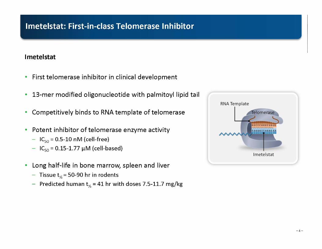
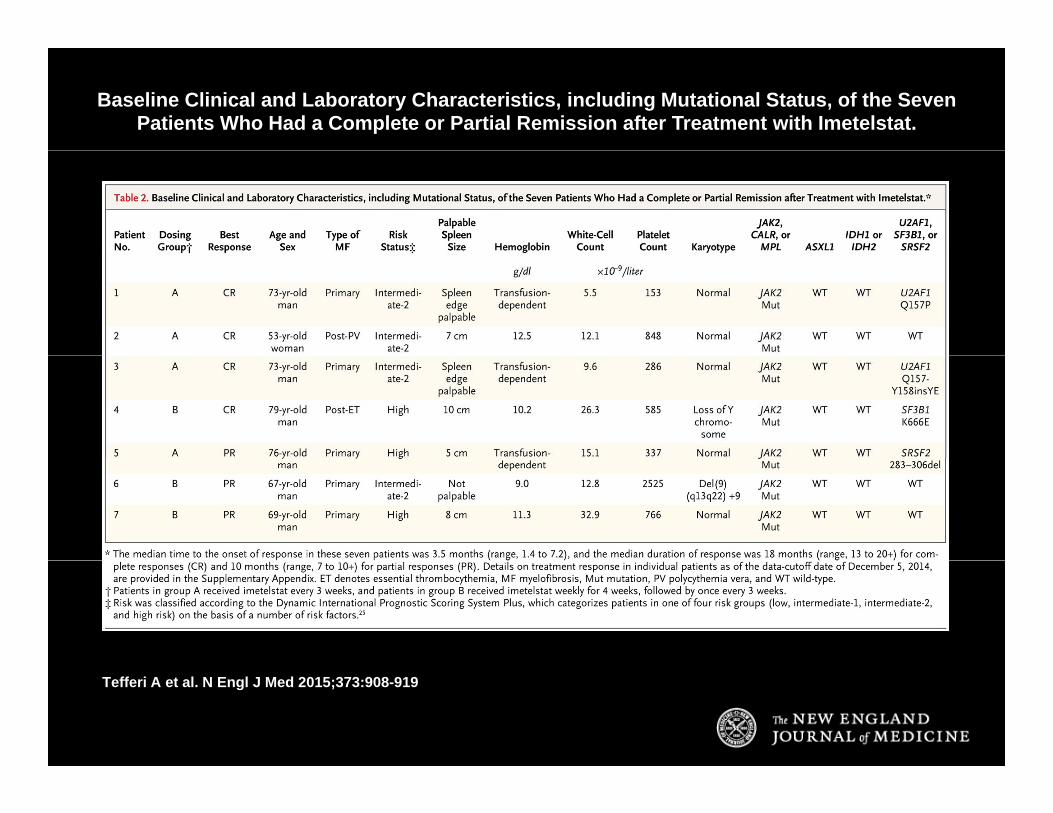
Baseline Clinical and Laboratory Characteristics, including Mutational Status, of the Seven Patients Who Had a Complete or Partial Remission after Treatment with Imetelstat.
Tefferi A et al. N Engl J Med 2015;373:908-919

ConclusionsConclusions
• Imetelstat was found to be active in patients with myelofibrosis but also had the potential to cause clinically significant myelosuppression.
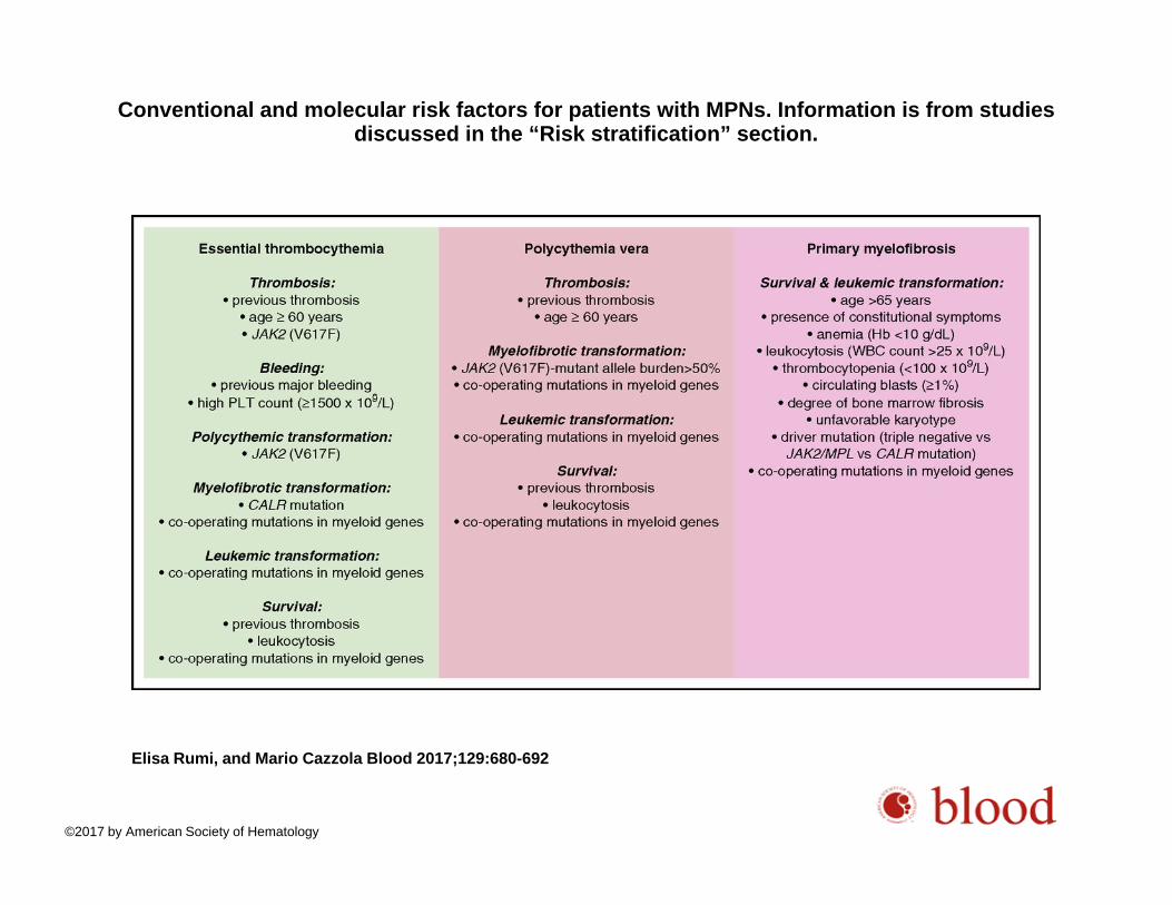
Conventional and molecular risk factors for patients with MPNs. Information is from studies discussed in the “Risk stratification” section.
Elisa Rumi, and Mario Cazzola Blood 2017;129:680-692
©2017 by American Society of Hematology