Motor systems spinal-cortex
20
Motor systems 389 The control of voluntary movements is complex. Many different systems across numerous brain areas need to work together to ensure proper motor control. We will start a journey through these areas, beginning at the spinal cord and progressing up the brain stem and eventually reaching the cerebral cortex. Then to complete the picture we’ll add two “side loops,” the basal ganglia and the cerebellum. This is relatively difficult material because the pathways are complex and our understanding of the nervous system decreases as we move up to higher CNS structures. Please keep up and complete all of the practice questions, and you will really enjoy this section. Remember, this material is important for you as future physicians because many of your patients will exhibit signs and symptoms associated with motor diseases. SPINAL LOWER MOTOR NEURONS Lower motor neurons (LMNs; also called alpha motor neurons) are directly responsible for the generation of force by the muscles they innervate. A motor unit is a single LMN and all of the muscle fibers it innervates. The collection of LMNs that innervate a single muscle (triceps) is called a motor neuron pool. You should remember from the spinal cord and brain stem module that LMNs that control proximal and distal muscles are found mainly in the cervical and lumbosacral segments of the spinal cord. At these levels, the LMNs are distributed such that neurons controlling axial muscles lie medial to those LMNs controlling distal muscles, and motor neurons controlling flexors lie dorsal to those controlling extensors. In the thoracic cord below T1, the LMNs are associated only with axial musculature. Motor Systems
Transcript of Motor systems spinal-cortex

Motor systems389
The control of voluntary movements is complex. Many different systems across numerousbrain areas need to work together to ensure proper motor control. We will start a journey throughthese areas, beginning at the spinal cord and progressing up the brain stem and eventually reachingthe cerebral cortex. Then to complete the picture we’ll add two “side loops,” the basal ganglia andthe cerebellum. This is relatively difficult material because the pathways are complex and ourunderstanding of the nervous system decreases as we move up to higher CNS structures. Pleasekeep up and complete all of the practice questions, and you will really enjoy this section.Remember, this material is important for you as future physicians because many of your patients willexhibit signs and symptoms associated with motor diseases.
SPINAL LOWER MOTOR NEURONS
Lower motor neurons (LMNs; also called alpha motor neurons) are directly responsible forthe generation of force by the muscles they innervate. A motor unit is a single LMN and all of themuscle fibers it innervates. The collection of LMNs that innervate a single muscle (triceps) is calleda motor neuron pool.
You should remember from the spinal cord and brain stem module that LMNs that controlproximal and distal muscles are found mainly in the cervical and lumbosacral segments of thespinal cord. At these levels, the LMNs are distributed such that neurons controlling axial muscleslie medial to those LMNs controlling distal muscles, and motor neurons controlling flexors liedorsal to those controlling extensors. In the thoracic cord below T1, the LMNs are associated onlywith axial musculature.
Motor Systems

Motor systems390
LMNs respond to, and are therefore controlled by, inputs from three sources: dorsal rootganglia, spinal interneurons (cells that do not project out of the area), and projections from highercenters such as the brain stem and cerebral cortex.
Stretch Reflex
Reflexes are short latency, relatively automatic responses to sensory stimulation. Spinalreflexes are a basic building block of movement. Dorsal root inputs provide the sensory input forspinal reflexes, and the LMNs provide the motor output pathway.
One of the simplest and best studied reflexes is the stretch reflex - stretch a muscle and thereflex circuit leads to contraction of the same muscle. Stretch reflexes work to resist lengthening ofa muscle. They are functionally efficient because they allow weight-bearing muscles to adjust to achanging load at the level of the spinal cord, without the information having to go all the way up tocortex for a decision. This is accomplished with a muscle spindle (our old friend from spinal cordand Physiology lectures) and a few neurons. You should recall the Ia and II fibers in the dorsalroots. Ia fibers are associated mainly with the nuclear bag intrafusal fibers and carry informationregarding the length and change in length of the muscle. The II fibers are primarily concernedwith the constant length of the muscle and thus bring information into the spinal cord from thenuclear chain fibers. You recall from the spinal cord module that these fibers synapse on cells inClarke’s column and accessory cuneate nucleus, which give rise to the spino- and cuneocerebellaraxons. The Ia and II fibers make additional synapses that are important in stretch reflexes. Stretchof the intrafusal muscle fibers increases activity in the Ia and II fibers, which excite alpha LMNs.This results in contraction of the extrafusal muscle fibers in the same (homonymous) muscle. Thisis a monosynaptic reflex, in that there is only one synapse between the incoming information andthe outgoing signal to contract the muscle. This monosynaptic reflex takes about 1 ms; the synapticdelay between the sensory input and the LMN is between 0.5 and 0.9 ms. In the cat, a single Ia fibermakes excitatory connections with all homonymous motor neurons. This Ia divergent signalproduces a very strong excitatory drive to the muscle within which it originates. This is calledautogenic excitation. The Ia fiber also makes monosynaptic connections with LMNs that innervatemuscles that are synergistic to (cooperate with) the homonynous muscle.
The incoming Ia fiber also synapses on a Ia inhibitory interneuron that in turn inhibitsLMNs that innervate the antagonist muscles (stretch biceps——inhibit triceps; notice this is NOT a
Motor systems391
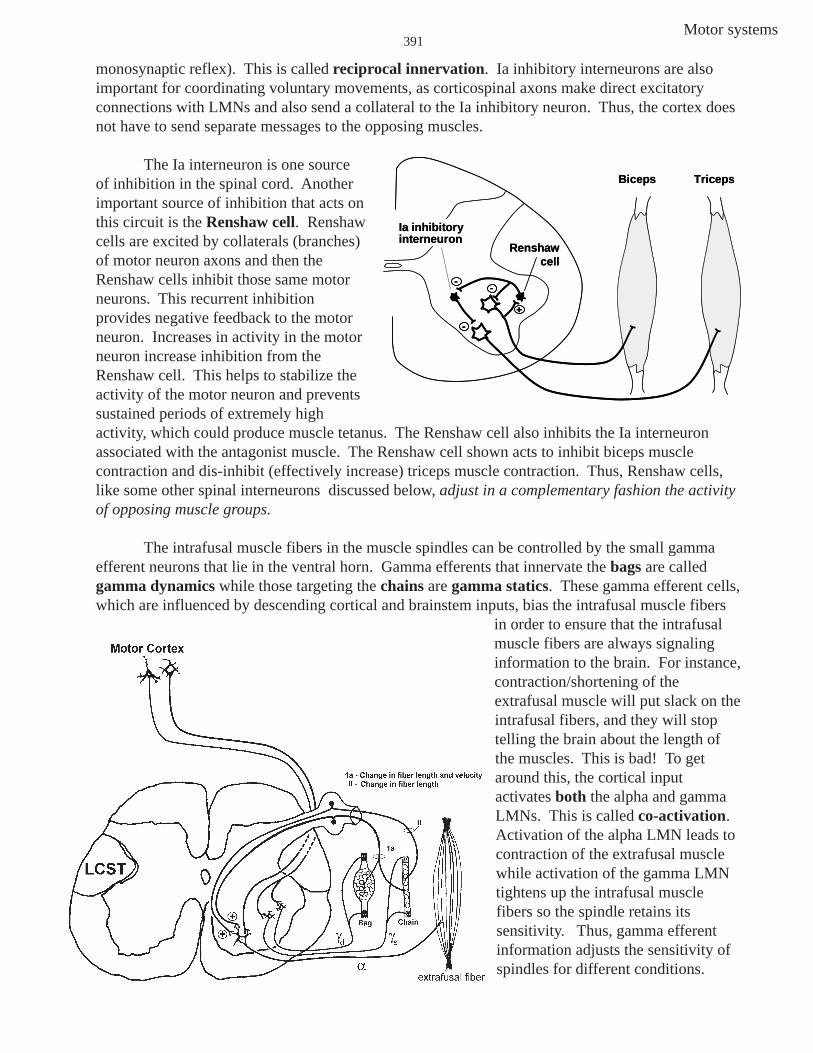
monosynaptic reflex). This is called reciprocal innervation. Ia inhibitory interneurons are alsoimportant for coordinating voluntary movements, as corticospinal axons make direct excitatoryconnections with LMNs and also send a collateral to the Ia inhibitory neuron. Thus, the cortex doesnot have to send separate messages to the opposing muscles.
The Ia interneuron is one sourceof inhibition in the spinal cord. Anotherimportant source of inhibition that acts onthis circuit is the Renshaw cell. Renshawcells are excited by collaterals (branches)of motor neuron axons and then theRenshaw cells inhibit those same motorneurons. This recurrent inhibitionprovides negative feedback to the motorneuron. Increases in activity in the motorneuron increase inhibition from theRenshaw cell. This helps to stabilize theactivity of the motor neuron and preventssustained periods of extremely highactivity, which could produce muscle tetanus. The Renshaw cell also inhibits the Ia interneuronassociated with the antagonist muscle. The Renshaw cell shown acts to inhibit biceps musclecontraction and dis-inhibit (effectively increase) triceps muscle contraction. Thus, Renshaw cells,like some other spinal interneurons discussed below, adjust in a complementary fashion the activityof opposing muscle groups.
The intrafusal muscle fibers in the muscle spindles can be controlled by the small gammaefferent neurons that lie in the ventral horn. Gamma efferents that innervate the bags are calledgamma dynamics while those targeting the chains are gamma statics. These gamma efferent cells,which are influenced by descending cortical and brainstem inputs, bias the intrafusal muscle fibers
in order to ensure that the intrafusalmuscle fibers are always signalinginformation to the brain. For instance,contraction/shortening of theextrafusal muscle will put slack on theintrafusal fibers, and they will stoptelling the brain about the length ofthe muscles. This is bad! To getaround this, the cortical inputactivates both the alpha and gammaLMNs. This is called co-activation.Activation of the alpha LMN leads tocontraction of the extrafusal musclewhile activation of the gamma LMNtightens up the intrafusal musclefibers so the spindle retains itssensitivity. Thus, gamma efferentinformation adjusts the sensitivity ofspindles for different conditions.
Ia inhibitory interneuron
Renshawcell
--
-
+
Biceps Triceps
Ia inhibitory interneuron
Renshawcell
----
--
++
Biceps Triceps

Motor systems392
Inverse Stretch (myotatic) Reflex
Ib fibers carry information from Golgi tendon organs (GTOs). Each GTO is comprised ofcollagen fibers intertwined with a Ib fiber. Thus, stretching of the tendon “squeezes” the Ib fiber, andit begins to fire. While muscle spindles are sensitive to changes in length of a muscle, GTOs are mostsensitive to changes in muscle tension and thus signal the force in a muscle. GTOs provide thenervous system with precise information about the state of contraction of the muscle.
The inverse stretch reflex is also known as the inverse myotatic reflex or Golgi tendonreflex. This reflex involves Ib afferents, Ib inhibitory interneurons, and LMNs. Increased firing ofthe Ib results in the inhibition of the homonymous muscle (autogenic inhibition).
The inverse stretch reflex is polysynaptic, meaning that it involves more than one synapse.This reflex is therefore slower than the stretch reflex. However, the inverse stretch reflex can over-ride the stretch reflex. If there is a very large stimulus, such as a strong blow to the patella tendonwith a hammer, the quadriceps muscles will contract due to the stretch reflex. To prevent damage tothe tendon due to the muscle pulling too hard on it, the inverse stretch reflex is initiated by increasingtension in the tendon, and the contraction of the muscle is inhibited. The inverse stretch reflex istherefore damping down the effect of the stretch reflex.

Motor systems393
Flexion/withdrawal reflex
Pain and temperature fibers in the dorsal roots (Cs and deltas) play a role in the flexionreflex, also known as the withdrawal reflex (you already know the pathway over which this painand temperature information reaches cortex via the ALS, etc.). At the spinal level, this reflexresponds to noxious stimuli, such as a hot object on the skin. The result is a rapid flexion, aprotective withdrawal mechanism that moves the body part away from the noxious stimulus.Another example of this reflex is stepping on a pin. The pin will be sensed by delta and C fibers,which synapse with a series of excitatory and inhibitory interneurons to produce a flexion response.The excitatory interneurons excite LMNs to the hamstring, while the inhibitory interneurons inhibitLMNs to the quadriceps (reciprocal inhibition).
The flexion reflex is often accompanied by a crossed extension reflex acting on thecontralateral limb. Using the example of stepping on a hot match, the crossed extension reflexwould brace the other leg, helping to maintain balance. In this case, excitatory interneurons exciteLMNs innervating the contralateral quadriceps, while inhibitory interneurons inhibit LMNs thatproject to the contralateral hamstrings.
You can see from the flexion/crossed extensor reflex that flexion withdrawal is a complete,albeit simple, motor act. While this reflex is reasonably stereotyped, the spatial extent and force ofmuscle contraction is dependent upon stimulus intensity. For instance, a moderately painfulstimulus will result in the production of a moderately fast withdrawal of your finger and wrist, whilea real painful stimulus results in the forceful withdrawal of the entire limb. Thus reflexes are notsimply repetitions of a stereotyped movement pattern, but instead are modulated by properties ofthe stimulus.

Motor systems394
It is important to understand that reflexes are adaptable and control movement in apurposeful manner. For example, a perturbation (a disturbance of motion, course, arrangement, orstate of equilibrium) of the left arm can cause contraction of the opposite elbow extensor in onesituation (right arm used to prevent the body from being pulled forward) but not in the situationwhere the opposite (right) arm holds a cup and the perturbation causes an inhibition of oppositeelbow extensor. Also, don’t forget that spinal reflexes are functionally efficient because they enableadjustments of movements to be made at the level of the spinal cord, without having to involve (andwait for) decisions from higher levels of the motor systems.
Table Support Hold CupDon’t tip over Don’t spill it
perturbationperturbation
tricepsactivated
tricepsinhibited
Table Support Hold CupDon’t tip over Don’t spill it
perturbationperturbation
tricepsactivated
tricepsinhibited

Motor systems395
SPINAL CORD NEURONAL NETWORKS (CENTRAL PATTERN GENERATORS)
You now understand some basic reflexesinvolving the LMNs. Let’s move “up” slightlyin the motor “hierarchy,” in particular, toneuronal networks in the spinal cord thatgenerate rhythmic alternating activity. A centralpattern generator (CPG) is a neuronal networkcapable of generating a rhythmic pattern ofmotor activity. Normally, these CPGs arecontrolled by higher centers in the brain stemand cortex. A simple spinal cord circuit isshown. This circuitry underlies alternatingflexion and extension --- when some cells areactive, the others are inhibited. These cells lie inthe ventral horn on the same side of the spinalcord and include flexor and extensor motorneurons, together with their associatedinterneurons. Descending inputs from higherlevels provide continuous input to the excitatoryinterneurons. However, because of randomfluctuations in excitability or other inputs, oneside of the circuit initially dominates and inhibitsthe other. Let’s say for example that excitatoryinterneuron #1 turns on first. It will not onlyexcite the flexor LMN and cause flexion, but itwill also turn on inhibitory interneuron #2,which inhibits excitatory interneuron #2 so thatthe extensor LMN is inhibited. Inhibitoryinterneuron #2 also inhibits itself, so the flexion will switch to extension when the inhibitoryinterneuron #2 stops firing. Remember, the tonic inputs are exciting excitatory neuron #2. This willmean that now excitatory interneuron #2 will fire and excite inhibitory interneuron #1, whichinhibits excitatory interneuron #1, and the pattern continues. You do not have to understand thedetails of such circuits. What you need to know is that there are circuits of cells in the spinal cord,composed of LMNs and interneurons, that generate basic patterns of locomotion.
Sometimes these CPGs can be activated below the level of a spinal cord transection. Forinstance, if a quadriplegic’s hips are extended, spontaneous uncontrollable rhythmic movements(flexion and extension) of the legs occur. Moreover, if babies are held erect and moved over ahorizontal surface, rhythmic stepping movements take place. Granted, these stepping rhythmicactivities in babies involve sensory input, but the circuitry for the rhythmicity of the movements is inthe spinal cord. These two examples indicate that basic neuronal circuitry for locomotion isestablished genetically.
An important point to remember is that descending inputs (e.g., frombrainstem or cortex) can act on these spinal circuits to modify their associatedmovements and even initiate the movements.

Motor systems396
INFLUENCE OF “HIGHER” CENTERS UPON LMNs
At this point in our examination of the motor systems we know that there is reflex and centralpattern program circuitry within the spinal cord and that certain reflexes and movements can occurusing this intrinsic circuitry of the spinal cord.
The next level of organization in the motor systems “hierarchy” is descending inputs from anumber of different nuclei in the brain stem and primary motor cortex to the spinal cord circuitry.Descending pathways to the spinal cord can be divided into dorsolateral and ventromedial systemsbased upon which spinal cord funiculus the pathway travels in and the termination pattern of itsaxons in the spinal cord grey. These two systems target different musculature.
Dorsolateral system—control of distal musculature
You already know about the lateral corticospinal tract (LCST), and this is one of twodorsolateral pathways. The LCST is especially important for the control of distal musculature andfor steering extremities and manipulating the environment. The other pathway in the dorsolateralsystem is the rubrospinal tract, which arises from our old friend the “ruber-duber.” The rubrospinaltract courses next to the LCST within the lateral funiculus and is also important for the control ofdistal limb musculature. Axons of the dorsolateral system reach the LMNs that lie more laterally inthe ventral horn. They also terminate in only one or two spinal segments; this precise targeting byindividual axons allows precise control over muscle groups. Thus the dorsolateral pathways areinvolved in the finer motor control of the distal musculature (finger movements for example).
Lesions involving both dorsolateral pathways (LCST and rubrospinals) result in the inabilityto make fractionated (independent) movements of the wrist or fingers. Moreover, voluntarymovements are slower

Motor systems397
and less accurate. However, the patient still has the ability to sit upright, and stand withrelatively normal posture (via functioning pathways in the ventromedial system discussed next).
A lesion of only the LCST initially resembles a combined lesion of the LCST and rubrospinaltract, but there is considerable recovery of function over time. The principle remaining deficit isinability to move the fingers independently (fractionated movements). Evidently, the functioningrubrospinal tract accounts for the recovered function of the wrist but it does not contribute toindependent finger movements.
Ventromedial system—control of axialand proximal musculature
You already know thatthe tecto- and vestibulospinaltracts (MVST and LVST)travel in the ventral funiculusand terminate on LMNs thatcontrol axial and proximalmusculature. Thus, they keepthe head balanced on theshoulders as the bodynavigates through space, andthe head turns in response tonew sensory stimuli.
The pontine andmedullary reticulospinaltracts (PRST and MRST) alsotravel in the ventral funiculus.(These pathways were notpresented in the spinal cordand brain stem lectures, soyou have not forgotten them).The PRST, which lies medialto the MRST in the ventralfuniculus, arises from cells inthe pons that lie around andnear the PPRF (paramedianpontine reticular formation atthe level of the abducensnucleus and motor VII; level5). In contrast, the MRST hasits origin from cells dorsal tothe inferior olive (level 3).The reticular formationconsists of those cells in thebrain stem that do not

Motor systems398
comprise the sensory and motor nuclei that you so carefully learned earlier in the course.
The PRST enhances the anti-gravity reflexes of the spinal cord. Thus, this pathway excitesupper limb flexors and lower limb extensors. This is an important component of motor control,since most of the time the activity of ventral horn cells maintains, rather than changes, muscle lengthand tension. PRST cells are spontaneously active but they also receive excitatory input from thecerebellum (soon to be discussed) and the cerebral cortex
The MRST has the opposite effect of the PRST, in that it inhibits anti-gravity reflexes ofspinal cord (inhibits upper limb flexors and lower limb extensors). MRST cells, like those in thePRST also receive excitatory input from the cerebellum and the cerebral cortex.
All of these ventromedial pathways (MVST,LVST, TST, MRST, PRST) innervate LMNs andinterneurons that control axial and proximal limbmuscles. Moreover, the axons in the ventromedialpathway distribute collaterals to many segments alongtheir distribution in the spinal cord (for the coordinationof intersegmental movements). You can see that such adistribution is not well suited for the discrete control of afew muscles but rather for control of groups of musclesinvolved in posture and balance.
Lesions of the ventromedial pathways in animalsresult in difficulty in righting to a sitting or standingposition and immobility of the body axis and proximalparts of the extremities. However, the hands and distalparts of the extremities are still active, because thedescending pathways controlling LMNs that innervatemore distal muscles travel in the dorsolateral part of thespinal cord.
Don’t forget the important organizational rule forof the descending motor pathways in the spinal cord:
dorsolateral system = distal musculatureventromedial system = axial and proximal musculature
You now know the main descending motor pathways. One important point to remember isthat there is more than one way for cortical information to reach the spinal cord; damage to onepathway can sometimes be compensated by a surviving pathway. There are smaller pathways I havenot discussed that follow the ventromedial-dorsolateral organizational rule. For example, a smallpercentage of corticospinal axons travel in the ventral funiculus. Unlike their famous LCST cousinsthat travel in the lateral funiculus and influence distal musculature, these ventral funiculus axonstarget spinal neurons related to axial and proximal muscles. This example is only meant to reinforcethe dorsolateral:distal, ventromedial:axial/proximal distinction. We won’t discuss this particularventromedial corticospinal tract further.
Spinal CordLMNs, Interneurons, CPGs
ruberreticular nuclei
superior colliculusvestibular nuclei
Cor
ticos
pina
l(la
t)
dorso-lateral
pathways
ventro-medial
pathways
Motor Cortex
Spinal CordLMNs, Interneurons, CPGs
ruberruberreticular nuclei
superior colliculusvestibular nuclei
Cor
ticos
pina
l(la
t)
dorso-lateral
pathways
ventro-medial
pathways
Motor Cortex

Motor systems399
PRIMARY MOTOR CORTEX
Motor cortex acts on the spinal and brainstem motor neurons and pathways to produce themore elaborate voluntary movements. Primary motor cortex provides the descending signals toexecute the movements.
The primary motor cortex is also called area 4 of Brodmann and motor I (MI).Corticospinal cells in area 4 project directly to the lower motor neurons (LMNs) in the spinal cordand brain stem and tell these LMNs, and in turn the muscles they innervate, what to do. Cells in MIare the classic upper motor neurons (UMNs). MI is located in the precentral gyrus, which lies infront (rostral) of the central sulcus. MI occupies most of this gyrus along both its lateral and medialsurfaces and is bound caudally by the central sulcus.
Human motor map. Left: The entire lateral surface of the right hemisphere is seen while only thedorsal and medial aspect of the left hemisphere is visible. A coronal (or frontal) section is cutthrough area 4 or MI. Right: The map of the body (homunculus) as represented on the precentralgyrus in this brain section. Medial is to the left, lateral to the right.
Somatotopic organization of MIIn the late 1950s Wilder Penfield studied the somatotopic organization of MI in patients by
stimulating MI with relatively large electrodes (the patients gave their permission to do this as partof their surgery). He found a topographically organized representation of the entire head and body,which is schematically shown above and to the right as the classic homunculus (L. little man). Thehead and forelimbs are represented laterally, and the hind limb and feet medially. The cortical areasallotted to different bodily regions are of unequal size, with the degree of representationproportional to the amount of skilled movement required of the respective part of the body. Thus, inhumans the face (particularly the tongue and lips used in speaking), thumb, and hand receive adisproportionately large representation, giving these structures a correspondingly exaggerated
Tongue
Face
Thum
bFing
ers
Tru
nk
Toes
Lateral(Sylvian)fissure
Elb
owSwallowing
Tongue
Face
Thum
bFing
ers
Tru
nk
Toes
Lateral(Sylvian)fissure
Elb
owSwallowing

Motor systems400
appearance. Movements of limbs evoked by stimulation of MI are strictly contralateral, but somemovements of the face (remember corticobulbars) may be bilateral.
The main outputs of MI are the now familiar corticospinal and corticobulbar pathways,which arise from cells that are pyramidal in shape and lie in layer 5 of MI (most of the cerebralcortex has 6 layers, 1 being most superficial and 6 the deepest). The largest corticospinal cells arecalled Betz cells.
Several corticospinal axons are shown in the drawing above as they course through theposterior limb of the internal capsule, the cerebral peduncle, pyramids of the medulla and pyramidaldecussation. Once in the spinal cord, these corticospinal axons comprise the LCST. You will learnmore about the internal capsule in a future lecture. For now, all you need to know is that the internalcapsule is 1) a large fiber bundle that contains axons going to and coming from the cerebral cortex,

Motor systems401
2) that it has an anterior and posterior limb separated by a genu (L. bended knee) and 3) thatthe corticospinal tract axons course through the posterior limb, while the corticobulbars arefound in the genu.
Physiology of corticospinal neurons
Much of what we know about the physiology of cells that project into the corticospinal tractcomes from single neuron recordings in awake monkeys trained to make specific movements.Corticospinal neurons were studied in a classic experiment in which monkeys were trained to move ahandle by flexing and extending the wrist (a nice distal motor task). The monkey was cued by a lightto either flex or extend its wrist. The experiment revealed several basic properties of corticospinalneurons:
First, corticospinal neurons are sensitive to thedirection of movement. Some are most active for wristextension. Others, as illustrated in the figure, are mostactive for wrist flexion. Second, the MI corticospinalresponse precedes the muscle electromyographic (EMG)activity, as expected since it is producing the motorcommand for that movement. Third, corticospinal activityis related to the force required to make the movement.The cell produces more action potentials with the additionof resistance that requires more force to make themovement. Conversely, the neurons are less active inconditions when the movement is assisted, requiring lessforce (not illustrated). Finally, the same neuronal activityis seen even when the movement is done repeatedly manyhundreds of times (i.e., there is no adaptation orhabituation of the cellular activity).
Functions of MI
Studies of the type illustrated have shown that corticospinal cells are telling LMNs in thespinal cord what to do. In fact, corticospinal cells convey a lot of details about amplitude, force anddirection of limb movement. In other words, MI neurons are NOT leaving these decisions up to theLMNs, as LMNs are too dumb. Thus, MI cells act on the LMNs (and in turn certain muscles) todetermine specific aspects of the movement: what direction to move, how far to move (amplitude)and how much force to use. The corticospinals in MI are specifying some pretty basic stuff instead ofjust sitting back and saying “grab that glass of water, arm and hand.” Remember, MI’s contributionsto motor control include controlling the number of muscles and movement forces and trajectories.Thus, MI cells are mainly concerned with the actual execution of the movements.
Primary Motor Cortex - Execution of Movements
Cortico-spinal axon
Flexor EMG
Extensor EMG
Cortico-spinal axon
Response during flexion
Flexor EMG
Response during flexion against a resistance
Extensor EMG
A
B
Corticospinal activity correlates with muscle force
Lever Position
Extension
Flexion
flexion movement onset
Cortico-spinal axon
Flexor EMG
Extensor EMG
Cortico-spinal axon
Response during flexion
Flexor EMG
Response during flexion against a resistance
Extensor EMGExtensor EMG
A
B
Corticospinal activity correlates with muscle force
Lever Position
Extension
Flexion
flexion movement onset
Lever Position
Extension
Flexion
flexion movement onset

Motor systems402
Effects of lesions of MI
You already know from spinal cord and brainstem lectures that MI lesions affect thecontralateral body. Relatively small lesions affect localized parts of the body because MI istopographically organized (remember the homunculus). With larger lesions there will behemiplegia. Much of the homunculus is devoted to the hand, so skilled, independent movements ofthe fingers are often affected. Also, a Babinski sign would be present if the lesion involves UMNsof the lower limb.
An important consequence of multiple descending pathways is that a surviving pathway cansometimes compensate for the loss of another pathway. A nice example of this is the partialrecovery of wrist and hand function that follows a complete lesion of LCST. The corticorubrospinalpathway appears to assume some of the jobs formerly done by LCST (although it isn’t much helpwhen it comes to the more refined movements of thefingers). As you might expect, very little recoveryoccurs if the lesion involves the LCST and thecorticorubrospinal pathway.
Separate cortical neurons generally comprisethe beginning of each of the descending motorpathways. This means that one set of cortical neuronsgives rise to the corticospinals, another set to thecorticorubers, another set to the corticoreticulars, etc.Thus, a small lesion in motor cortex could affect cellsof just one projection, producing symptoms associatedwith only that pathway.
However, you need to know that lesions ofmotor cortex are usually large - large enough to affectcortical neurons of the direct (e.g., LCST) and indirect(e.g., corticorubrospinal, corticoreticulospinal)descending pathways involved in voluntary motorcontrol. This leads to compromised voluntarymovement and the classic signs associated with UMNlesions. You already know these. Don’t forget them!
Classic signs of UMN lesion:weakness, hypertonia, hyper-reflexia, spasticity, Babinski sign
and NO muscle atrophy
Spinal CordLMNs, Interneurons, CPGs
ruberreticular nuclei
superior colliculusvestibular nuclei
Cor
ticos
pina
l(la
t)
dorso-lateral
pathways
ventro-medial
pathways
Motor Cortex
Distalmusculature
axial/proximalmusculature
Spinal CordLMNs, Interneurons, CPGs
ruberruberreticular nuclei
superior colliculusvestibular nuclei
Cor
ticos
pina
l(la
t)
dorso-lateral
pathways
ventro-medial
pathways
Motor Cortex
Distalmusculature
axial/proximalmusculature

Motor systems403
Blood Supply of MI
Area 4 or MI receives itsblood supply from the MiddleCerebral Artery and AnteriorCerebral Artery. Note that theanterior cerebral artery feeds the partof MI that extends down the medialsurface of the hemisphere, whichcontrols the lower limbs. The middlecerebral artery feeds the lateralsurface of the hemisphere, whichcontains the upper limb and headportions of MI.
AFFECTED ARTERY AREA AFFECTED SIDE
Ant. Cerebral Lower limb Contra
Mid. Cerebral Upper limb and Head Contra
Caudal Rostral
middlecerebralartery
posteriorcerebralartery
anteriorcerebralartery
Corpus callosum
Cerebral Arteries
Medial View
Lateral View
Caudal Rostral
middlecerebralartery
posteriorcerebralartery
anteriorcerebralartery
Corpus callosum
Cerebral Arteries
Medial View
Lateral View

Motor systems404
MOTOR ASSOCIATION/PREMOTOR CORTICAL AREAS
Studies in the late 1800’s showed that electrical stimulation of frontal cortex elicitedmovements of body parts. We now know this happens most easily in MI. Stimulation of MIrequires the lowest intensity current to evoke a movement. This probably reflects the directprojection of MI corticospinal neurons to LMNs. MI-stimulated movements are relatively simpleand involve a single joint, consistent with the role of MI in the execution of simple movements.
Electrical stimulation of cortex immediately rostral to MI also elicits movements, but withimportant differences. The current intensity has to be higher to produce an effect, and themovements are more complex, involving multiple joints, such as reaching or grasping movements.The areas in front of MIassociated with more complexmotor function are called motorassociation or premotor areas.
Premotor cortex isinvolved with making complex,multiple-joint movements andin the planning of movements.This is accomplished largely bytelling MI what movements toexecute. The total size of thesepremotor areas is considerablylarger than area 4. These areasprove to be particularlyimportant in the control ofhuman voluntary movement andare often compromised in diseases. The two premotor areas we will talk about are thesupplementary motor area (SMA) and the lateral premotor area (PMl). Remember, both arepart of cortical area 6.
Lateral premotor area: sensory motor interactions
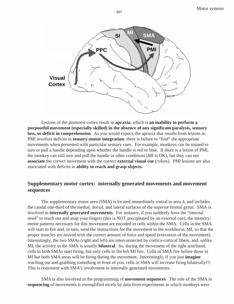
The lateral premotor area (PMl) lies on the lateral convexity of the hemisphere. Manymotor actions are responses to visual or auditory cues, and cells in PMl are active during suchexternally cued movements. For instance, you see an apple (external cue), and you reach out andgrasp it. Pathways involved in this sensorimotor transformation (seeing it is “sensory” and reachingfor and grasping it is “motor”) are seen in the next figure. The visual information (apple) falls firston the retina, and the retinal signal eventually reaches the primary visual cortex in the back of thebrain. From visual cortex, the visual information is conveyed rostrally and bilaterally to what istermed the posterior parietal cortex (PPC) in areas 5 and 7. The PPC helps to transform visualinformation about the location of the apple in extrapersonal space into the direction of a reachingmovement. Moving a body part relative to another body part is an example of a movement inintrapersonal space, while movements in extrapersonal space are movements executed in a threedimensional coordinate system fixed by points in the environment —whew! Information from PPCis conveyed (via several pathways, but we won’t sweat the details) to PMl. PMl acts on MI whichconveys the descending information to LMNs to execute the reaching movement.
Motor systems405
Lesions of the premotor cortex result in apraxia, which is an inability to perform apurposeful movement (especially skilled) in the absence of any significant paralysis, sensoryloss, or deficit in comprehension. As you would expect, the apraxia that results from lesions inPMl involves deficits in sensory-motor integration: there is failure to “find” the appropriatemovements when presented with particular sensory cues. For example, monkeys can be trained toturn or pull a handle depending upon whether the handle is red or blue. If there is a lesion of PMl,the monkey can still turn and pull the handle in other conditions (MI is OK), but they can notassociate the correct movement with the correct external visual cue (colors). PMl lesions are alsoassociated with deficits in ability to reach and grasp objects.
Supplementary motor cortex: internally generated movements and movementsequences
The supplementary motor area (SMA) is located immediately rostral to area 4, and includesthe caudal one-third of the medial, dorsal, and lateral surfaces of the superior frontal gyrus. SMA isinvolved in internally generated movements. For instance, if you suddenly have the “internalneed” to reach out and snap your fingers (this is NOT precipitated by an external cue), the memory/motor patterns necessary for this movement are encoded in cells within the SMA. Cells in the SMAwill start to fire and, in turn, send the instructions for the movement to the workhorse, MI, so that theproper muscles are moved with the correct amount of force and speed (execution of the movement).Interestingly, the two SMAs (right and left) are interconnected by cortico-cortical fibers, and, unlikeMI, the activity in the SMA is usually bilateral. So, during the movement of the right arm/hand,cells in both SMAs start firing, but only cells in the left MI fire. Cells in SMA fire before those inMI but both SMA areas will be firing during the movement. Interestingly, if you just imaginereaching out and grabbing something in front of you, cells in SMA will increase firing bilaterally!!!This is consistent with SMA’s involvment in internally generated movements.
SMA is also involved in the programming of movement sequences. The role of the SMA insequencing of movements is exemplified nicely by data from experiments in which monkeys were
posterior parietal cortex
PMl
SMA
Visual Cortex
MI
PPC
SI
posterior parietal cortex
posterior parietal cortex
PMl
SMA
Visual Cortex
MI
PPC
SI

Motor systems406
trained to do three movements; push, pull or turn a manipulandum (a joystick like you use withvideo games) in four different orders (e.g. push, turn, pull, would be one choice). Cells were foundthat increased their firing to a particular sequence (turn, push, pull) but not to a different sequence(turn, push, turn). Other SMA cells were active during other sequences. In the above task with themanipulandum, the visual appearance of the apparatus (an external cue) gave no clue as to whichmovement to make or even the order of the movements (unlike the case in movements involvingPMl).
Bottom line: SMA is big on internally generated sequences of movements.
Motor Cortex function revealed by imaging cerebral flood flow
Additional information about SMA and its differences from MI are provided by studies thatlook at the regional cerebral blood flow (rCBF) during motor tasks. In these studies an increase inrCBF means the cortical area is more active. In one rCBF study, subjects are asked to makemovements that differ in complexity. The simplest motor task requires subjects to make fastflexions of the index finger against a spring-loaded movable cylinder. This leads to increased rCBFin the finger area of the contralateral MI and SI (primary somatosensory cortex). The increase inrCBF in the somatosensory cortex reflects activation of peripheral tactile and proprioceptivereceptors caused by the finger flexions. More importantly, no higher cortical motor area (like PMlor SMA) exhibits an increase in rCBF in this task, just MI. Thus, MI is the primary motor cortical area involved in the execution of simple repetitivemovements.
Simple Repetitive MovementsSimple Repetitive MovementsIncreased rCBFin MI and SIIncreased rCBFin MI and SI

Motor systems407
When the same subjects are asked to carry out, from memory, sophisticated and complexmovements, pre-motor cortical area(s) also exhibit an increase in rCBF. For example, the thumb, inquick succession, must touch the index finger twice, the middle finger once, the ring finger threetimes, and the little finger two times. Then, with the thumb opposed to the little finger, the wholesequence is reversed!!! (Don’t worry, you will not be asked to do the sequence for the boards). Thisis a complex series of movements guided by memory and carried out in intrapersonal space(moving a body part relative to another body part). Again, there is increased rCBF in thecontralateral MI and SI (a bigger area since more fingers are involved). In addition there isincreased blood flow within SMA bilaterally. Thus, a complex task that consists of a sequence ofremembered movements involves the activity of both SMAs and MI and SI contralateral to theactive fingers. Of course, MI is needed for the execution. When the subject is asked to just imaginethe finger sequence movement, SMA lights up bilaterally, consistent with its role in the planning ofinternally generated sequences. However, MI is inactive (no execution, no MI activity).
Lesions of SMA lead to apraxia, which we have already defined for PMl lesions as theinability to produce purposeful movement even in the absence of muscle weakness or sensory loss.There is NO paralysis, only problems in planning. As you would expect, the apraxia following SMAlesions involve a deficit in performing sequences of movements. For instance, monkeys who hadlearned how to open a latch box by opening three different latches in a particular sequence aregreatly impaired following either uni- or bilateral lesions of SMA. Remember, the two SMAs areconnected across the midline. A lesion of one affects the proper functioning of the other. Forinstance, a lesion of the right SMA means the right MI is not getting proper planning information.This affects the left arm/hand. In addition, the left SMA has lost important input from the rightSMA so the left SMA input to the left MI is not normal. In the end, bi-manual coordination isaffected.
SMA
SMA
SMA MI and SI
SMA
Increased rCBF in labeled areas
think about doing complex task
complex motor sequence task
SMA
SMA
SMA MI and SI
SMA
Increased rCBF in labeled areas
think about doing complex task
complex motor sequence task

Motor systems408
The premotor areas are more specializedthan the “execution” area, MI. Both SMA and PMlare involved in planning of complex movements.However, the premotor area involved depends onthe type of movement and whether the impetus forthe movement is generated internally (SMA) orbased on an external cue (PMl). The movementcan be the same, but the areas that activate andtell MI which muscles to turn on for executiondiffer.
IN SUMMARY
MI - descending commands to execute basic movementsVentro-medial pathways - axial/proximal musculatureDorso-lateral pathways - distal musculature
Premotor cortex - planning of complex movementsSMA - sequences, bimanual coordination
- internally-generated movementsPMl - sensory-motor associations
- externally initiated and guided movements
frontal & prefrontal cortexsensory cortex
Spinal CordLMNs, Interneurons, CPGs
ruberreticular nuclei
superior colliculusvestibular nuclei
Cor
ticos
pina
l(la
t)
dorso-lateral
pathways
ventro-medial
pathways
area 6area 4PPC PMlat
SMA
frontal & prefrontal cortexsensory cortex
Spinal CordLMNs, Interneurons, CPGs
ruberruberreticular nuclei
superior colliculusvestibular nuclei
Cor
ticos
pina
l(la
t)
dorso-lateral
pathways
ventro-medial
pathways
area 6area 4PPC PMlat
SMA