MORPHOLOGY AND MORPHOMETRIC STUDY OF PLACENTA...
122
Dissertation on MORPHOLOGY AND MORPHOMETRIC STUDY OF PLACENTA AND UMBILICAL CORD WITH ITS VASCULAR PATTERN AND COMPARATIVE ANATOMY Submitted in partial fulfillment for M.S.DEGREE EXAMINATION BRANCH – V ANATOMY Upgraded Institute of Anatomy Madras Medical College & Research Institute, Chennai – 600 003 THE TAMILNADU Dr.M.G.R. MEDICAL UNIVERSITY CHENNAI – 600 003 TAMIL NADU
-
Upload
nguyenmien -
Category
Documents
-
view
247 -
download
0
Transcript of MORPHOLOGY AND MORPHOMETRIC STUDY OF PLACENTA...
Dissertation on
MORPHOLOGY AND MORPHOMETRIC STUDY OF PLACENTA AND UMBILICAL CORD WITH ITS
VASCULAR PATTERN AND COMPARATIVE ANATOMY
Submitted in partial fulfillment for
M.S.DEGREE EXAMINATION BRANCH – V ANATOMY
Upgraded Institute of Anatomy Madras Medical College & Research Institute,
Chennai – 600 003
THE TAMILNADU Dr.M.G.R. MEDICAL UNIVERSITY CHENNAI – 600 003
TAMIL NADU
MARCH 2009
CERTIFICATE
This is to certify that the dissertation work on MORPHOLOGY AND
MORPHOMETRIC STUDY OF PLACENTA AND UMBILICAL CORD
WITH ITS VASCULAR PATTERN AND COMPARATIVE ANATOMY
is the bonafide work done by Dr.R.SUDHA in the Institute of Anatomy,
Madras Medical College, Chennai – 600 003 during the year 2006 – 2009
under my supervision and guidance in partial fulfillment of the regulation laid
down by The Tamil Nadu Dr.M.G.R Medical University, for the M.S.,
Anatomy branch V examination to be held in March 2009.
Dr.T.P.Kalaniti M.D., Dr.Christilda Felicia Jebakani M.S., Dean Director Madras Medical College, Institute of Anatomy, Chennai – 600 003 Madras Medical College Chennai 600 003 Date: Date: Station: Station:

ACKNOWLEDGEMENT
I sincerely express my gratitude and indebtness to my most respected
teacher and guide DR.CHRISTILDA FELICIA JEBAKANI, M.B.,B.S.,
M.S., Director and Professor, Institute of Anatomy, Madras Medical College
who motivated me to perform this study and provided her invaluable guidance,
without which this work would not have been possible.
I earnestly thank DR. T.P.KALANITI M.D., Dean. Madras Medical
College, Chennai for permitting me to avail the facilities in this institution for
my study.
. I express my thanks to DR.K.SARASWATHI M.D., D.G.O,
Director and Superintendent, Institute of Obstetrics and Gynaecology, Egmore,
Chennai-08 for giving me permission to collect the placental specimens for my
study.
I express my gratitude to DR.C.SOUNDARAJAN, M.V.SC. Associate
Professor, Madras Veterinary College,Chennai-7, for giving his expertise,
helping me to collect the animal specimens, without whom comparative
anatomy wouldn’t have been made possible. I am very much indebted to him
for his timely help.
My gratefulness to DR. T.S. SWAMINATHAN, M.D., D.M.R.D.,
Director and Professor of Barnard Institute of Radiology, Madras Medical
College, Chennai, DR. Sundaresen and other faculty members for extending
their support in angiographic studies.

I am thankful to DR.USHA KOTHANDARAMAN M.S., Anatomy
Professor and H.O.D, Meenakshi Medical College & RI, for helping me to
learn the techniques of silicone gel cast method for the study of vascular
pattern of the placenta.
I thank DR.GUNAPRIYA.R, M.S., Anatomy, Associate Professor, and
Savitha Medical College, for being a backbone to me throughout this study
.Her help cannot be quantified.
I sincerely thank Dr.I.Jayaraj, Tmt.M.S.Thenmozhi, Dr.B.Chezhian,
Dr.V.Loganayaki, Dr.V.Sathyalaksmi, Dr.M.Vijayalakshmi and
Dr.P.Murugesan for their valuable suggestions and encouragement.
I would like to thank my seniors Dr.SumathiLatha, Dr Preethi
Ramya, Dr.Anitha, Dr.JayanthiGnanaDeepam & Dr.Jayanthi, for
encouraging me throughout this study.
I would like to thank my colleagues Dr.V.Anandhi, Dr.K.Rajeswari,
and Dr.Mukundan, who helped me throughout this study .I thank my friend
Dr.V.Rajapriya and my Juniors for their immense support and Comments.
I would like to express my thanks to the technical staff for their help.
I am indebted and being grateful to my parents, husband & children for
their sacrifice, enduring patience, and moral support, which made this, study a
reality.
Above all I thank God and pray him to continue his mercy upon me.

CONTENTS S.NO TITLE PAGE NO 1. INTRODUCTION 1 2. AIM OF THE STUDY 2 3. REVIEW OF LITERATURE 5 4. DEVELOPMENTAL ANATOMY 22 5. MATERIALS AND METHODS 24 6. OBSERVATION 30 7. DISCUSSION 44 8. CONCLUSION 70 9. BIBLIOGRAPHY

1
INTRODUCTION
‘Chief Nourisher in life’s feast’
‘Shakespeare: Macbeth II’
The word placenta is a Latin word meaning a flat plate or cake.
Placenta represents an extremely intimate parabiotic union of maternal
and foetal tissues for the inevitable requirement of the embryo.
After the delivery of the foetus the placenta becomes separated from the
uterine wall and is expelled with the membranes as the "after birth". Expelled
discoidal mass has two surfaces -maternal and foetal and a peripheral margin.
Maternal surface is finely granular mapped into 15-30 lobes by fissures
or grooves. Lobes correspond in large measure to the major branches of
distribution of the umbilical vessels.
Fetal surface is smooth, shiny and transparent. It is covered by amnion
with the umbilical cord attached to its center. The umbilical vessels radiate out
on the fetal surface from the umbilical cord below the amnion.
The peripheral margin is continuous with the fetal membrane, which
consists from outside inwards of fused decidua parietalis, decidua capsularis,
chorion laeve and amnion. Placenta begins to meet the demand of the embryo
as early as the 3rd week of intrauterine life, even before the mother is aware of
her pregnancy.
Examination of the placenta in utero as well as post-partum gives us
information about the congenital anomalies, its well being and nutritional status
that can be anticipated in the fetus.

2
AIM AND OBJECTIVE
Hospital medical board generally mandate that all tissues removed from
patients be sent for examination and a glaring exception has long been
placentae, which are usually discarded or reprocessed after at most a cursory
examination by the delivering physicians.
It is gratifying now to see improving quality of placental examination.
This newfound interest in the placenta is of multiple etiologies.
Malformations and disruptions of placental and body wall development
are common. These include body stalk anomalies, limb-body wall complex
with cord agenesis (Fig. 1), acardia, malformations related to monozygotic
twinning (Fig. 2), conjoined twins, twin-twin transfusion syndrome, limb
reduction defects and chorion villous sampling, amnion band syndrome
(Fig. 3), early &prolonged rupture of membranes, oligohydramnios sequence,
sirenomelia (Fig. 4), confined placental mosaicism and uniparental disomy.
In multiple pregnancies placentation and embryonic development are
more complex with twins. Dizygotes would individually develop as singleton
each with a distinct chorion, amnion and feto-placental circulation.
Monozygotic twins have the potential to develop as dizygotes or share their
chorion, amniotic sac or vascular connections.
Examination of the placentae may be very useful in medico legal cases.
The modern era of surgery on the placenta and cord began in 1988 when
a team of physicians at university of Utah performed the first fetoscopic laser
occlusion of chorioangiopagous vessels (FLOC PROCEDURE) (Fig. 5) in a
monochorionic twin pregnancy affected by twin-twin transfusion syndrome.
Three abnormalities of pregnancy, which have their pathogenesis in the
placenta rather than the fetus, which need surgical intervention, are (1)

3
Variations of twin-twin transfusion syndrome (TTTS) in monochorionic (MC)
twins (Fig. 8) (2) Heart failure of the normal pump twin in a monochorionic
acardius twin pregnancy and (3) Chorioangiomas (Fig. 9) large enough to
cause hydramnios and fetal hydrops. The treatment of these placental
abnormalities has been proposed by minimally invasive video fetoscopy linked
with either the photocoagulative properties of laser light or direct ligation of the
vasculature with clamps or suture as well as ultrasound guided transcutaneous
placement of intravascular occluding devices and solutions.
Percutaneous umbilical cord blood sampling (Fig.7) is done to analyse
the blood gas level of the fetus. Chorionic villous sampling (Fig. 6) at 8-9
weeks of gestation suggests that an early placental hemorrhage can lead to
characteristic pattern of malformations in the fetus. It also has taught us that
2% of placentae show confined placental mosaicism that can affect fetal
growth and survival.
All these procedures need expert knowledge about the morphological
anatomy and vascular pattern of the placenta in a detail manner.
Physicians and surgeons have been hampered to become more familiar
with anatomic variations of the placenta and the limitations of working in the
intrauterine environment.
Due to the above reasons I have made an effort to study the morphology
and morphometric analysis of the placenta and umbilical cord with vascular
pattern and comparative anatomy.
The present study is done with the following parameters:
1. Shape, diameter and thickness of the placenta.
2. Maternal surface – number of cotyledons.
3. Placental weight
-Compared with fetal weight, sex and maturity.

4
4. Fetal surface
-Vascular pattern of the placenta.
-Type of insertion of the umbilical cord.
5 Length and diameter of the umbilical cord.
6. Spiral turns (twist) of the umbilical cord.
-Umbilical coiling index.

5
REVIEW OF LITERATURE
1) Shape, diameter and thickness of the placenta: (Fig. 10)
Henry Gray (1858): He quoted that the expelled placenta is a flattened
discoidal mass with an approximately circular or oval outline and the average
diameter is about 185 mm, range 150 –200 mm and the average thickness is
23mm, range 10-40mm.
Leslie. B. Arey (1924): He stated that the expelled placenta is typically
a thick circular disc. Departure from a circular shape is quite common ranging
from an oval contour to other variant forms (ie. Spindle, pear, heart, crescent,
ring), which are more rarely encountered. The placenta may be notched, lobed
or even divided completely. Accessory placentae of smaller size than the main
placenta are not unusual. Fused placentae (Fig. 12) result when ordinary twins
become too closely implanted. Placenta measures 7 inches (17.5cm) in
diameter and one inch (2.5cm) in thickness.
Williams (1930): In his book of obstetrics he described that there are a
number of abnormal placental shape variations: -
i) Multiple placentae with a single fetus: Placenta may be separated
into lobes when the division is incomplete and the vessels of fetal origin extend
from one lobe to the other before uniting to form the umbilical cord, the
condition is termed placenta biparita or bilobata (Fig. 13).
ii) Succenturiata placenta: This variation results when one or more
small accessory lobes are developed in the membranes at a distance from the
periphery of the main placenta to which they usually have vascular connections
of fetal origin.
iii) Ring shaped placenta: This is a rare anomaly seen in fewer than 1
in 6000 deliveries. The placenta is annular in shape.

6
iv) Membranaceous placenta: All of the fetal membranes are covered
by functioning villi and the placenta develops as a thin membranous structure
occupying the entire periphery of the chorion. Placenta membranacea is also
referred to as placenta diffussa (Fig. 14).
v) Fenestrated placenta: In this anomaly the central portion of the
discoidal placenta is missing.
vi) Extrachorial placenta - Circumvallate placenta: (Fig. 11) The
chorionic plate on the fetal side of the placenta is smaller than the basal plate
and the fetal surface of such a placenta presents a central depression
surrounded by a thickened grayish white ring, it is called a circumvallate
placenta. When the ring coincides with the placental margin the condition is
sometimes described as circummarginate placenta.
Bradley. M. Patten (1946): He quoted that the placental portion of the
after-birth is a rounded disc. The placenta may exhibit a bilobed shape.
Augero. O (1957): He reported 3.26% of incidence of placenta bi-
lobulata or biparita in his study.
Roth .L.G (1957): He had written that succenturiate lobes may occur
singly or in multiples.
Scott. J.S (1960): He studied 3,161 cases and observed 18.3% of
circumvallate placenta.
Ziel .H.A (1963): He stated that he observed 0.62% of circumvallate
placentae when he studied 40,143 cases.
T.W.Sadler (1963): He said that at full term the placenta has a discoid
shape and is approximately 3 cm thick.

7
J. Bazso (1966): He quoted that he found an incidence of 5.8% for
circumvallate placenta in his series.
K. Benirschke et al (1967): They had written in their book ‘The
Pathology of Human placenta’ that the average thickness of the placenta in the
center is 2.0 cm.
Wilson. D & Paalman R.J (1967): They quoted that in their study of
10,927 placentae, they had observed 1% of circumvallate placenta.
Went worth. P (1968): He reported that when he studied 895 placentae,
he observed circumvallate placenta of about 6.5 %.
Torpin.R (1969): He mentioned that full term delivered placenta is in
more than 90% of the cases a disc – like flat, round to oval organ .It has
abnormal shapes such as placenta bilobata, and placenta succenturiata.
J.D. Boyd & W.J. Hamilton (1970): They described in their book ‘The
human placenta’ that the full term placenta is flat with a round or oval outline.
The placenta may be bidiscoidal (i.e., bipartite), trilobed (i.e. tripartite),
succenturiate, extrachorial or membranaceous.
A succenturiate placenta is one in which an accessory lobule or lobe or
more than one such lobule or lobe is separated by some distance from the
placenta or lies close to the main placental mass. Extra chorial placentae may
be circumvallate or marginate. In the circumvallate form (complete or
incomplete) the lateral edge of the placenta is undercut at its junction with
decidua parietalis so that there is a ditch round the periphery of the placenta. In
the marginate condition the edge of the placenta is raised to form the so-called
closing ring. The average diameter of the placenta is 185 mm (18.5cm) and
thickness is 23 mm (2.3cm).
8
Fujikura et al (1970): They had found bipartite placenta of about 4.2%
in 8505 specimens collected in the collaborative perinatal study.
Fox.H & Sen .D.K (1972): They reported 2.4% of circumvallate
placenta in a study of 3000 placentae.
Keith. L. Moore & T.V.N Persaud (1973): They said that the shape of
the placenta is circular. The placenta measures 15 – 20cm in diameter and
thickness of about 2 - 3 cm.
Richard. S. Snell (1973): He described that placenta is flattened and
circular in shape. Its diameter is about 8 inches (20cm) and it is one inch
(2.5cm) in thickness.
Fox.H (1978): He pointed out that placenta biparita or bilobata’s
incidence varies widely and it is cited at about 1 of 350(0.2%) deliveries.
Sarojamma (1986): She found that in a study of 100 placentae the
thickness of the placenta ranged from 1.5 to 2cm
J.P.E. Judson (1986): He reported in a study of 20 placentae, the
average diameter of the placenta to be 169.7mm (16.97cm)
Cynthia. G. Kaplan (1996): She stated the thickness of the placenta to
be 2-2.5cm.
GunaPriya.R (2001): She quoted that in a study of 100 placentae, she
observed 93% circular and 7% oval shapes of placentae. The average diameter
of the placenta was 17.4cm, range 12 - 22.5cm and the thickness was 2.1cm,
range 1.5 - 2.7cm.

9
2. Maternal surface: Cotyledons
Henry Gray (1858): He quoted that the maternal surface of the placenta
is finely granular and mapped into some 15-30 lobes.
T.W.Sadler (1963): He described that when the placenta is viewed from
the maternal side 15-20 slightly bulging areas, the cotyledons, covered by a
thin layer of deciduas basalis are clearly recognizable.
K.Benirschke et al (1967): They said that an incomplete system of
grooves subdivides the basal surface of the placenta into 10 to 40 slightly
elevated areas called maternal cotyledons (lobes or lobules).
Allan C.Barnes (1968): He mentioned that the cotyledons are indistinct
lobulations about 30 in number, visible on the maternal surface of the placenta.
J.D. Boyd &W.J.Hamilton (1970): They had written that the mature
placenta shows a variable number of cotyledons from 10 to 38 of slightly
elevated convex areas called lobes or when small, lobules.
Sarojamma (1986): she found in a study of 100 placentae, that the
average number of cotyledons to be 18 with a range 3 - 24.
Gunapriya.R (2001): She stated that in her study of 100 placentae, the
number of cotyledons in the maternal surface varies from 12 to 24. The average
being 18 in number.
Majumdar et al (2005): They studied 100 placentae and reported mean
number of cotyledons in normal control group to be 19 and 18 in hypertensive
group.
Sultana. S et al (2007): In a study of total 45 placentae, they found that
compared to the controls there was less placental diameter and cotyledon
number in eclampsia.

10
3) Weight of the placenta:
Henry gray (1858): He stated that the average weight of the expelled
placenta is 470 gms, range 200-800gms.
Adair.F.L & Thelander.H (1940): They found that in general, a
normal placenta is about one seventh of the weight of the fetus with which it is
associated.
Ghosh.L & Chandrasekhar.C (1948): They reported that the weight of
the placenta of mature male and female infants on an average was 486.3gms
and 479 gms in contrast to premature infants where the figures were 391.1gms
and 379.1 gms respectively.
Little (1960): He studied the placentae of 956 mature infants and
concluded that a placental co-efficient of less than 0.10 and greater than 0.18
should be considered to indicate a relatively small or large placenta and that
less than 0.08 and more than 0.2 should be considered definitely abnormal.
Wiggles worth (1962): He found that the placental weight ranged from
360 - 570 gms.
Dockery, J.L (1960): He reported a case of giant placenta weighing
1,984 gms with hemoglobin value of 4.1 gms per 100ml due to severe iron
deficiency.
Wong, T.C.& Lartour, J.A.P (1966): They reported a lower placental
weight of 399gms for growth retarded infants as compared to 463gms for
normal infants.
K.Benirschke et al (1967): They reported that the average weight of the
placenta was found to be of 470 gms.

11
N.A. Beischer et al (1968): They found that in a study of 490 patients
with anaemia, the incidence of placentae weighing more than 900gms was 2%.
In diabetes mellitus and erythroblastosis approximately 10 percent of the
placenta weighed over 900gms.
Saigal. S & Srivastava, J.R (1970): They reported that the weight of
the placenta was 531.5gms at 38 weeks and 475.9gms at 42 weeks and above.
J.D.Boyd &W.J.Hamilton (1970): They studied over 1000 placentae
delivered at term and quoted that the average weight of the placenta was 508
gms.
Fujikura et al (1970): They examined a total of 8505 placentae and
reported that biparitite placenta represented 4.2% and the placental weight was
greater in the bipartite group (mean weight 473.3 gms) than in other groups
(mean wt 448 gms).
Gupta et al (1972): In a study of 469 placentae the mean weight of the
placenta was found to be higher for males as compared to females. The mean
placental weight of infants below 37 weeks gestation was 449.9 gms and 405.3
gms for males and females respectively, while it was 521.9gms and 510.4gms
respectively in babies of 37 weeks gestation and above.
Keith. L. Moore & T.V.N.Persaud (1973): They had written that
placenta weighed 500 to 600 gms, which is about one sixth of the weight of the
average conceptus.
Richard S.Snell (1973): He stated that the placenta weighs about 1 lb
(500 gm).
Singla P.N et al (1978): They quoted that in a study of 69 anaemic
mothers (hemoglobin < 110g/l) and 16 mothers without anaemia (hemoglobin
≥ 110 g/l) the birth weight and placental weight were significantly reduced in

12
the severely anaemic mothers and it had direct relationships with the maternal
hemoglobin levels.
Shameer singh et al (1979): They reported in a study of 3500 placentae
that the mean weight of the placenta was found to be 475 gms.
Kher & Zawar (1981): They mentioned that a significant reduction in
foeto-placental weight ratio was observed in toxemia of pregnancy.
Godfrey, K.M et al (1991): In a study of 8684 pregnant women they
observed large placental weight, which was associated with a low maternal
hemoglobin.
Lurie.S et al (1999): In study of human feto-placental ratio in 431
deliveries, they reported the mean newborn weight was 3,382gms and the mean
placental weight was 613 gms. Mean feto placental weight ratio was 5.6 and
this ratio did not differ in male (5.7) and female (5.6) infants.
J.P.E. Judson (1986): He found that in a study of 20 placentae, the
average weight of the placenta was noted to be of 479.17gms.
Damania et al (1989): They had studied sixty placentae of hypertension
disorders of pregnancy and they had reported that the birth weight, placental
weight and feto placental ratio were less in hypertensive cases than in the
normotensive controls.
Rath.G et al (2000): They studied 218 hypertensive mothers and stated
that the weight of the placenta and the infant in hypertensive group were found
to be lower than the normal group.
Majumdar et al (2005): They observed that in a study of 100 placentae
the mean placental weight in control group was found to be 485.85 gms and in

13
hypertensive group to be 399.10gms. The mean foetoplacental weight ratio was
found to be 5.89 in control group and 6.23 in hypertensive group.
Swanson.L.D& Bewtrac.C (2008): They studied the placenta of live
singleton deliveries and reported that the mean weight of the mature term
placenta has increased over years from 499 to 537gms.
Kucuk. M, Doymaz. F (2008): They stated that placental weight and
placental weight to birth weight ratio are increased in diet and exercise treated
gestational diabetes mellitus (GDM) subjects. They observed that birth weight
in GDM to be 3288.3 gms and 3207.06 gms in control group. Placental weight
in GDM was found to be 694.8gms and 610.2gms in control group. Placental
coefficient (PW/BW) was noted to be 0.21 in GDM and 0.18 in control group.
4. Foetal surface: - a) Vascular pattern of placenta
Shordania. J (1929): He considered that the arteries in all human
placenta can be subdivided into two main groups according to the pattern
followed by their branching and he introduced the terms disperse and magistral
to describe them. In the disperse type with approximately central attachment of
the cord, the two arteries divide dichotomously several times into a number of
smaller vessels rapidly diminishing in caliber (Fig. 15). In the magistral type
the two arteries extend almost to the margin of the placenta before diminution
in their caliber occurs (i.e. longer undivided branches) (Fig. 19).
P.Bacsich & C.F.V. Smout (1937): They studied the foetal vessels of
50 human placentae with corrosion technique (Fig. 17). According to the
pattern made by the arteries, vascular pattern of placenta can be divided into
two groups (i) a disperse type in which the blood vessels divide dichotomously
(Fig. 15) and (ii) a magistral type in which the two main arteries extend as far
as the margin of the placenta and give off branches of small size only.
14
Edith. L. Potter (1952): He pointed out that normally the umbilical
cord contains 2 arteries and 1 vein embedded in Wharton’s jelly.
Keith.L.Moore (1973): He quoted that umbilical cord contains two
arteries and one vein and they are surrounded by mucoid connective tissue.
Paul Wentworth (1965): He reported in his study of 642 placentae that
he had observed in 20 placentae (3.1%) the foetal veins crossed over the foetal
arteries.
Kishore.N & Sarkar. S.C (1967): They viewed that the disperse type
of vessel distribution is more commonly present (61.8%).
Andrade. A (1968): He had mentioned that disperse type of vascular
pattern to be more frequently found in placentae with centrally inserted cords.
Bhargava.I & Raja.P.T.K (1969): They suggested that foetal veins
may occasionally cross over foetal arteries and this pattern is correlated
significantly with abnormal fetal development.
Z.Gordon et al (2007): They studied the anthropometry of fetal
vasculature in 15 placentae and revealed that the branching architecture of the
chorionic vessel is a combination of dichotomous (disperse) and monopodial
(magistral) pattern i.e. mixed type (Fig. 16). He also stated that the vascular
architecture was mostly monopodial for the marginal cord insertion and mostly
dichotomous for the central insertion (Fig.18).
Fox. H (1997): In his study he found increased number of vascular
profiles more commonly in cords from stillbirths and he associated this
anomaly with a history of maternal cigarette smoking.

15
Foetal surface- b) Insertion of the umbilical cord:
Hyrtl.J (1870): He observed 16% central type (Fig. 20), 54% eccentric
type, and 19% marginal type of cord insertion.
Chiari et al (1895): They reported 3.3% central type, 91.2% eccentric
type, 5% marginal type and 0.5% velamentous type of cord insertion.
Lefevre.G (1896): He stated out of 15,894 placental specimens, he
observed 0.84% velamentous type of cord insertion (Fig. 21).
Noldeke.H (1934): In a study of 10,000 placentae of singleton
pregnancies, he found 1.1% of velamentous type of cord insertion.
Grieco.A (1936): He said that in a study of 23,469 placentae of
singleton pregnancies, he observed 0.41% of velamentous type of cord
insertion.
Earn A.A. (1951): He stated that out of 5412 specimens, he found 56%
central, 28% eccentric, 15% marginal and 1.1% velamentous type of cord
insertions.
Simon Brody & David.A.Frenkel (1953): They quoted that out of 512
deliveries, they observed 6.2% of marginal insertion of the cords.
Diterlizzi &Rossi G.R. (1955): They had found that in a study of
15,416 placentae of singleton pregnancies, 1.0% of velamentous type of cord
insertion.
Shanklin D.R (1958): He observed that out of 500 specimens, he
observed 11% central type, 89% eccentric type (Fig. 15), 1.9% marginal type
and 0.78% velamentous type of cord insertion.

16
Scott.J.S (1960): He reported in a study of 3,161 placentae of singleton
pregnancies, 2% of marginal type and 1.5% velamentous type of cord insertion.
Krone.H.A (1961): In a study of 5214 specimens, he observed 25% of
central type, 64% eccentric, 10% marginal and 1% velamentous types of cord
insertion.
Corkill.T.F (1961): In a study of 12,695 placentae obtained from
singleton pregnancies, he reported 0.024% of velamentous type of cord
insertion.
Thomas. J (1963): He stated that of 18,316 placentae he studied, he
observed 5.2% marginal type, 1.3% velamentous type of cord insertion.
Monie I.W (1965): She reported that out of 183 specimens, she
observed 70% eccentric type of cord insertion, 14.7% marginal and 15.3%
velamentous type of cord insertion.
Torpin.R. & Barfield.W.E (1968): They found that in one third of
bilobed placenta the cord inserts on the larger lobe and in two thirds it has
velamentous insertion.
Eastman N.J & Hellmann.L.M (1966): They reported that out of 200
specimens they studied, they found central type of cord insertion in 18%,
eccentric in 73%, marginal in 7% and velamentous types of cord insertion in
1.25%.
Scheffel.T & Ranganke. D (1970): They quoted that out of 37,963
placentae obtained from singleton pregnancies, they observed 0.22% of
velamentous type of cord insertion.

17
Uyanwah et al (1977): They mentioned that in a study of 1000
placentae obtained from singleton pregnancies, they found 5.6% marginal type
of cord insertion and 1.6% velamentous type of cord insertion.
Robinson et al (1983): They stated that in a study of 44,677 placentae,
they observed 8.5% marginal type, 1.5% velamentous type of cord insertion
Sarojamma (1986): She studied 100 placentae and observed 40%
central type of cord insertion, 53% eccentric, 2% marginal and 4%
velamentous type of cord insertions.
J.P.E. Judson (1986): He reported that in a study of 20 placentae, he
observed 20% central type of cord insertion, 70% eccentric, 10%marginal and
0% velamentous type of cord insertion.
Nordenvall et al (1988): In a study of 330 placentae, they observed that
marginal cord insertions were correlated to extrachorial and bilobate placentae.
Gunapriya.R (2001): In 100 placentae she observed 86% eccentric, 5%
central, 9% battle dore and 0% velamentous type of cord insertion.
5. Length and diameter of the umbilical cord:
Henry Gray (1858): He said that the fully developed umbilical cord
measures on an average of about 50cm in length and 1-2 cm in diameter. Its
length varies from 20-120 cm.
Gardiner J.P (1922): He obtained an average length of 55 cm for
normal cords. He considered cord length less than 32 cm as absolutely short
and length more than 32 cm as relatively short.
Shordania J. (1929): He recorded an average length of 60 cm in a
series of 421 cords with range of about 35 -104 cm and the cord diameter to
be 1-2 cm.
18
Williams (1930): He quoted that the average length of the cord was
observed to be 55 cm, range 30 to 100 cm, and the diameter to be 0.8 to 2.0 cm.
Fog. J (1930): He measured an average cord length of 54.8cm for 1,467
premature infants and 60.7 cm for 6,533 full term infants. The average cord
length for the 8000 cases in the combined series was 59.6 cm.
Schmidt -Elemndorf. H.R (1952): They stated that the length of the
mature umbilical cord ranged from 50 to 70 cm and the diameter from 15 to
20 mm.
Edith L.Potter (1952): He measured the length of the umbilical cord to
be around 50 cm and the diameter to be 1-2 cm.
W.F.Rayburn et al (1981): They had observed 536 term deliveries and
defined a short cord as all cords measuring less than 35 cm in length.
Mossinger et al (1986): They quoted that the infants with Down
syndrome have significantly shorter cords (45.1 cm, versus 57.3 cm for
controls) and they speculated this to be due to the reduced fetal activity in
uterus.
Soernes.T & Bakke.T (1986): They reported that in breech presentation the cord length measures 53.78cm in males and 52.51 cm in females.
Sarojamma (1986): In a study of 100 placentae she found the average length of the cord to be 38.5 cm and range to be 25 - 85 cm.
Gunapriya.R (2001): In a study of 100 placentae she observed the average length of the cord in both sexes to be 53.5cm, range 30 - 70cm. The average length of the cord in male baby was 54.6 cm and in female baby it was 52.1cm. The diameter of the umbilical cord ranged from 0.6 - 1.1 cm.

19
6. Spiral turns (twist) of the umbilical cord:
Neugebauer L.A (1858): He reported in a total of 160 cases the
direction of the spiral twist of the umbilical cord was noted to be sinistral type
(Left) in 114 cases (71%), dextral type (right) (24%) in 39 cases and nil type
in 7 cases (4.3%).
Read. W (1860): He stated that in total cases of 54, the direction of
the twist of the umbilical cord is sinistral in 42 cases (77%), dextral in 8 cases
(14%), nil in 7 (4.3%) cases. He reported 1 case of complicated twist (1.85%).
Henry.W.Edmonds (1954): He observed in a series of 100 umbilical
cords from singleton pregnancies that the major twist was found to be sinistral
in 82%, dextral in 12% and nil in 6% (Fig. 22).
Ronald Lacro (1987): In a study of 2801 live born singletons, he
observed left twist of the umbilical cord to be 83%, right twist of the umbilical
cord to be 12% and he noted absent twist of the umbilical cord in 5%.
Thomas. H et al (1993): They reported that 38 (4.3%) of 894 fetuses
were born with non-coiled umbilical vessels and they were at increased risk for
perinatal morbidity and mortality.
Rana et al (1995): They evaluated 635 placentae and reported that the
umbilical coiling index in their study was found to be 0.19coils /cm.
20
ANIMAL PLACENTA (Fig. 23)
1.Domestic cattle-Cow Placenta
KurtBenirschke (2007):
He quoted that the polycotyledonary cow placenta weighed around 4 - 5
kg. The umbilical cord has 4 large blood vessels, two arteries, two veins and an
allantoic duct.
2.Domestic Pig Placenta
KurtBenirschke (2007):
He found that the umbilical cord of the domestic pig contains three
blood vessels and a widely patent allantoic duct.
3.Sheep Placenta
Reynolds.S.R (1952):
He had written that the umbilical cord of the sheep has no spirals.
Kleeman et al (2001):
He provided the weight of the sheep placenta to be around 600 gms.
KurtBenirschke (2007):
He quoted that the sheep placenta is polycotyledonary in shape with 60
to 100 cotyledons. The umbilical cord has four large allantoic blood vessels
(2Artery, 2Vein) and a large patent allantoic duct. The umbilical cord measures
27 cm in length.

21
4.Goat Placenta (Fig. 24)
KurtBenirschke (2007):
He says that the goat placenta at term weighs around 200gms and the
umbilical cord measures around 10cm in length and 1 cm in diameter with four
blood vessels and a large allantoic duct. There were no spirals found in the
cord.
5.Domestic Rabbit Placenta
Martensson. L (1984):
He stated that the average placental weight of the rabbit is 4gms.
KurtBenirschke (2007): (Fig. 25)
He says that there are two umbilical arteries and an umbilical vein in the
cord. The umbilical cord is 2 cm long and has no spirals.

22
DEVELOPMENT OF PLACENTA AND UMBILICAL CORD
The placenta and the fetal membranes develop from the blastocyst wall
and the embryo from the inner cell mass. Syncytial and cytotrophoblastic
elements may be distinguished by the time the conceptus is embedded in the
endometrium, 7½ days after conception. Lacunar spaces appear within the
syncytium at 9 days; these are the forerunners of the intervillous space
(Fig. 26).
Formation of the villi begins at 11-12 days. Finger like projections of
the cytotrophoblast push out into the lacunar spaces, carrying a head of them a
covering of the syncytium. By 13 days the villi contains shallow cores of
mesoblast and angioblastic cells and by 16 days the villi have become
branched. Villi develop over the entire surface of the conceptus but greatest
development occurs basally which persists to form the definitive placenta and
the villi on the superficial portions of the conceptus degenerate. The fetal blood
vessels develop from the angioblasts in the villous core and they grow rapidly
and co-alesce.
Communications between the intra embryonic vascular system and the
vessels in the developing placenta is established and the heart starts to beat on
about 22 days of life. Development of the maternal circulation in the placenta
begins with the development of the lacunar spaces in syncytiotrophoblast,
which are filled with the maternal blood by 11½ days, and they communicate
with endometrial capillaries and venous sinusoids. By 22 days some spiral
vessels open into the labyrinth of the clefts in peripheral cytotrophoblastic
shell and they communicate indirectly with the intervillous space. Placenta
attains its definitive architectural form by the end of the 1st Trimester.
By Day 13 of post conception, the embryo is composed of two cavities,
an aminotic cavity lined by ectoderm and a primary yolk sac lined by

23
endoderm. By 18th day endoderm fully surrounds the yolk sac and an
exocoelm has cavitated within the extra embryonic mesoderm, which lines
the trophoblastic shell and the two embryonic cavities. The two portions of
the extra embryonic mesoderm are connected basal to the amniotic cavity. This
mesenchymal bridge forms the connecting stalk or the umbilical cord (Fig.27).
The embryo rotates and the yolk sac faces the implantation site.
Amniotic cavity enlarges and surrounds the embryo. Embryonic disc bends in
the antero-posterior direction and rolls up in the lateral direction thus herniating
into the amniotic cavity, which divides the yolk sac into an intra embryonic
part (intestinal) and an extra embryonic part (Omphalomesentric duct) with
accompanying vessels and secondary yolk sac, forming the umbilical cord. 2
allantoic arteries and 2 allantoic veins are initially formed, later the right
umbilical vein disappears at 6mm stage (end of 2 months). Wharton’s jelly
derived from the extra embryonic mesoderm surrounds the blood vessels.

24
MATERIALS AND METHODS
A total number of 100 freshly delivered placentae with umbilical cord
were collected from the Institute of Obstetrics and Gynaecology, Egmore,
Chennai, after obtaining consent from the individuals.
The placentae were collected soon after their expulsion from normal
deliveries and caesarean section.
Placenta and umbilical cord were collected from
1) Normal uncomplicated primigravida and multigravida cases – 69
specimens of which male conceptus placentae were 33 and female
conceptus placentae were 36.
2) Pathological conditions and factors complicating pregnancies – 31
specimens of which 14 are male conceptus and 17 are female conceptus.
i) Pregnancy induced hypertension –
a) Mild hypertension with blood pressure: 120/90 – 130/99 mm
of Hg – 5 specimens (2 male, 4 female).
b) Moderate hypertension with blood pressure: 140/100 −170
/110 mm of Hg – 3 specimens (1 male, 2 female).
c) Severe hypertension (Eclampsia) with blood pressure: ≥
170/120mm of Hg – 3 specimens (2 male, 1 female).
ii) Diabetes mellitus – 2specimens (1 male, 1 female).
iii) Anaemia – 2 specimens with Hb 8 & 7 gms % (2 male).
iv) Rh-isoimmunisation – 1 specimen from an Rh-negative mother with
a Rh- postive conceptus (1female).

25
v) Prematurity – 6 specimens (including intrauterine death) (2 male, 4
female) from 28-36 weeks.
vi) Post maturity – 3 specimens (1 male, 2 female) from 41 - 42 weeks
of pregnancy.
Vii) Abruptio placentae –2 specimens (2 male)
viii) Twin pregnancy –1 specimen of fused placenta of dichorionic-
diamniotic pregnancy (2 male)
ix) Abnormal presentations (Breech, Transverse lie) – 2 specimens
(2 female)
x) Placenta praevia – 1 specimen (1 female)
In each case a preliminary history was elicited from the mother regarding:
1. Age 2. Parity 3. Period of amenorrhoea 4. History of bleeding per vaginum 5. Previous obstetric history 6. H/o hypertension, diabetes mellitus and toxaemia of pregnancy.
A neat tabulation of all the above parameters was made for all the
specimens collected.
The conceptus whose placentae were obtained were also examined for
the following facts:
1. Sex of the baby
2. Weight of the baby
3. Any visible anomalies of the baby
4. Maturity of the baby
26
The collected placentae were washed in tap water; membranes were
examined and then trimmed. The specimens were then transported to the
Institute of Anatomy, Madras Medical College, Chennai, in formalin filled
plastic containers.
Fresh animal placentae of cow, pig, sheep, goat, and rabbit were
examined in Live Stock Research Station, Katupakkam, Kanchipuram district.
They were washed in tap water and membranes were trimmed. These placentae
were examined and their morphometric values were recorded and photographs
were taken.
Methods
I. Morphometric values of the placenta were recorded.
1) Shape, diameter and thickness:
i) Shape - was noted and recorded.
ii) Diameter - of the placenta was measured with an inch tape.
iii) Thickness - was noted by vernier calipers.
Initially the vernier calipers were checked for zero error with jaws
closed. The jaws of the calipers were placed on either side of the peripheral
margin of the placenta with firm pressure on the placental surface. When both
the locking screws of the calipers were tightened the calipers was removed
from the placenta and the readings were recorded after the measurement in the
main scale of the calipers was read to the nearest tenth of the centimeter.
2) Maternal surface: The number of cotyledons were counted and recorded.

27
3) Placental weight: Weight was recorded using a weighing scale.
Babies (conceptus) whose placenta were obtained were also examined for the
following facts-
i) Sex of the baby was recorded.
ii) Weight of the baby recorded by weighing scale.
iii) Maturity of the baby was noted.
Preterm conceptus is babies born before 37 weeks of gestation. Term
conceptus is babies born from 38 - 40 wks of gestation. Post term conceptus is
babies born beyond 40 week of gestation.
4) Fetal surface: Vascular pattern – disperse; magistral and mixed pattern was
recorded. Type of insertion of the umbilical cord was noted. Number of
umbilical arteries and umbilical vein were noted.
5) Length and diameter of the umbilical cord: Cord was measured in the
delivery room with an inch tape and they were recorded. The segments
attached to the baby and to the placenta were measured and the results were
added.
-Diameter of the umbilical cord was recorded by vernier calipers.
6) Spiral turns (twist) of the umbilical cord: The vessels of the cord are wound
as cylindrical helices. They remain equidistant from its central axis and retain a
constant curvature. In a dextral spiral held vertically the portions of the spiral
lying between the axis and observer, as the observer might say, "in front of the
axis"-will appear to slant from point above on the right to a point below on the
left.
In a sinistral spiral so held the reverse will appear; the anterior parts of
the spiral will course from left above to right below. In other words, the course

28
of the anterior portion of a dextral spiral will be parallel to the right-hand limb
of a letter V, while the course of the corresponding portions of a sinistral
special will be parallel to the left-hand limb of a letter V. This will be true no
matter in which position a spiral is held for consideration. Spiral turns were
counted and recorded (Fig.22).
-Presence of true knots and false knots were recorded.
II Histological study of placenta and the umbilical cord:
Two bits of tissues from the placenta and the umbilical cord from
normal pregnancy as well as pathological cases such as hypertension, diabetes
mellitus, and anaemia were taken. Tissue bits from the animal placenta and
cord were also taken. The bits of tissues were fixed with 10% formalin.
Dehydration of the tissues was done with ascending grades of alcohol, clearing
done with xylol, impregnation and embedding of tissues were done with
paraffin wax. Section cutting was done with microtome. Thin sections of
tissues were mounted on the slides by using albumin solution (glycerin + egg
white). Fixed section were stained with eosin and haematoxylin stain and
mounted on the slide with dpx and the covering was done with a cover slip.
The tissues were examined under a microscope and the photographs were taken
after examination under photomicroscope with the help of a computer.
III. Silicone Gel Cast technique to study the placental vasculature:
Materials required:
1. Silicone gel 100ml – Black colour -1 & White colour -1.
2. Heparin Injection (5000) - 1 Amp
3. Butterfly needle No: 17 - 3
4. Caulking gun to instill the silicone gel by shooting with force - 1

29
Method:
Five placentae were taken from IOG, Egmore, Chennai, washed in
tap water, and the membranes were trimmed. The umbilical cord was cut close
to its site of insertion and the vessels were flushed with first tap water and then
with heparinised solution using a butterfly needle. Then 20-30ml of silicone gel
black colour for vein and white colour for artery was injected through the
butterfly needle with the help of a caulking gun into the vessels one by one.
The cast was allowed to set for 24 hrs. The specimens were kept in plastic
containers and placed under thin stream of running water for 24 hrs. After 24
hrs the specimens were boiled for three hours in an aluminium vessel. The
macerated tissues were removed and the cast was studied.
IV. Contrast study of the placental vasculature by Angiogram:
5 placental specimens were taken to the Radiology department,
Government General hospital, Chennai-3, from Institute of Obstetrics and
Gynaecology, Egmore, Chennai-8. The specimens were washed thoroughly and
blotted dry with cloth. 3 - 17 no: butterfly needle was introduced into each
umbilical vessel. 5ml - 10ml of omnipaque (contrast) was injected into each
umbilical vessel one by one and then they were clamped. The entire procedure
was done under fluoroscopic guidance and serial radiological pictures were
taken and studied.

30
OBSERVATION
In the present study of 100 placental specimens, 69 specimens were
collected from uncomplicated pregnancies and 31 specimens from
complicating pregnancies (Table 1 &2).
1. Morphology and Morphometry of the placenta:
I) Shape, diameter and thickness of the placenta:
1) Shape of the placenta:
(Table 3) (Chart: A) (Chart: 14)
a. Circular: (Pic: 1) In the present study of 100 specimens, 55 circular
shape placentae were observed. Out of 55 circular placentae, 42 circular shape
placentae was found in uncomplicated pregnancies (17 male, 25 Female) and
13 circular shape placentae was observed in complicating pregnancies such as
mild pregnancy induced hypertension, prematurity, post maturity in male
conceptus and diabetes mellitus, Rh isoimmunisation, prematurity, and post
maturity in female conceptus.
b. Oval: (pic: 2) In the present study 30 oval shape placentae were noted, of
which 24 oval shaped placentae were observed in uncomplicated pregnancies
(12 Male, 12 Female) and 8 from complicating pregnancies such as moderate
pregnancy induced hypertension, anaemia, abruptio placentae in male
conceptus and mild pregnancy induced hypertension, abnormal presentations
from female conceptus.
c. Triangular:(pic: 3) 8 triangular placentae were observed in this study
of which 4 from uncomplicated pregnancies (4 male) and 4 from complicated
pregnancies (2 Male, 2 Female) such as moderate and severe pregnancy
induced hypertension.

31
d. Placenta succensuriata: (pic: 4 &10) Accessory lobe or lobules found in
the membrane or close to the placenta, which receives blood, supply from the
main placenta.
In the present study 2 succenturiata placentae were observed in male conceptus from mothers suffering from mild pregnancy induced hypertension.
e. Placenta circumvallata: (pic: 5) An anomaly of the chorionic plate of the human placenta in which transition from membranous chorion to villous chorion takes place not at the edge of the placenta but with in some distance from the circumference of the fetal surface. Marginal ring has double fold of amnion and chorion and the margin is raised and plicated.
In the present study one placenta circumvallata was observed from a case of abruptio placentae. Villous chorion on the fetal surface was observed. A double fold of amnion and chorion with raised edges were noted. The placenta and the conceptus were low in weight.
f. Fused placenta: (pic: 6) In the present study in a case of dichorionic -diamniotic (didi) twin pregnancy a fused placenta was observed. The placenta was not found to be associated with any malformations.
g. Kidney shape:(pic: 7) One specimen of kidney shape placenta with hyper-coiled cords was observed in a complicated case of a severe pregnancy induced hypertension from a female conceptus.
h. Heart shape:(pic:8) In the present study one heart shape placenta (female conceptus) was observed from a normal uncomplicated pregnancy.
i. Placenta biparita / bilobed shape :(pic: 9) Placenta may be separated into lobes and when the division is incomplete, the vessels of fetal origin extend from one lobe to the other before uniting to form the umbilical cord.
In the present study one specimen of placenta biparita was observed from a case of placenta praevia. Division of the lobes was incomplete and the

32
fetal vessels extended from one lobe to the other before they formed the umbilical cord. No congenital malformations were observed. The cord inserted marginally in one lobe.
2) Diameter of the placenta:
(Table 4 &14 a) (Pic: 11&12) (Chart: 14)
In the present study of 100 specimens the average diameter of the
placenta was observed as 17.73 cm, range being (11-28) cm. In male fetus
placenta the average diameter was 17.72 (11-28) cm. In female fetus placenta
the diameter averaged 17.74 (12-26) cm.
In 69 uncomplicated cases the average diameter of the placenta was
observed to be 17.97 cm, range being (14-22) cm. In male conceptus the
diameter averages 18.13 (14-22) cm and in female conceptus it is 17.81
(14-22) cm.
In 31 complicated pregnancies the diameter averages 17.16 (11-28) cm.
In male conceptus placenta the average diameter is 16.75 (11-28) cm and in
female conceptus the diameter is 17.58 (12-26) cm.
The least diameter was observed in severe pregnancy induced
hypertension (11 cm) and the maximum diameter were noted in twin (didi)
pregnancy (28cm). Male placental diameter is increased in uncomplicated cases
and decreased in complicated cases.
3) Thickness of the placenta:
(Table 5 &14 a) (Pic: 13 &14)(Chart-14)
The average thickness of the placenta in the present study is 1.9 cm,
range being 1- 3.1 cm. In male conceptus placenta the average is 1.9 (1-3) cm
and in female conceptus it is 1.9 (1-3.1) cm.

33
In 69 uncomplicated pregnancies the average thickness is 1.931 cm (1-
3.1). In male conceptus the average thickness is 1.96 (1-3) cm and in female
conceptus it is 1.9 (1.2-3.1) cm.
In 31 uncomplicated pregnancies the average thickness is 1.8 (1-2.8) cm. In males it is 1.7 (1-2.5) cm and in female it is 1.9(1-2.8) cm.
The thickness of the placenta is increased in factors complicating pregnancies such as diabetes mellitus, post maturity and twin pregnancy. The thickness is reduced in severe pregnancy induced hypertension, anaemia, and prematurity (including intra uterine death).
II. Maternal surface - Cotyledons:
(Table 6 & 14a) (Pic: 15 & 16)(Chart-14)
The average cotyledons in the present study is 18, range being (8-30). In male conceptus the average cotyledon number is 18 (8-30) and in female conceptus it is 18 (9-28).
In 69 uncomplicated pregnancies the average cotyledon number is 19 (12-26). In male conceptus placenta it is 19 (13-25) and in female conceptus it is 19 (14-26).
In 31 complicated pregnancies the average cotyledon number is 17(8-30). In male conceptus the cotyledon number averaged about 17 (8-30) and in female conceptus about 17(9-28).
The cotyledon number is reduced in factors complicating pregnancy such as severe pregnancy induced hypertension (least), prematurity, anaemia, and placenta praevia. Cotyledon number is increased in post maturity and twin pregnancy (maximum). In anaemia the cotyledons were ill defined (pic: 21).
34
III. a) Weight of the placenta:
(Table: 7 & 14 b) (Pic: 17 & 18) (Chart-14)
In the present study of 100 specimens the average weight of the placenta
is 472.49 gms, range being (150-850) gms. In male conceptus the average
weight in the present study is 468.29 (150-850) gms and in female conceptus
the average is 476.69 (275-675) gms.
In 69 uncomplicated pregnancies the average weight of the placenta is
490.58 (375-625) gms. In male conceptus the average weight in uncomplicated
pregnancies is 487.42 gms, range (400-625) gms. In female conceptus the
average weight is 493.75 gms, range (375-575) gms.
In 31 complicated pregnancies the average weight of the placenta is
432.74 gms, range (150-850) gms. In male conceptus it is 423.21 gms, range
(150-850) gms and in female conceptus the average weight is 440.5 (275-675)
gms.
The average weight of the placenta in complicated pregnancies like
moderate and severe pregnancy induced hypertension, anaemia, Rh
isoimmunisation and prematurity (IUD-least) were decreased and the average
weight of the placenta were increased in diabetes mellitus, postmaturity and
twin pregnancy (maximum).
b) Weight of the conceptus:
(Table- 8a, 8b & 14 b) (Pic: 17) (Chart: B &C) (Chart-14)
The average weight of the conceptus in the present study is 3.16 (0.475 -
4) kg. The average weight of the male conceptus is 3.005(0.475 - 3.9) kg and
female conceptus is 3.247 (2.1- 4) kg.

35
In 69 uncomplicated pregnancies the average weight is 3.28 (2.2- 3.8)
kg. Average weight of the male conceptus is 3.13(2.2-3.75) kg and female
conceptus is 3.4 (2.4-3.8) kg.
In 31 complicated pregnancies the average weight of the conceptus is
2.6 (0.475 - 4) kg. In male conceptus the average is 2.71 (0.475 - 3.9) kg and
female conceptus is 2.85 (2- 4) kg. The baby weighed 475 gms in prematurity
(IUD)(Least) and 4 Kg (maximum) in diabetes mellitus.
c) Fetoplacental Ratio (F: P):
(Table 8 c &14 b) (Chart-14)
Fetoplacental ratio is calculated by the ratio of fetal weight by placental
weight. J.D. Boyd & W.J. Hamilton (1970) says it is 6:1.
The average Fetoplacental ratio in the present study is 6.6:1.
Fetoplacental ratio is male conceptus is 6.6:1 and in female conceptus is 6.7:1.
In 69 uncomplicated pregnancies the average fetoplacental ratio is 6.7: 1
and in male conceptus the ratio is 6.6:1 and is female conceptus it is 6.7:1.
In 31 complicated pregnancies the average fetoplacental ratio is 6.5:1. In
male conceptus it is 6.6:1 and in female conceptus the ratio is 6.5:1. The feto
placental ratio is increased in Rh isoimmunisation (7.2) and decreased in twin
pregnancy (5.76)
d) Placental coefficient:
(Table 8 d &14 a) (Chart-14)
The placental coefficient is calculated by the ratio of placental weight by
fetal weight.

36
Little (1960) states "values less than 0.8 and more than 0.2 as
abnormal".
The average placental coefficient in the present study of 100 cases is
0.15. In males conceptus it is 0.14 and in female conceptus it is 0.15.
In 69 uncomplicated pregnancies the average placental coefficient is
0.14 and in the male and female conceptus also it is 0.14.
In 31 complicated cases the average placental coefficient is 0.16. In the
male conceptus it is 0.19 and in female conceptus it is 0.14. Placental
coefficient is reduced in Rh isoimmunisation (0.13), and increased in twin
pregnancy (0.17).
IV. Foetal surface: a) Vascular pattern of the placenta:
(Table 9) (Pic: 27 & 28) (Chart-14)
Vascular pattern of the placenta was studied by silicone gel cast
technique in 5 specimens and in other 95 specimens the vascular pattern was
observed after the removal of the amnion.
i) Disperse pattern: (Pic: 27) The two arteries divide dichotomously
several times into a number of smaller vessels rapidly before diminishing in
caliber.
In the present study of 100 cases with 101 cords, 41 cases (40%) showed
disperse pattern of which 3 were studied by corrosion cast technique and 38
from naked eye examination.
ii) Magistral pattern: (Pic: 27) The two arteries extend almost to the
margin of the placenta before diminution in their caliber occurs (longer
undivided branches). In the present study 15 cases (14%) were observed to
37
have magistral pattern of which 2 were studied by corrosion cast technique and
13 by naked eye examination.
iii) Mixed pattern : (Pic: 28) a combination of disperse and magistral pattern was observed in 45 (44%) cases by naked eye examination.
In (didi) twin pregnancy (fused) placenta, vascular anastomoses between the vessels of both the umbilical cord were not observed. In all the 100 specimens 2 umbilical arteries and 1 umbilical vein was present.
b. Insertion of the umbilical cord:
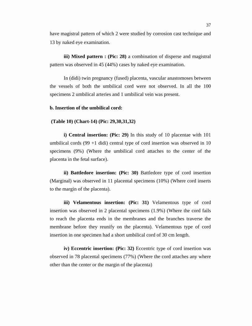
(Table 10) (Chart-14) (Pic: 29,30,31,32)
i) Central insertion: (Pic: 29) In this study of 10 placentae with 101 umbilical cords (99 +1 didi) central type of cord insertion was observed in 10 specimens (9%) (Where the umbilical cord attaches to the center of the placenta in the fetal surface).
ii) Battledore insertion: (Pic: 30) Battledore type of cord insertion (Marginal) was observed in 11 placental specimens (10%) (Where cord inserts to the margin of the placenta).
iii) Velamentous insertion: (Pic: 31) Velamentous type of cord insertion was observed in 2 placental specimens (1.9%) (Where the cord fails to reach the placenta ends in the membranes and the branches traverse the membrane before they reunify on the placenta). Velamentous type of cord insertion in one specimen had a short umbilical cord of 30 cm length.
iv) Eccentric insertion: (Pic: 32) Eccentric type of cord insertion was observed in 78 placental specimens (77%) (Where the cord attaches any where other than the center or the margin of the placenta)

38
V. Umbilical cord: (a) Length of the umbilical cord:
(Table 11 & 14 a) (Pic: 33) (Chart: 14)
In the present study of 100 placentae with 101 cords (99+1) the average
length of the cord is 47.95 cm, range being (30-60) cm. In male conceptus the
average length of the cord is 46.5cm, range (32- 55) cm and in female
conceptus the average length is 49.23 cm, range being (30-60) cm.
The average length of the cord in 69 uncomplicated cases is 48.07 cm
range being (30-60) cm, in male conceptus the average length is 46.54 cm
range (32-55) cm and in female conceptus it is 49.48 (30-60) cm.
The length of the cord in 31 complicated pregnancies averages about
47.68 cm, range (35 -57) cm. In male conceptus the average length is 46.46 cm,
range (35-55) cm and in female conceptus the length is 48.76 (38-57) cm.
Mean umbilical cord length in complicated pregnancies is 57cm in Rh-
isoimmunisation and 43cm in abruptio placentae.
b. Diameter of the umbilical cord:
(Table 12 &14 c) (Pic: 34)(Chart-14)
In the present study the average diameter of 101 cords is 1.132 cm,
range being (0.85-1.6) cm. In male conceptus the average diameter is 1.128 cm,
range being (0.85-1.5) cm and in female it is 1.136 (0.85-1.6) cm.
In the 69 uncomplicated pregnancies the average diameter of the cord is
1.138 cm, range (0.86-1.6) cm. In male conceptus the average diameter of the
cord is 1.120 cm, range (0.86-1.5) cm and in female conceptus the average is
1.156 cm, range (0.87-1.6) cm.
In 31 pregnancies with complication the average diameter of the cord is
1.112 cm, range being (0.85-1.5) cm. In male conceptus the average diameter is

39
1.146 (0.85-1.5) cm and in female conceptus the average is 1.094cm, range
(0.85-1.4) cm. The diameter of the complicated pregnancy is less than the
uncomplicated pregnancies. The umbilical cord diameter is increased in severe
hypertension (1.26cm) and decreased in post maturity (0.91cm). The umbilical
cord diameter of diabetes mellitus and anaemia is 1.2cm and 1.15cm.
VI. Spiral turns (twist) of the umbilical cord:
(Table-13 a &14 c) (Pic: 35)(Chart-14)
In the present study of 100 placentae with 101 cords in 96 (95%)
specimens, sinistral (left) twist of the umbilical cord was observed. In 3
specimens (2%) dextral (Right) twist of the umbilical cord was observed. In 2
specimens (1.9%) compound (Both) twist was observed and in 1 specimen
(0.9%) nil (absent) twist was observed.
1) Umbilical cord coiling index (UCI):
(Table-13 b &14 c) (Chart-14)
This is calculated by the formula -
Number of coils in an umbilical cord -------------------------------------------- = Umbilical coiling index Length of the cord Coils /cm.
In this study the average umbilical coiling index in 100 cases is 0.134
coils/cm.
In 69 complicated cases of pregnancy the average umbilical coiling
index is 0.131 coils/cm.
In 31 complicated cases of pregnancy the average coiling index is 0.139
coils/cm.

40
In complicated pregnancies the average umbilical coiling index is
slightly more than normal cases. In complicated pregnancies the umbilical
coiling index is increased in placenta praevia and minimum in diabetes
mellitus.
2) False knots:
(Table-13 c) (Pic: 36)
In the present study of 100 placentae, 15 false knots were observed.
True knots were not encountered.
2. Histological study of the placenta and the umbilical cord:
1) Normal Human Placenta: (pic: 37)
Human placenta showed chorionic villi with outer synctiotrophoblast
and inner layer of cytotrophoblast. Chorionic villi were separated from each
other by wide intervillous spaces. Blood vessels were seen.
Umbilical cord showed 2 arteries and 1 vein embedded in the amniotic
epithelium. In the vessels tunica intima, media and adventitia were well
appreciated.
2) Hypertension placenta: (pic: 38)
Showed pale villi with numerous syncitial knots and few blood vessels.
Intervillous spaces were reduced and fibrin deposits were observed.
Umbilical cord showed 2 arteries and 1 vein. Tunica media of the
arteries showed hypertrophy.

41
3) Diabetes mellitus: (pic: 39)
Large hypo vascular immature villi with wider intervillous spaces were
observed. Fibrin deposits were also found.
4) Anaemia: (pic: 40)
Villi with reduced intervillous spaces were observed .Few blood vessels
(reduced vascularity) with congestion and dilation of the vessels were noted.
Tunica media of the umbilical artery showed extravasated blood.
3. Corrosion cast technique – Silicon gel cast method:
(Table: 15) (Pic: 41,42,43,44)
By silicone gel cast technique in 5 specimens the vascular pattern of the
placenta was studied. Out of 5 specimens 3 had eccentric type of cord insertion
and 2 specimens had marginal type of insertion of cord. Observations revealed
3 disperse (dichotomous) type of vascular pattern and 2 types of magistral
(monopodial) pattern.
4. Contrast study of the placental vasculature by angiogram method:
(Table: 16) (pic: 45 & 46)
5 fresh specimens (3 had central cord insertion and 2 had marginal cord
insertion) were washed and injected with omnipaque (contrast). The placental
vessels showed a well delineated branching pattern. The branching pattern
was disperse in 3 specimens and magistral in 2 specimens.

42
OBSERVATION ANIMAL PLACENTA (Table-17)
1. Cow Placenta: (pic: 47,48, 57)
Cow placenta was observed to be polycotyledonary in shape. Placenta
measured 13 cm in breadth, 40 cm in length, 0.9 cm in thickness and 4 kg in
weight. Cotyledons were around 80 in number and concave in shape. The
umbilical cord measured 35 cm in length, 1 cm in diameter and contained 4
blood vessels, 2 umbilical arteries and 2 veins with an allantoic duct. No spiral
twist of the cord was observed.
2. Pig placenta: (pic: 49, 50, 58)
The pig placenta was observed to be diffuse in shape. It was fused
placentae of 4 piglets weighing 1.2 kg. Placenta measured averagely 15 cm in
diameter, 35 cm in length, and 1.2 cm in thickness. The placenta on the surface
formed small tufts compared to cotyledons in other group. The umbilical cord
measured 30 cm in length, 0.95 cm in diameter with 2 umbilical arteries, 1 vein
and an allantoic duct. The spiral turns were absent.
3. Sheep placenta: (pic: 51,52,59)
Polycotyledonary shaped sheep placenta was observed in this study.
The sheep placenta was 35 cm in length; 10 cm in breadth, 0.8 cm in thickness
and it weighed 800gms. Convex cotyledons around 60 were observed. The
umbilical cord measured averagely 20 cm in length, 1 cm in diameter with 2
umbilical arteries, 2 umbilical veins and an allantoic duct. There was no spiral
turns present in the cord.
4. Goat placenta: (pic: 53, 54, 60)
In this study Goat placenta was found to be polycotyledonary in shape,
breadth of the placenta was 13 cm, length 30 cm and thickness 0.75 cm. Goat
placenta weighed around 400gms. Cotyledons were convex shaped around 70

43
in number. The length of the umbilical cord measured 18 cm in length, 1cm in
diameter, with 2 umbilical arteries, 2 umbilical veins and an allantoic duct.
Spiral turns were absent.
5. Rabbit: (pic: 55,56,59)
Rabbit like the human placenta is discoidal in shape. The diameter of the
placenta was 5.5 cm and thickness 1.2 cm. Rabbit placenta weighed 5 gms.
Cotyledons were 4 in number. The umbilical cord was short, 2 cm in length
with diameter 0.75 cm. The umbilical cord contained 2 umbilical arteries and 1
umbilical vein. Allantoic duct was not visualized in this specimen. Spiral turns
of the cord were absent.

44
DISCUSSION
I. Shape, diameter and thickness of the placenta
1. Shape of the placenta
a) Circular shape: (Chart: 1) (Table: 18 a)
Leslie. B. Arey (1924), Bradley. M. Patten (1946), T.W. Sadler
(1963), Keith. L. Moore & T.V.N. Persaud (1973), Richard S. Snell (1973)
have mentioned placenta to be circular in shape.
Sarojamma (1986) says 57% placenta to be circular in shape,
Gunapriya. R (2001) reported the same to be 93%.
In the present study of 100 placentae 55 (55%) were circular shaped
which coincides with the statements of the above scientists. The incidence of
percentage is similar to Sarojamma (1986) and varies from Gunapriya R.
(2001).
b) Oval shape: (Table: 18 b)
Leslie B. Arey (1924) stated the placentae could be oval in shape.
Sarojamma (1986) observed 36% oval shaped placentae.
Gunapriya.R (2001) reported 7% oval shaped placenta.
In the present study of 100 placentae, oval shape placentae were observed in 30 specimens (30%). This finding concurs with the statements of the above authors but the percentage value is greater than Gunapriya. R (2001) and less than Sarojamma (1986).
c) Circular or oval shape:
Henry Gray (1858) quoted that the placenta is a flattened discoidal
mass approximately circular or oval in outline.
45
J.D. Boyd and W.J. Hamilton (1970) stated that the placenta is a flat,
round to oval in outline.
Torpin R (1969) mentioned 90% of the placentae were disk like flat
round to oval organ.
In the present study of 100 placentae 55 (55%) were circular in shape, 30 (30%) were oval in shape. This finding agrees with the statements of Henry Gray (1858), Torpin.R (1969) and J.D.Boyd & W.J.Hamilton (1970). The percentage value (55+30 = 85%) is closely similar to Torpin. R (1969).
d) Triangular shape: (Table: 18 c)
Sarojamma (1986) observed 7% triangular shaped placenta in her study of 100 placentae.
In the present study 8 (8%) placental specimens were triangular in shape, which perfectly coincides with the finding of Sarojamma (1986).
e) Placenta succensuriata: (Table: 18 d)
Leslie. B. Arey (1924), Williams (1930), Roth L.G. (1957), J.D. Boyd
& W.J. Hamilton (1970) and Torpin R. (1969) have mentioned about
accessory placentae and Gunapriya. R (2001) quoted 3% incidence of
placenta succensuriata in her study.
In this study 2 (2%) placentae succensuriata was observed. This finding
corresponds with above scientists and the percentage value is close to
Gunapriya R. (2001).
f) Extrachorial placenta - placenta circumvallata: (Chart: 2) (Table: 18 e)
Williams (1930) stated about the placenta circumvallata as one of the abnormal shapes.

46
J.D. Boyd &W.J. Hamilton (1970) quoted in circumvallate form; the
lateral edge of the placenta is undercut at its junction with decidua parietalis.
A double fold of amnion and chorion is present.
Scott J.S. (1960) observed 18.3% of circumvallate placenta,
Wentworth. P (1968) reported 6.5%, J. Bazso (1966) 5.8%, Fox. H & Sen.
D.K. (1972) 2.4%, Wilson D. & Paalman R.J (1967) 1% and Ziel H.A.
(1963) reported 0.62% of circumvallate placenta.
In this present study 1 (1%) circumvallate placenta was observed from
100 specimens with double fold of amnion and chorion. This finding concurs
with the statement of Williams (1930) and J.D. Boyd &W.J. Hamilton (1970).
The incidence is in confirmity with Wilson D & Paalman. R.J. (1967) and is of
higher value than Ziel H.A. (1963) and less than Scott J.S. (1960), J. Bazso
(1966), Wentworth P (1968) and Fox. H& Sen D.K. (1972).
g) Other forms:
Leslie B. Arey (1924) encountered variant forms of placentae rarely
(spindle, pear, heart, crescent, ring) and also mentioned about fused placenta.
In the present study of 100 placentae other variant forms observed were
1(1%) kidney shape, 1(1%) heart shape and 1(1%) fused placentae. This
finding agrees with Leslie B. Arey (1924).
h) Placenta Biparita (Bilobed shape/ Bilobata): (Chart: 3) (Table: 18 f)
Leslie B. Arey (1924), Williams (1930), Bradley .M. Patten (1946), Torpin R. (1969), J.D. Boyd & W.J. Hamilton (1970) have mentioned about placenta biparita, while Augero. O (1957) reported 3.26% and H .Fox (1978) 0.2% of placenta biparita.
47
In the present study of 100 placentae the incidence of placenta biparita is
1 (1%). This observation concurs with statement of the above authors but the
incidence value is greater than H. fox (1978) and less than Augero. O (1957).
2. Diameter of the placenta:
a) Average diameter of the placenta :
(Chart: 4) (Table: 19 a)
The average diameter of the placenta stated by Henry Gray (1858),
J.D. Boyd & W.J. Hamilton (1970) is 185 mm (18.5 cm), Leslie B. Arey
(1924) stated 7 inches (17.5 cm), Gunapriya R. (2001) 17.4 cm, J.P.E.
Judson (1986) 169.7mm, and Richard S. Snell (1973) reported 8 inches
(20cm).
In the present study of 100 placentae, the average diameter of the 100
placentae is 17.73 cm. This finding coincides with Leslie. B. Arey (1924) and
Gunapriya.R. (2001). This finding is elevated than that of J.P.E. Judson (1986)
and less than Henry Gray (1858), J.D. Boyd & W.J. Hamilton (1970) and
Richard.S. Snell (1973). The average placental diameter in 69 uncomplicated
normal pregnancies is 17.97 cm and in other 31 complicated pregnancies it is
17.20 cm. The average diameter of the complicated pregnancies is similar to
the normal uncomplicated pregnancies.
b) Range of placental diameter: (Table: 19 b)
The Range of diameter of the placenta quoted by Henry Gray (1858) is
150-200mm, T.W.Sadler (1963) 15-25cm, Keith L.Moore & T.V.N. persaud
(1973) 15-20 cm and Gunapriya. R (2001) is 12 -22.5 cm.
In the present study of 100 placentae the diameter of the placentae
ranges from 11-28 cm. This findings is close to T.W.Sadler (1963),
Gunapriya.R (2001) and similar to other scientists.

48
The diameter of the placenta in 69 uncomplicated pregnancies ranges
from 14-22 cm and in other 31 complicated pregnancies ranges from 11-28cm,
which has wider range, compared to the uncomplicated pregnancies.
c) Average diameter in Eclampsia:
Sultana. S et al (2007) observed less placental diameter in eclamptic
group compared to normal group in a study of 45 placentae.
In the present study of 100 placentae, the average diameter of the
placentae in severe (Eclampsia) hypertension was found to be 14 cm which is
less than the value of the normal uncomplicated pregnancies 17.97 cm. This
finding is in conformity with Sultana. S et al (2007).
3. Thickness of the placenta:
a) Average thickness of the placenta: (chart: 5) (Table: 20 a)
T.W. Sadler (1963) quoted the average thickness of the placenta to be 3
cm, Leslie, B. Arey (1924) and Richard. S. Snell (1973) stated the average
thickness to be l inch (2.5 cm), Henry Gray (1858) says 23mm, J.D. Boyd &
W.J. Hamilton (1970) and Gunapriya.R (2001) stated 2.1 cm (21 mm) and
K. Benirschke et al (1967) observed it as 2cm.
In the present study the average thickness of the placenta in 100
specimens was observed as 1.9cm. This finding is closely similar to K.
Benirschke et al (1967), J.D. Boyd & W.J. Hamilton (1970), and Gunapriya.R.
(2001) And varies from the rest of the other authors. The average thickness of
the placenta in 69 uncomplicated pregnancies is 1.9 cm and in 31 complicated
pregnancies it is 1.89 cm. The average thickness of the complicated
pregnancies is less than the normal pregnancies.

49
b) Range of thickness of the placenta: (Table: 20 b)
Henry Gray (1858) stated the thickness of the placenta ranges from 10-
40 mm; Keith L. Moore & T.V.N.Persaud (1973) says 2-3 mm, Guna Priya.
R (2001) found 1.5-2.7 cm, Cynthia G. Kaplan (1996) said 2 - 2.5 cm and
Sarojamma (1986) states the thickness ranges from 1.5 - 2 cm.
In the present study the thickness of the placenta ranges from 1 - 3.1 cm.
This finding coincides with Henry gray (1858), Gunapriya. R (2001) and varies
from others.
The thickness ranges from 1 - 3.1 cm in 69 uncomplicated pregnancies
and 1 - 2.8 cm in 31 complicated pregnancies. The thickness range in
complicated pregnancies is slightly less than the uncomplicated pregnancies.
II. Maternal surface-cotyledons:
a) Average cotyledon number: (Table: 21 a)
The average number of maternal cotyledons of the placenta quoted by
various authors like Allan C. Barnes (1968) is 30, Majumdar et al (2005) 19,
Sarojamma (1986) and Gunapriya. R (2001) is 18.
In the present study of 100 placentae, the average number of maternal
cotyledon is 18. This finding is in confirmity with Sarojamma (1986),
Gunapriya. R (2001), Majumdar et al (2005) and less than Allan C. Barnes
(1968).
The average number of cotyledon in 69 uncomplicated pregnancies is 19
and in 31complicated pregnancies are 17. The cotyledon number is less in
complicated pregnancies as compared to normal pregnancies.

50
b) Range of cotyledon Number: (Table: 21 b)
K.Benirschke et al (1967) quoted the cotyledon number ranges from 10
- 40, J.D.Boyd & W.J.Hamilton (1970) says 10-38, Henry Gray (1858) says
15-30, T.W.Sadler (1963) 15-20, Gunapriya. R (2001) stated it as 12 - 24 and
Sarojamma (1986) quoted the range of the cotyledon number as 3 - 24.
In the present study the cotyledon number ranges from 8 - 30. This
finding is similar to Henry Gray (1858) and differs from others.
The range of cotyledon in 69 uncomplicated cases is 12 - 26 and in 31
complicated cases is 8 - 30. The cotyledon number has a wider range in
uncomplicated pregnancies compared to normal pregnancies.
c) Cotyledon number in Hypertension: (Table: 21 c)
Majumdar et al (2005) observed mean number of cotyledons in
hypertension group to be 18. Sultana S. et al (2007) reported less cotyledon
number in eclamptic group when compared to normal group.
In the present study the mean number of cotyledon in hypertension
group is 14. (Mild-17, moderate-14, and severe (eclampsia)-11). The mean
number of cotyledon in normal group (69 uncomplicated cases) is 19. The
mean number of cotyledon in hypertension group is less than the normal group.
This finding corresponds with Majumdar et al (2005) but the value is less
compared to them. The mean cotyledon number in eclamptic group is 11,
which is less than the normal group 19. This finding is in confirmity with
Sultana S. et al (2007)

51
III. WEIGHT OF THE PLACENTA:
a) Average weight of the placenta: (Chart: 6) (Table: 22 a)
The average weight of the placenta quoted by Lurie. S et al (1999) is
613 gms, Kucuz M Doymaz. F (2008) is 610 gms, Swanson. LD, Bewtrac. C
(2008) is 537 gms and Saigal. S & Srivastava J.R. (1970) is 531.5 gms.
Majumdar et al (2005) quotes the average weight to be 485.85 gms,
Shameer Singh et al (1979) 475 gms, Henry gray (1858) 470 gms, Wong
T.C. & J.A.P. Lartour (1966) 463 gms, Fujikura et al (1970) 448 gms and
Rath G et al (2000) says it is 382.148 gms.
In the present study the average weight of the placentae in 100
specimens is 472.49 gms. This finding is similar to Henry Gray (1858),
Shameer Singh et al (1979) and Majumdar et al (2005). This finding is greater
than Wong T.C., & J.A.P.Lartour (1966), Fujikura et al (1970), Rath. G et al
(2000) and less than Lurie. S et al (1999), Kucuz M; Doymaz. F. (2008), Saigal
S & Srivastava. J.R. (1970) and Swanson LD, Bewtrac.C (2008).
The average weight of placenta in 69 uncomplicated pregnancies is
490.58 gms and in 31 complicated pregnancies it is 432.74 gms. Average
weight in complicated cases in less than the normal uncomplicated group.
b) Range of Placental Weight: (Table: 22 b)
The range of the placental weight quoted by various authors is
Henry Gray (1858) 200-800 gms, Wiggles Worth. J.S. (1962) 360-570 gms
and Hoseman. H (1949) 400-1000 gms.
In the present study of 100 placentae the placental weight ranges from
(150-850) gms. This finding is similar to Henry Gray (1858) and varies from
other authors.

52
The placental weight ranges from 375-675 gms in 69 uncomplicated
pregnancies and 150-850 gms in 31 complicated pregnancies. Complicated
cases have a wider range of placental weight compared to the normal cases.
c) Placental weight of Male and Female mature conceptus:
(Table: 22 c)
Gosh L.V. & Chandrasekhar. C (1948) reported the average weight of
the placenta in mature male and female conceptus to be 486.3 gms and 479
gms.
S.P.Gupta et al (1972) reported the same to be 521.9 gms and 510.4
gms. He also stated that the male placentae are heavier than the female
placentae.
In the present study of 100 placentae, the average weight of the placenta
in mature male and female conceptus is 468.29 gms and 476.69 gms. This
finding is closely similar to Gosh. L.V.& Chandrasekhar. C (1948) and varies
from S.P.Gupta et al (1972) and similarly the female placentae in this study
weigh more than the male placentae.
The average placental weight in male and female placenta in 69
uncomplicated pregnancies is 487.42 gms and 493.75 gms, which are higher
than the average placental weight of male and female conceptus in 31
complicated pregnancies, which is 423.21 gms, and 440.5 gms.
Female placentae weigh more than the male placentae.
d) Placental weight in Relation with fetal weight:
Adair.F.L. &Thealander.H (1946) stated the normal placental weight
is about one seventh the weight of the conceptus.

53
Keith L. Moore & T.V.N. Persaud (1973) quoted that the placental
weight to be about one sixth the weight of the average conceptus.
In the present study the average weight of the placenta and conceptus is
472.49 gms and 3.13 kg. Placental weight is 1/6.6 the weight of the conceptus.
This finding coincides with Adair.F.L & Thelander.H (1925) and is slightly
more than Keith L. Moore & T.V.N.Persaud (1973). The placental weight in 69
uncomplicated pregnancies is 1/6.6 of the weight of the conceptus and in 31
complicated pregnancies it is 1/6.3 of the weight of the conceptus.
e) Feto placental (F: P) Ratio in Normal cases: (Table: 22 d)
Lurie. S et al (1999) observed feto placental ratio as 5.6 in his study and
mentioned it did not vary much in male (5.7) and female (5.6)
Majumdar et al (2005) reported feto placental ratio to be 5.89 in
normal pregnancies.
In the present study the average fetoplacental ratio is 6.6. It is 6.6 in
male and 6.7 in female. This finding is greater than the both the authors. Male
and female feto placental ratio does not vary much and this finding agrees with
Lurie. S et al (1999). Fetoplacental ratio in 69 uncomplicated pregnancies is 6.7
and it is 6.5 in 31 complicated pregnancies. They also do not differ much.
f) Feto placental ratio is Hypertension: (Table: 22 e)
Kher A.V and Zawar M.P. (1981) reports reduction in fetoplacental
(F: P) ratio in toxemia of pregnancy, Damania K.R. et al (1989) states that F:
P ratio is less in hypertensive pregnancies when compared to the normal
uncomplicated pregnancies.
Majumdar et al (2005) observed F: P ratio 6.23 in hypertensive
pregnancies as compared to 5.89 in norm tension pregnancies.

54
In the present study the fetoplacental ratio in normal pregnancies is 6.6.
In mild, moderate and severe hypertension feto-placental ratio is 6.4, 6, and
6.98.The average F: P ratio in hypertension is 6.47, which is less than the
normal uncomplicated pregnancies. This finding corresponds with Damania
K.R. et al (1989).
The F: P ratio in severe hypertension is greater than the normal. The
value and the finding coincide with Majumdar et al (2005) and vary from Kher
A.V.& Zawar M.P (1981). Feto-placental ratio in premature, post mature
placenta is 6.6, 6.3 and in diabetes mellitus it is 6.7.
g) Placental coefficient in normal pregnancies: (Table: 22 f)
Little (1960) reported the placental coefficient <0.08 and >0.2 as
abnormal. Kucuk. M. Doymaz. F (2008) stated the placental coefficient to be
0.18 in normal cases.
In the present study the placental coefficient is 0.15. This value is within
0.08-0.2 as stated by Little (1960). This finding coincides with Little (1960).
The observed value is less than Kucuk. M. Doymaz. F (2008). The placental
coefficient in 69 uncomplicated pregnancies is 0.14, which is less than the
value of 31 complicated pregnancies 0.16.
h) Placental coefficient in Diabetes Mellitus: (Table: 22 f)
Kucuk. M. Doymaz F (2008) stated the placental coefficient in
gestational diabetes mellitus to be 0.21.
In the present study the average placental coefficient in 2 cases of
diabetes mellitus is 0.14. This is less than the value of Kucuk.M Doymaz.
F (2008).

55
i) Placental weight in Hypertension pregnancies:
(Chart-7a) (Table: 22 g)
Damania K.R. et al (1989) mentioned the placental weight to be less in hypertension pregnancies as compared to the normal pregnancies.
Rath G. et al (2000) observed mean placental weight in mild, moderate and severe hypertension as 351.308 gms, 338.024 gms and 332.410 gms.
Majumdar et al (2005) reported the mean placental weight in hypertension as 399.10 gms.
In the present study the average placental weight observed in mild hypertension (5 cases), moderate (3 cases) and severe hypertension (3 cases) is 475 gms, 375 gms and 316 gms. The mild and moderate hypertension value is greater than Rath G. et al (2000) and the severe hypertension value is less than Rath G. et al (2000).
The average weight of the hypertensive placenta is 388.6 gms, which is
less than the weight of the normal pregnancies (490.58 gms). This finding
coincides with Damania K.R. et al (1989) and Majumdar et al (2005).
j) Placental weight in Diabetes Mellitus: (Table: 22 h)
N.A. Beischer et al (1968) observed approximately that the weight of the placenta to be more than 900 gms in diabetic pregnancy.
Kucuz. M, Doymaz. F (2008) stated the average placental weight from Gestational diabetes mellitus to be 694.8 gms.
In the present study the mean placental weight in 2 specimens from
gestational diabetes case was observed as 587.5 gms. This value is closer to
Kucuz .M. Doymaz .F (2008) and differs from N.A. Beischer et al (1968).

56
k) Placental weight in Anaemia: (Table: 22 i)
Dockery J.L (1960) reported a placental specimen of weight 1,984 gms
from anaemia complicating pregnancy with Hb% 4.1.
Singla P.N. et al (1978) observed the placental weight to be reduced in
severe anaemia and the quoted that it is related directly to the hemoglobin
levels.
N.A. Beischer et al (1968) reported 2% of the placentae weigh more
than 900 gms in 420 cases of anaemia.
Godfrey KM et al (1991) mentioned that anaemic cases have larger
placental weight with low hemoglobin value.
In the present study of 100 placentae, 2 placentae obtained from anaemia pregnancies with hemoglobin percentage 8 and 7 measured mean weight of 387.5 gms which was less compared to the normal weight of 490.58 gms in 69 uncomplicated pregnancies. This finding is similar to Singla. P.N. et al (1978) and varies from Dockery J.L (1960), N.A.Beischer et al (1968) and Godfrey KM et al (1991)
l) Placental weight in Rh-isoimmunisation: (Table: 22 j)
N.A. Beischer et al (1968) reported erythroblastosis placental weight to be approximately more than 900 gms.
In the present study the average weight of the placental specimen from Rh-ve mother with Rh+ve baby without complications was observed to be 400 gms, which is much less than the author.

57
m) Placental weight in premature male and female conceptus:
(Chart-7b) (Table: 22 k)
Gosh L.V. & Chandrasekhar (1946) stated that the placental weight of
the male and female premature conceptus is 391.1 gms and 379.1 gms
S.P.Gupta et al (1972) reported the same to be 449.9 gms and 405.3
gms. He also stated that the male placentae are heavier than female placentae.
In this study of 100 placentae, in 6 premature placentae the average
placental weight in 2 male and 4 female conceptus is 237.5 gms and 341.25
gms.
This finding differs from both the authors and in this study premature
weight of the placenta in the female is greater than that of the male.
n) Placental weight in post mature placenta: (Table: 22 l)
Saigal. S & Srivastava J.R. (1970) observed the average placental
weight at 42 wks and above to be 475.9 gms.
In the present study the average placental weight in 3 post mature
placentae is 583.5 gms. This weight is much higher than the finding of Saigal.
S & Srivastava J.R. (1970).
o) Weight of the placenta in bipartite (bilobed) placenta: (Table:22 m)
Fujikura et al (1970) stated the weight of 4.2% of bipartite placenta to
be 473 gms.
In the present study 1 (1%) placenta biparita was observed with
placental weight 475 gms. This finding is in confirmity with Fujikura et al
(1970).

58
IV) Foetal surface: a) vascular pattern of the placenta:
Shordania. J (1929) mentioned about the disperse and magistral type of
vascular pattern. He also stated that centrally attached cords have disperse
pattern. P.Bascish& C.F.V.Smout (1937) also observed magistral and
disperse type. Andrade. A (1969) quoted disperse type was frequently
associated with centrally inserted cords. Z.Gordon et al (2007) mentioned a
combination of dichotomous (disperse) and monopodial (magistral) pattern. He
stated dichotomous (disperse) pattern was seen in centrally inserted cords and
monopodial (magistral) pattern in marginal inserted cords.
In present study of 100 placentae (99+1 didi) disperse type was
observed in 41 specimen (40%) in which central insertion of cord was observed
in 9% and Eccentric cord insertion in 30%. Magistral type was observed in 15
specimen (14%) with 11(10%)marginal cord insertion and 4(3%) with
eccentric cord insertion. Mixed type was seen in 45 (44%) specimens with
eccentric type of cord insertion. This finding coincides with Shordania J.
(1929), P.Basich and C.F.V.Smout (1937), and Z.Gordon et al. (2007) and is
less similar from Andrade. A (1969) as disperse type is also seen in eccentric
cords apart from centrally inserted cords.
Kishore. N & Sarkar. SC (1967) observed disperse type in 61.8% but
in the present study disperse type was observed only in 40% which varies from
Kishore N & Sarkar.sc (1967). (Table: 23 a)
Bhargava.I &Raja P.T.K. (1969) quoted that foetal veins occasionally
cross-fetal arteries and it is correlated with abnormal fetal development.
Paul Wentworth (1965) reported 3.1% fetal veins crossing foetal
arteries. In the present study of 100 placentae 1 specimen (1%) showed foetal
veins crossing arteries and it was observed with normal development of the
foetus. (Pic: 56)(Table 23 b)
59
Foetal surface: b) Insertion of the umbilical cord:
1) Central type: (Chart:8) (Table: 24 a)
Sarojamma (I986) observed 40% central type of cord insertion, Earn
A.A. (1951) in 28%, Krone H. A. (1961) in 25% and J. P.E.Judson (1986) in
20%.
Eastman N.J.& Hellmann L.M. (1966) reported in 18% central type,
Hyrtl.J (1870) in 16%, Shanklin D.R (1958) in 11%, Gunapriya.R (2001)
5% central type and Chiari et al (1895) observed 3.3% central type of cord
insertion.
In the present study of 100 specimens (99+1(didi) with 101 cords) in 10
specimens (9%) central type of cord insertion was observed. This incidence
value is similar to Shanklin D.R. (1958), higher than Gunapriya R. (2001) and
Chiari et al (1895). This value is less than the other authors.
2) Battle Dore (marginal) Type: (Chart: 9) (Table: 24 b)
The following are authors who observed battledore (Marginal) type of
cord insertion - Hyrtl. J (1870) in 19%, Earn A.A (1951) in 15%, Monie I.W
(1965) 14.7%, Krone H.A. (1961) in 10%, and J. P.E.Judson (1986) in 10%.
Gunapriya. R (2001) reported marginal type in 9%, Eastman N.J. and
Hellmann L.M (1966) in 7%, S.Brody & D.A.Frenkel (1953) in 6.2%
Uyanwah et al (1977) in 5.6%, Chiari et al (1895) in 5%, and Thomas.
J. (1963) in 5.2%.
Scott J.S (1960) observed 2% marginal type insertion, Sarojamma
(1986) 2%, Shanklin D.R (1958) 1.9% and Robinson et al (1983) reported in
1.5% marginal type of cord insertion.

60
In the present study 13 specimens (12%) were observed to have
battledore/marginal type of cord insertion in 101 cords. This incidence value is
similar to Krone. H.A. (1961), J.P.E.Judson (1986) and is less than Earn A.A.
(1951), Monie I.W (1965), and higher than the other authors.
3) Velamentous type of cord insertion: (Chart: 10) (Table: 24 c)
Monie I.W (1965) reported 15.3% velamentous type of cord insertion;
Sarojamma (1986) mentioned the same to be 4%.
Uyanwah et.al (1977) observed 1.6%, Robinson et al (1983), Scott J.S
(1960) each reported 1.5%, Thomas. J (1963) 1.3%, Eastman N.J. &
Hellmann L.M (1966) 1.25% of velamentons type of Cord insertion.
Noldeke.H (1934), Earn A.A (1951), Diterlizzi.G & Rossi.G.F (1955)
and Krone.H.A (1961) each reported 1% of velamentous type of Cord
insertion, Lefevre.G (1896) stated 0.84%, Shanklin J.R. (1958) 0.78%,
Greico. A. (1936) 0.4%, Scheffel T. & Langanke. D (1970) 0.22% and Cork
ill T.F (1961) found 0.024% of velamentous type of cord insertion.
In the present study of 100 placentae in 2 specimens (1.9%)
velamentous type of cord insertion was observed. This finding is almost similar
to Uyanwah et al (1977), Robinson et al (1983), Scott J.S (1961) and it is less
than Monie.I.W (1965) and Sarojamma (1986). This incidence value is greater
than other scientist.
4) Eccentric type of Cord insertion: (Chart: 11) (Table: 24 d)
Eccentric type of Cord insertion was found by Chiari et al (1895) in
91.2%, Shanklin J.R (1958) in 89%, Gunapriya R (2001) in 86%, Eastman
N.J&Hellmann L.M (1966) in 73%, Monie.I.W (1965) in 70%, J.P.E.Judson
(1986) in 80% and Krone H.A (1961) in 64%.
61
Earn A.A (1951) reported Eccentric type in 56%, Hyrtl. J (1870) in
54% and Sarojamma (1986) in 53%.
In the present study eccentric type of cord insertion was observed in 76
specimens (75%). This finding is closely similar to Eastman N.J.& Hellmann
L.M. (1966), Monie I.W (1965), J. P.E.Judson (1986). It is less than Chairi et al
(1895), Shanklin J.R (1958), Gunapriya R. (2001) and it is higher than the
other authors.
5) Insertion of cord in Bilobed Placentae:
Torpin R. & Barfield W.E. (1968) observed that in 1/3rd of the bilobed
placenta the cord inserts on the larger lobe and in 2/3 it has velamentous type
of insertion.
In the present study of 100 placentae 1 case (1%) of placenta biparita
was observed in which cord inserts on the larger lobe, which coincides with
Torpin R. and Barfield W.E. (1968).
Nordenvall. M et al. (1988) reported marginal type of cord insertion in
extrachorial and bilobed placentae. In the present study 1 specimen of
extrachorial placenta and 1 specimen of bilobed placentae was observed of
which only the bilobed placentae was associated with marginal (battledore)
type of cord insertion and extrachorial had central type of cord insertion.
6) Foetal Blood vessels of the umbilical cord:
Edith.L.Potter (1952) and Keith.L.Moore & T.V.N.Persaud (1973)
observed 2 umbilical arteries and 1 umbilical vein.
Stephen A. Heifetz (1996) quoted super numerarary vessels are less
common than single umbilical artery. H. Fox (1997) stated increased vascular
profiles in stillbirth associated with maternal cigarette smoking.

62
In the present study of 100 placentae in all the specimens (including
intrauterine death) 2 umbilical arteries and 1 umbilical vein was observed. No
case of single umbilical artery or supernumerary vessels was observed.
V) Length and diameter of the umbilical cord:
1. Length of the umbilical cord:
a) Average of the length of the umbilical cord:
(Chart: 12) (Table: 25 a)
The average length of the umbilical cord quoted by Fog J. (1930) is 59.6
cm, Shordania. J (1929) is 60 cm, Mossinger et al (1986) is 57 Cm, Williams
(1930) is 55 cm, Gardiner J.P (1922) is 55 cm, Gunapriya.
R (2001) is 53.5 cm, Henry gray (1858) is 50 cm, Edith.L.Potter (1952) is 50
cm, and Sarojamma (1986) is 38.5 cm.
In the present study the average length of the umbilical cord is 47.95 cm
in 100 specimens. This finding is closer to Henry Gray (1858), Edith. L. Potter
(1952) and higher than Sarojamma (1986). This value is less than the other
authors.
Fog. J. (1930) quoted the average length of the cord in term pregnancy is
60.7 cm. The average length of the umbilical Cord in 69 uncomplicated
pregnancies is 48.06 cm, which is less than Fog. J (1930). The average length
of the cord in 31 complicated pregnancies is 47.71cm, which is less than the
uncomplicated pregnancies.
b) Range of the Length of the umbilical cord: (Table: 25 b)
The range of length of the umbilical cord quoted by Henry gray (1858)
is 20-120 cm; Sarojamma (1986) is 25-85 cm, Gunapriya. R (2001) is 30-70
cm, Williams (1930) is 30-100 cm, Shordania. J (1929) is 35-104 cm;
Schmidth-Elmendorf (1952) is 50-70 cm and Fox. H (1997) is 54-61 cm.

63
In the present study the length of the umbilical cord ranges from 30-60
cm, which coincides with Gunapriya. R (2001) and varies from others. The
length of the cord ranges from 30-60 cm in 69 uncomplicated pregnancies and
35-57 cm in 31 complicated pregnancies, which is similar to the normal
uncomplicated pregnancies.
c) Short length of the umbilical cord:
Gardiner J.P. (1922) states cord length < 32 cm as absolutely short. In
the present study one specimen with cord length < 32 cm was observed which
measured 30 cm in length with velamentous type of cord insertion.
W.F. Rayburn et al (1981) quotes that cord length less than 35 cm as
short. In the present study 2 cases of short cord with length 30 cm and 32 cm
was observed.
Mossinger et al (1986) quoted infants with Down’s syndrome have shorter cords (45.1 cm). In the present study no case of Down’s syndrome was observed.
d) Length of the cord in Male and Female conceptus: (Table: 25 c)
Gunapriya. R (2001) quoted the average length of the cord in male conceptus is 54.6 cm and in female conceptus is 52.1 cm.
In the present study of 100 placentae the average length of the cord in male conceptus is 46.54 cm and in female is 49.23 cm. The length of the cord in female conceptus is longer than the male conceptus, which differs from Gunapriya. R (2001).
In 31 complicated pregnancies the average length of the cord in male and female conceptus is 46.6 cm and 48.70 cm, which is similar to the normal uncomplicated pregnancies.

64
e) Length of the cord in preterm conceptus: (Table: 25 d)
Fog. J (1930) stated the length of the cord in preterm conceptus to be 54.8 cm, which is less than the term value, which is 60.7 cm.
In the present study also the average cord length of the preterm conceptus (43.1 cm) is shorter than that of normal term pregnancies 48.06 cm as quoted by Fog. J. (1930).
f) Length of the cord in Breech presentation: (Table: 25 e)
Soernes T. & Bakke T. (1986) quoted the length of the cord in breech presentation in male and female conceptus to be 53.78 cm and 52.51cm.
In the present study 1 placental specimen (female conceptus) was obtained through breech delivery. The cord length was 41 cm, which is less than the value of Soernes. T. & Bakke. T. (1986).
2. Diameter of the umbilical cord: (Table: 26)
Diameter of the umbilical cord stated by Henry Gray (1858), Shordania. J (1929) and Edith. L. Potter (1952) each ranges from 1-2 cm (10-20 mm), schmidt-Elemendorf (1952) says 15-20mm, Williams (1930) stated 0.8-2 cm and Gunapriya. R (2001) quoted it as 0.6-1.1 cm.
In the present study the diameter of the placenta in 100 specimens ranges from 0.85 - 1.6cm, which coincides with Williams (1930) and differs from others. The range of the umbilical cord diameter in 31 complicated pregnancies is (0.85-1.5cm) similar to 69 normal uncomplicated pregnancies (0.86-1.6 cm).

65
VI) Spiral Twist(Turns) of the umbilical cord:
a) Sinistral twist (Left) : (Chart-13) (Table: 27 a)
The percentage of sinistral twist quoted by Ronald. V. Lacro (1987) is 83, Henry. W. Edmonds (1954) is 82, Read. W (1860) is 77 and Neugebauer L.A (1858) is 71.
In the present study of 100 placentae (99+1(didi) with 101cords) sinistral twist was observed in 96 specimens (95%), which is higher than the value of the above authors.
b) Dextral twist (Right) : (Table: 27 b)
The percentage of dextral (right) twist reported by various authors like
Neugebauer. L.A (1858) is 24, Read. W (1860) 14, Henry W.Edmonds
(1954) 12 and Ronald. V. Lacro (1987) 12.
In the present study dextral (right) twist was observed in 3 specimens
(2%), which is much less compared to the above authors.
c) Compound twist (Right and Left) : (Table: 27 c)
Read. W (1860) stated 1.85% incidence of compound twist.
In the present study 2 specimens of compound twist (1.9%) was observed which coincides with Read. W (1860).
d) Nil Twist: (Table: 27 d)
The percentage of nil twist (absent twist) observed by various authors like Henry. W.Edmonds (1954) is 8, Ronald. V.Lacro (1987) 5, Thomas et al (1993) 4.3, Neugebauer. L.A (1858) 4.3 and Read.W (1860) 1.8.

66
In the present study 1 specimen (0.9%) had no twist and it was shorter in length with velamentous cord insertion. This value is much less when compared to the above authors.
e) Umbilical coiling index : (Table: 27 e)
Rana et al (1995) reported the average umbilical coiling index in their study of 635 placentae was 0.19 coils/cm
In this study of 100 placentae the average umbilical coiling index is 0.134 coils /cm. This value is less similar to Rana et al (1995).
67
DISCUSSION (Animal Placenta)
Domestic cattle - cow : (Table: 28 a)
Wild (1964):
Stated the cotyledons in cow placenta ranges from 80-120.
In the present study the cow placenta contained 80 cotyledons, which is
similar to Wild (1964).
KurtBenirschke (2007):
Quoted cow placenta weigh 4 - 5 kg with 4 blood vessels and no spiral
twist in the umbilical cord.
In the present study the cow placenta weighed 4 kg and the cord
contained 4 blood vessels with absent spiral twist, which concurs with findings
of KurtBenischke (2007).
Domestic Pig: (Table: 28 b)
Stephens J. Roberts (1971):
Says the pig placenta is diffuse in type.
In the present study 1 fused pig placenta with no discrete cotyledon was
observed which corresponds with findings of Stephen. J. Roberts.
KurtBenirschke (2007):
Quotes that the pig placenta contains 3 blood vessels with a patent
allantoic duct.

68
In the present study the umbilical cord of the pig placenta measured
around 40 cm, with 3 blood vessels and an allantoic duct, which matches with
the findings of KurtBenirschke (2007).
Sheep Placenta: (Table: 28 c &d)
Kleeman et al (2001):
Reported the weight of the sheep placenta to be around 600 gms.
In the present study the weight of the sheep placenta is 800 gms, which
is greater than Kleeman et al (2001).
KurtBenirschke (2007):
Stated the polycotyledonary sheep placenta contained around 60-100
cotyledons. Umbilical cord measured 27 cm in length with 4 blood vessels and
an allantoic duct.
In the present study the sheep placenta is polycotyledonary in shape with
60 cotyledons. The length of the cord is 20 cm with 4 blood vessels and an
allantoic duct, which is similar to KurtBenischke (2007).
Reynolds S.R.M. (1952):
Quoted umbilical cord of the sheep had no spirals.
In the present study also spirals were absent in the umbilical cord.
Goat placenta: (Table: 28 e)
KurtBenirschke (2007):
States that goat placenta weigh around 200 gms. The umbilical cord
measured 10 cm in length, 1 cm in diameter with 4 blood vessels and an
allantoic duct with absent spirals of the umbilical cord.

69
In the present study the goat placenta weighed around 400 gms and the
length of the umbilical cord measured 17cm, which differs from Kurt
Benirschke (2007). The umbilical cord without spirals measures 0.95cm in
diameter with 4 blood vessels and an allantoic duct, which is similar to Kurt
Benirschke (2007).
Domestic Rabbit: (Table: 28 f)
Martensson.L (1984):
Stated the average placental weight of the rabbit to be 4 g.
In the present study the rabbit placenta weighed 5 gms, which is similar
to Martensson.L (1984).
KurtBenirschke (2007):
Stated that rabbit umbilical cord measured 2 cm in length, with absent
spirals and contained within it 2 umbilical arteries and 1 umbilical vein.
In the present study the umbilical cord measured 2 cm in length with 2
umbilical arteries, 1 vein, and with absent spirals. This finding agrees with Kurt
Benirschke (2007).
70
CONCLUSION
In 100 placentae the circular shape was the common form (55%) and
oval shape was the next common (30%). Triangular shape (8%),
succensuriata (2%), circumvallata (1%), bilobedplacenta (1%), kidney
shape (1%), heart shape (1%) and fused placenta (1%) were also
observed in this study.
The mean diameter of the placenta in 100 specimens is 17.73 cm. The
mean diameter of the placenta in complicated pregnancies (17.20 cm) is
similar to the uncomplicated pregnancies (17.97 cm). The mean
placental diameter is increased in diabetes mellitus and decreased in
anaemia and hypertension.
The average thickness of the placenta in the present study in 1.9 cm. The
thickness of the placenta in complicated pregnancies (1.89 cm) is
slightly less than the uncomplicated pregnancies (1.9cm). The thickness
of the diabetes mellitus placenta (2.6cm) is greater than the
uncomplicated pregnancies and the thickness in hypertension and
anaemia is less than the uncomplicated pregnancies (1.5cm).
The average cotyledon number in the present study is 18, in normal
uncomplicated pregnancies it is 19 and in complicated pregnancies it is
17. The cotyledon number is increased in diabetes mellitus (19) and
reduced in hypertension (14). In anaemia ill-defined cotyledons were
observed.
The average weight of the placenta in 100 cases is 472.49 gms, in
complicated pregnancies it is 432.74 gms and in uncomplicated
pregnancies it is 490.58 gms. In diabetes mellitus (587.5 gms), weight of
the placenta is greater than the uncomplicated pregnancies (490.58 gms).

71
The weight of the placenta in hypertension (388.66 gms) and anaemia
(387.5 gms) is less than the uncomplicated pregnancies (490.58 gms).
The mean feto-placenta ratio in this study is 6.6 (Complicated
pregnancies – 6.5 and uncomplicated pregnancies – 6.7). The feto
placental ratio is not much altered in anaemia (6.3), hypertension (6.4)
and diabetes mellitus (6.7) when compared to uncomplicated
pregnancies.
The placental coefficient in this study is 0.15 (complicated-0.16 and
uncomplicated is 0.14). The placental coefficient is increased in factors
complicating pregnancy such as hypertension (0.15), anaemia (0.15) and
not altered in diabetes mellitus (0.14) when compared to normal
pregnancies.
The average weight of the conceptus in the present study is 3.16 kg
(complicated – 2.6 kg and uncomplicated – 3.28 kg). The average
weight of the conceptus is reduced in factors complicating pregnancy
like intrauterine death, anaemia, and hypertension and increased in
diabetes mellitus.
In the present study the vascular pattern of the placenta observed were
disperse type 40%, magistral 14% and mixed type 44%.
In the present study the type of cord insertion observed was central 9%,
battledore (marginal) 12%, velamentous type 1.9% and eccentric type
75%, which is the most common in the present study.
In all the 100 specimens 2 umbilical arteries and 1 umbilical vein was
present.
The average length of the umbilical cord is 47.95 cm. The length of the
cord in complicated pregnancies is 47.71 cm, which is shorter compared
to the normal pregnancies 48.06 cm.

72
The average diameter of the umbilical cord in the present study is 1.132
cm (complicated pregnancies- 1.112 cm, uncomplicated pregnancies-
1.138 cm).
In the present study dextral twist was observed to be (right) 2%,
compound twist 1.9%, nil twist 0.9%, and sinistral twist (Left) was 95%
which was most commonly observed.
In the present study 15 % of false knots were observed.
Histological study –In hypertensive placenta histology showed
numerous syncytial knots, hypermature villi with medial hypertrophy of
the blood vessel. Diabetic placenta showed hypovascular large villi with
fibrin deposits. Placenta in anaemia showed small villi with narrow
intervillous spaces.
Silicone gel cast technique revealed 3 disperse type and 2 magistral type
of vascular pattern.
Contrast study of placental vasculature by angiogram method revealed 3
disperse type and 2 magistral type of vascular pattern.
Sheep, goat and cow placentae have cotyledonary type of placenta
where as pig has diffuse type of placenta. Only rabbit has discoidal
placenta similar to the humans. Weight of the cow placenta is heavier
than the human placenta.
Morphology and morphometric study of the placenta and umbilical cord
with vascular pattern will be definitely useful to the vascular surgeons
operating intrauterinely to correct the congenital defects, and the
variations observed will be useful to the respective clinicians and
anatomist, who are doing further research in this field. Comparative
anatomy done in this study will help the Veterinary doctors in their
research.
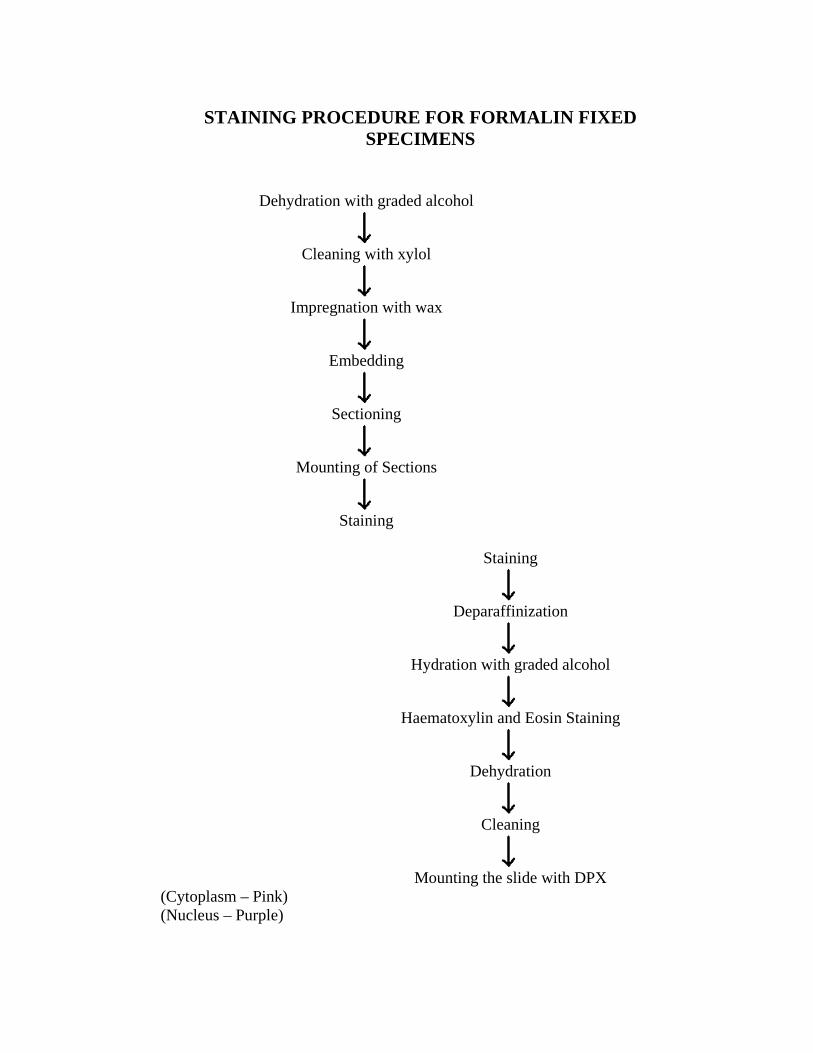
STAINING PROCEDURE FOR FORMALIN FIXED SPECIMENS
Dehydration with graded alcohol
Cleaning with xylol
Impregnation with wax
Embedding
Sectioning
Mounting of Sections
Staining
Staining
Deparaffinization
Hydration with graded alcohol
Haematoxylin and Eosin Staining
Dehydration
Cleaning
Mounting the slide with DPX
(Cytoplasm – Pink) (Nucleus – Purple)
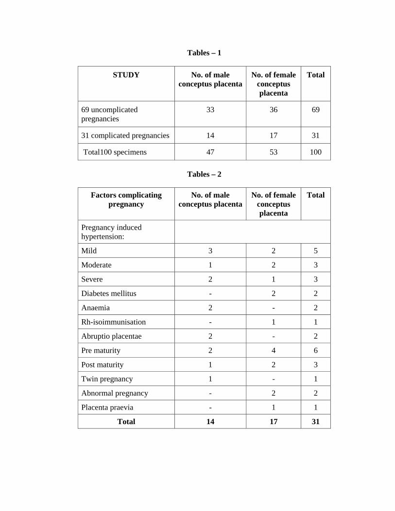
Tables – 1
STUDY No. of male conceptus placenta
No. of female conceptus placenta
Total
69 uncomplicated pregnancies
33 36 69
31 complicated pregnancies 14 17 31
Total100 specimens 47 53 100
Tables – 2
Factors complicating
pregnancy No. of male
conceptus placentaNo. of female
conceptus placenta
Total
Pregnancy induced hypertension:
Mild 3 2 5
Moderate 1 2 3
Severe 2 1 3
Diabetes mellitus - 2 2
Anaemia 2 - 2
Rh-isoimmunisation - 1 1
Abruptio placentae 2 - 2
Pre maturity 2 4 6
Post maturity 1 2 3
Twin pregnancy 1 - 1
Abnormal pregnancy - 2 2
Placenta praevia - 1 1
Total 14 17 31
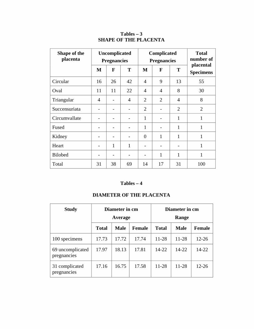
Tables – 3 SHAPE OF THE PLACENTA
Uncomplicated
Pregnancies Complicated Pregnancies
Shape of the placenta
M F T M F T
Total number of placental
Specimens
Circular 16 26 42 4 9 13 55
Oval 11 11 22 4 4 8 30
Triangular 4 - 4 2 2 4 8
Succensuriata - - - 2 - 2 2
Circumvallate - - - 1 - 1 1
Fused - - - 1 - 1 1
Kidney - - - 0 1 1 1
Heart - 1 1 - - - 1
Bilobed - - - - 1 1 1
Total 31 38 69 14 17 31 100
Tables – 4
DIAMETER OF THE PLACENTA
Diameter in cm Average
Diameter in cm Range
Study
Total Male Female Total Male Female
100 specimens 17.73 17.72 17.74 11-28 11-28 12-26
69 uncomplicated pregnancies
17.97 18.13 17.81 14-22 14-22 14-22
31 complicated pregnancies
17.16 16.75 17.58 11-28 11-28 12-26

Tables – 5
THICKNESS OF THE PLACENTA
Thickness in cm Average
Thickness in cm Range
Study
Total Male Female Total Male Female
100 specimens 1.9 1.9 1.9 1-3.1 1-3 1-3.1
69 uncomplicated pregnancies
1.93 1.96 1.9 1-3.1 1-3 1.2-3.1
31 complicated pregnancies
1.8 1.7 1.9 1-2.8 1-2.5 1-2.8
Tables – 6
MATERNAL SURFACE -COTYLEDON
Cotyledon number Average
Cotyledon number Range
Study
Total Male Female Total Male Female
100 specimens 18 18 18 8-30 8-30 9-28
69 uncomplicated normal pregnancies
19 19 19 12-26 13-25 14-26
31 complicated pregnancies
17 17 17 8-30 8-30 9-28

Tables – 7
WEIGHT OF THE PLACENTA
Placental weight in (gms)
Average
Placental weight in (gms)
Range
Study
Total Male Female Total Male Female
100 specimens 472.49 468.29 476.69 150-850 150-850 275-675
69 uncomplicated normal pregnancies
490.58 487.42 493.75 375-625 400-625 375-575
31 complicated pregnancies
432.74 423.21 440.5 150-850 150-850 275-675
Tables – 8 a
WEIGHT OF THE CONCEPTUS
Weight of the conceptus in (Kg)
Average
Weight of the conceptus in (Kg)
Range
Study
Total Male Female Total Male Female
100 specimens 3.16 3.005 3.247 .475-4 0.475-3.9 2.1-4
69 uncomplicated normal pregnancies
3.28 3.13 3.4 2.2-3.8 2.2-3.75 2.4-3.8
31 complicated pregnancies
2.6 2.71 2.85 0.475-4 0.475-3.9 2-4

Table: 8 b
WEIGHT OF THE PLACENTA IN RELATION TO THE WEIGHT OF THE CONCEPTUS
Weight of the
Conceptus kg
Weight of the
Placenta gms Sex of the conceptus
No of conceptus
Average Range Average Range
Male 47 3.005 .475-3.9 468.29 150-850
Female 53 3.247 2.1-4 476.69 275-675
Tables –8c
FETO PLACENTAL RATIO
Feto placental ratio in (FW/PW) (FP) Average
Study
Total Male Female
100 specimens 6.6 6.6 6.7
69 uncomplicated pregnancies 6.7 6.6 6.7
31 complicated pregnancies 6.5 6.6 6.5
Tables – 8 d
PLACENTAL COEFFICIENT
Placental co-efficient (PW/FW) Average
Study
Total Male Female
100 specimens 0.15 0.14 0.15
69 uncomplicated pregnancies 0.14 0.14 0.14
31 complicated pregnancies 0.16 0.19 0.14

Tables – 9
VASCULAR PATTERN OF THE PLACENTA
S.No. Type of pattern Number of
vascular pattern %
1 Disperse 41 40
2 Magistral 15 14
3 Mixed 45 44
Tables – 10 INSERTION OF THE UMBILICAL CORD
S.No. Type of Insertion Number of
specimens %
1. Central 10 9
2. Battle dore (Marginal) 11 10
3. Velamentous 2 1.9
4. Eccentric 78 77
Tables – 11
UMBILICAL CORD LENGTH
Umbilical cord length in (Cm) Average
Umbilical cord length in (Cm) Range
Study
Total Male Female Total Male Female
100 specimens 47.95 46.54 49.23 30-60 32-55 30-60
69 uncomplicated pregnancies
48.06 46.54 49.48 30-60 32-55 30-60
31complicated pregnancies
47.68 46.46 48.76 35-57 35-55 38-57

Tables – 12 DIAMETER OF THE UMBILICAL CORD
Umbilical cord
diameter in (Cm) Average
Umbilical cord diameter in (Cm) Range
Study
Total Male Female Total Male Female
100 specimens 1.132 1.128 1.136 0.85-1.6 0.85-1.5 0.85-1.669 uncomplicated pregnancies
1.138 1.120 1.156 0.86-1.6 0.86-1.5 0.87-1.6
31 complicated pregnancies
1.112 1.146 1.094 0.85-1.5 0.85-1.5 0.85-1.4
Tables –13 a SPIRAL TWIST OF THE UMBILICAL CORD
S.No. Type of Spiral Twist Number of Spiral
Twist %
1. Sinistral 96 95 2. Dextral 3 2 3. Compound 2 1.9 4. Nil 1 0.9
Tables – 13 b
UMBILICAL COILING INDEX
S.No. Study Average umbilical coiling index per cm
1. 100 specimens 0.134 2. 69 uncomplicated pregnancies 0.131 3. 31 complicated pregnancies 0.139
Tables – 13 c KNOTS
S.No. Type of Knot Number of Knots
1. False knots 15 2. True knots Absent

Table 14 c
MORPHOMETRIC VALUES OF THE FACTORS COMPLICATING PREGNANCY
Factors complicating pregnancy
Average diameter of
umbilical cord Cm
Range of umbilical
cord diameter Cm
Umbilical coiling
index per cm
Pregnancy induced hypertension
i) Mild 1.2 1-1.5 .14
ii) Moderate 1.01 .89-1.2 .17
iii) Severe 1.26 1.1-1.4 .133
Diabetes mellitus 1.2 1.1-1.3 .085
Anaemia 1.15 1.1-1.2 .0925
Rhiso immunisation 1.1 - .07
Prematurity 1.25 .85-1.4 .13
Post maturity .91 .85-1 .10
Abruptio placentae 1.05 1-1.1 .097
Twin pregnancy 1.2 1.1-1.3 .13
Abnormal presentation 1.15 1.1-1.2 .15
Placenta praevia 1.1 - .18

Table: 15 VASCULAR PATTERN OF THE PLACENTA
BY SILICONE GEL CAST METHOD
Specimen Type of vascular pattern
1 Disperse
2 Magistral
3 Disperse
4 Magistral
5 Disperse
Table: 16
VASCULAR PATTERN OF THE PLACENTA BY ANGIO GRAM METHOD
Specimen Type of vascular pattern
1 Disperse
2 Magistral
3 Disperse
4 Magistral
5 Disperse

Table – 17
MORPHOMETRIC VALUES OF THE ANIMAL PLACENTA
Parameters Cow Pig Sheep Goat Rabbit
1. Shape of the placenta
Poly
cotyledonary
Diffuse Poly
cotyledonary
Poly
cotyledonary
Discoidal
2. Length of the placenta
40 cm 35 cm 35 cm 30cm -
3.Breadth, Diameter of the placenta
13 cm 15 cm 10 cm 13 cm 5.5cm
4. Thickness of the placenta
0.9 cm 1.2 cm 0.8 cm 0.75 cm 1.2 cm
5. Weight of the placenta
4 kg 1.6kg 800gms 400gms 5gms
6. Cotyledon number
80 concave shaped
Absent 60 convex shaped
70 Convex shaped
4
7. Length of the umbilical cord
35 cm 30 cm 20 cm 17 cm 2 cm
8. Diameter of the umbilical cord
1cm .95 cm 1 cm 1 cm 0.75 cm
9. Umbilical blood vessels
2 arteries, 2 veins
2 arteries 1 vein
2 arteries
2 vein
2 arteries 2 vein
2 arteries 1 vein
10. Allantoic duct Present Present Present Present Absent
11. Spiral Twist of the umbilical cord
Absent Absent Absent Absent Absent
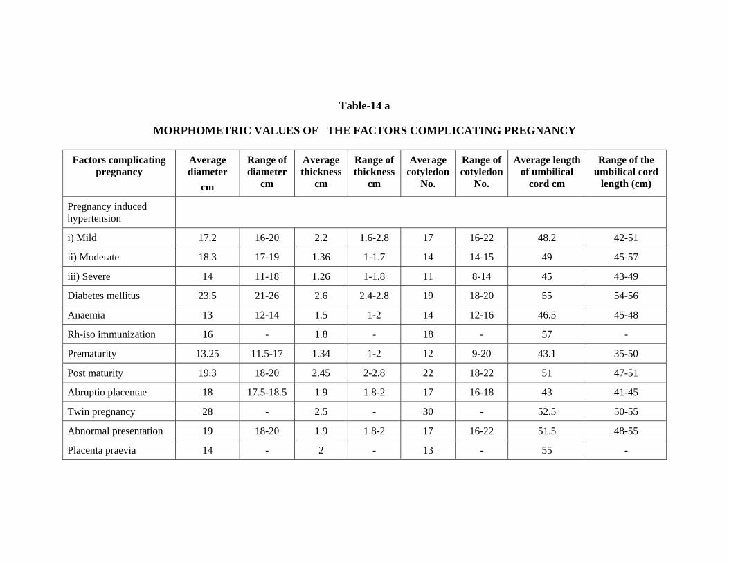
Table-14 a
MORPHOMETRIC VALUES OF THE FACTORS COMPLICATING PREGNANCY
Factors complicating pregnancy
Average diameter
cm
Range of diameter
cm
Average thickness
cm
Range of thickness
cm
Average cotyledon
No.
Range of cotyledon
No.
Average length of umbilical
cord cm
Range of the umbilical cord
length (cm)
Pregnancy induced hypertension
i) Mild 17.2 16-20 2.2 1.6-2.8 17 16-22 48.2 42-51
ii) Moderate 18.3 17-19 1.36 1-1.7 14 14-15 49 45-57
iii) Severe 14 11-18 1.26 1-1.8 11 8-14 45 43-49
Diabetes mellitus 23.5 21-26 2.6 2.4-2.8 19 18-20 55 54-56
Anaemia 13 12-14 1.5 1-2 14 12-16 46.5 45-48
Rh-iso immunization 16 - 1.8 - 18 - 57 -
Prematurity 13.25 11.5-17 1.34 1-2 12 9-20 43.1 35-50
Post maturity 19.3 18-20 2.45 2-2.8 22 18-22 51 47-51
Abruptio placentae 18 17.5-18.5 1.9 1.8-2 17 16-18 43 41-45
Twin pregnancy 28 - 2.5 - 30 - 52.5 50-55
Abnormal presentation 19 18-20 1.9 1.8-2 17 16-22 51.5 48-55
Placenta praevia 14 - 2 - 13 - 55 -

Table-14 b
MORPHOMETRIC VALUES OF THE FACTORS COMPLICATING PREGNANCY
Factors complicating pregnancy
Average placental
weight gms
Range of placental
weight gms
Average weight the of
conceptus Kg
Range of weight of the
conceptus Kg
Feto placental
ratio
Placental coefficient
Pregnancy induced hypertension
i) Mild 475 425-525 3.322 3-3.8 6.4 .14
ii) Moderate 375 1350-400 2.266 2-2.7 6 .16
iii) Severe 316.66 2275-350 2.025 1.4-2.4 6.98 .15
Diabetes mellitus 587.5 -575-600 3.95 3.9-4 6.7 .14
Anaemia 387.5 375-400 2.45 2.2-2.7 6.3 .15
Rh- iso immunisation 400 - 2.9 - 7.2 .13
Prematurity 306.6 150-390 1.945 .475-2.7 6.34 .15
Post maturity 585 500-575 3.73 3.5-3.7 6.3 .15
Abruptio placentae 425 350-475 2.7 2.2-3.2 6.5 .15
Twin pregnancy 425 - 2.45 - 5.76 .17
Abnormal presentation 437.5 425-450 3 3-3.2 6.8 .14
Placenta praevia 475 - 3.3 - 6.9 .14

Table: 18 a
COMPARISON OF INCIDENCE OF CIRCULAR
SHAPE OF THE PLACENTA
AUTHORS %
SAROJAMMA (1986) 57
GUNAPRIYA.R (2001) 93
PRESENT STUDY 55
Table: 18 b
COMPARISON OF INCIDENCE OF OVAL SHAPE OF THE PLACENTA
AUTHORS %
SAROJAMMA (1986) 36
GUNAPRIYA.R (2001) 7
PRESENT STUDY 30
Table: 18 c
COMPARISON OF INCIDENCE OF TRIANGULAR SHAPE OF THE PLACENTA
AUTHORS %
SAROJAMMA (1986) 7
PRESENT STUDY 8
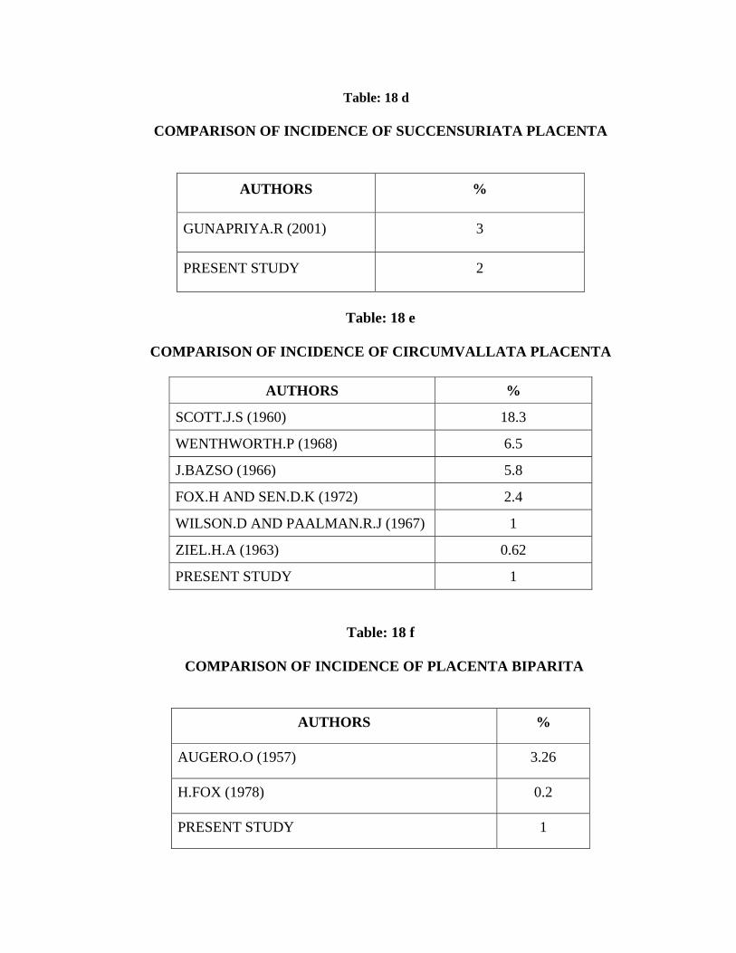
Table: 18 d
COMPARISON OF INCIDENCE OF SUCCENSURIATA PLACENTA
AUTHORS %
GUNAPRIYA.R (2001) 3
PRESENT STUDY 2
Table: 18 e
COMPARISON OF INCIDENCE OF CIRCUMVALLATA PLACENTA
AUTHORS %
SCOTT.J.S (1960) 18.3
WENTHWORTH.P (1968) 6.5
J.BAZSO (1966) 5.8
FOX.H AND SEN.D.K (1972) 2.4
WILSON.D AND PAALMAN.R.J (1967) 1
ZIEL.H.A (1963) 0.62
PRESENT STUDY 1
Table: 18 f
COMPARISON OF INCIDENCE OF PLACENTA BIPARITA
AUTHORS %
AUGERO.O (1957) 3.26
H.FOX (1978) 0.2
PRESENT STUDY 1

Table: 19a
COMPARISON OF AVERAGE DIAMETER OF THE PLACENTA
AUTHORS DIAMETER IN CM
HENRY GRAY (1858) 18.5
J.DBOYD AND W.J HAMILTON (1970) 18.5
LESLIE.B.AREY (1924) 17.5
GUNAPRIYA.R (2001) 17.4
J.P.E.JUDSON (1986) 16.9
RICHARD.S. SNELL (1973) 20
PRESENT STUDY 17.73
Table: 19 b
COMPARISON OF RANGE OF PLACENTAL DIAMETER
AUTHORS DIAMETER IN CM
HENRY GRAY (1858) 10-40
T.W.SADLER (1963) 15-25
KEITHL.L.MOORE & T.V.N PERSAUD (1973) 15-20
GUNAPRIYA.R (2001) 12-22.5
PRESENT STUDY 11-28

Table: 20 a
COMPARISON OF AVERAGE THICKNESS OF THE PLACENTA
AUTHORS THICKNESS IN CM
T.W.SADLER (1963) 3
LESLIE .B. AREY (1924) 2.5
RICHARD.S.SNELL (1973) 2.5
HENRY GRAY (1858) 2.3
J.DBOYD AND W.J HAMILTON (1970) 2.1
GUNAPRIYA.R (2001) 12-22.5
PRESENT STUDY 1.9
Table: 20 b
COMPARISON OF RANGE OF PLACENTAL THICKNESS
AUTHORS THICKNESS IN CM
HENRY GRAY (1858) 10-40
T.W.SADLER (1963) 15-25
KEITHL.L.MOORE & T.V.N PERSAUD (1973) 2-3
GUNAPRIYA.R (2001) 1.5-2.7
CYNTHIA..G.KAPALAN (1996) 2-2.5
SAROJAMMA (1986) 1.5-2
PRESENT STUDY 1-3.1

Table: 21 a
COMPARISON OF AVERAGE COTYLEDON NUMBER
AUTHORS COTYLEDON NO:
ALLAN.C.BARNES (1968) 30
MAJUMDAR ET AL (2005) 19
SAROJAMMA (1986) 18
GUNAPRIYA.R (2001) 18
PRESENT STUDY 18
Table: 21 b
COMPARISON OF RANGE OF COTYLEDON NUMBER
AUTHORS COTYLEDON NO:
K.BENIRSCHKE ET AL (1967) 10-40
J.DBOYD AND W.J HAMILTON (1970) 10-38
HENRY GRAY (1858) 15-30
T.W.SADLER (1963) 15-20
SAROJAMMA (1986) 3-24
GUNAPRIYA.R (2001) 12-24
PRESENT STUDY 8-30
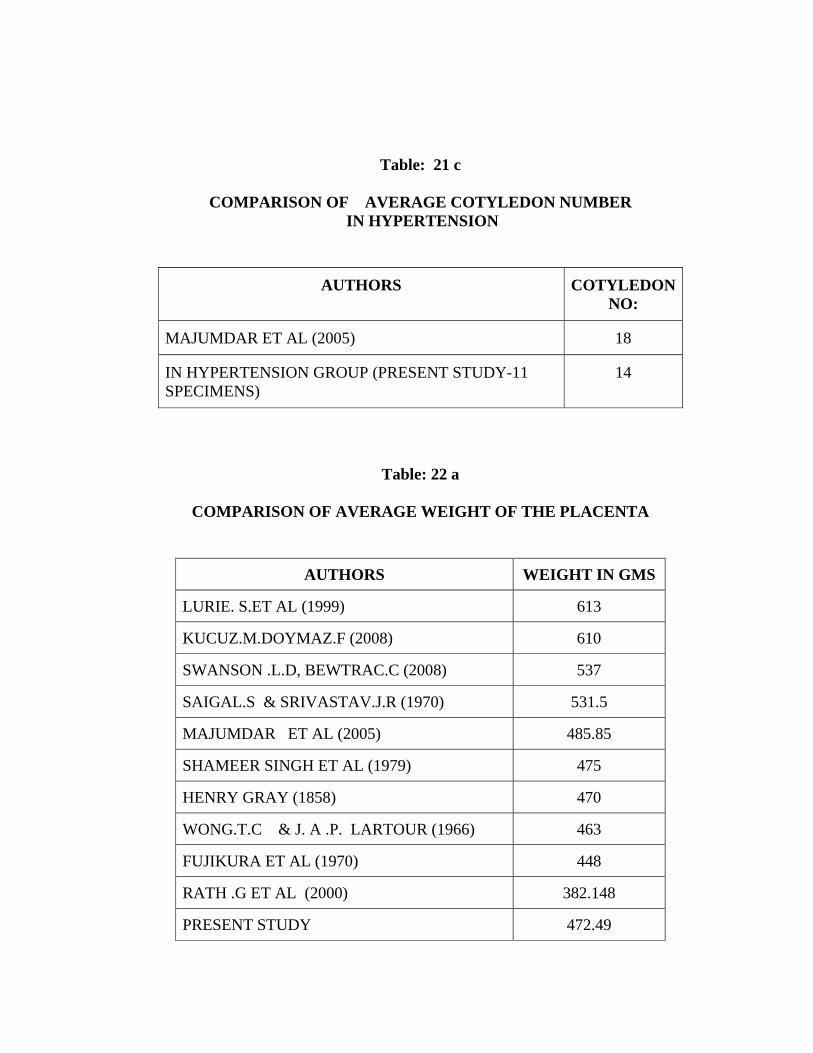
Table: 21 c
COMPARISON OF AVERAGE COTYLEDON NUMBER IN HYPERTENSION
AUTHORS COTYLEDON NO:
MAJUMDAR ET AL (2005) 18
IN HYPERTENSION GROUP (PRESENT STUDY-11 SPECIMENS)
14
Table: 22 a
COMPARISON OF AVERAGE WEIGHT OF THE PLACENTA
AUTHORS WEIGHT IN GMS
LURIE. S.ET AL (1999) 613
KUCUZ.M.DOYMAZ.F (2008) 610
SWANSON .L.D, BEWTRAC.C (2008) 537
SAIGAL.S & SRIVASTAV.J.R (1970) 531.5
MAJUMDAR ET AL (2005) 485.85
SHAMEER SINGH ET AL (1979) 475
HENRY GRAY (1858) 470
WONG.T.C & J. A .P. LARTOUR (1966) 463
FUJIKURA ET AL (1970) 448
RATH .G ET AL (2000) 382.148
PRESENT STUDY 472.49
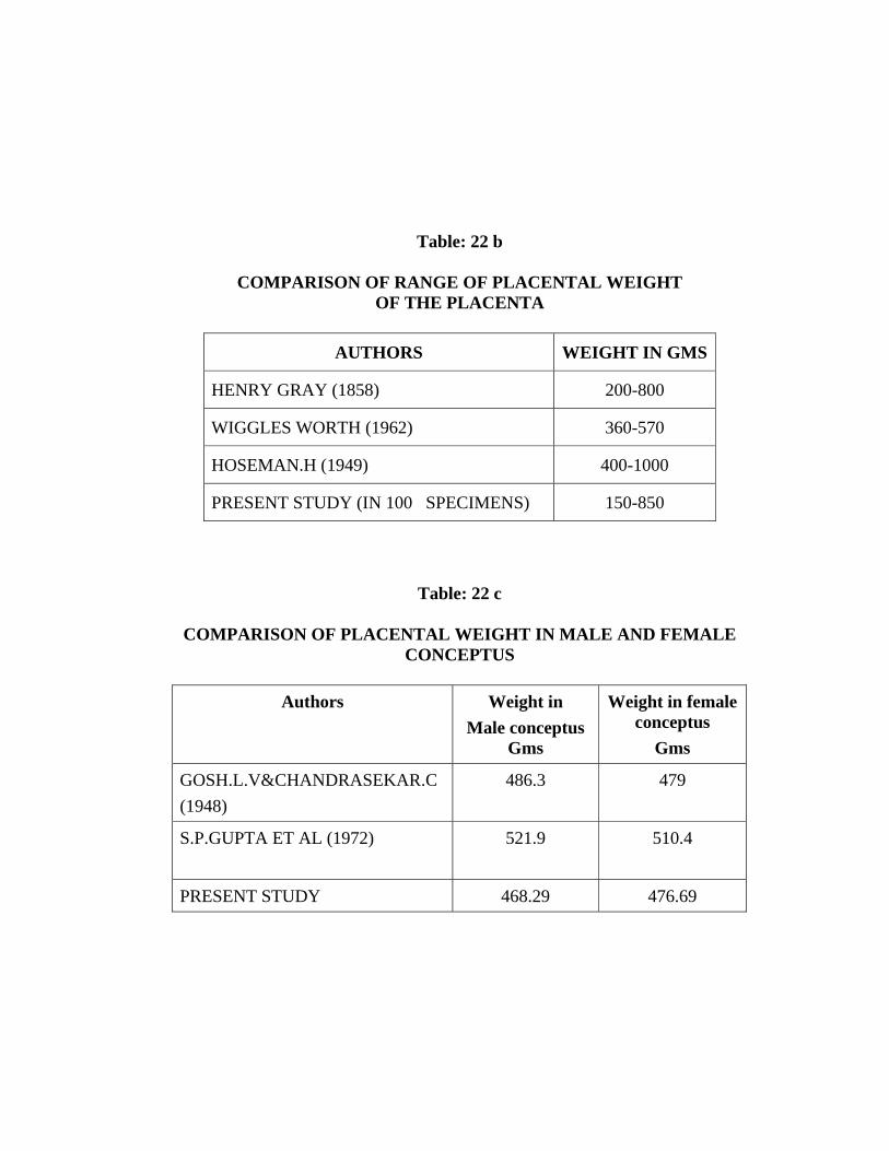
Table: 22 b
COMPARISON OF RANGE OF PLACENTAL WEIGHT OF THE PLACENTA
AUTHORS WEIGHT IN GMS
HENRY GRAY (1858) 200-800
WIGGLES WORTH (1962) 360-570
HOSEMAN.H (1949) 400-1000
PRESENT STUDY (IN 100 SPECIMENS) 150-850
Table: 22 c COMPARISON OF PLACENTAL WEIGHT IN MALE AND FEMALE
CONCEPTUS
Authors Weight in Male conceptus
Gms
Weight in female conceptus
Gms
GOSH.L.V&CHANDRASEKAR.C (1948)
486.3 479
S.P.GUPTA ET AL (1972) 521.9 510.4
PRESENT STUDY 468.29 476.69

Table: 22 d
COMPARISON OF FETO-PLACENTAL WEIGHT RATIO IN NORMAL CASES
Authors Average feto-placental
weight
Average feto-placental
weight in males
Average feto-placental weight in females
KUCUZ. M.DOYMAZ.F (2008)
5.6 5.7 5.6
PRESENT STUDY 6.6 6.6 6.7
Table: 22 e
COMPARISON OF FETO-PLACENTAL WEIGHT RATIO IN HYPERTENSION PREGNANCIES
Authors Average feto-
placental weight in normal pregnancies
Average feto-placental weight in
hypertensive pregnancies
MAJUMDAR ET AL (2005)
5.89 6.23
PRESENT STUDY 6.6 6.47

Table: 22 f
COMPARISON OF PLACENTAL COEFFICIENT IN NORMAL
PREGNANCIES AND DIABETES MELLITUS
Authors Placental coefficient in normal
pregnancies
Placental coefficient in
diabetic pregnancies
KUCUZ. M.DOYMAZ.F (2008) 0.18 0.21
PRESENT STUDY 0.15 0.14
Table: 22 g
COMPARISON OF PLACENTAL WEIGHT IN HYPERTENSION
Authors Weight in mild
hypertension Gms
Weight in moderate
hypertension Gms
Weight in severe
hypertension Gms
RATH.G ET AL (2000) 351.308 338.308 332.410
PRESENT STUDY 475 375 316
Table: 22 h COMPARISON OF PLACENTAL WEIGHT IN DIABETES MELLITUS
Authors Weight in gms
N.A BEISCHER ET AL (1968) 900
KUCUZ. M.DOYMAZ.F (2008) 694.8
PRESENT STUDY 587.5

Table: 22 i
COMPARISON OF PLACENTAL WEIGHT IN ANAEMIA
AUTHORS WEIGHT IN GMS
DOCKERY.J.L (1960) 1,984
N.A BEISCHER ET AL (1968) 900
PRESENT STUDY 387.5
Table: 22 j
COMPARISON OF PLACENTAL WEIGHT IN RH-ISOIMMUNISATION
AUTHORS WEIGHT IN GMS
N.A BEISCHER ET AL (1968) 900
PRESENT STUDY 400
Table: 22 k
COMPARISON OF PLACENTAL WEIGHT IN PREMATURE MALE AND FEMALE CONCEPTUS
Authors Weight in
Male conceptus
gms
Weight in Female
conceptus gms
GOSH.L.V & CHANDRASEKAR.C (1948) 391.1 379.1
S.P.GUPTA ET AL (1972) 449.9 405.3
PRESENT STUDY 237.5 341.5

Table: 22 l
COMPARISON OF PLACENTAL WEIGHT IN
POST-MATURE CONCEPTUS
AUTHORS WEIGHT IN GMS
SAIGAL.S & J.R SRIVASTAVA (1970)
475.9
PRESENT STUDY 585
Table: 22 m
COMPARISON OF WEIGHT OF PLACENTA BIPARITA
AUTHORS WEIGHT IN GMS
FUJIKURA ET AL (1970) 473
PRESENT STUDY 475
Table: 23 a
COMPARISON OF INCIDENCE OF DISPERSE TYPE
OF VASCULAR PATTERN
AUTHORS %
KISHORE .N & SARKAR.S.C (1967) 61.8
PRESENT STUDY 40

Table: 23 b COMPARISON OF INCIDENCE OF FOETAL VEINS
CROSSING FOETAL ARTERIES
AUTHORS %
PAUL WENTHWORTH (1965) 3
PRESENT STUDY 1
Table: 24 a
COMPARISON OF INCIDENCE OF CENTRAL TYPE
OF CORD INSERTION
AUTHORS %
SAROJAMMA (1986) 40
EARN.A.A (1951) 28
KRONE.H.A. (1961) 25
J.P.E JUDSON (1986) 20
EASTMAN .N.J & HELLMANN.L.M (1966) 18
HYRTL. J (1870) 16
SHANKLIN.D.R (1958) 11
GUNAPRIYA.R (2001) 5
CHIARI ET AL (1895) 3.3
PRESENT STUDY 9

Table: 24 b
COMPARISON OF INCIDENCE OF BATTLE DORE (MARGINAL) TYPE OF CORD INSERTION
AUTHORS %
HYRTL. J (1870) 19
EARN.A.A (1951) 15
MONIE.I.W (1965) 14.7
KRONE.H.A. (1961) 10
J.P.E JUDSON (1986) 10
GUNAPRIYA.R (2001) 9
EASTMAN .N.J & HELLMANN.L.M (1966) 7
S.BRODY & D.A.FRENKEL (1953) 6.2
UYANWAH ET AL (1977) 5.6
CHIARI ET AL (1895) 5
THOMAS.J (1963) 5.2
SCOTT.J.S (1960) 2
SAROJAMMA (1986) 2
SHANKLIN.D.R (1958) 1.9
ROBINSON ET AL (1983) 1.5
PRESENT STUDY 12
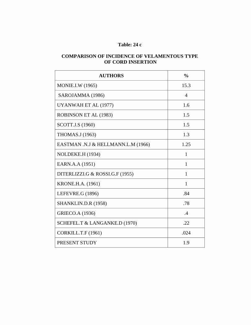
Table: 24 c
COMPARISON OF INCIDENCE OF VELAMENTOUS TYPE OF CORD INSERTION
AUTHORS %
MONIE.I.W (1965) 15.3
SAROJAMMA (1986) 4
UYANWAH ET AL (1977) 1.6
ROBINSON ET AL (1983) 1.5
SCOTT.J.S (1960) 1.5
THOMAS.J (1963) 1.3
EASTMAN .N.J & HELLMANN.L.M (1966) 1.25
NOLDEKE.H (1934) 1
EARN.A.A (1951) 1
DITERLIZZI.G & ROSSI.G.F (1955) 1
KRONE.H.A. (1961) 1
LEFEVRE.G (1896) .84
SHANKLIN.D.R (1958) .78
GRIECO.A (1936) .4
SCHEFEL.T & LANGANKE.D (1970) .22
CORKILL.T.F (1961) .024
PRESENT STUDY 1.9

Table: 24 d
COMPARISON OF INCIDENCE OF ECCENTRIC TYPE OF CORD INSERTION
AUTHORS %
CHIARI ET AL (1895) 91.2
SHANKLIN.D.R (1958) 89
GUNAPRIYA.R (2001) 86
EASTMAN .N.J & HELLMANN.L.M (1966) 73
MONIE.I.W (1965) 70
J.P.E JUDSON (1986) 70
KRONE.H.A. (1961) 64
EARN.A.A (1951) 56
HYRTL. J (1870) 54
SAROJAMMA (1986) 53
PRESENT STUDY 75
Table: 25 a
COMPARISON OF AVERAGE LENGTH OF THE UMBILICAL CORD
AUTHORS LENGTH IN CM
FOG.J (1930) 59.6 SHORDANIA .J (1929) 60 MOSSINGER ET AL (1986) 57 WILLIAMS (1930) 55 GARDINER .J.P (1922) 55 GUNPRIYA.R (2001) 53.5 HENRY GRAY (1858) 50 EDITH.L.POTTER (1952) 50 SAROJAMMA (1986) 38.5 PRESENT STUDY 47.95

Table: 25 b
COMPARISON OF RANGE OF LENGTH OF THE UMBILICAL CORD
AUTHORS LENGTH IN CM
HENRY GRAY (1858) 20-120
SAROJAMMA (1986) 25-85
GUNAPRIYA.R (1986) 30-70
WILLIAMS (1930) 30-100
SHORDANIA.J (1929) 35-104
SCHMIDTH-ELMENDORF (1952) 50-70
FOX.H (1997) 54-61
PRESENT STUDY 30-60
Table: 25 c
COMPARISON OF LENGTH OF THE UMBILICAL CORD IN MALE AND FEMALE CONCEPTUS
Authors Average length
of the cord cm
Average length of the cord in males
cm
Average length of the cord in
females cm
GUNAPRIYA.R (2001)
53.5 54.6 52.1
PRESENT STUDY 47.95 46.54 49.23
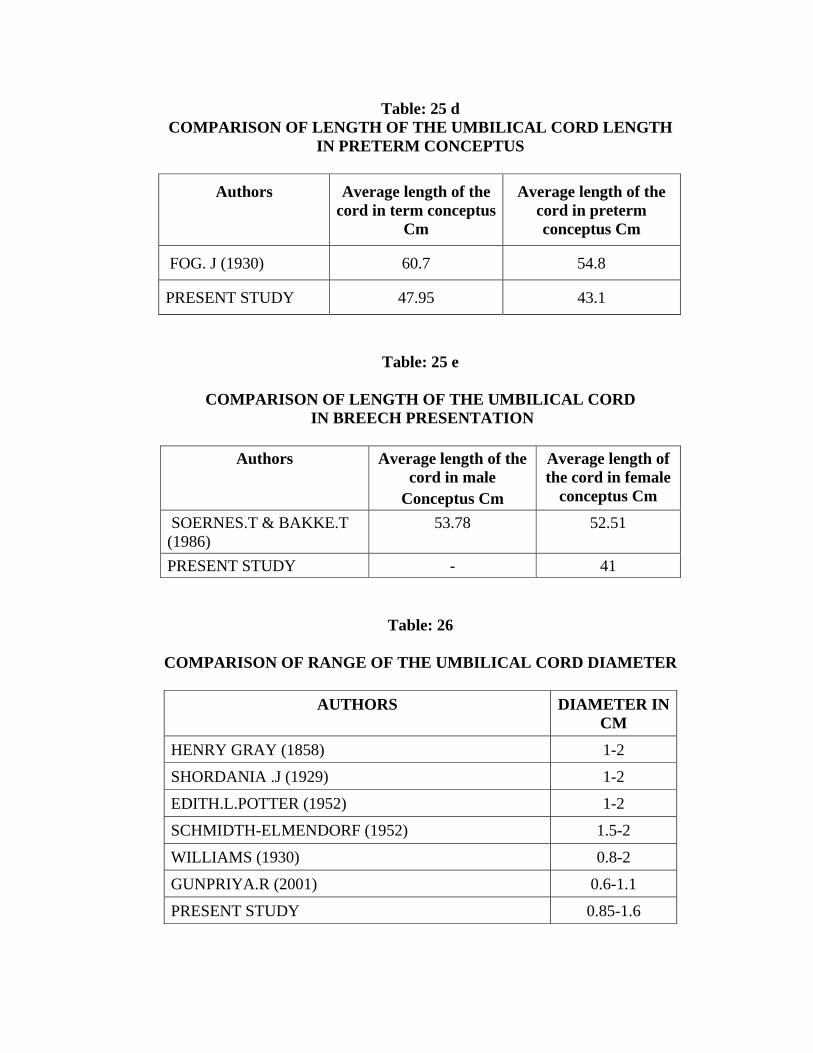
Table: 25 d COMPARISON OF LENGTH OF THE UMBILICAL CORD LENGTH
IN PRETERM CONCEPTUS
Authors Average length of the cord in term conceptus
Cm
Average length of the cord in preterm conceptus Cm
FOG. J (1930) 60.7 54.8
PRESENT STUDY 47.95 43.1
Table: 25 e
COMPARISON OF LENGTH OF THE UMBILICAL CORD IN BREECH PRESENTATION
Authors Average length of the
cord in male Conceptus Cm
Average length of the cord in female
conceptus Cm
SOERNES.T & BAKKE.T (1986)
53.78 52.51
PRESENT STUDY - 41
Table: 26
COMPARISON OF RANGE OF THE UMBILICAL CORD DIAMETER
AUTHORS DIAMETER IN CM
HENRY GRAY (1858) 1-2
SHORDANIA .J (1929) 1-2
EDITH.L.POTTER (1952) 1-2
SCHMIDTH-ELMENDORF (1952) 1.5-2
WILLIAMS (1930) 0.8-2
GUNPRIYA.R (2001) 0.6-1.1
PRESENT STUDY 0.85-1.6

Table: 27 a
COMPARISON OF INCIDENCE OF SINISTRAL (LEFT) TYPE OF UMBILICAL CORD TWIST
AUTHORS %
RONALD.V.LACRO (1987) 83
HENRY.W.EDMONDS (1954) 82
READ.W (1860) 77
NEUGEBAUER .L.A (1858) 71
PRESENT STUDY 95
Table: 27 b
COMPARISON OF INCIDENCE OF DEXTRAL (RIGHT) TYPE
OF UMBILICAL CORD TWIST
AUTHORS %
NEUGEBAUER .L.A (1858) 24
READ.W (1860) 14
HENRY.W.EDMONDS (1954) 12
RONALD.V.LACRO (1987) 12
PRESENT STUDY 2
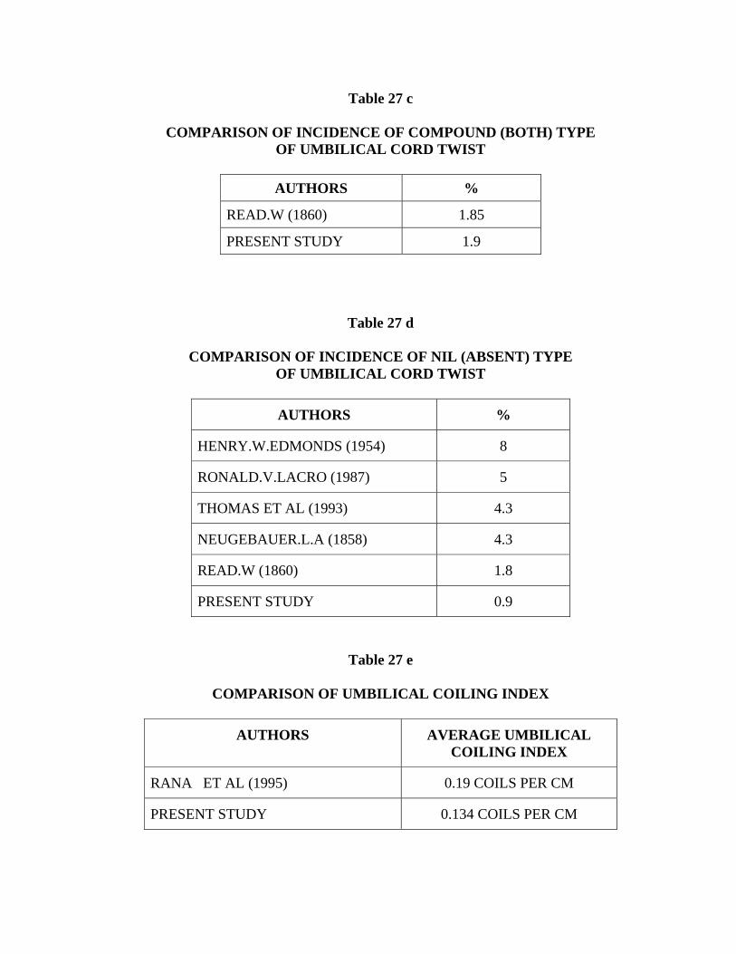
Table 27 c
COMPARISON OF INCIDENCE OF COMPOUND (BOTH) TYPE OF UMBILICAL CORD TWIST
AUTHORS %
READ.W (1860) 1.85
PRESENT STUDY 1.9
Table 27 d
COMPARISON OF INCIDENCE OF NIL (ABSENT) TYPE OF UMBILICAL CORD TWIST
AUTHORS %
HENRY.W.EDMONDS (1954) 8
RONALD.V.LACRO (1987) 5
THOMAS ET AL (1993) 4.3
NEUGEBAUER.L.A (1858) 4.3
READ.W (1860) 1.8
PRESENT STUDY 0.9
Table 27 e
COMPARISON OF UMBILICAL COILING INDEX
AUTHORS AVERAGE UMBILICAL COILING INDEX
RANA ET AL (1995) 0.19 COILS PER CM
PRESENT STUDY 0.134 COILS PER CM

ANIMAL PLACENTA
Table 28 a
COW PLACENTA- COMPARISON OF COTYLEDON NUMBER, UMBILICAL BLOOD VESSELS AND
LENGTH OF THE UMBILICAL CORD
Authors Shape of the placenta
Weight of the placenta
kg
Cotyledon
Number
Umbilical Blood Vessels
Length of the umbilical
cord cm
WILD.A (1964) - - 80 - 120 - -
KURT BENIRSCHKE (2007)
Poly
cotyledonary
4 – 5 - 4, (2 Arteries
2 Veins)
-
PRESENT STUDY Poly
Cotyledonary
4 60 4, (2 Arteries 2 Veins)
20
Table: 28 b
PIG PLACENTA- COMPARISON OF COTYLEDON NUMBER,
UMBILICAL BLOOD VESSELS AND LENGTH OF THE UMBILICAL CORD
Authors Shape of the
placenta Weight of
the placentaKg
Cotyledon Number
Umbilical blood vessels
Length of the umbilical
cord cm
STEEPHEN.J. ROBERTS (1971)
Diffuse - No discrete cotyledons
- -
KURT BENIRSCHKE (2007)
- - - 3, (2 Arteries 1 Vein)
-
PRESENT STUDY Diffuse 1.6 No cotyledons
3, (2 Arteries, 1 Vein)
30

Table: 28 c
SHEEP PLACENTA- COMPARISON OF WEIGHT OF THE PLACENTA
Authors Weight of placenta in gms
KLEEMAN ET AL (2001) 600
PRESENT STUDY 800
Table: 28 d
SHEEP PLACENTA- COMPARISON OF COTYLEDON NUMBER, UMBILICAL BLOOD VESSELS AND
LENGTH OF THE UMBILICAL CORD
Authors Cotyledon Number
Umbilical blood vessels
Length of the umbilical cord
cm
KURT BENIRSCHKE (2007)
60-100 4, (2 Arteries 2 Veins)
27
PRESENT STUDY 60 4 (2 Arteries 2 Veins)
20
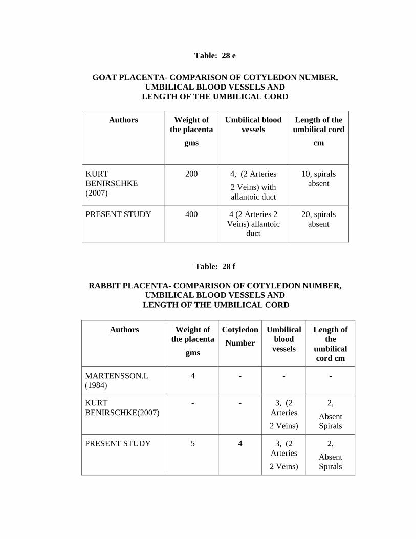
Table: 28 e
GOAT PLACENTA- COMPARISON OF COTYLEDON NUMBER, UMBILICAL BLOOD VESSELS AND
LENGTH OF THE UMBILICAL CORD
Authors Weight of the placenta
gms
Umbilical blood vessels
Length of the umbilical cord
cm
KURT BENIRSCHKE (2007)
200 4, (2 Arteries 2 Veins) with allantoic duct
10, spirals absent
PRESENT STUDY 400 4 (2 Arteries 2 Veins) allantoic
duct
20, spirals absent
Table: 28 f
RABBIT PLACENTA- COMPARISON OF COTYLEDON NUMBER, UMBILICAL BLOOD VESSELS AND
LENGTH OF THE UMBILICAL CORD
Authors Weight of the placenta
gms
CotyledonNumber
Umbilical blood vessels
Length of the
umbilical cord cm
MARTENSSON.L (1984)
4 - - -
KURT BENIRSCHKE(2007)
- - 3, (2 Arteries 2 Veins)
2, Absent Spirals
PRESENT STUDY 5 4 3, (2 Arteries 2 Veins)
2, Absent Spirals

BIBLIOGRAPHY
1. Adair. F.L& Thelander. H: study of the weight and dimensions of the human placenta in its relation to the weight of the newborn infant, Am.J.Obst. & Gynec.10: pg: 172,1925.
2. Allan.c. Barnes: Intra-uterine development. Pg: 36. 1968
3. Andrade. A: Anatomical and radiographic study on the anastomosis and branching of the umbilical arteries in white and non-white Brazilians. Acta Anat (Basel) 70: 66-75 (1968).
4. Augero. O: Anomalias Mortologicas dela placenta Ysu significado clinico Artergrafa caracas. 1957
5. Bascish. P and Smout. C.F.V : Some Observations on the foetal vessels of the human placenta with an account of the corrosion technique. Jol.of Anatomy Vol. XXII, Oct- 1937-July 1938. pg: 358-364.
6. Bazso. J: Retardation of intrauterine growth and its causes. Gynec. Prac: pg: 17, 293. 1966.
7. Bhargva.I and Raja.P.T.K: Arteriovenous crossings on the chorial surface of the human placenta in abnormal pregnancy and development. Experientia 25: 831-832. 1969.
8. Boyd. J.D. and W.J. Hamilton: The human placenta. Pg: 114, 1970.
9. Bradley M. Pattern: Human Embryology pg: 167, 173 1946.
10. Chiari.J, Braun.C and Sparth.J: Klinik der Geburtshiffe and Gyankologie Enke, Erlangen.1895.
11. Corkill. T.F: The infant's vulnerable life-line. Aust. N.Z.J. Obstet Gynaecol. 1: 154-160, 1961.
12. Damania. I et al: The placenta in hypertensive disorder in pregnancy. J. Obs. and Gynaecol. Ind: 1989: 39-28-31.

13. Diterlizzi and Rossi: G.F. Studio clinico - statistics sulle anomalie del funiculo. Ann Obstet Clinicol. 77: 459-474, 1955.
14. Dockery J.L.: Am J. Obstet.&gynec 79: 138. 1960.
15. Earn A.A: The effect of congenital abnormalities of the umbilical cord and placenta on the newborn and mother: A survey of 5676 consecutive deliveries. J. Obs Gynaecol. Brit. Emp. 58: 456-459, 1951.
16. Eastman. N.J and Hellmann. L.M: Williams Obstetrics ed. 13, Newyork, 1966, Appleton Centery - Crofts, Inc.
17. Edith. L. Potter: Pathology of the fetus 1952
18. Fog. J: Die Lange der Nabelschnur. Acta Obstet. Gynec. Scand; 9, 132-149.
19. Fox . H: Pathology of placenta 2ed. W.B. Saunders, London.1997.
20. Fox. H. And Sen D.K: Placenta Extrachorialis: A clinic-pathologic study. J. Obstet. Gynaecol. Brit: Common Wealth 79: 32-35. 1972.
21. Fox. H: Pathology of placenta monograph, Vol.VII Philadelphia Saunders, pg: 828, 1978.
22. Gardiner J.P: The umbilical cord: normal length: length in cord complications etiology and frequency of coiling. surg. Gynecol. Obstet. 34: 252-256, 1922.
23. Godfrey.KM, Redman.CW, Baiker.DJ, Osmond.C: The effect of maternal anaemia and iron deficiency on the ratio of fetal weight to placental weight. Br.J. Obs. Gynaecol. 1991, Sep; 98(9): 886-91.
24. Gordon. Z et al: Anthropometry of fetal vasculatare in the chorionic plate. J. Anat (2007).
25. Gosh.L.V and Chandrasekhar.C: A study of the standards of prematurity of Indian infants. Govt of India Press, New Delhi.1946.

26. Greico.A: Pilievi clinic-statistics Sulla inserzore velamentosa ed a racchetta del cordone obellicale monit. Onstet-Ginecol. Endocrin. Melatol 8: 89-102, 1936.
27. Gunapriya.R: The morphology, morphometeric study of placenta and umbilical cord with vascular pattern and clinical correlations.pg: 28 –43, 2001.
28. Gupta. S.P, Bahl. L. and S.K. Dikshit: A study of placenta in relation to birth weight and gestational age.1972.
29. Henry Gray: Grays Anatomy, The anatomical basis of clinical practice, 39th Edition pg: 1341- 1348,1858.
30. Henry. W. Edmonds: The spiral twist of the normal umbilical cord in Twins and in singletons. Pg: 102-119. Vol.67, No.1, Am.J. Obs. Gynec. Janu. 1954.
31. Hoseman.H: Duration of pregnancy and weight of the placenta. Archieves of Gynaecology 176: 453.1949.
32. Hyrtl. J: Die Blutgefasse der Menschlichen. Nachgeburt in Normalen and Abnormen Verhaltnissen. Braumuller, Vienna.1970.
33. Judson. J.P. E: The placental vasculature: pg: 62, 1986.
34. Keith L Moore and T.V.N. Persaud: The developing Human- Clinically Oriented Embryology, pg: 134, 124 (1973)
35. Kher A.V. and Zawar: Indian J. Path Microbiology, 24: 245, 1981.
36. Kishore.N and Sarkar.SC: The arterial patterns of placenta. A post partum radiological study. J. Obs. Gynaecol. India. 17: 9-13 (1967).
37. Krone H.A: Diebedutung der Eibett - storungen fur die Entstehung menschlicher Missbildungen. Fischer, Stuttgart.1961.

38. Kucuz, M. Doymay F: Placental weight and placental weight to birth weight ratio are increased in diet - and exercise -treated gestational diabetes mellitus subjects but not in subjects with one abnormal value on 100-g oral glucose tolerance test. J. Diabetes Complications. 2008. April 15.
39. Kurt Benirschke, Peter Kaufmann, Rebecca Baergen: The pathology of human placenta. 5th edition.pg: 13.1967.
40. Lefevre. G: These deparis 1896.
41. Leslie Braninerd Arey: Developmental anatomy. Pg: 122, 123, 1924.
42. Lurie S. Feinstein, M.Mament.Y: Human fetal - placental weight ratio in normal singleton near term pregnancies. Gynecol. obstet. Invest. 1999; 48(3): 155-7.
43. Majumdar.s et al: A study of placenta in normal and hypertensives pregnancies. 2005, Journal of Anat. Soc. India. Vol. 54, No.2.
44. Monie I.W: Am. j. Obst. & Gynec. 93: 276, 1965.
45. Mossinger et al: Umbilical cord length in Down's syndrome. Amer. J. Dis. Child. 140: 1276-1277, 1986.
46. N.A. Beischer, M. Holsman, W.H. Kitchen: Relations of various forms of anaemia to placental weight. Am.J. Obs. Gyn, July 15, Vol. 101, No.6, 1968, 801-809.
47. Neugebaurer L.A: Morphologic der monschlichen, Nabelschneu, Breslau 1858, W.G. Korn.
48. Noldeke.H: Geburtskomplikationen, bei, Insertio velamentosa, Zenbratbl gynackol. 58: 351-356, 1934.
49. Nordenvall. M, Sandstedt B, Ulmsten.U: Relationship between placental shape, cord insertion, lobes and gestational outcome. Acta Obstet. Gynecol Scand. 1988; 67(7): 611-6.

50. Paul Wentworth: Some anomalies of the foetal vessels of the human placenta. J. Anat (1965). 99, 2, pg: 273-282.
51. Rana et al: Adverse perinatal outcome in patients with an abnormal umbilical coiling index.obstetrics and gynecology, vol 85,no: 4,april (1995)
52. Rath.G. Garg. K. Sood M: Insertion of umbilical cord on the placenta in Hypertensive mother. J. Ant. Soc.l India. 49(2): 149-152 (2000).
53. Rayburn, W.F., Beynen A, Brinkman DL: Umbilical cord length and intrapartum complications. Obstet. Gynecocl. 57: 450-452, 1981.
54. Read. W: Boston. M & S.J. 62: 497, 1860.
55. Richard L. Naeye: Umbilical cord length: clinical significance: J. Pediatr. 107: 278-281, 1985.
56. Richard S. Snell: Clinical anatomy pg: 372, 7th ed, 1973
57. Robinson.L.K, Jones K.L. and Benirschke.K: The nature of structural defects associated with velamentous and marginal insertion of the umbilical cord. Am.J. Obs.Gynecol. 146: 191-193, 1983.
58. Ronald.V.Lacro: The umbilical cord twist: origin, direction and relevance. pg 833-838, Oct. 1987. Am.J.Obs. Gynecol. 1987.
59. Roth L.G: Central Placenta praevia due to succenturiate Lobe. Am.J.Obs Gynecol 14: 447-449, 1957.
60. Sadler. T.W.: Langman's Medical Embryology, pg: 105, 1963.
61. Saigal S. and Srivastava J.R: The significance of examination of the placentae in the evaluation of the new born infant. Indian pediat: 7, 68,1970
62. Sarojamma: Morphological and histological study of the placenta. Pg: 75-78,1986.

63. Scheffel T.and Langanke.D: `Die Niabel - Schnurkomplikation an der Universitats Fraunklinik von 1955 bis 1967 zentrabl .gynakol: 429-434, 1970.
64. Schmidt – Elmendorf. H.R: Anatomic, Physiologica and patholgoic du and Nabelschnur, inschlieblich vorzitigen, Blasenprungs In Biologic and pathologic des, weibes, p.282, seitz - Americh.1952.
65. Scott. J.S: (Placenta Marginata and Circumvallata) Placenta extrachorialis: a factors in antepartum hemorrhage. J. Obst. Gynaecol. Brit. Emp. 67: 904-918.
66. Shameer Singh et al: abstract from Twenty seventh annual conference, Anatomical society of India, 1979, p: 31.
67. Shanklin D.R: Obst & Gynec. 11: 129, 1958.
68. Shordania.J: Der architektonische Aafbau der getasse der enschlichen Nachgeburt and ihre Bezichuagen Zur Entwicklung der trucht Arch. Gynack, 135, 568-598, 1929.
69. Simon Brody and David. A. Frenkel: Marginal Insertion of the cord and premature Labour.1953.
70. Singla P.N. Chand.S, Khanna.S, Agarwal.K.N: Effect of maternal anaemia on the placenta and the newborn infant. Acta Paediatric: Scand. 1978. Sep: 67(5); 645-8.
71. Soernes T. and Bakke.T: The length of the human umbilical cord in vertex and breech presentation. Am.J. Obs. Gynecocl. 154: 1086-1087, 1986.
72. Stephen. A. Heifetz : The umbilical cord obstetrically important lesions. Clinical obstetrics and gynecology, Vol. 39, Number 3, Pg: 571-587 (1996).
73. Sultana. S et al: Changes of placental diameter, thickness and cotyledons in eclampsia. Mymensingh Med. Journal 2007 Jul 16(22): pg: 127-31.

74. Swanson LD Bewtrac: Increase in normal placental weight related to increase in maternal body mass index. J. Matern Fetal Neonatal Med. 2008. Feb; 21(2): 111-3.
75. Thomas H. Strong, John P. Elliot Tari-G. Radin: Non-coiled umbilical Blood vessles. A. New marker for the fetus at Risk. Vol. 81, No.3, Obst. Gynecol. 1993: 81-409-11.
76. Thomas. J.: Die Entwicklung Von. Tetas and placenta bei Nabelgefea Banomalien. Arch Gynakol. 198: 216-223. 1963.
77. Torpin R. and Barfield W.E: Placenta duplex. J.Med. Association Ga: 57: 78-80, 1968.
78. Torpin R: The human placenta, Thomas spring field IL, 1969.
79. Toshio Fujikura, Ralph.C, Benson Shirley.G.Dricoll: The Bipartite placenta and its clinical features. Amj. Obs.Gyn. Vol.107 No.7, 1970, pg: 1013-1017.
80. Uyanwah, - Akpom P.O. and Fox. H: The clinical significance of marginal or velamentous insertion of Cord. Brit. J. Obstet Gynaecol: 84: 941-943, 1977.
81. Wentworth. P: circumvallate and circummarginate placentas. Amer. J. Obst. Gynaecol. 102:pg: 44-47, 1968.
82. Wiggles worth. J.S: The gross and microscopic pathology of the prematurely delivered placenta. Journal of Obstet. Gynaeco. Of British Common wealth. 69: 934-943.1962
83. Williams: Williams’s obstetrics, 22 Edition, pg: 828. 1930.
84. Wilson D. and Paalman R.J: Clinical significance of circumvallate placenta. Obstet. Gynecol. 29: 774-778. 1967.

85. Wong T.C, Lartour J.P.A: Microscopoic measurement of the placenta components in an attempt to assess the malnourished newborn infants. Am.J. Obs. Gynec. 9, pg: 942, 1966.
86. Ziel. H.A: circumvallate placenta a cause of antepartum bleeding premature delivery and perinatal mortality obstet.gynecol.22: pg: 798-802.1963.
Animal placenta 1. Kleeman et al: Fetoplacental growth in sheep administered progesterone
during just 3 days of pregnancy placenta 22: 14-3 2001.
2. KurtBenirschke: Comparative placentation- Domestic sheep goat, pig, rabbit, cow, and mht: http: comparative placentation. UCS. (2007)
3. Martensson.L: The pregnant rabbit, guinea pig, sheep and rhesus monkey as models in reproductive physiology. Europ. J. Obst. Gynecol. 18: 169-182, 1984.
4. Reynolds. S.R.M: The proportion of whartons’ jelly in the umbilical cord in relation to distension of the umbilical arteries and vein with observations on the folds of Hoboken. Anat. Rec. 119: 365-377, 1952.
5. Steephen. J. Roberts: Veterinary Obstetric and Genital diseases. (Theriogenology) pg. 42-47(1971).
6. Wild. A: Interscuhungen uberden Aufbau der placenta fetalis des Rindes and it Auswirkung auf die gestundhit des Kalbes. Zbl. Veterinary Mediz. 11: 60-89, 1964.

LEGEND
AC-ACCESSORY LOBE
AE-AMNIOTIC EPITHELIUM
AL-ALLANTOIC DUCT
AM-AMNIOTIC MEMBRANE
AV-ANCHORING VILLI
B-BATTLEDORE (MARGINAL) INSERTION
BC-BLOOD CLOT
BI-BILOBED SHAPE
BV-BLOOD VESSEL
C-COTYLEDON
CC-CONVEX SHAPED COTYLEDON
CI-CENTRAL INSERTION
COC-CONCAVE SHAPED COTYLEDON
CR-CIRCULAR SHAPE
CT-COMPOUND TWIST
CV-CHORIONIC VILLI
CVV-CIRCUMVALLATE SHAPE
CY-CYTOTROPHOBLAST
D-DISPERSE (DICHOTOMOUS)
DAC-DOUBLE FOLD OF AMNION AND CHORION
E-ECCENTRIC INSERTION
F-FEMALE
FA- FETAL ARTERY
FO-FOLD
FD-FIBRIN DEPOSIT
F: P- FW/BW-FETOPLACENTAL RATIO
FN-FALSE KNOT
FS -FETAL SURFACE
FU-FUSED SHAPE
FV-FETAL VEIN

G-GROOVE
HCUC-HIGHLY COILED UMBILICAL CORD
H-HAEMORRAGE
HE-HEART SHAPE
HO-HORN
IC-ILLDEFINED COTYLEDON
IVS-INTERVILLOUS SPACE
K-KIDNEY SHAPE
KN-KNOT
L-LUMEN
LT-LEFT TWIST
M-MALE
ME-MEMBRANE
MA-MAGISTRAL (MONOPODIAL)
MI-MIXED PATTERN
MS-MATERNAL SURFACE
O-OVAL SHAPE
PM- PERIPHERAL MARGIN
PW/BW-PLACENTAL WEIGHT/BABY WEIGHT-PLACENTAL COEFFICIENT
RT-RIGHT TWIST
SKN-SYNCITIAL KNOTS
ST-SPIRAL TWIST
SU-SUCCENTURIATE SHAPE
SY-SYNCYTOTROPHOBLAST
T-TRIANGULAR SHAPE
TA-TUNICA ADVENTITIA
TI-TUNICA INTIMA
TM-TUNICA MEDIA
UC-UMBILICAL CORD
V-VELAMENTOUS INSERTION