Morphology and evolution of caudal fin in lamniform sharks
48
Via Sapientiae: e Institutional Repository at DePaul University College of Liberal Arts & Social Sciences eses and Dissertations College of Liberal Arts and Social Sciences 3-2010 Morphology and evolution of caudal fin in lamniform sharks Sun H. Kim DePaul University is esis is brought to you for free and open access by the College of Liberal Arts and Social Sciences at Via Sapientiae. It has been accepted for inclusion in College of Liberal Arts & Social Sciences eses and Dissertations by an authorized administrator of Via Sapientiae. For more information, please contact [email protected], [email protected]. Recommended Citation Kim, Sun H., "Morphology and evolution of caudal fin in lamniform sharks" (2010). College of Liberal Arts & Social Sciences eses and Dissertations. 9. hps://via.library.depaul.edu/etd/9
Transcript of Morphology and evolution of caudal fin in lamniform sharks

Via Sapientiae:The Institutional Repository at DePaul University
College of Liberal Arts & Social Sciences Thesesand Dissertations College of Liberal Arts and Social Sciences
3-2010
Morphology and evolution of caudal fin inlamniform sharksSun H. KimDePaul University
This Thesis is brought to you for free and open access by the College of Liberal Arts and Social Sciences at Via Sapientiae. It has been accepted forinclusion in College of Liberal Arts & Social Sciences Theses and Dissertations by an authorized administrator of Via Sapientiae. For more information,please contact [email protected], [email protected].
Recommended CitationKim, Sun H., "Morphology and evolution of caudal fin in lamniform sharks" (2010). College of Liberal Arts & Social Sciences Theses andDissertations. 9.https://via.library.depaul.edu/etd/9

i
Morphology and Evolution of Caudal Fin in Lamniform Sharks
A Thesis Presented in
Partial Fulfillment of
the Requirements for the Degree of
Master of Science
December 2009
By
Sun H. Kim
Thesis Advisor: Kenshu Shimada, Ph.D.
Department of Biological Sciences
College of Liberal Arts and Sciences
DePaul University
Chicago, Illinois
ii
Table of Contents
Approval Page ……………………………………………………………………………………………………….…………….……i Table of Contents……...………………………………………………………………………………………………….…….…….ii List of Figures……………………………………………………………………………………………………………….……..……iii List of Tables…………………………………………………………………………………………………………….……….……..iv Acknowledgements………………………………………………………………………………………………………….….……v Abstract……………………..…………………………………………………………………………………………………….…..….vi Introduction……………………………………………………………………………………………………………………….…..…1 Materials and Methods……………………………………………………………………………………………………….….…4
Examined Specimens………………………………………………………………………………………….…...…….4 Anatomical Examination………………………………………………………………………………..………..…….5 Caudal Fin Terminology………………………………………………………………………………….………..…….6 Measurements……………………………………………………………………………………………….……………...6 Character Mapping………………………………………………………………………………………….…………..…8
Results…………………………………………………………………………………………………………………………………....10 Scyliorhinus retifer………………………………………………………………………………………………….…...10 Mitsukurina owstoni…………………………………………………………………………………..……………..…10 Carcharias taurus………………………………………………………………………………………..………….……11 Odontaspis ferox……………………………………………………………………………….…………..……….……11 Odontaspis noronhai………………………………………………………………………….……………..….…..…11 Pseudocarcharias kamoharai……………………………………………………….……………………...………12 Megachasma pelagios………………………………………………………………………………….….…..………12 Alopias pelagicus……………………………………………………………………………….……….……..…………12 Alopias superciliosus……………………………………………………………….………………….………..………13 Alopias vulpinus………………………………………………………………………………………….…………..……13 Cetorhinus maximus……………………………………..……………………………..…………….……………..…14 Carcharodon carcharias……………………………………………………….…………………….………………..14 Isurus oxyrinchus………………………………………………………………………………………..………………..14 Isurus paucus………………………………………………………………………………………….…….……………..15 Lamna ditropis……………………………………………………………………………….…………….………….…..15 Lamna nasus…………………………………………………………………………………..…………….……………..15
Discussion…………………………………………………………………………………………..…………………….……………..17 Phylogenetic Mapping of Caudal Fin Data……………………………………………………….….………..17 Three Caudal Fin Types in Lamniforms and Their Evolutionary History…………….…………..19 Functional Differences Based on Caudal Fin Types………………………………………………….…...21
References……………………………………………………………………………………………………….……….………….….25 Appendix 1……………………………………………………………………………………………………….……….…………….40 Appendix 2……………………………………………………………………………………………………….……….…………….41

iii
List of Figures
Figure 1 All 15 lamniform species……………………………………………………………………….………….30
Figure 2 Medical imaging technology used to radiographically………………………………………31 examine shark specimens
Figure 3 Schematic drawings of lamniform caudal fin showing………………………………………32 nomenclature and measured variables
Figure 4 Examples of radiographs showing caudal fin anatomy………………………………………33 in one carcharhiniform and 15 lamniform species
Figure 5 Mapping of average values of Cobb’s angle and hypochordal…………………………..35 angle onto morphology-based phylogenetic tree of lamniforms along with schematic illustrations of caudal fin showing its outline and skeletal arrangement
Figure 6 Mapping of average values of Cobb’s and hypochordal…………………………………….36 angle onto molecular-based phylogenetic tree of lamniforms along with schematic illustrations of caudal fin showing its outline and skeletal arrangement
Figure 7 Three types of caudal fin in lamniforms and their possible……………………………….32 evolutionary scenarios

iv
List of Tables
Table 1 List of examined specimens in this study………………………………………………………….38
Table 2 Average value of heterocercal angle, hypocercal angle,…………………………………..39 Cobb’s angle, and hypochordal angle data for each species
v
Acknowledgements
I thank the following individuals who were involved in the acquisition, loan, or
transportation of examined specimens: A. Y. Suzumoto (BPBM); M. A. Rogers, K. Swagel, M. W.
Westneat, P. Willink (FMNH); K. Nakaya (HUMZ); J. A. Seigel (LACM); K. E. Hartel, A. Williston
(MCZ); C. Klepadlo, P. A. Hastings, H. J. Walker (SIO); L. M. Page, R. H. Robins (UF); D. W. Nelson
(UMMZ); J. Finan, L. Palmer, S. Raredon, S. Smith; E. Wilbur, D. Pitassy, and J. T. Williams
(USNM); M. Miya (Natural History Museum and Institute, Chiba, Japan), S. J. Arceneaux, R. L.
Humphreys, Jr. (Pacific Islands Fisheries Science Center, National Marine Fisheries Service,
National Oceanic and Atmospheric Administration); S. R. Van Sommeran (Pelagic Shark
Research Foundation, Capitola, California); B. L. Beatty (New York College of Osteopathic
Medicine); and J. L. Castillo-Geniz (Instituto Nacional de la Pesca, Baja California, México).
Special thanks go to K. Gray, B. Karl, J. Hickey, P. Myefski, A. Nicholas, C. Rigsby, L. Wansk
(Children's Memorial Hospital, Chicago, Illinois) for assisting me with CT scanning and x-ray
shooting of examined specimens. Financial support was made by the Environmental Science
Program, Department of Biological Sciences, and University Research Council at DePaul
University, Chicago, Illinois, that were provided to, or awarded to, K. Shimada. I would also like
to thank M. Silliker for her continued support since I came to DePaul. I also thank A. Ippolito
and W. Aguirre for sitting on my thesis committee. Special thanks to K. Shimada for his
patience and guidance as my mentor and supervising professor of this project. Finally, I would
like to thank my friends and family for supporting me throughout all these years.

vi
Abstract
Sharks have a distinct asymmetrical caudal fin referred to as heterocercal tail that is a
key characteristic of the group and has diversified within sharks in ways that are correlated with
lifestyle. However, practically no study examining the evolutionary trend and history of the
caudal fin morphology within a specific shark group exists. Here, I examined the caudal fin
morphology and evolution of the shark order Lamniformes that consists of 15 extant species
with diverse behaviors and lifestyles. The goals of this study are to describe the skeletal
morphology of the caudal fin in each lamniform species based primarily on radiographic
analysis, to examine the evolutionary pattern and history of the caudal fin through phylogenetic
mapping, and to relate different caudal fin types observed in lamniforms to their known
behaviors and life styles. This study suggests that caudal fins with a more horizontally directed
curvature of the vertebral column are plesiomorphic, whereas those with a large dorsally
directed curvature of the vertebral column are apomorphic within Lamniformes. It also shows
that caudal fins with posteriorly directed hypochordal rays are plesiomorphic, and that those
with ventrally directed hypochordal rays are apomorphic within Lamniformes. Three basic
caudal fin types are recognized in extant lamniforms on the basis of these skeletal variables.
One important discovery form the recognition of the three fin types is that the evolution of
external morphology of caudal fin does not necessarily correspond to the evolution of its
internal (skeletal) anatomy in lamniform sharks. Certain behaviors and lifestyles seen in
different lamniforms are correlative with the different caudal fin types. A less asymmetrical tail
is a derived feature in lamniforms that evolved for fast swimming to capture fast swimming
prey.

1
Introduction
The structure and biomechanics of the caudal fin of fishes have been studied by many
researchers (e.g., Agassiz, 1833; Ryder, 1884; Garman, 1913; Thomson, 1976; Thomson &
Simanek, 1977; Lauder, 2000). In particular, the caudal fin of sharks (Chondrichthyes:
Elasmobranchii) have received considerable attention due to the asymmetrical form referred to
as heterocercal tail, or heterocercy (e.g. Thomson, 1976; Thomson & Simanek, 1977; Lauder,
1989, 2000; Liao & Lauder, 2000a, 2000b; Lauder et al., 2003; Lingham-Soliar, 2005a, 2005b).
Heterocercy in sharks occurs because the enlarged, dorsoposteriorly directed dorsal (upper)
lobe of the caudal fin relative to the ventroposteriorly directed ventral (lower) lobe as the
notochord, or vertebral column, extends into the dorsal lobe and forms its axis (Goodrich,
1958). It is related to swimming that produces forces acting on the center of balance to give
sharks fine control for climbing, diving, and turning (Thomson, 1976, 1990).
Understanding the sequence of anatomical modification over the course of evolution is
a central theme in comparative morphology. For example, the heterocercal tail is regarded to
be a characteristic of early fishes (Thomson, 1976; Lauder, 2000), and chondrichthyans are the
only group to have retained heterocercy for 350 million years (Thomson & Simanek, 1977).
Thus, analyzing the heterocercal tail in sharks is important to understand their evolutionary
success (Blake, 1991). Whereas features of the caudal fin in sharks have been used as
phylogentic characters (e.g., Shirai, 1996; Shimada, 2005), yet practically no work has
specifically examined the evolutionary trend and history of the caudal fin morphology within a
shark group. Therefore, I attempt to fill this gap by focusing on the skeleton of the caudal fin in
lamniform sharks (Lamniformes, also referred to as mackerel sharks (Fig. 1)).

2
Lamniformes is an order of sharks that emerged 200 million years ago during the
Jurassic and radiated during the Cretaceous (Maisey et al., 2004). The extant lamniforms
consist of 15 species (Fig. 1) that are placed in ten genera and seven families: Alopiidae
(Alopias), Cetorhinidae (Cetorhinus), Lamnidae (Carcharodon, Isurus, and Lamna),
Megachasmidae (Megachasma), Mitsukurinidae (Mitsukurina), Odontaspididae (Carcharias and
Odontaspis), and Pseudocarchariidae (Pseudocarcharias) (Compagno, 2001). These lamniforms
are large (>3 m) active pelagic sharks with the exception of the small (ca. 1 m) pseudocarchariid
shark. It is widely accepted that Lamniformes is monophyletic on the basis of morphological
and molecular data, although the exact interrelationships of taxa within the order is still in
debate (Compagno, 1990; Martin, 1996; Shirai, 1996; Martin & Naylor, 1997; Naylor et al.,
1997; Martin et al., 2002; Shimada, 2005). Morphologically, the lamniform monophyly is
supported by the following synapomorphies: 1) lamnoid tooth pattern (Compagno, 1990) or
upper and lower dental bullae (Shimada, 2002, 2005), 2) elongated ring-type intestinal valve
with over 15 turns (Compagno, 1990; Shirai, 1996; Carvalho, 1996), and 3) endochordal radii
radiating from the notochordal sheath (Compagno, 1990; Shirai, 1996).
Lamniforms are a relatively small elasmobranch group, but many show highly
specialized anatomy and ecological functions in which their caudal fin plays important roles.
For example, Cetorhinus and Megachasma (basking and megamouth sharks) have minute teeth
and are filter feeders (planktivorous) with low cruising speeds (Compagno, 2001; Shimada,
2007), whereas many other species have prominent teeth for feeding on a variety of fishes and
require fast swimming for hunting (LeMier, 1951; Taylor et al., 1983; Long, 1991; Casey &
Kohler, 1992; Holts & Bedford, 1992). Alopias spp. (thresher sharks) have an elongated caudal

3
fin that is apparently used as a whip to stun and kill their prey (Stillwell & Casey, 1976; Castro &
Huber, 1992; Compagno, 2001). Furthermore, some lamniforms are highly migratory and may
travel long distances (e.g., Nelson et al., 1997; Jorgensen et al., 2009; Skomal et al., 2009).
Despite the wide variation in observed behaviors and lifestyles that are presumably affected by
the caudal fin morphology, very little is known about the anatomy and evolution of their caudal
fin. Therefore, the goals of this study are to: 1) describe the skeletal morphology of the caudal
fin in each lamniform species, 2) to examine the evolutionary pattern and history of the caudal
fin by mapping caudal fin characters onto previously proposed phylogenetic trees of
lamniforms, and 3) to relate different caudal fin types observed in lamniforms to their known
behaviors and life styles.

4
Materials and Methods
Examined specimens.—Lamniform specimens are generally not common in museum
collections. The fact that many species live in open or deep marine environments makes the
acquisition of new specimens difficult and rare. Also, some lamniform species are on the
endangered or threatened species list due to human exploitation (Compagno, 2001). Even if
specimens are collected, many lamniform specimens held in museum collections are
incomplete commonly due to their large sizes. Despite these circumstances, I was able to
examine the caudal fin of all 15 known modern species. They are: Alopias pelagicus (pelagic
thresher), A. superciliosus (bigeye thresher), A. vulpinus (common thresher), Carcharias taurus
(sand tiger), Carcharodon carcharias (great white), Cetorhinus maximus (basking shark), Isurus
oxyrinchus (shortfin mako), I. paucus (longfin mako), Lamna ditropis (salmon shark), L. nasus
(porbeagle), Megachasma pelagios (megamouth), Mitsukurina owstoni (goblin shark),
Odontaspis ferox (small tooth sand tiger), O. noronhai (bigeye sand tiger), and Pseudocarcharias
kamoharai (crocodile shark). In addition, I examined the caudal fin of Scyliorhinus retifer
(Carcharhiniformes: Scyliorhinidae) for comparison (see below for rationale). Table 1 lists all
the examined specimens, which are all preserved (in ethanol), non-embryonic free-swimming
individuals. They are all housed in the following nine institutions: Bernice P. Bishop Museum
(BPBM), Honolulu, USA; Field Museum of Natural History (FMNH), Chicago, USA; Museum of
Zoology, Hokkaido University (HUMZ), Japan; Natural History Museum of Los Angeles (LACM),
California, USA; Museum of Comparative Zoology (MCZ), Harvard University, Cambridge,
Massachusetts, USA; Scripps Institution of Oceanography (SIO), University of California at San
Diego, La Jolla, USA; Florida Museum of Natural History, University of Florida (UF), Gainesville,

5
USA; Museum of Zoology, University of Michigan (UMMZ), Ann Arbor, USA; and United State
National Museum (USNM), Washington, D.C., USA.
Anatomical examination.—I examined the shape and other external features of the
caudal fin in each specimen based on direct observation. To examine the skeletal morphology
of the caudal fin, I primarily used radiographic images generated by medical imaging
techniques. Medical imaging techniques were chosen to preserve the structural integrity of the
examined samples because lamniform specimens are generally rare and thus destructive
examinations (e.g., dissection) were avoided whenever possible. Radiographic data were
collected at the Children’s Memorial Hospital, Chicago, Illinois, using a Siemens Medical
Systems’ SOMATOM Sensation® 64-slice computer tomographic (CT) scanner (Fig. 2A). Multiple
CT images showing skeletal elements of the specimens were generated using Siemens’ InSpace
software. In addition, conventional medical X-ray machines (e.g., Swissray DR unit: Fig. 2B)
were used to supplement the examination of caudal skeletal elements which were effective for
specimens with weakly calcified skeleton. X-ray images were commonly compared with CT
images to ensure that the CT images reflect true anatomy in order to avoid misinterpretation
due to digital artifact. Whereas the image quality of many CT and X-ray images were optimized
or enhanced using Adobe Photoshop CS3, the image quality of each skeletal element allowed
me to roughly determine its level of calcification (for skeletal elements with strong and weak
images in radiographs are here described as ‘high’ and ‘low’ calcification levels, respectively).
6
Caudal fin terminology.—Nomenclature of vertebral structure and associated elements
primarily follows that of Thomson (1976) and Little & Bemis (2004). In a typical shark,
externally, the caudal fin consists of dorsal (‘upper’) and ventral (‘lower’) lobes (Fig. 3A).
Internally, the vertebral column extends from the body towards the terminal end of the dorsal
lobe. Individual vertebral centra in the vertebral column are calcified and are separate, except
at the posterior-most tip where they are represented by a cartilaginous rod. The dorsal lobe is
further divided into the ventral and dorsal ‘fin webs.’ The dorsal fin web contains cartilaginous
neural spines that are referred to as epichordal rays, whereas the ventral fin web does not
contain any skeletal element and is represented by a thin dermal flap. A small ventral
projection occurs within the thin dermal flap near the posterior end of the dorsal lobe, and it is
termed the subterminal lobe. The ventral lobe of the caudal fin is represented by a dermal flap
that is commonly strengthened by broad prolongations of the hemal spines, known as
hypochordal rays. Although variation does exist, in general, each vertebral centrum is
associated with one epichordal ray and one hypochordal ray.
Measurements.—Thomson (1976) quantified the shape of caudal fin in sharks by
measuring the angle of upward bend of the notochordal axis (heterocercal angle) from the
midline body axis and the angle between the body axis and an imaginary line extending from
the base (anterior end) of the caudal fin to the tip of the ventral lobe (hypochordal angle). I
followed Thomson’s (1976) method by measuring these two angles although Thomson’s
‘hypochordal angle’ is here referred to as hypocercal angle (Fig. 3B). The sum of the
heterocercal angle and hypocercal angle gives the total ‘caudal spread’ of the caudal fin, and it
can be viewed as an approximation of caudal fin symmetry. The horizontal plane of the body to

7
define the heterocercal and hypocercal angles was determined by the overall alignment of the
main body axis with the horizontal line passing through the center of gravity of each examined
specimen. Horizontal plane for specimens which are heavily distorted through preservation or
have the caudal fin amputated from the body was determined from images of intact specimens
of the same species or Compagno’s (2001) illustration of the entire body for each species.
Thomson’s (1976) method describes the overall outline (external form) of the caudal
fin. In particular, the heterocercal angle shows the upward bend of the dorsal lobe of the
caudal fin, but it does not describe the curvature seen in the vertebral column (or notochordal
axis) of the caudal fin. Therefore, I also examined the curvature of the vertebral column by
means of Cobb’s angle that is a measure of vertebral curvature typically used in diagnosing
scoliosis in human patients from radiographic images (Cobb, 1948; Fig. 3C). The anterior end of
the caudal fin skeleton was determined for each specimen according to Little & Bemis (2004) as
the first vertebra (i.e., anterior-most caudal vertebra) to bear a hypochordal ray that supports
the ventral lobe of the caudal fin. The anterior-most caudal vertebra is used to determine the
angle most similar to that exhibited by vertebrae that follow the midline of the body. The
vertebra which exhibits the most rotation relative to the anterior-most caudal vertebra is
identified and designated as the ‘posterior caudal vertebra’ for the purpose of Cobb’s analysis.
Lines of equal distance used as ‘reference markers’ are then drawn perpendicular to the
anterior-most and posterior caudal vertebra. Lines are then drawn perpendicular to the
reference markers, and the angle formed at the point of intersection is measured as the Cobb’s
angle (Fig. 3C). The obtained angle reflects the curvature of the examined vertebral segment.
I also examined the orientation of the hypochordal rays. I measured an angle between

8
the longest hypochordal ray and the vertebral column in the caudal fin for each specimen, here
called the hypochordal angle (Fig. 3D). The rationale for choosing the longest hypochordal ray
is that it is assumed to be the most functional ray for supporting the ventral lobe.
Character mapping.—Character mapping is a phylogenetic technique to determine
patterns of character change along an evolutionary lineage (Felenstein, 1985; Harvey & Pagel,
1991; Brooks & McLennan, 1991). I used this technique to examine the evolutionary pattern of
caudal fin skeleton through lamniform phylogeny. The average value of two main skeletal
measurements of the caudal fin, Cobb’s angle and hypochordal angle, was mapped for the 15
lamniform species in previously proposed morphology-based and molecular-based cladograms.
For the morphology-based tree, I chose the cladogram presented by Compagno (1990) which
was the first proposed hypothesis about the phylogenetic interrelationships of all extant
lamniform species. Compagno’s tree has some shortcomings (Shimada, 2005), but subsequent
morphology-based phylogenetic studies by Shirai (1996) and Shimada (2005) showed little
conflict with Compagno’s arrangement of taxa although their trees were less resolved. For the
molecular-based cladogram, I used Martin et al.’s (2002) tree because it represents the most
recently proposed molecular-based tree that includes all lamniform genera (cf. Martin and
Naylor, 1997; Naylor et al., 1997). For comparative purpose, I also examined the caudal fin of
Scyliorhinus retifer which belongs to Carcharhiniformes, a clade generally considered to be
sister to Lamniformes (e.g., Shirai, 1996, Maisey et al., 2004). Although Scyliorhinus retifer was
not included in the original work by Compagno (1990) and Martin et al. (2002), the species is
depicted as an outgroup in the cladograms I used. Where Compagno’s (1990) tree is
constructed ‘noncomputer method of clustering derived taxa’ with tenuous methods (see

9
Shimada, 2005), it is important to note that the tree does not depend on caudal fin characters
for its support. Caudal fin characters are also independent from the construction of Martin et
al.’s (2002) molecular-based tree.

10
Results
Raw measurements of the heterocercal angle, hypocercal angle, caudal spread, Cobb’s
angle, hypochordal angle, and presence or absence of keeled caudle peduncle are found in
Appendix 1. The average values of these measurements for each species are given in Table 2.
In Appendix 2, I note the calcification level of vertebral centra, epichordal rays, and
hypochordal rays in each species. Below, I describe the data and other observations by species,
including the examined carcharhiniform species, Scyliorhinus retifer.
Scyliorhinus retifer (Fig. 4A).—This carchariniform species exhibits low calcification
levels in its caudal skeleton. The total count of caudal vertebrae is about 41 with the posterior-
most hypochordal ray terminating at the 28th caudal vertebra. Epichordal rays are very small
such that one caudal vertebra may span two to three epichordal rays. Fusions of two or more
epichordal rays were not detected. This species exhibits the lowest heterocercal angle (1.6°),
lowest Cobb’s angle (1.1°), and lowest hypochordal angle (59.5°) of all species examined in this
study (Table 2). The hypocercal angle is 39.1° and the caudal spread is 40.7°.
Mitsukurina owstoni (Fig. 4B).—This species shows the least level of calcification of the
vertebral centra and hypochordal rays among all the examined taxa. Due to extremely low
calcification levels, the caudal fin skeleton (especially hypochordal rays) of FMNH 117742 was
exposed through dissection to supplement radiographic observations. The estimated total
count of the caudal vertebrae for both specimens is 45. This species shows the average
heterocercal angle of 4.3° and Cobb’s angle of 5.7°, which are the second lowest values in
examined lamniforms for these two measurements. The hypocercal angle is 36.1°, the lowest
value of all examined specimens, and the caudal spread is 40.4°. The hypochordal angle is

11
61.2°. Due to poor calcification, the terminal hypochordal and epichordal rays were not
discernable.
Carcharias taurus (Fig. 4C).—Both examined specimens show very low calcification
levels and the total number of caudal vertebrae is 76. This species possesses the lowest
average heterocercal angle (3.2°) with a high hypocercal angle (42.8°) compared to other
examined taxa with low heterocercal angles. The caudal spread is 46.0°. Cobb’s angle is
relatively low (7.9°) and only Mitsukurina owstoni, Alopias pelagicus, and A. vulpinus exhibit
lower angles. The hypocercal angle is 42.8° and the hypochordal angle is 66.5°. Due to the low
calcification level, epichordal rays are not visible, and hypochordal rays are only discernable up
to the 21st caudal vertebra for MCZ 436 and up to the 16th one for FMNH 16136.
Odontaspis ferox (Fig. 4D).—The caudal skeleton of this species shows moderate levels
of calcification. There are 64 caudal vertebrae and the hypochordal rays terminate at about the
57th vertebra. This species possesses a low heterocercal angle (8.2°) and low Cobb’s angle
(15.0°). It also has a low hypochordal angle (60.9°) such that the hypochordal rays extend
posteriorly from their junction to the caudal vertebrae. The hypocercal angle is 45.8°, and the
caudal spread is 54.0°. Epichordal rays are long compared to other lamniforms (about half the
length of the corresponding hypochordal ray), and are visible up to the 57th caudal vertebra.
Odontaspis noronhai (Fig. 4E).—The skeleton of the caudal fin in this species appears to
be well calcified. There are 58 caudal vertebrae in all, and hypochordal rays can be counted up
to the 28th vertebra. The species possesses a low heterocercal angle (13.5°), low Cobb’s angle
(16.4°), and low hypochordal angle (68.5°). The hypocercal angle (38.0°) measured in this
species was low compared to other species examined. The caudal spread is 51.5°. Hypochordal

12
rays corresponding to the 6th and 7th caudal vertebrae are fused whereas all other rays are
separate. Epichordal rays are present from the 7th to 38th caudal vertebrae and show no
specific pattern as many are irregularly fused together with irregularly shapes.
Pseudocarcharias kamoharai (Fig. 4F).—The caudal fin skeleton is well calcified overall
in this species. The total number of caudal vertebrae is 63, and hypochordal rays as well as
epichordal rays terminate at the 40th caudal vertebra. This species has a low heterocercal
angle (7.4°), low Cobb’s angle (16.5°), and low hypochordal angle (59.7°) (note: Cobb’s angle
could not be measured for USNM 303207 due to heavy damage in the pre-caudal vertebral
column). The hypocercal angle observed in this species is 48.1°, and the caudal spread is 55.4°.
Epichordal rays occur posteriorly from the 10th through 12th caudal vertebrae in examined
specimens. They are not tabular plates as seen in many other lamniforms but are rather
represented by thin rods radiating dorsally from the caudal vertebrae.
Megachasma pelagios (Fig. 4G).—This species exhibits very low calcification levels of
the caudal vertebrae, hypochordal rays, and epichordal rays. There are about 84 caudal
vertebrae. It has a relatively low heterocercal angle (6.4°), Cobb’s angle (8.1°), and hypocercal
angle (39.6°) of all examined species. The hypochordal angle is 65.0°. The caudal spread (46.0°)
is low compared to other examined taxa. Two or more epichordal rays appear to be fused
between the first and 20th caudal vertebra, but the rest appear not to be fused.
Alopias pelagicus (Fig. 4H).—The examined specimen is well calcified with about 293
caudal vertebrae. It exhibits a low heterocercal angle (11.0°) and an extremely low Cobb’s
angle (5.5°). On the other hand, it has an extremely high hypochordal angle (137.1°), second to
only A. superciliosus (see below) and exhibits the highest hypocercal angle (60.0°) of all

13
specimens examined. The caudal spread is 71°. Hypochordal rays are unclear in radiographs
past the 80th caudal vertebra, but regularly arranged, tabular epichordal rays are clearly visible
up to near the posterior-most caudal vertebrae.
Alopias superciliosus (Fig. 4I).—The caudal fin skeleton is well calcified in the examined
specimen. The total count of caudal vertebrae is approximately 286. This species exhibits a
high heterocercal angle (33.8°) and high Cobb’s angle (20.0°), whereas it displays a relatively
low hypocercal angle (31.7°) with a caudal spread of 65.5°. It is noteworthy that this taxon has
the highest measured hypochordal angle (137.4°) among all the species examined in this study.
Epichordal rays are represented by broad tabular plates, each spanning approximately two to
four caudal vertebrae. This species is distinct among all the examined lamniforms in that the
epichordal rays and hypochordal rays become acutely elongate just anterior to the subterminal
margin of the caudal fin and immediately taper off towards the posterior tip.
Alopias vulpinus (Fig. 4J).—This species shows high levels of calcification. There are
approximately 272 caudal vertebrae. It exhibits a very low heterocercal angle (8.2°) as well as a
low Cobb’s angle (6.4°). It also demonstrates the second lowest hypocercal angle (58.1°) and
the third highest hypochordal angle (128.6°) among the specimens examined. For this species
the caudal spread is 66.3°. Similar to other Alopias species, the epichordal rays are represented
by broad tabular plates in which each spans approximately two to four vertebrae. Hypochordal
rays and epichordal rays are continuously present along the length of the vertebral column in
the caudal fin.

14
Cetorhinus maximus (Fig. 4K).—The caudal fin skeleton is not well calcified overall. The
total count of caudal vertebrae is approximately 74. USNM 197870 exhibits hypochordal rays
which are continuous from the caudal fin origin to the posterior end of the dorsal lobe.
Epichordal rays show very little calcification and appear to be present throughout the length of
the dorsal lobe, but their exact pattern was not discernable. The hypochordal angle (74.5°) is
relatively low and this species is the only taxon with a low hypochordal angle and a high
heterocercal angle (29.6°) and high Cobb’s angle (29.1°; note that this value is based only on
MCZ 54413 because adequate images were not obtained for USNM 197870). The hypocercal
angle (42.1°) is high with the caudal spread of 71.1°.
Carcharodon carcharias (Fig. 4L).—The caudal skeleton of this species is overall well
calcified. There are approximately 69 caudal vertebrae. At least in LACM 43804, the
hypochordal rays terminate at the 58th caudal vertebra, whereas the epichordal rays are
discernable up to the 39th caudal vertebrae (note: only CT images are available for FMNH
38335 in which its hypochordal rays are visible but epichordal rays are not). The heterocercal
angle (33.4°), Cobb’s angle (28.5°), and hypochordal angle (111.5°) are all high. The hypocercal
angle was 42.1°, and the caudal spread was 75.5°. Epichordal rays are not in close proximity to
the caudal vertebrae as seen in most other lamniform taxa; rather, they are located away from
the vertebral column by a distance related to the height of the corresponding vertebra.
Isurus oxyrinchus (Fig. 4M).—This species possesses a well-calcified caudal fin skeleton
except for the epichordal rays. The total number of caudal vertebrae is approximately 72. The
taxon exhibits a high heterocercal angle (33.5°) and a high Cobb’s angle (37.3°; note that this
measurement excludes UMMZ 94726, UMMZ 177116, and USNM 185940, because their caudal

15
fin images are too distorted for accurate angle measurements). The hypocercal angle is 47.3°,
which gives a caudal spread of 80.8°. All specimens of this species exhibite a high hypochordal
angle (106.3°).
Isurus paucus (Fig. 4N).—The only examined specimen of this species shows moderate
levels of calcification of the caudal skeleton, except the epichordal rays and the posterior two-
thirds of the hypochordal rays. This species has approximately 80 caudal vertebrae. The
heterocercal angle (33.6°), Cobb’s angle (30.5°), and hypochordal angle (94.3°) in this species
are high. The hypocercal angle (40.8°) is relatively low, and the total caudal spread is 74.4°.
Lamna ditropis (Fig. 4O).—This species exhibits high levels of calcification except for the
epichordal rays. The total number of caudal vertebrae is approximately 73. This species has a
high heterocercal angle (29.7°; note that this measurement is based only on USNM 201731
because SIO 50-114 is preserved with too much physical distortion). The Cobb’s angle (29.3°) is
high, and the hypochordal angle is 97.7°. The hypocercal angle is relatively low (38.9), and the
total caudal spread is 68.5°. Hypochordal rays are continuous from the caudal origin up to near
the posterior end of the vertebral column. The hypochordal rays in the anterior one-third of
the caudal fin are distinctly broad and may be represented by multiple fused hypochordal rays.
Lamna nasus (Fig. 4P).—This species has high levels of calcification except for the
epichordal rays. There are approximately 69 caudal vertebrae. A high heterocercal angle
(34.3°), high Cobb’s angle (29.7°), and high hypochordal angle (115.9°) are present. The
hypocercal angle is 47.1°. The caudal spread for this species is 81.4°. It is worth noting that
hypochordal rays in the anterior one-third of the caudal fin show very broad hypochordal rays
16
spanning the length of multiple caudal vertebrae in both examined specimens, and they
probably represent multiple fused hypochordal rays.

17
Discussion
Phylogenetic mapping of caudal fin data.—Thomson and Simanek (1977) surveyed the
body form, fin positions, and the shape of caudal fins in various shark taxa. They recognized
two caudal fin types in Lamniformes: 1) a high aspect ratio tail with a high heterocercal angle
and a hypocercal angle characterized by Carcharodon, Cetorhinus, and Isurus, and 2) a
moderate heterocercal angle characterized by Alopias and Carcharias. However, their study did
not include all the lamniform species, the characterization of the caudal fin morphology was
limited to external anatomy, and they did not discuss their data in phylogenetic terms. In
contrast, I examine the caudal fin of 15 lamniform species, focusing primarily on their skeleton,
and map my caudal fin data on to previously proposed phylogenetic trees to investigate the
evolutionary trends in the skeletal morphology of their caudal fin.
The two cladograms I used for mapping, the morphology-based tree presented by
Compagno (1990) and the molecular-based tree proposed by Martin et al. (2002), differ
significantly in the position of Alopias. The morphology-based tree (Fig. 5) shows that Alopias is
sister to a clade uniting Cetorhinus and Lamnidae (Carcharodon, Isurus, and Lamna), and
indicates that they are more derived relative to Mitsukurina, Carcharias, Odontaspis,
Pseudocarcharias, and Megachasma. This tree topology contrasts with the molecular-based
tree (Fig. 6) where Alopias is separated from the clade comprising Cetorhinus and Lamnidae.
Instead, Alopias is clustered with a clade uniting Odontaspis, Pseudocarcharias, and
Megachasma. The systematic position of Carcharias remains unclear, but the molecular-based
tree shows that that it is sister to a lineage uniting Cetorhinus and Lamnidae. Regardless, both
trees show that Mitsukurina represents the most basal taxon among extant lamniforms and

18
that the clade uniting Cetorhinus and Lamnidae are the most (in the morphology-based tree), or
one of the most (in the molecular-based tree), derived taxon among extant lamniforms.
The mapping of Cobb’s angle for each species shows a certain trend in both cladograms
(Figs. 5, 6). For example, Mitsukurina, recognized as the basal most lamniform taxon, exhibits
an extremely low Cobb’s angle along with the carcharhiniform Scyliorhinus, which exhibits the
lowest Cobb’s measurement of all examined species in this study. A relatively low Cobb’s angle
is also exhibited by other lamniforms that are often considered less derived, such as Alopias,
Carcharias, Megachasma, Odontaspis, and Pseudocarcharias (e.g., Fig. 5). On the other hand,
Cetorhinus and Lamnidae, the most, or one of the most, derived assemblages in lamniform
phylogeny exhibit extremely high Cobb’s angles (Figs. 5, 6). The fact that low Cobb’s angles are
present in Mitsukurina and Scyliorhinus suggests that low Cobb’s angles (i.e., more horizontally
directed curvature of the vertebral column) are plesiomorphic conditions. The presence of high
Cobb’s angles (i.e., large dorsally directed curvature of the vertebral column) in Cetorhinus and
Lamnidae that are derived lamniforms indicates that high Cobb’s values are apomorphic.
Like Cobb’s angle, the mapping of the hypochordal angle also shows a recognizable
pattern (Figs. 5, 6). Where the carcharhiniform Scyliorhinus exhibits the lowest hypochordal
angle measured in this study, Mitsukurina as well as other lamniforms that are often regarded
as less derived forms, such as Alopias, Carcharias, Cetorhinus, Odontaspis, and
Pseudocarcharias (e.g., Fig. 5), possess low hypochordal angles. On the other hand, taxa that
are considered to be most (Fig. 5), or one of the most (Fig. 6), derived clade in Lamniformes,
‘Cetorhinus + Lamnidae’ exhibit high hypochordal angles. The low hypochordal angles in
Scyliorhinus and basal taxa as well as the high hypochordal angle in derived taxa strongly

19
suggest that, within Lamniformes, a caudal fin with a low hypochordal angle (i.e., with
posteriorly directed hypochordal rays) is a plesiomorphic condition, and a caudal fin with a high
hypochordal angle (i.e., with ventrally directed hypochordal rays) is an apomorphic condition.
Three caudal fin types in lamniforms and their evolutionary history.—Based on my
skeletal data (Cobb’s angle and hypochordal angle) mapped on the two phylogenetic trees, I
recognize three types of skeletal pattern in the caudal fin of modern lamniform taxa. They are
referred to as Types 1, 2, and 3 (Fig. 7A). Type 1 is characterized by a caudal fin with a low
Cobb’s angle (≤20°) and a low hypochordal angle (<80°). This type is found in Mitsukurina,
Carcharias, Odontaspis, Pseudocarcharias, Alopias, and Megachasma. Type 1 is considered to
be a less derived (plesiomorphic) feature in Lamniformes and this interpretation is supported
by the fact that Type 1 condition is also present in the carcharhiniform Scyliorhinus. Type 2 is
characterized by a caudal fin with a high Cobb’s angle (>20°) and a low hypochordal angle
(<80°), and it is recognized only in Cetorhinus. Type 3 is characterized by a caudal fin with a
high Cobb’s angle (>20°) and a high hypochordal angle (>80°). Type 3 is recognized only in
Lamnidae (Carcharodon, Isurus, and Lamna).
Despite the topological differences between the morphology-based and molecular-
based trees, the two cladograms provide similar evolutionary scenarios in terms of the three
identified fin types in lamniforms (Fig. 7A). Figure 7B and Figure 7C are the morphology-based
and molecular-based cladograms, respectively (cf. Figs. 5, 6), showing the evolutionary pattern
of the caudal fin morphology in terms of the three fin types in each phylogenetic hypothesis.
Both trees suggest that Type 1 caudal fin likely already existed in the first lamniform that
emerged because Type 1 is present in the basal-most extant lamniform, Mitsukurina.
20
Type 2 caudal fin is unique to Cetorhinus. Thus, it may represent an autapomorphy of
Cetorhinus (2’ in Fig. 7B, C). However, there is an alternative hypothesis in light of the fact that
Cetorhinus is sister to Lamnidae (Carcharodon, Isurus, and Lamna) with the most derived caudal
fin type, Type 3, in both morphology-based and molecular-based trees. Because Type 2 shows
a mosaic of features between Type 1 (i.e., with low hypochordal angles) and Type 2 (i.e., with
high Cobb’s angles), it is equally possible that Type 2 can be regarded as a ‘transitional form’
between Type 1 and Type 3 (2 in Fig. 7B, C). In this evolutionary scenario, the caudal fin
evolved from Type 1 to Type 2, and from Type 2 to Type 3 through lamniform phylogeny.
It is noteworthy that data from the heterocercal angle (Table 2) correspond quite well to
each tail type (Fig. 7A) in relation to Cobb’s angle although the heterocercal angles were not
used in my character mapping. For example, like Cobb’s angle, low heterocercal angles (<20°)
are exhibited in taxa with Type 1 caudal fin, Mitsukurina, Carcharias, Odontaspis,
Pseudocarcharias, and Alopias, with the exception of A. superciliosus that has a high
heterocercal angle. A similar trend is present for other taxa with high heterocercal angles
where those taxa have a Type 2 or Type 3 caudal fin. Alopias pelagicus and A. vulpinus exhibit
low heterocercal angles characteristic of Type 1, but A. superciliosus exhibits a high
heterocercal angle seen in Type 3. The distinctness of A. superciliosus suggests a unique
specialization in its caudal fin morphology (see below for further discussion).

21
Functional differences based on caudal fin types.—Heterocercy in sharks is related to
swimming ability that affects their motion in aquatic environments (see Introduction).
Therefore, the difference among the three caudal fin types in lamniforms must also be related
to their locomotion and related activities. Below, I make an attempt to relate each of the three
caudal fin types with known and inferred behaviors and lifestyles in different lamniforms.
Lamniform taxa with Type 1 caudal fin include Mitsukurina, Carcharias, Odontaspis,
Pseudocarcharias, Alopias, and Megachasma. Although very little is known of their biology,
Mitsukurina and Odontaspis are considered slow swimmers and presumably prefer small soft-
bodied prey (e.g., other fishes and squids) based on their narrow, delicate teeth (Compagno,
1984, 2001). Carcharias feeds on a variety of small prey (e.g., small bony fishes, squids, and
lobsters) and is known to be a slow mid-water swimmer with the ability to hover motionless in
the water (Compagno, 2001). Megachasma, a presumed suction-based filter-feeder that prey
on epipelagic and mesopelagic euphausiid shrimp, copepods, and sea jellies with diel vertical-
migrating behavior, is likewise considered to be a slow swimmer (Lavenberg & Seigel, 1985;
Compagno 1990, 2001). Pseudocarcharias is the smallest living lamniform (<1.1 m TL), and its
documented diet consists of slow-swimming prey such as shrimps and small fishes (Compagno,
1984, 2001). Unlike other species with Type 1 caudal fin, members of Alopias are said to be
relatively strong swimmers, but their most unique aspect is their caudal fin with a greatly
elongated dorsal lobe that is used as a whip to stun and kill small fishes (Stillwell & Casey, 1976;
Castro & Huber, 1992; Compagno, 2001). The elongated caudal fin in Alopias is a unique
adaptation for its specialized hunting strategy, but one common aspect of lamniforms with
Type 1 caudal fin is that they all feed on small prey that cannot move fast in the waters.

22
Type 2 caudal fin is found only in Cetorhinus. Cetorhinus is a large filter-feeding shark
that relies on the passive flow of water through its pharynx generated by slow cruising for
filtration (Compagno, 1984; 2001). Cetorhinus maintains cruising for long distances for prey
capture and seasonal migration (Compagno, 2001; Skomal et al., 2009). A large Type 2 caudal
fin in Cetorhinus apparently allows its forward motion for filter feeding with powerful strokes
and permits its wide-ranging seasonal movement.
Type 3 caudal fin is found in species of Lamnidae. Lamnids are known to be fast efficient
swimmers (Compagno, 2001). Carcharodon shows a fluid and powerful style of cruising
allowing it to efficiently cruise for long periods (e.g., migration: Compagno, 2001; Bonfil et al.,
2005). It is also an active shark capable of sudden high-speed dashes and is known to breach
completely out of water in pursuit of fast-swimming prey (e.g., pinnipeds and cetaceans:
Compagno, 2001). Isurus is also known for being extremely active and may be the fastest
swimming elasmobranch (Compagno, 1984). It has an estimated top speed of almost 10 m/sec
per hour that enables it to feed on fast swimming prey, such as bluefish, swordfish, and
mackerels, and to leap high out water (Grey, 1934; Carey and Teal, 1969; Stillwell & Kohler,
1982). Lamna prey on fast-swimming, moderate-sized prey (e.g., Pacific salmon, mackerel, and
herring) with very active and strong swimming when in pursuit of such prey (Compagno, 2001).
Thus, it appears clear that the Type 3 caudal fin is suited for fast swimming to capture fast-
swimming prey. It is also noteworthy that lamnids can maintain a body temperature above the
ambient water (e.g., Carey & Teal, 1969; Carey et al., 1981; Emery, 1985, 1986; Lai et al., 1997;
Anderson & Goldman, 2001). It is likely that the evolution of Type 3 caudal fin (i.e., fast
swimming ability) in lamnids is correlated with the endothermic body temperature regulation.

23
Thomson & Simanek (1977) recognized the similarity in the caudal fin shape between
Cetorhinus and Lamnidae (at least in Carcharodon and Isurus) and noted the similarity to be
odd. My study that examined the skeletal anatomy of the caudal fin was able to distinguish
Cetorhinus from Lamnidae on the basis of the hypochordal angle (Type 2 vs. Type 3). One
important corollary from this result is that the evolution of external morphology does not
necessarily correspond to the evolution of internal (skeletal) anatomy in the caudal fin of
sharks. In this instance, it also means that a symmetrical tail (i.e., with a high Cobb’s angle)
does not necessarily indicate fast swimming. In addition, it is noteworthy that Cetorhinus and
lamnids all have a keeled, caudal peduncle at their caudal fin base (Compagno, 2001; Appendix
1). However, my observation shows that Cetorhinus has a weakly calcified caudal fin skeleton,
whereas the overall calcification level of the caudal fin in lamnids is high (see Results). The poor
calcification in Cetorhinus and high calcification level in Lamnidae may be another factor that
allows the latter to be able to swim fast by calcification-based stiffening of the caudal fin
compared to the former.
As pointed out above, Alopias superciliosus is unique in that it has a caudal skeletal
pattern that sets it apart from the other Alopias species. Alopias superciliosus possess a low
Cobb’s angle but a high hypochordal angle that is not seen in A. pelagicus or A. vulpinus. This
condition is characteristic of Type 3 caudal fin in Lamnidae if Cobb’s angle was used as a
criterion to define the fin types. Because Type 3 in lamnids are interpreted to allow fast
swimming (see above), the presence of ‘Type 3-like’ caudal fin in A. superciliosus may suggest
that A. superciliosus could swim faster than the other two Alopias species.

24
There are some areas that may be worthy of further investigation. For example, due to
the difficulty in acquiring lamniform specimens (see Materials and Methods), this study did not
allow adequate examination of individual variation in caudal fin morphology within each
species. Thus, the range of morphological variation of the caudal fin in each species is yet to be
demonstrated. It would also be interesting to examine the caudal fin anatomy of many other
elasmobranch taxa to investigate the caudal fin evolution of Lamniformes in a broader
phylogenetic context. Whereas the calcification level of the caudal fin skeleton varies from
species to species, and from component to component (Appendix 2), a comparative study of
‘stiffness’ of caudal fin structures (e.g., by quantifying calcification levels) may also yield useful
information to understand the evolution of swimming mode and behavior of each lamniform
species. Nevertheless, along with previous broad studies on the caudal fin morphology in
sharks, such as the work by Thomson & Simanek (1977) and Little & Bemis (2004), my
examination of morphological variation in the skeletal anatomy of the caudal fin in lamniforms
demonstrates that skeletal features of the caudal fin in sharks may be regarded as valuable
mapping characters as well as potential phylogenetic characters. In particular, measuring
Cobb’s angle and hypochordal angle may prove to be a useful approach to quantify the caudal
fin morphology in other groups of sharks. These methods may also be incorporated in the
analysis of fossil fishes, and if so would be very useful for adding to knowledge of the evolution
of the extinct group.

25
References Agassiz, L. (1833 [1833-1843]): Recherches sur les poissons fossiles [5 vol.]. Imprimerie de
Patitpierre, Neuchâtel. Anderson, S.D. and Goldman, K.J. (2001): Temperature measurements from salmon sharks,
Lamna ditropis, in Alaskan waters. Copeia, Vol. 2001, p. 794-796. Blake, R. (1991): Efficiency and Economy in Animal Physiology. Cambridge University Press. Bonfil, R., Meyer, M., Scholl, M., Johnson, R., O’Brien, S., Oosthuizen, H., Swanson, S., Kotze, D.
and Paterson M. (2005): Transoceanic migration, spatial dynamics and population linkages of white sharks. Science, Vol. 310, p. 100-103.
Brooks, D.R. and McLennan, D.A. (1991): Phylogeny, Ecology, and Behavior: A Research
Program in Comparative Biology. University of Chicago Press. Carey, F.G. and Teal, J.M. (1969): Mako and porbeagle: warm-bodied sharks. Comparative
Biochemistry and Physiology, Vol. 28, p. 199-204. Carey, F.G., Teal, J.M. and Kanwisher, J.W. (1981): The visceral temperatures of mackerel
sharks (Lamnidae). Physiological Zoology, Vol. 54, p. 334-344. Carvahlo, M.R., de. (1996): Higher-level elasmobranch phylogeny, basal squaleans, and
paraphyly. In Stiassny, M.L.J., Parenti, L.R. and Johnson, G.D. (eds.), Interrelationships of Fishes, p. 35-62. Academic Press, San Diego.
Casey, J.G. and Kohler, N.E. (1992): Tagging studies on the shortfin mako shark (Isurus
oxyrinchus) in the western North Atlantic. Australian Journal of Marine & Freshwater Research, Vol. 43, p. 45-60.
Castro, P. and Huber, M.E. (1992): Marine Biology. Mosby-Year Book, Saint Louis. Cobb, J.R. (1948): Outline for the study of scoliosis. Instructional course lectures. Academy of
Orthopedic Surgeons, Vol. 5, p. 261-75. Compagno, L.J.V. (1984): FAO species catalogue. Sharks of the world. An annotated and
illustrated catalogue of shark species known to date. FAO Fisheries Synopsis, No. 125, Vol. 4, p. 1-655.
Compagno, L.J.V. (1990): Relationships of the megamouth shark, Megachasma pelagios
(Lamniformes: Megachasmidae), with comments on its feeding habits. NOAA Technical Report NMFS, Vol. 90, p. 357-379.

26
Compagno, L.J.V. (2001): Sharks of the world: an annotated and illustrated catalogue of shark species known to date. Vol. 2. Bullhead, mackerel and carpet sharks (Heterodontiformes: Lamniformes and Orectolobiformes). FAO Species Catalogue for Fishery Purposes, No. 1, Vol. 2, p. 1–269.
Emery, S.H. (1985): Hematology and cardiac morphology in the great white shark,
Carcharodon carcharias. Southern California Academy of Science, Mem. 9, p. 73-80. Emery, S.H. (1986): Hematological comparisons of endothermic vs. ectothermic elasmobranch
fishes. Copeia, Vol. 1986, p. 700-705. Felsenstein, J. (1985): Phylogenies and the comparative method. American Naturalist, Vol.
125, p. 1–15. Garman, S. (1913): The Plagiostoma. Memoirs of the Museum of Comparative Zoology, Vol.
36, p. 1-515. Benthis Press, Los Angeles. [Reprint] Goodrich, E.S. (1958): Studies on the Structure and Development of Vertebrates. Vol. 1. Dover
Publications, New York. Grey, Z. (1934): The great mako. Natural History, Vol. 34, p. 221-233. Harvey, P.H. and Pagel, M.D. (1991): The Comparative Method in Evolutionary Biology. Oxford
University Press. Holts, D.B. and Bedford, D.W. (1992): Horizontal and vertical movements of the shortfin mako
shark Isurus oxyrinchus, in the southern California Bight. Australian Journal of Marine and Freshwater Research, Vol. 44, p. 901-909.
Jorgensen, S.J., Reeb, C.A., Chapple, T.K., Anderson, S., Perle, C., Van-Sommeran, S.R., Fritz
Cope, C., Brown, A.C., Klimley, A.P. and Block, B.A. (2009): Philopatry and migration of Pacific white sharks. Proceedings of the Royal Society B, doi:10.1098/rspb.2009.1155.
Lai, N.C., Korsmeyer, K.E., Katz, S., Holts, D.B., Laughlin, L.M. and Graham, J.B. (1997).
Hemodynamics and blood properties of the shortfin mako shark (Isurus oxyrinchus). Copeia 1997, p. 424-428.
Lauder, G.V. (1989): Caudal fin locomotion in ray-finned fishes: historical and functional
analyses. American Zoologist, Vol. 29, p. 85-102. Lauder, G.V. (2000): Function of the caudal fin during locomotion in fishes: kinematics, flow
visualization, and evolutionary patterns. American Zoologist, Vol. 40, p. 101-122.

27
Lauder, G.V., Drucker, E.G., Nauen, J.C. and Wilga, C.D. (2003): Experimental hydrodynamics and evolution: caudal fin locomotion in fishes. In V. Bels, J.-P. Gasc, and A. Casinos (eds.), Vertebrate Biomechanics and Evolution, p. 117-135. BIOS Scientific Publishers, Oxford.
Lavenberg R.J. and Seigel J.A. (1985): The Pacific's megamystery megamouth. Terra, Vol. 23,
No. 4, p. 29-31. LeMier, E.H. (1951): Recent records of the great white shark, Carcharodon carcharias, on the
Washington coast. Copeia, Vol. 1951, p. 259. Liao, J. and Lauder, G.V. (2000a): Wake Dynamics of the heterocercal tail in freely swimming
sturgeon (Acipenser transmontanus). American Zoologist, Vol. 39, p. 55a. Liao, J. and Lauder, G.V. (2000b): Function of the heterocercal tail in white sturgeon: flow
visualization during steady swimming and vertical maneuvering. Journal of Experimental Biology, Vol. 203, p. 3585-3594.
Lingham-Soliar, T. (2005a): Caudal fin in the white shark, Carcharodon carcharias (Lamnidae):
A dynamic propeller for fast, efficient swimming. Journal of Morphology, Vol. 264, p. 233-252.
Lingham-Soliar, T. (2005b): Caudal fin allometry in the white shark Carcharodon carcharias:
implications for locomotory performance and ecology. Naturwissenschaften, Vol. 5, p. 231-236.
Little, C.D. and Bemis, W.E. (2004): Observations of the skeleton of the heterocercal tail of
sharks (Chondrichthyes: Elasmobranchii). In G. Arratia, M.V.H. Wilson and R. Cloutier (eds.), Recent Advances in the Origin and Early Radiation of Vertebrates, p. 563-573. Verlag Dr. Friedrich Pfeil, München.
Long, D.J. (1991): Apparent predation by a white shark Carcharodon carcharias on a pygmy
sperm whale Kohia breviceps. Fishery Bulletin, Vol. 89, p. 538-540. Maisey, J.G., Naylor, G.J.P. and Ward, D. (2004): Mesozoic elasmobranchs, neoselachian
phylogeny, and the rise of modern neoselachian diversity. In G. Arratia and A. Tintori (eds.), Mesozoic Fishes III. Systematics, Paleoenvironments and Biodiversity, p. 17-56. Verlag Dr. Friedrich Pfeil, München.
Martin, A.P. (1996): Systematics of the Lamnidae and the origination time of Carcharodon
carcharias inferred from the comparative analysis of mitochondrial DNA sequences. In A.P. Klimley and D.G. Ainley (eds.), Great White Sharks: the Biology of Carcharodon carcharias, p. 49-53. Academic Press, San Diego.

28
Martin, A.P. and Naylor, G.J.P. (1997): Independent origin of filter-feeding in megamouth and basking sharks (order Lamniformes) inferred from phylogenetic analysis of cytochrome b gene sequences. In K. Yano, J. F. Morrissey, Y. Yabumoto and K. Nakaya (eds.), Biology of Megamouth Shark, p. 39-50. Tokai University Press, Tokyo.
Martin, A.P., Pardini, A.T., Noble, L.R. and Jones, C.S. (2002): Conservation of a dinucleotide
simple sequence repeat locus in sharks. Molecular and Phylogenetic Evolution, Vol. 23, p. 205-213.
Naylor, G.J.P., Martin, A.P., Mattison, E.G. and Brown, W.M. (1997): Interrelationships of
lamniform sharks: testing phylogenetic hypotheses with sequence data. In T.D. Kocher and C.A. Stepien (eds.), Molecular Systematics of Fishes, p. 199-218. Academic Press, San Diego.
Nelson, D.R., McKibben, J.N., Strong, W.R. Jr., Lowe, C.G., Sisneros, J.A., Schroeder, D.M. and
Lavenberg, R.J. (1997): An acoustic tracking of a megamouth shark, Megachasma pelagios: a crepuscular vertical migratory. Environmental Biology of Fishes, Vol. 49, p. 389-399.
Ryder, J.A. (1884): On the origin of heterocercy and the evolution of the fins and fin-rays of
fishes. Annual Report by the U.S. Commision on Fish and Fisheries for 1886, p. 981-1107. Shimada, K. (2002): Dental homologies in lamniform sharks (Chondrichthyes: Elasmobranchii).
Journal of Morphology, Vol. 251, p. 38-72. Shimada, K. (2005): Phylogeny of lamniform sharks (Chondrichthyes: Elasmobranchii) and the
contribution of dental characters to lamniform systematics. Paleontological Research, Vol. 9, p. 55-72.
Shimada, K. (2007): Mesozoic origin for megamouth shark (Lamniformes: Megachasmidae).
Journal of Vertebrate Paleontology, Vol. 27, p. 512-516. Shirai, S. (1996): Phylogenetic interrelationships of neoselachians (Chondrichthyes:
Euselachii). In M. L. J. Stiassny, L. R. Parenti and G. D. Johnson (eds.), Interrelationships of Fishes, p. 9-34. Academic Press, San Diego.
Skomal, G.B., Zeeman, S.I., Chisholm, J.H., Summers, E.L., Walsh, H.J., McMahon, K.W. and
Thorrold, S.R. (2009): Transequatorial migrations by basking sharks in the western Atlantic Ocean. Current Biology, Vol. 19, p. 1–4.
Stillwell, C.E. and Casey, J.G. (1976): Observations on the bigeye thresher shark, Alopias
superciliosus, in the western North Atlantic. Fishery Bulletin, Vol. 74, p. 221-225.

29
Stillwell, C.E. and Kohler, N.E. (1982): Food, feeding habits, and estimates of daily ration of the shortfin mako (Isurus oxyrinchus) in the northwest Atlantic. Canadian Journal of Fisheries and Aquatic Sciences, Vol. 39, p. 407-414.
Taylor, L.R., Compagno, L.J.V. and Struhsaker, P.J. (1983): Megamouth – a new species, genus
and family of lamnoid shark (Megachasma pelagios, Family Megachasmidae) from the Hawaiian Islands. Proceedings of the California Academy of Sciences, Vol. 43, No. 8, p. 87-110.
Thomson, K.S. (1976): On the heterocercal tail of sharks. Paleobiology, Vol. 2, p. 19-38. Thomson, K.S. (1990). The shape of a shark’s tail. American Scientist, Vol. 78, p. 499-501. Thomson, K.S. and Simanek, D.E. (1977): Body form and locomotion in sharks. American
Zoologist, Vol. 17, p. 343-354.
30
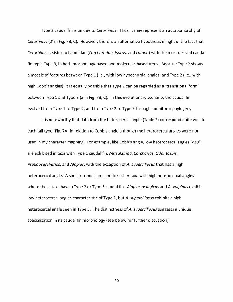
Figure 1. All fifteen modern lamniform species (after Shimada, 2005, fig. 1; bar scale = 50 cm). A, goblin shark (Mitsukurina owstoni); B, sandtiger shark (Carcharias taurus) C, smalltooth sandtiger (Odontaspis ferox); D, bigeye sandtiger (Odontaspis noronhai); E, crocodile shark (Pseudocarcharias kamoharai); F, megamouth shark (Megachasma pelagios); G, pelagic thresher (Alopias pelagicus); H, bigeye thresher (Alopias superciliosus); I, common thresher (Alopias vulpinus); J, basking shark (Cetorhinus maximus); K, great white shark (Carcharodon Carcharias); L, shortfin mako (Isurus oxyrinchus); M, longfin mako (Isurus paucus); N, salmon shark (Lamna ditropis); O, porbeagle (Lamna nasus).

31
Figure 2. Medical imaging technology used to radiographically examine shark specimens. A, CT scanner; B, X-ray machine.
A
B

32
Figure 3. Schematic drawings of lamniform caudal fin showing nomenclature and measured variables (anterior to left; see text for detail). A, terminology used in this study (abbreviations: DL, dorsal lobe; ER, epichordal ray; HR; hypochordal ray; PT, posterior tip; SM, subterminal margin; VC, vertebral column; VL, ventral lobe; VT, ventral tip); B, heterocercal angle (Het) and hypocercal angle (Hyp) that describes overall fin shape and spread; C, Cobb’s angle (CA) that describes curvature of vertebral column; D, hypochordal angle that describes orientation of longest hypochordal ray.
A B
C D
33
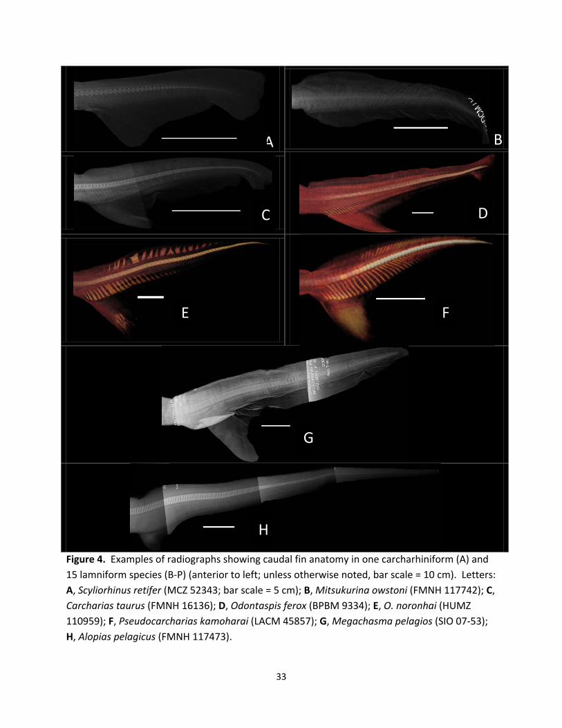
A B
Figure 4. Examples of radiographs showing caudal fin anatomy in one carcharhiniform (A) and 15 lamniform species (B-P) (anterior to left; unless otherwise noted, bar scale = 10 cm). Letters: A, Scyliorhinus retifer (MCZ 52343; bar scale = 5 cm); B, Mitsukurina owstoni (FMNH 117742); C, Carcharias taurus (FMNH 16136); D, Odontaspis ferox (BPBM 9334); E, O. noronhai (HUMZ 110959); F, Pseudocarcharias kamoharai (LACM 45857); G, Megachasma pelagios (SIO 07-53); H, Alopias pelagicus (FMNH 117473).
H
D C
E F
G

34
Figure 4 (continued). I, Alopias superciliosus (UF 160188); J, A. vulpinus (SIO 95-14); K, Cetorhinus maximus (MCZ 54413); L, Carcharodon carcharias (FMNH 38335); M, Isurus oxyrinchus (USNM 179570); N, I. paucus (UF 160174); O, Lamna ditropis (SIO 50-114); P, L. nasus (UMMZ 60412).
K L M
N O P
I
J

35
Figure 5. Mapping of average values of Cobb’s angle and hypochordal angle onto morphology-based phylogenetic tree of lamniforms (with one carcharhiniform taxon added as outgroup: see text for detail) along with schematic illustrations of caudal fin showing its outline and skeletal arrangement (not to scale). Abbreviations: SR, Scyliorhinus retifer; MO, Mitsukurina owstoni; CT, Carcharias taurus; OF, Odontaspis ferox; ON, Odontaspis noronhai; PK, Pseudocarcharias kamoharai; MP, Megachasma pelagios; AP, Alopias pelagicus; AS, A. superciliosus; AV, A. vulpinus; CM, Cetorhinus maximus; CC, Carcharodon carcharias; IO, Isurus oxyrinchus; IP, I. paucus; LD, Lamna ditropis; and LN, L. nasus.

36
Figure 6. Mapping of average values of Cobb’s and hypochordal angle onto molecular-based phylogenetic tree of lamniforms (with one carcharhiniform taxon added as outgroup: see text for detail) along with schematic illustrations of caudal fin showing its outline and skeletal arrangement (not to scale). Abbreviations: SR, Scyliorhinus retifer; MO, Mitsukurina owstoni; CT, Carcharias taurus; OF, Odontaspis ferox; ON, Odontaspis noronhai; PK, Pseudocarcharias kamoharai; MP, Megachasma pelagios; AP, Alopias pelagicus; AS, A. superciliosus; AV, A. vulpinus; CM, Cetorhinus maximus; CC, Carcharodon carcharias; IO, Isurus oxyrinchus; IP, I. paucus; LD, Lamna ditropis; and LN, L. nasus.

37
Figure 7. Three types of caudal fin in lamniforms (A) and their possible evolutionary scenarios (B, C; each number indicates each fin type; 2 and 2' indicate two alternative hypothesis; see text for detail). A, schematic diagram showing Type 1, 2, and 3 caudal fins (see text for detail; not to scale); B, morphology-based phylogenetic tree with three caudal fin types mapped; C, molecular-based phylogenetic tree with three caudal fin types mapped.
A Type 1 Type 2 Type 3
B C

38
Table 1. List of examined specimens in this study.
Species (Sample size) Catalogue Number TL (mm) Sex
Mitsukurina owstoni (n=2)
FMNH 117742 SIO 07-46
1,265 1,150
Female Male
Carcharias taurus (n=2)
FMNH 16136 MCZ 436
910 1,000
Male Female
Odontaspis ferox (n=1) BPBM 9334 1,900 Female
Odontaspis noronhai (n=1) HUMZ 110959 2,168 Male
Pseudocarcharias kamoharai (n=4) FMNH 117474 LACM 45857 USNM 303206 USNM 303207
1,011 922 930 1,020
Male female Male Male
Megachasma pelagios (n=1) SIO 07-53 2,149 Female
Alopias pelagicus (n=1) FMNH 117473 1,690 Female
Alopias superciliosus (n=1) UF 160188 1,872 Male
Alopias vulpinus (n=2) SIO 78-138 SIO 95-14
1,310 1,270
Male Female
Cetorhinus maximus (n=2) MCZ 54413 USNM 197870
3,850 Unknown
Female Unknown
Carcharodon carcharias (n=2) FMNH 38335 LACM 43805-1
2,714 1,261
Female Male
Isurus oxyrinchus (n=6) UMMZ 177116 UMMZ 94726 USNM 179570 USNM 185940 USNM 201733 USNM 201915
890 854 1,073 1,160 766 1,052
Female Male Male Male Female Female
Isurus paucus (n=1) UF160174 1,250 Male
Lamna ditropis (n=2) SIO 50-114 USNM 201731
749 791
Female Female
Lamna nasus (n=2) MCZ 37028 UMMZ 60412
1,150 1,060
Male Male
Scyliorhinus retifer (Carcharhiniformes: n=1) MCZ 52343 415 Male

39
Table 2. Average value of heterocercal angle, hypocercal angle, Cobb’s angle, and hypochordal angle data for each species (see Appendix 1 for raw measurements from each examined specimen). Heterocercal and hypocercal angles describe overall caudal fin shape whereas Cobb’s and hypochordal angles are skeletal measurements (see text for detail).
Species Heterocercal Angle Hypocercal Angle Cobb’s Angle Hypochordal Angle
Mitsukurina owstoni 4.3 36.1 5.8 61.2
Carcharias Taurus 3.2 42.8 7.9 66.5
Odontaspis ferox 8.2 45.8 15.0 60.9
Odontaspis noronhai 13.5 38.0 16.4 68.5
Pseudocarcharias kamoharai 7.4 48.1 16.5 59.7
Megachasma pelagios 6.4 39.6 8.1 65.0
Alopias pelagicus 11.0 60.0 5.5 137.1
Alopias superciliosus 33.8 31.7 20.0 137.4
Alopias vulpinus 8.2 58.1 6.4 128.6
Cetorhinus maximus 29.6 38.1 29.1 74.6
Carcharodon carcharias 33.4 42.1 28.5 111.6
Isurus oxyrinchus 33.2 47.3 37.3 106.3
Isurus paucus 33.6 40.8 30.5 94.3
Lamna ditropis 29.7 38.8 29.3 97.7
Lamna nasus 68.5 47.1 29.7 115.9
Scyliorhinus retifer (Carcharhiniformes) 1.6 39.1 1.1 59.5

40
Appendix 1. Heterocercal angle (Het), hypocercal angle (Hyp), caudal spread (CS: sum of heterocercal and hypocercal angles), Cobb’s angle (CA), hypochordal angle (HA), and presence (P) or absence (A) of keeled caudal peduncle (KCP) for each examined specimen.
Species Specimen Het (°) Hyp (°) CS (°) CA (°) HA (°) KCP
Mitsukurina owtoni SIO 07-46 FMNH 117742
3.9 4.7
36.6 35.5
40.5 40.2
5.6 5.9
* 61.2***
A A
Carcharias Taurus FMNH 16136 MCZ 436
4.5 1.8
42.3 43.3
46.8 45.1
6.9 8.9
66.8 66.1
A A
Odontaspis ferox BPBM 9334 8.2 45.8 54.0 15.0 60.9 A
Odontaspis noronhai HUMZ 110959 13.5 38.0 51.5 16.4 68.5 A
Pseudocarcharias kamoharai LACM 45857 USNM 303206 USNM 303207 FMNH 117474
6.8 5.8 8.3 8.5
44.1 44.4 50.1 53.6
50.9 50.2 58.4 62.1
17.0 17.0
* 15.5
60.9 57.7 60.2 60.1
A A A A
Megachasma pelagios SIO 07-53 6.4 39.6 46.0 8.1 65.0 A
Alopias pelagicus FMNH 117473 11.0 60.0 71.0 5.5 137.1 A
Alopias superciliosus UF 160188 33.8 31.7 65.5 20.0 137.4 A
Alopias vulpinus SIO 78-138 SIO 95-14
9.0 7.3
53.3 62.9
62.3 70.2
10.25 2.5
133.1 124.1
A A
Cetorhinus maximus USNM 197870 MCZ 54413
34.5 24.7
40 36.1
74.5 60.8
* 29.1
71.30*** 77.8
P P
Carcharodon carcharias LACM 43804 FMNH 38335
33.2 33.6
44.8 39.3
78.0 72.9
28.5 28.4
114.2 108.9
P P
Isurus oxyrinchus UMMZ 94726 UMMZ 177116 USNM 179570 USNM 185940 USNM 201733 USNM 201915
30.6 30.3 39.0 31.8 37.9 29.3
44.2 46.6 45.0 50.9 41.0 56.0
74.8 76.9 84.0 82.7 78.9 85.3
* *
39.0 *
28.5 44.5
94.3** 120.6**
111.3 103.2**
106.1 102.1
P P P P P P
Isurus paucus UF 160174 33.6 40.8 74.4 30.5 94.3 P
Lamna ditropis SIO 50-114 USNM 201731
* 29.7
* 38.8
* 68.5
28.0 30.5
100.4 94.9
P P
Lamna nasus UMMZ 60412 MCZ 37028
36.9 31.6
46.0 48.1
82.9 79.7
31.4 27.9
122.1** 109.6
P P
Scyliorhinus retifer (Carcharhiniformes)
MCZ 52343 1.6 39.1 40.7 1.1 59.5 A
* = unavailable data
**= approximation
*** = specimen examined by dissection

41
Appendix 2. Qualitative analysis of calcification level of caudal fin skeleton in each examined specimen. The calcification level (high or low) is reported here for caudal vertebrae, epichordal rays, and hypochordal rays. Analysis is based on radiograph images produced by CT and X-ray.
Species Catalogue Number Vertebrae Epichordal Rays Hypochordal Rays
Mitsukurina owstoni SIO 07-46 FMNH 117742
Low Low
Low Low
Low Low
Carcharias Taurus FMNH 16136 MCZ 436
High High
High High
High High
Odontaspis ferox BPBM 9334 High High High
Odontaspis noronhai HUMZ 110959 High High High
Pseudocarcharias kamoharai LACM 45857 USNM 303206 USNM 303207 FMNH 117474
High High High High
High High High High
High High High High
Megachasma pelagios SIO 07-53 Low Low Low
Alopias pelagicus FMNH 117473 High High High
Alopias superciliosus UF 160188 High High High
Alopias vulpinus SIO 78-138 SIO 95-14
High High
High High
High High
Cetorhinus maximus USNM 197870 MCZ 54413
Uncertain Low
Low Low
Low Low
Carcharodon carcharias LACM 43804 FMNH 38335
High High
Low Low
High High
Isurus oxyrinchus UMMZ 94726 UMMZ 177116 USNM 179570 USNM 185940 USNM 201733 USNM 201915
High High High High High High
Low Low Low Low Low Low
High High High High High High
Isurus paucus UF 160174 High Low High
Lamna ditropis SIO 50-114 USNM 201731
High High
Low Low
High High
Lamna nasus UMMZ 60412 MCZ 37028
High High
Low Low
High High
Scyliorhinus retifer (Carcharhiniformes)
MCZ 52343 High High High