Morphological Transformation of PS- b -PEO Diblock Copolymer by...
5
Morphological Transformation of PS-b-PEO Diblock Copolymer by Selectively Dispersed Colloidal CdS Quantum Dots Siao-Wei Yeh and Kung-Hwa Wei* Department of Materials Science and Engineering, National Chiao Tung University, Hsinchu, Taiwan 30049, ROC Ya-Sen Sun, U-Ser Jeng, and Keng S. Liang National Synchrotron Radiation Research Center, 101 Hsin-Ann Road, Science-Based Industrial Park, Hsinchu, Taiwan 30077, ROC Received June 11, 2003 Revised Manuscript Received August 28, 2003 Block copolymers are versatile platform materials because they can self-assemble into various nanostruc- tures with period thicknesses between 10 and 100 nm under the appropriate compositions and conditions, owing to microphase separation between incompatible blocks. 1-3 In recent years, a number of studies involving applications of nanostructured block copolymer as nanotemplate, 4-6 nanomasks for lithography, 7 and pho- tonic crystal 8 have been reported. Specifically, the work on nanometer-scale surface pattern with long-range order created by crystallization of asymmetric PB-b- PEO by Reiter 4b,c is an interesting one. In another case, sorting out different sizes of CdSe nanoparticles into PS- b-PMMA porous template with capillary force was carried out by Russell et al. 5a Several groups also reported the use of block copolymer as nanotemplates to grow Co, Ag, and Au nanowires 5b-d or to control the spatial locations of nanoparticles 6b,d such as TiO 2 and Pd. Moreover, block copolymer lithography with large area nanoscale patterning by Register et al. 7a,b has also been reported. These studies motivated us to investigate the interaction between the block copolymer and nano- particles, particularly on the effect of nanoparticles in the morphology of block copolymers. For semiconductor nanoparticles with sizes close to their Bohr exciton radius (typically between 1 and 10 nm), the size-dependent band gap results in tunable optical properties. 9,10 These semiconductor nanoparticles are termed quantum dots (QDs) because their tunable optical properties can be predicted by quantum mechan- ics. Nanoparticles that are not treated with a surfactant or bonded to polymer chains will, however, form large aggregates. The use of nanostructured block copolymers as tem- plates to selectively control the spatial location of semiconductor nanoparticles in one of the blocks may lead to several potential applications. For instance, periodic high refractive index contrast domains in phase-separated block copolymers can be used in pho- tonic crystal applications. Nanoparticles with highly efficient luminescence can be combined with organic layers in light-emitting devices. 11 The incorporation of nanoparticles into block copolymers, however, would lead to more complicated block copolymer morphologies than their pristine state as predicted by Balazs’group, 12 which used a self-consistent-field theory and a density functional theory for describing the polymer and the nanoparticles, respectively, to predict the morphology and phase diagram. In this Communication, we report the selective distribution of CdS QDs in the PEO block of a diblock copolymer, PS-b-PEO, and the resultant morphological changes. Specifically, CdS QDs induce the PEO domains to change from hexagonally packed cylinders to body-centered-cubic or simple-cubic spheres, as shown in Scheme 1. To our knowledge, this is the first study concerning the morphological transformation of block copolymer by nanoparticles. For the present study, an asymmetric polystyrene-b- poly(ethylene oxide) diblock copolymer (PS-b-PEO) with a molecular weight ratio of 125K/16.1K was purchased from Polymersource Inc. The volume fraction of PEO in this PS-b-PEO is 0.11, with a polydispersity of 1.04. CdS nanoparticles were synthesized with mercaptoet- hanol as the surfactant by reacting cadmium acetate dehydrate (Cd(Ac) 2 ‚2H 2 O), sodium sulfide (Na 2 S), and mercaptoethanol (HSC 2 H 4 OH) in methanol, following a modification of the kinetic trapping method. 10 After filtration, CdS QDs were collected and then dispersed in N,N-dimethylformamide (DMF). In our case, surfac- tant-modified CdS containing a chemically active hy- droxyl surface, as shown in Scheme 1, became hydro- philic, and their basic properties are given in Table 1. In Table 1, the averaged size of CdS QDs is about 2.5 nm as determined from SAXS curves of CdS/PS-b-PEO in Figure 1 because in the higher Q region (Q > 0.07 Å -1 ) where the scattering is dominated by form factor, the SAXS curves can be modeled using a sphere form factor of radius 2.5 nm. The sizes of CdS calculated from the onset absorption of the UV-vis spectrum typically represent the larger size in the size distribution curve of CdS in DMF. 6 Moreover, the CdS size obtained from the X-ray diffraction is the crystal size, which is the smallest and corresponds to the fact that we have polycrystal CdS. Subsequently, CdS/DMF was added to a previously prepared PS-b-PEO/DMF solution under stirring. This mixture was dried slowly under vacuum at 323 K and then maintained at 383 K for 24 h, after which the CdS/PS-b-PEO nanocomposite film was ob- tained. Preparation of pure PS-b-PEO films is similar to that of CdS/PS-b-PEO, except the lack of CdS. Thermal gravity analysis had been used to check the presence of the residual DMF solvent, and there was * To whom correspondence should be addressed: Tel 886-35- 731871; Fax 886-35-724727; e-mail [email protected]. Scheme 1. Morphological Transformation of PS-b-PEO Diblock Copolymer by Selectively Dispersed Colloidal CdS QDs 7903 Macromolecules 2003, 36, 7903-7907 10.1021/ma034800g CCC: $25.00 © 2003 American Chemical Society Published on Web 09/18/2003
Transcript of Morphological Transformation of PS- b -PEO Diblock Copolymer by...
Morphological Transformation of PS-b-PEODiblock Copolymer by Selectively DispersedColloidal CdS Quantum Dots
Siao-Wei Yeh and Kung-Hwa Wei*
Department of Materials Science and Engineering, NationalChiao Tung University, Hsinchu, Taiwan 30049, ROC
Ya-Sen Sun, U-Ser Jeng, and Keng S. Liang
National Synchrotron Radiation Research Center,101 Hsin-Ann Road, Science-Based Industrial Park,Hsinchu, Taiwan 30077, ROC
Received June 11, 2003Revised Manuscript Received August 28, 2003
Block copolymers are versatile platform materialsbecause they can self-assemble into various nanostruc-tures with period thicknesses between 10 and 100 nmunder the appropriate compositions and conditions,owing to microphase separation between incompatibleblocks.1-3 In recent years, a number of studies involvingapplications of nanostructured block copolymer asnanotemplate,4-6 nanomasks for lithography,7 and pho-tonic crystal8 have been reported. Specifically, the workon nanometer-scale surface pattern with long-rangeorder created by crystallization of asymmetric PB-b-PEO by Reiter4b,c is an interesting one. In another case,sorting out different sizes of CdSe nanoparticles into PS-b-PMMA porous template with capillary force wascarried out by Russell et al.5a Several groups alsoreported the use of block copolymer as nanotemplatesto grow Co, Ag, and Au nanowires5b-d or to control thespatial locations of nanoparticles6b,d such as TiO2 andPd. Moreover, block copolymer lithography with largearea nanoscale patterning by Register et al.7a,b has alsobeen reported. These studies motivated us to investigatethe interaction between the block copolymer and nano-particles, particularly on the effect of nanoparticles inthe morphology of block copolymers.
For semiconductor nanoparticles with sizes close totheir Bohr exciton radius (typically between 1 and 10nm), the size-dependent band gap results in tunableoptical properties.9,10 These semiconductor nanoparticlesare termed quantum dots (QDs) because their tunableoptical properties can be predicted by quantum mechan-ics. Nanoparticles that are not treated with a surfactantor bonded to polymer chains will, however, form largeaggregates.
The use of nanostructured block copolymers as tem-plates to selectively control the spatial location ofsemiconductor nanoparticles in one of the blocks maylead to several potential applications. For instance,periodic high refractive index contrast domains inphase-separated block copolymers can be used in pho-tonic crystal applications. Nanoparticles with highlyefficient luminescence can be combined with organiclayers in light-emitting devices.11 The incorporation ofnanoparticles into block copolymers, however, wouldlead to more complicated block copolymer morphologiesthan their pristine state as predicted by Balazs’group,12
which used a self-consistent-field theory and a density
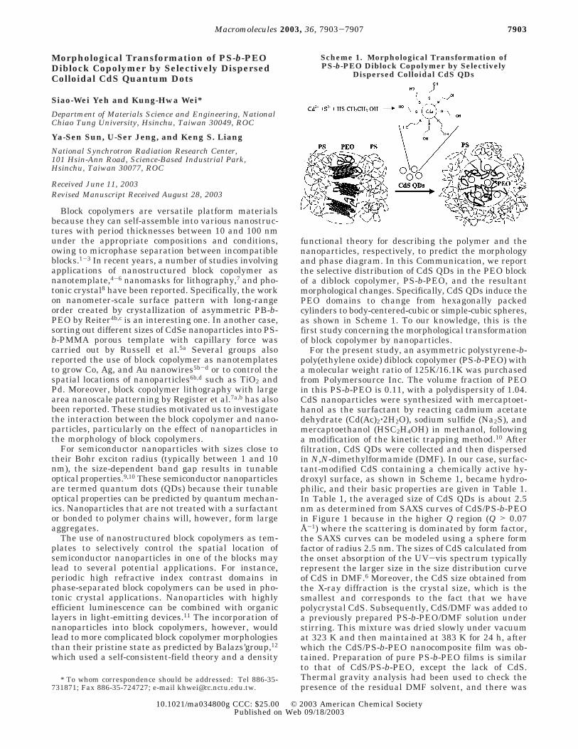
functional theory for describing the polymer and thenanoparticles, respectively, to predict the morphologyand phase diagram. In this Communication, we reportthe selective distribution of CdS QDs in the PEO blockof a diblock copolymer, PS-b-PEO, and the resultantmorphological changes. Specifically, CdS QDs induce thePEO domains to change from hexagonally packedcylinders to body-centered-cubic or simple-cubic spheres,as shown in Scheme 1. To our knowledge, this is thefirst study concerning the morphological transformationof block copolymer by nanoparticles.
For the present study, an asymmetric polystyrene-b-poly(ethylene oxide) diblock copolymer (PS-b-PEO) witha molecular weight ratio of 125K/16.1K was purchasedfrom Polymersource Inc. The volume fraction of PEOin this PS-b-PEO is 0.11, with a polydispersity of 1.04.CdS nanoparticles were synthesized with mercaptoet-hanol as the surfactant by reacting cadmium acetatedehydrate (Cd(Ac)2‚2H2O), sodium sulfide (Na2S), andmercaptoethanol (HSC2H4OH) in methanol, followinga modification of the kinetic trapping method.10 Afterfiltration, CdS QDs were collected and then dispersedin N,N-dimethylformamide (DMF). In our case, surfac-tant-modified CdS containing a chemically active hy-droxyl surface, as shown in Scheme 1, became hydro-philic, and their basic properties are given in Table 1.In Table 1, the averaged size of CdS QDs is about 2.5nm as determined from SAXS curves of CdS/PS-b-PEOin Figure 1 because in the higher Q region (Q > 0.07Å-1) where the scattering is dominated by form factor,the SAXS curves can be modeled using a sphere formfactor of radius 2.5 nm. The sizes of CdS calculated fromthe onset absorption of the UV-vis spectrum typicallyrepresent the larger size in the size distribution curveof CdS in DMF.6 Moreover, the CdS size obtained fromthe X-ray diffraction is the crystal size, which is thesmallest and corresponds to the fact that we havepolycrystal CdS. Subsequently, CdS/DMF was added toa previously prepared PS-b-PEO/DMF solution understirring. This mixture was dried slowly under vacuumat 323 K and then maintained at 383 K for 24 h, afterwhich the CdS/PS-b-PEO nanocomposite film was ob-tained. Preparation of pure PS-b-PEO films is similarto that of CdS/PS-b-PEO, except the lack of CdS.Thermal gravity analysis had been used to check thepresence of the residual DMF solvent, and there was
* To whom correspondence should be addressed: Tel 886-35-731871; Fax 886-35-724727; e-mail [email protected].
Scheme 1. Morphological Transformation ofPS-b-PEO Diblock Copolymer by Selectively
Dispersed Colloidal CdS QDs
7903Macromolecules 2003, 36, 7903-7907
10.1021/ma034800g CCC: $25.00 © 2003 American Chemical SocietyPublished on Web 09/18/2003

no residual DMF present in these block copolymernanocomposites.
Small-angle X-ray scattering experiments were per-formed on wiggler beamline BL-17B1 at the NationalSynchrotron Radiation Research Center, Taiwan. Wide-angle X-ray diffraction on samples was collected byusing a conventional rotating anode source. For trans-mission electron microscopy (TEM) and atomic forcemicroscopy (AFM), the thin specimens were microtomedwith Leica Ultracut Uct. TEM images were obtainedwith Hitachi H-600 transmission electron microscope.AFM measurements were performed in tapping modewith a Digital Nanoscope IIIa under ambient conditions.The glass transition temperatures (Tg) and meltingpoint (Tm) were obtained from a Dupont DSC 2910 at aheating rate of 20 °C/min. Photoluminescence spectra(PL) were obtained with a Hitachi F4500 fluorescencespectrophotometer at room temperature.
Figure 1 shows one-dimensional small-angle X-rayscattering patterns (SAXS) of PS-b-PEO and CdS/PS-b-PEO nanocomposites by synchrotron radiation. Forpure PS-b-PEO, four peaks appear at Q ) 0.016, 0.027,0.032, and 0.042 Å-1, corresponding to a ratio of 1:31/2:41/2:71/2. This ratio indicates typical scattering by hex-agonally packed cylinders (HEX). The intercylinderdistance (D) was determined to be 45.3 nm by eq 1.
where d100 ) 2π/Q100 and Q100 ) 0.16 nm-1. In the caseof PS-b-PEO containing 0.35% CdS, the scattering peaksare located at Q ) 0.0114, 0.0158, 0.0203, 0.0228,0.0281, and 0.0399 Å -1, which gives a ratio of 1: 21/2:31/2:41/2:61/2:121/2. This ratio implies that the scatteringis caused by either body-centered-cubic packed spheres
(bcc) or simple-cubic spheres (sc). The interspheredistance is 67.5 nm, as determined from eq 2.
where d110 ) 2π/Q110 and Q110 ) 0.114 nm-1. SAXSresults confirm that the nanostructured HEX structureof pure PS-b-PEO has been transformed to a bcc or scmorphology due to the presence of CdS QDs. The sizeof CdS QDs in the block copolymer is about 2.5 nm, asderived from the structure form of the SAXS curve.
Parts a and b of Figure 2 show the transmissionelectron microscopy (TEM) images of PS-b-PEO stainedwith OsO4 and CdS/PS-b-PEO without staining, respec-tively. PEO domains appear in dark phase in Figure2a, owing to selective staining, and display as shortcylinders. In the case of CdS/PS-b-PEO, however, peri-odic dark spherical phases of CdS-included PEO appear,and no pure PEO domains without CdS QDs could beobserved. The location of CdS in the PEO domain isrevealed by energy dispersive spectrometry. Dark phasesare caused by the higher electron density of cadmiumrelative to that of PS-b-PEO. The selective distributionof mercaptoethanol-modified CdS in the PEO domainis quite possibly due to dipole-dipole interactionsbetween the hydroxyl groups of mercaptoethanol andthe PEO block. The diameter of CdS-included PEOspheres is approximately 23 nm and the interspheredistance is about 60 nm, as estimated from their TEMimages. To cover all PEO domains, there must be somedistributions of CdS QDs in each PEO domain becausethe volume fraction of added CdS with respect to thePEO block is about 2.7%, which is not enough to covereach PEO domain fully. It is, however, not possible todetect them with the current techniques.
The results from TEM analysis are consistent withthose by SAXS. Further evidence of the two differentmorphologies can be found in phase-contrast atomicforce microscopy images (AFM) of PS-b-PEO and CdS/PS-b-PEO samples, as shown in parts c and d of Figure2, respectively. A diamond knife used during the mi-crotoming process causes the oblique lines in thesefigures. The difference in the images of pure PS-b-PEOby TEM and AFM (Figure 2a,c) could be explained bythe fact that the dark PEO domains which appear asinclined short cylinders in bulk can be projected intocylindrical shapes in the transmission electron micros-copy study, whereas the topology of the same micro-tomed slice would shows hairy spherical images by thetapping mode of AFM.
Figure 3a shows the thermal analysis results of PS-b-PEO and CdS/PS-b-PEO by differential scanningcalorimetry. A crystal melting peak at 42.9 °C associatedwith the PEO domain and a glass transition tempera-ture (Tg) at 99 °C attributed to the PS domain appearin the pure PS-b-PEO case. (The amorphous phase ofPEO is rather small and undetectable.) In the presenceof CdS, the crystal melting peak of PEO depressed anddiminished to a small kink, with an apparent Tg of-56.6 °C. The PS domain, however, maintains a Tg ofabout 99 °C. The difference between the two cases canbe explained by a loss of crystallinity in the PEO domainby the infiltration of CdS QDs into the PEO domain.Moreover, the wide-angle X-ray diffraction results shownin Figure 3b,c also support the corresponding crystal-to-amorphous change of the PEO domain. In the caseof pure PS-b-PEO, two sharp peaks at 1.35 and 1.63 Å-1
Table 1. Characteristic Properties of CdS Nanoparticles
absorptiononset (λo)a
particle sizein DMFb
emissionwavelengthc
crystalsized
av particlesizee
447 nm 3.37 nm 650 nm 1.33 nm 2.5 nma The onset absorption wavelength of CdS nanoparticles in DMF
as obtained from UV-vis spectra. b The sizes of CdS nanoparticlesin DMF were calculated with the onset absorption obtained byUV-vis spectra using the following equation:6,9,10Eg,os - Eg,ref )∆Eg ≈ (p2π2/2R2)(1/µ) - (1.8e2/εR). c The emission wavelength isdetermined from the photoluminescence. d Crystal sizes are cal-culated using the Debye-Scherrer equation. e The averaged sizeof CdS was obtained from SAXS curves of CdS/PS-b-PEO in Figure1.
Figure 1. Synchrotron SAXS curves of PS-b-PEO and CdS/PS-b-PEO.
D ) (4/3)1/2d100 (1)
D ) (3/2)1/2d100 (2)
7904 Communications to the Editor Macromolecules, Vol. 36, No. 21, 2003

represent the crystallinity peaks of the PEO domainafter deconvolution, as shown in Figure 3b. Figure 3cshows amorphous X-ray diffraction curves when CdSis incorporated into the PEO domain, indicating thechange from crystalline to amorphous PEO. This resultis also consistent with those obtained by DSC. Thepossible scenario for this phenomenon is due to therelatively small size of CdS to the contour length of PEOblock (2.5 nm vs 130 nm) and the dipole-dipole interac-tion between the angling hydroxy group of surface-attached mercaptoethanol on CdS and ethylene oxidein the PEO domain; CdS is tethering to the PEO chainand appears to destroy the crystallinity of the PEOdomain, as shown in Scheme 1. This enables CdS-infiltrated PEO domains to be amorphous and tominimize their surface energy by forming either bcc orsc structures.
When only the volume fraction of the block copolymerPS-b-PEO is considered, it is true that in the equilib-rium state the pure PS-b-PEO should be in the sphericalregion with body-centered-cubic packing due to strong
segregation.13 The morphology of asymmetric amor-phous-crystalline block copolymers, however, dependson both the microphase separation of the two blocks andthe crystallization kinetics of the crystallizable block asdemonstrated in the work by de Jeu et al.14 In theirstudy, lamellar or hexagonally perforated lamellarstructures of PS-b-PEO were obtained even though thevolume fraction of PS-b-PEO indicates that it is typicallywithin the morphological range of sphere or cylinders.In our study, the hexagonally packed cylindrical mor-phology of PS-b-PEO represents a compromise betweenthe microphase separation involving PS and PEO blocksand the crystallization kinetics of the PEO block.13,14
The cylindrical morphology of pure PS-b-PEO is in ametastable state due to the fast crystallization of thePEO block. The addition of CdS quantum dots into thediblock copolymer inhibits the crystallization of PEOblock. The resultant morphology of CdS/PS-b-PEOsample, therefore, is determined largely by the mi-crophase separation involving PS block and CdS/PEOblock, (i.e., the crystallization effect no longer exists).
Figure 2. (a) TEM image of PS-b-PEO stained by OsO4. The dark regions correspond to PEO phases stained with OsO4. (b) TEMimage of CdS/PS-b-PEO without staining. (c) AFM images of thin films microtomed from bulk PS-b-PEO and (d) AFM images ofthin films microtomed from bulk CdS/PS-b-PEO.
Macromolecules, Vol. 36, No. 21, 2003 Communications to the Editor 7905

Theoretically, it is possible to observe the morphologicaltransformation of pure PS-b-PEO, providing two re-quirements are satisfiedsat high temperature (farabove Tm of PEO and Tg of PS) and long time (diffusionrate of polymer melt is very small). In reality, thisphenomenon may not happen for pure PS-b-PEO sincethe block copolymer might start to degrade if it weremaintained at high temperature for too long.
Photoluminescence spectra of CdS QDs in DMF andthe CdS/PS-b-PEO nanocomposite film are shown inFigure 4. CdS QDs in DMF emit red light with amaximum intensity located at 650 nm. The peakmaximum of CdS in the PEO block of PS-b-PEO is red-shifted by 30 nm compared to CdS in DMF. The red shiftin the luminescence of CdS QDs might be caused byaggregation or the different chemical environment.10
In conclusion, semiconductor CdS QDs can be selec-tively dispersed in the PEO domain of PS-b-PEO blockcopolymer by using a surfactant. The CdS-infiltratedPEO domains are transformed from originally hexago-nally packed cylindrical structures to bcc or sc struc-tures because CdS inhibits the crystallization andminimizes the surface energy of CdS-infiltrated PEOphase. The photoluminescence of CdS is slightly affectedby their incorporation in PS-b-PEO.
Acknowledgment. The authors acknowledge theNational Science Council for funding this work throughNSC 91-2120-M-009-001. Mr. Tsung-Lun Wu is appreci-ated for his help in experiments and discussions.
References and Notes
(1) (a) Bates, F. S.; Fredrickson, G. H. Annu. Rev. Phys. Chem.1990, 41, 525. (b) Bates, F. S. Science 1991, 251, 898.
(2) (a) Zhu, L.; Stephen, Z. D.; Cheng; Huang, P.; Ge, Q.; Quirk,R. P.; Thomas, E. L.; Lotz, B.; Hsiao, B. S.; Yeh, F.; Liu, L.Adv. Mater. 2002, 14, 31. (b) Choi, S.; Lee, K. M.; Han, C.D.; Sota, N.; Hashimoto, T. Macromolecules 2003, 36, 793.(c) Buck, E.; Fuhrmann, J. Macromolecules 2001, 34, 2172.
(3) (a) Loo, Y. L.; Register, R. A.; Ryan, A. J. Macromolecules2002, 35, 2365. (b) Loo, Y. L.; Register, R. A.; Ryan, A. J.;Dee, G. T. Macromolecules 2001, 34, 8968. (c) Rottele, A.;Thurn-Albrecht, T.; Sommer, J. U.; Reiter, G. Macromol-ecules 2003, 36, 1257.
(4) (a) Kim, D. H.; Lin, Z.; Kim, H. C.; Jeong, U.; Russell, T. P.Adv. Mater. 2003, 15, 811. (b) Reiter, G.; Castelein, G.;Sommer, J. U.; Rottele, A.; Thurn-Albrecht, T. Phys. Rev.Lett. 2001, 87, 226101. (c) Reiter, G.; Castelein, G.; Hoerner,P.; Riess, G.; Blumen, A.; Sommer, J. U. Phys. Rev. Lett.1999, 83, 3844. (d) Boker, A.; Knoll, A.; Elbs, H.; Abetz, V.;Muller, A. H. E.; Krausch, G. Macromolecules 2002, 35,1319.
(5) (a) Misner, M. J.; Skaff, H.; Emrick, T.; Russell, T. P. Adv.Mater. 2003, 15, 221. (b) Thurn-Aibrecht, T.; Schotter, J.;Castle, G. A.; Emley, N.; Shibauchi, T.; Krusin-Elbaum, L.;Guarini, K.; Black, C. T.; Touminen, M. T.; Russell, T. P.Science 2000, 290, 2126. (c) Lopes, W. A. Phys. Rev. E 2002,65, 31606. (d) Lopes, W. A.; Jaeger, H. M. Nature (London)2001, 414, 735.
(6) (a) Moffitt, M.; Vali, H.; Eisenberg, A. Chem. Mater. 1998,10, 1021. (b) Weng, C. C.; Wei, K. H. Chem. Mater. 2003,15, 2936. (c) Bockstaller, M. R.; Lapetnikov, Y.; Margel, S.;Thomas, E. L. J. Am. Chem. Soc. 2003, 125, 5276. (d) Ribbe,A. E.; Okumura, A.; Matsushige, K.; Hashimoto, T. Macro-molecules 2001, 34, 8239.
(7) (a) Park, M.; Chaikin, P. M.; Register, R. A.; Adamson, D.H. Appl. Phys. Lett. 2001, 79, 257. (b) Park, M.; Harrison,
Figure 3. (a) DSC analysis of CdS/PS-b-PEO and PS-b-PEO.The samples were heated from -90 to 130 °C at 10 °C/min.(b) Deconvolution curves of the WAXS curve of crystallinityPS-b-PEO. (c) WAXS of PS-b-PEO and CdS/PS-b-PEO aftercrystallization at -20 °C for 18 h.
Figure 4. Photoluminescence of CdS and CdS/PS-b-PEO afterexcitation with 430 nm light.
7906 Communications to the Editor Macromolecules, Vol. 36, No. 21, 2003

C.; Chaikin, P. M.; Register, R. A.; Adamson, D. H. Science1997, 276, 1401. (c) Kim, H. C.; Jia, X.; Stafford, C. M.; Kim,D. H.; McCarthy, T. J.; Tuominen, M.; Hawker, C. J.;Russell, T. P. Adv. Mater. 2001, 13, 795. (d) Cheng, J. Y.;Ross, C. A.; Thomas, E. L.; Smith, H. I.; Vancso, G. J. Appl.Phys. Lett. 2002, 81, 3657.
(8) Urbas, A. M.; Maldovan, M.; DeRege, P.; Thomas, E. L. Adv.Mater. 2002, 14, 1850.
(9) (a) Zhang, J. Z. Acc. Chem. Res. 1997, 30, 423. (b) Brus, L.J. Phys. Chem. 1986, 90, 2555.
(10) (a) Veinot, J. G. C.; Ginzburg, M.; Pietro, W. J. Chem. Mater.1997, 9, 2117. (b) Herron, N.; Wang, Y.; Eckert, H. J. Am.Chem. Soc. 1990, 112, 1322.
(11) (a) Gao, M. Y.; Richter, B.; Kirstein, S. Adv. Mater. 1997, 9,802. (b) Gao, M. Y.; Richter, B.; Kirstein, S.; Mohwald, H.J. Phys. Chem. B 1998, 102, 4096.
(12) (a) Thompson, R. B.; Ginzburg, V. V.; Matsen, M. W.; Balazs,A. C. Science 2001, 292, 2469. (b) Thompson, R. B.; Gin-zburg, V. V.; Matsen, M. W.; Balazs, A. C. Macromolecules2002, 35, 1060. (c) Lee, J. Y.; Thompson, R. B.; Jasnow, D.;Balazs, A. C. Macromolecules 2002, 35, 4855.
(13) Hamley, I. W. The Physics of the Block Copolymers, OxfordUniversity Press: New York, 1998.
(14) Li, L.; Serero, Y.; Koch, M. H. J.; de Jeu, W. H. Macromol-ecules 2003, 36, 529.MA034800G
Macromolecules, Vol. 36, No. 21, 2003 Communications to the Editor 7907