Morphological and molecular modifications induced by heat...
16
/. Embryol. exp. Morph. 77, 167-182 (1983) \(fj Printed in Great Britain © The Company of Biologists Limited 1983 Morphological and molecular modifications induced by heat shock in Drosophila melanogaster embryos By GIORGIO GRAZIOSI 1 , FRANCO DE CRISTINI, ANGELO DI MARCOTULLIO, ROBERTO MARZARI, FULVIO MICALI AND ADRIANO SAVOINI From the Istituto di Zoologia e Anatomia Comparata, Universitd di Trieste, Italy SUMMARY The early embryo of Drosophila melanogaster did not survive treatment at 37 °C (heat shock) for 25 min. The histological analysis of eggs treated in this way showed that the heat shock caused disintegration of nuclei and of cytoplasmic islands, displacement and swelling of nuclei and blocked mitoses. These effects were not observed in embryos treated after blasto- derm formation. After this stage, we noticed that development was slowed down. The heat shock proteins (hsp 83,70 and 68) were, under shock, synthesized at all developmental stages. There was little or no synthesis of hsp 70 and 68 in unfertilized eggs, but synthesis increased in proportion to the number of nuclei present. Most probably, hsp 70 synthesis was directed by zygotic mRNA. DNA synthesis was not blocked by the heat shock though the overall incorporation of pH]thymidine was substantially reduced, presumably because of the block of mitoses. We did not find a direct relation between survival pattern and hsp synthesis. We concluded that some, at least, of the heat shock genes can be activated at all developmental stages and that heat shock could be used for synchronizing mitoses. INTRODUCTION The usual temperature for raising Drosophila melanogaster is around 24 °C though this organism can also live at temperatures ranging from 15 to 34 °C (Hedman & Krogstad, 1963; Powsner, 1935). Thisflycan also survive relatively short exposure to more extreme temperatures such as 4°C or 40 °C. During the exposure to high temperature, usually around 37 °C, Drosophila cells undergo considerable molecular and physiological changes (Ashburner & Bonner, 1979, review). Transcription and translation of the genes which were active before the heat shock are halted (Bonner & Pardue, 1976; Lindquist, 1980,1981; McKen- zie, Henikoff & Meselson, 1975; Spradling, Penman & Pardue, 1975), while a newly synthesized set of RNAs is translated into at least seven different proteins (Mirault et al. 1978; Tissieres, Mitchell & Tracy, 1974; Henikoff & Meselson, 1977; Spradling, Pardue & Penman, 1977). 1 Author's address: Istituto di Zoologia e Anatomia Comparata, Casella Universita, 34100 Trieste, Italy.
Transcript of Morphological and molecular modifications induced by heat...
/ . Embryol. exp. Morph. 77, 167-182 (1983) \(fj
Printed in Great Britain © The Company of Biologists Limited 1983
Morphological and molecular modifications inducedby heat shock in Drosophila melanogaster embryos
By GIORGIO GRAZIOSI1, FRANCO DE CRISTINI, ANGELODI MARCOTULLIO, ROBERTO MARZARI, FULVIO
MICALI AND ADRIANO SAVOINI
From the Istituto di Zoologia e Anatomia Comparata, Universitd di Trieste,Italy
SUMMARY
The early embryo of Drosophila melanogaster did not survive treatment at 37 °C (heatshock) for 25 min. The histological analysis of eggs treated in this way showed that the heatshock caused disintegration of nuclei and of cytoplasmic islands, displacement and swelling ofnuclei and blocked mitoses. These effects were not observed in embryos treated after blasto-derm formation. After this stage, we noticed that development was slowed down. The heatshock proteins (hsp 83,70 and 68) were, under shock, synthesized at all developmental stages.There was little or no synthesis of hsp 70 and 68 in unfertilized eggs, but synthesis increasedin proportion to the number of nuclei present. Most probably, hsp 70 synthesis was directedby zygotic mRNA. DNA synthesis was not blocked by the heat shock though the overallincorporation of pH]thymidine was substantially reduced, presumably because of the blockof mitoses. We did not find a direct relation between survival pattern and hsp synthesis. Weconcluded that some, at least, of the heat shock genes can be activated at all developmentalstages and that heat shock could be used for synchronizing mitoses.
INTRODUCTION
The usual temperature for raising Drosophila melanogaster is around 24 °Cthough this organism can also live at temperatures ranging from 15 to 34 °C(Hedman & Krogstad, 1963; Powsner, 1935). This fly can also survive relativelyshort exposure to more extreme temperatures such as 4°C or 40 °C. During theexposure to high temperature, usually around 37 °C, Drosophila cells undergoconsiderable molecular and physiological changes (Ashburner & Bonner, 1979,review). Transcription and translation of the genes which were active before theheat shock are halted (Bonner & Pardue, 1976; Lindquist, 1980,1981; McKen-zie, Henikoff & Meselson, 1975; Spradling, Penman & Pardue, 1975), while anewly synthesized set of RNAs is translated into at least seven different proteins(Mirault et al. 1978; Tissieres, Mitchell & Tracy, 1974; Henikoff & Meselson,1977; Spradling, Pardue & Penman, 1977).
1 Author's address: Istituto di Zoologia e Anatomia Comparata, Casella Universita, 34100Trieste, Italy.
168 G. GRAZIOSI AND OTHERS
Such a response has been described for tissue culture cells and for variousorgans at various developmental stages of Drosophila (Chomyn, Moller &Mitchell, 1979; McKenzie et al. 1975; Lewis, Helmsing & Ashburner, 1975;Peterson, Moller & Mitchell, 1979) and it has been found in all the eucarioticorganisms so far tested (Barnett, Altschuler, McDaniel & Mascarenhas, 1980;Fink & Zuethen, 1970; Giudice, Roccheri & Di Bernardo, 1980; Kelley, Aliperti& Schlesinger, 1980; McAlister & Finkelstein, 1980). Early embryos are the onlyknown possible exception to this response: the Drosophila embryo before blasto-derm formation (Dura, 1981; Graziosi et al. 1980), and the sea urchin embryobefore gastrulation (Giudice et al. 1980) synthesize little or no heat shockproteins. The fly embryo response to heat shock also differs from otherDrosophila systems in other respects. Hsp 83 has been found in large amountsas a cold protein in eggs kept at normal temperature, although its synthesis canbe elicited by a heat shock of 30min (Graziosi et al. 1980; Savoini et al. 1981).Furthermore, early embryos are extremely sensitive to the exposure to hightemperatures: 15min of treatment cause a very high mortality rate as well asphenocopies (Mitchell & Lipps, 1978; Santamaria, 1979).
We undertook this study to investigate this high mortality rate in early em-bryos and, by quantitating the [35S]methionine-labelled hsps, to correlate thesurvival pattern with hsp synthesis.
MATERIALS AND METHODS
Egg harvesting and fly maintenance
Eggs were collected from a population cage of roughly 5000 flies. The layingpopulation was kept under a fixed regime of food supply and of light/dark cycleto ensure the deposition of a large amount of eggs between 10 am and 4 pm. Thelight was switched on at 5 am and switched off at 5 pm. Fresh baker's yeastdeposited on Petri dishes of standard Drosophila food was offered every hour;starting at 9 am until 5 pm. The feeding dishes were also used for harvesting eggs,but they were left in the cage for lOmin only. An average age of 5min wasassumed when the eggs were withdrawn from the cage. The mass collectionswere used for the survival curve and they contained 5-10 % of eggs in advanceddevelopmental stages. The eggs used for two-dimensional electrophoresis andfor the histological analysis were laid over a period of 5 min and were individuallychecked for age.
A small cage containing a few hundred flies was prepared to collect eggs fromthe cross of heterozygote flies Df (3R) 229, Df (3R) kar33 (Ish-Horowicz et al.1979).
Egg labelling and heat shock
Upon removal of the chorion membrane with 20 % sodium hypochlorite andseveral washes in Drosophila saline, the eggs were permeabilized according to
Modifications induced by heat shock in Drosophila embryos 169the method of Limbourg & Zalokar (1973). Then the eggs were covered with30 fi\ of incubation medium and transferred either to 24 °C (controls), or to 37 °C.[35S]methionine or [3H]thymidine (final concentrations of 1 mCi/ml and 0-5 mCi/ml respectively) were added 10 min after the transfer of the eggs to the desiredtemperature. Similarly the controls had a preincubation of 10 min at 24 °C beforeaddition of the label. At the end of the incubation, the eggs were washed in coldmedium and either fixed for the histological analysis or sonicated for proteinextraction.
Electrophoresis and fluorography
Protein extracts underwent two-dimensional electrophoresis as reported byO'Farrell (1975). After sonication, the samples were freeze-dried, resuspendedin 50 /il of lysis buffer, centrifuged for 30 min at 50 000 g and 3 x 1 //I aliquots ofthe supernatant were used to count the total radioactivity in the TCA precipit-able material. All first dimensions were loaded with 100 000 c.p.m. All the otherelectrophoretic parameters were the same as reported by Savoini et al. (1981).
Since hsps 70 and 68 did not always appear as stainable spots, their foci wereidentified matching the stained gels with autoradiographs run for other purposes(Graziosi et al. 1980; Savoini et al. 1981). Hsp 83 and /3-tubulin were alwaysrecognized as consistent stainable spots. The protein spots were cut out, placedin 1 ml of 30 % H2O2 and evaporated at 60 °C until dry, resuspended in scintilla-tion liquid and counted.
Single eggs were analysed on 9 % polyacrylamide slab gels using the discon-tinuous sodium dodecyl sulphate (SDS) method of Laemmli (1970). These gelsunderwent fluorography as reported by Bonner & Laskey (1974) using Kodakfilm RP X-Omat.
Histology and autoradiography
Both chorion and vitelline membranes were removed from the eggs for his-tological analysis. After fixation using the method of Zalokar & Erk (1977), theeggs were embedded in paraffin and sectioned at 8 /im. The sections were stainedwith haematoxilin and eosin for 10 and 2 min respectively and permanentlymounted in Canada balsam.
The autoradiographs were prepared by dipping the slides in the photographicemulsion Ilford K5 diluted 1:2 with water at 37 °C. The excess of emulsion wasdrained by leaving the slides vertical until completely dry. All slides were ex-posed for 7 days and developed by standard methods. The histological sectionsused for the autoradiography were stained after exposure and development.
RESULTS
Embryonic viability
A total of 5452 eggs, subdivided into 43 experimental groups, was heat
170 G. G R A Z I O S I A N D O T H E R S
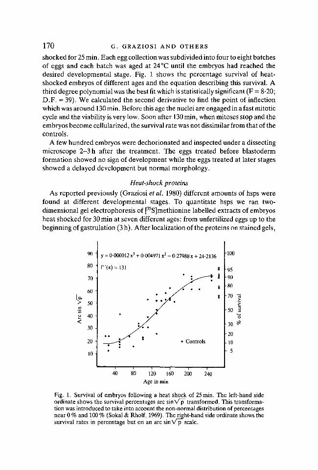
shocked for 25 min. Each egg collection was subdivided into four to eight batchesof eggs and each batch was aged at 24 °C until the embryos had reached thedesired developmental stage. Fig. 1 shows the percentage survival of heat-shocked embryos of different ages and the equation describing this survival. Athird degree polynomial was the best fit which is statistically significant (F = 8-20;D.F. = 39). We calculated the second derivative to find the point of inflectionwhich was around 130 min. Before this age the nuclei are engaged in a fast mitoticcycle and the viability is very low. Soon after 130 min, when mitoses stop and theembryos become cellularized, the survival rate was not dissimilar from that of thecontrols.
A few hundred embryos were dechorionated and inspected under a dissectingmicroscope 2-3 h after the treatment. The eggs treated before blastodermformation showed no sign of development while the eggs treated at later stagesshowed a delayed development but normal morphology.
Heat-shock proteins
As reported previously (Graziosi et al. 1980) different amounts of hsps werefound at different developmental stages. To quantitate hsps we ran two-dimensional gel electrophoresis of p5S]methionine labelled extracts of embryosheat shocked for 30 min at seven different ages: from unfertilized eggs up to thebeginning of gastrulation (3 h). After localization of the proteins on stained gels,
90
80
70
60 -
50 -
H 40 -
30 -
20 -
10 -
•>*
y = 0-000012 x3 + 0-004971 x2 - 0-27988 x + 24-2136
f"(x)= 131 g
o Controls
100
959080
70 13>
50 3o
30 ^
20
10
• 5
40 80 120 160
Age in min
200 240
Fig. 1. Survival of embryos following a heat shock of 25 min. The left-hand sideordinate shows the survival percentages arc sinV^T transformed. This transforma-tion was introduced to take into account the non-normal distribution of percentagesnear 0 % and 100 % (Sokal & Rholf, 1969). The right-hand side ordinate shows thesurvival rates in percentage but on an arc sin V p scale.
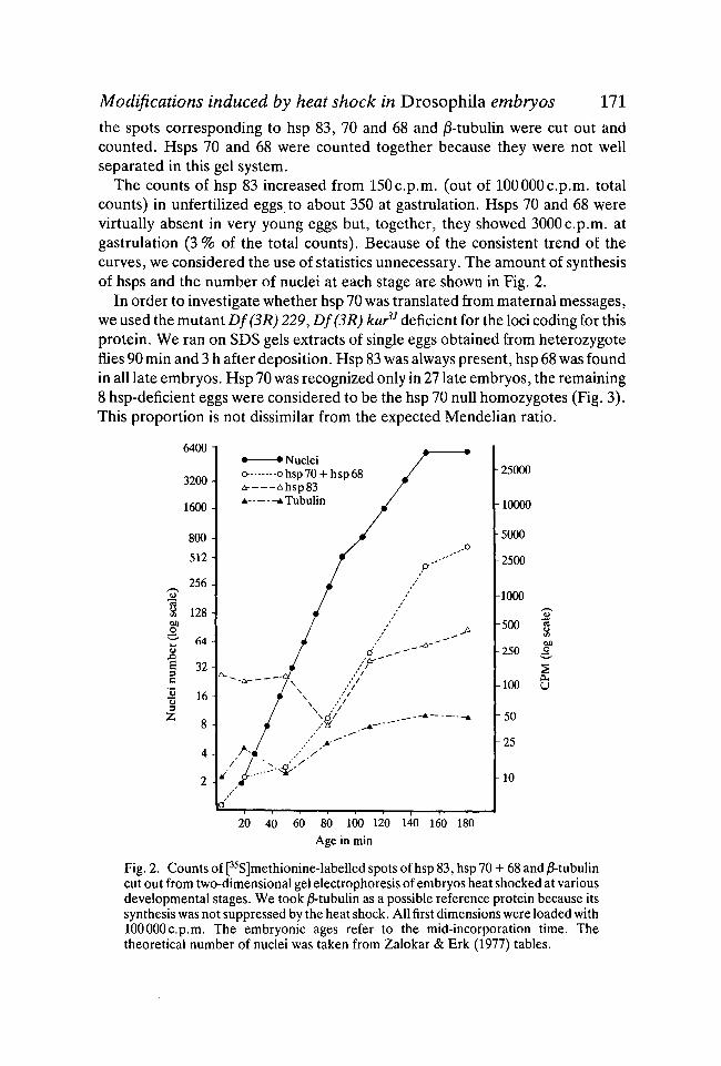
Modifications induced by heat shock in Drosophila embryos 111the spots corresponding to hsp 83, 70 and 68 and /3-tubulin were cut out andcounted. Hsps 70 and 68 were counted together because they were not wellseparated in this gel system.
The counts of hsp 83 increased from 150c.p.m. (out of 100000c.p.m. totalcounts) in unfertilized eggs to about 350 at gastrulation. Hsps 70 and 68 werevirtually absent in very young eggs but, together, they showed 3000c.p.m. atgastrulation (3 % of the total counts). Because of the consistent trend of thecurves, we considered the use of statistics unnecessary. The amount of synthesisof hsps and the number of nuclei at each stage are shown in Fig. 2.
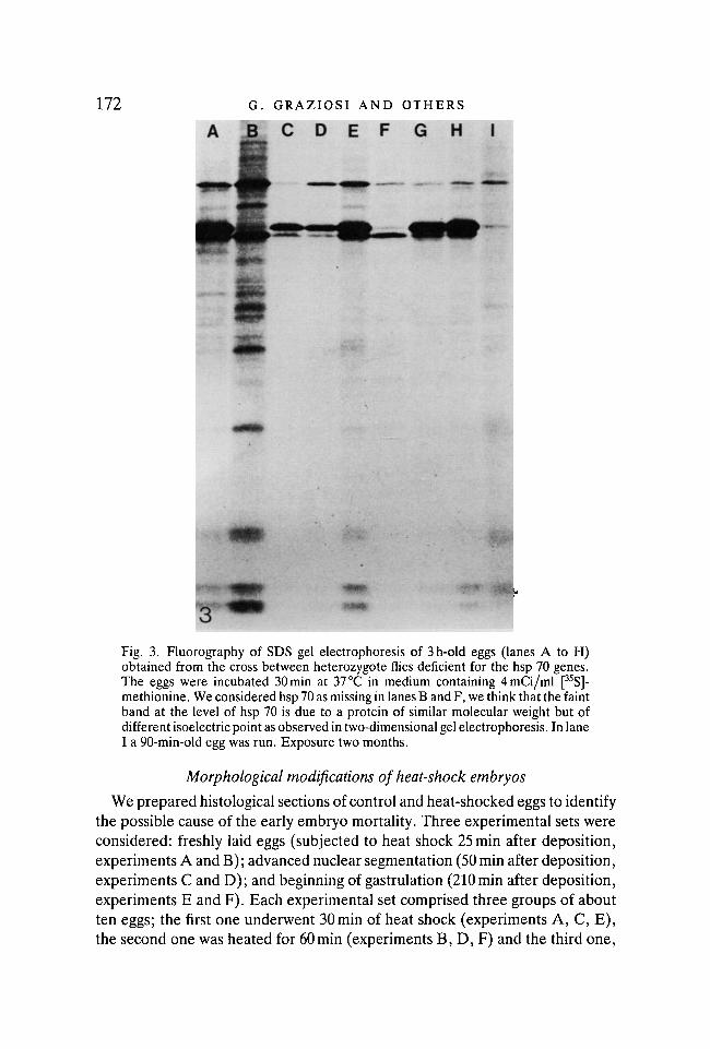
In order to investigate whether hsp 70 was translated from maternal messages,we used the mutant Df(3R) 229, Df(3R) kar31 deficient for the loci coding for thisprotein. We ran on SDS gels extracts of single eggs obtained from heterozygoteflies 90 min and 3 h after deposition. Hsp 83 was always present, hsp 68 was foundin all late embryos. Hsp 70 was recognized only in 27 late embryos, the remaining8 hsp-deficient eggs were considered to be the hsp 70 null homozygotes (Fig. 3).This proportion is not dissimilar from the expected Mendelian ratio.
6400-
3200-
1600 -
800
512
256-/—\
$ 128-00or 64-
32-
•§ 16-11 8 ^
• • Nucleio o hsp 70 + h s p 68A- Ahsp83A ATubulin
-25000
10000
5000
2500
-1000
-500 Ioo
250 2
-100
50
25
10
20 40 60 80 100 120 140 160 180
Age in min
Fig. 2. Counts of [35S]methionine-labelled spots of hsp 83, hsp 70 + 68 and /3-tubulincut out from two-dimensional gel electrophoresis of embryos heat shocked at variousdevelopmental stages. We took j3-tubulin as a possible reference protein because itssynthesis was not suppressed by the heat shock. All first dimensions were loaded with100000 c.p.m. The embryonic ages refer to the mid-incorporation time. Thetheoretical number of nuclei was taken from Zalokar & Erk (1977) tables.
172 G. GRAZIOSI AND OTHERS
C D E F G H I
m
Fig. 3. Fluorography of SDS gel electrophoresis of 3h-old eggs (lanes A to H)obtained from the cross between heterozygote flies deficient for the hsp 70 genes.The eggs were incubated 30 min at 37 °C in medium containing 4mCi/ml [35S]-methionine. We considered hsp 70 as missing in lanes B and F, we think that the faintband at the level of hsp 70 is due to a protein of similar molecular weight but ofdifferent isoelectric point as observed in two-dimensional gel electrophoresis. In laneI a 90-min-old egg was run. Exposure two months.
Morphological modifications of heat-shock embryos
We prepared histological sections of control and heat-shocked eggs to identifythe possible cause of the early embryo mortality. Three experimental sets wereconsidered: freshly laid eggs (subjected to heat shock 25 min after deposition,experiments A and B); advanced nuclear segmentation (50 min after deposition,experiments C and D); and beginning of gastrulation (210 min after deposition,experiments E and F). Each experimental set comprised three groups of aboutten eggs; the first one underwent 30min of heat shock (experiments A, C, E),the second one was heated for 60min (experiments B, D, F) and the third one,
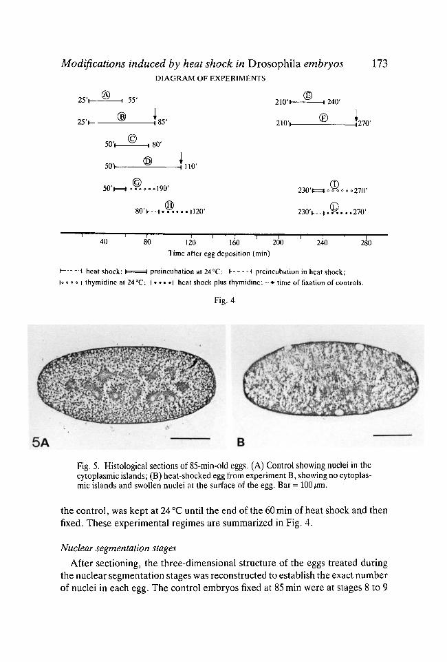
Modifications induced by heat shock in Drosophila embryos 173DIAGRAM OF EXPERIMENTS
25V
25V
50V
50V
50'
55'
is,80'
4110'
.190'
21O'i
210V
23O'l
H240'
H 2 7 0 '
= 270'
.270'
40 80—I—120 160 2C
Time after egg deposition (min)
240
1 heat shock; I 1 preincubation at 24°C; J- * preincubation in heat shock;
o o o | thymidine at 24 °C; ! • • • • ! heat shock plus thymidine; —• time of fixation of controls.
Fig. 4
5A B
Fig. 5. Histological sections of 85-min-old eggs. (A) Control showing nuclei in thecytoplasmic islands; (B) heat-shocked egg from experiment B, showing no cytoplas-mic islands and swollen nuclei at the surface of the egg. Bar = 100 fim.
the control, was kept at 24 °C until the end of the 60 min of heat shock and thenfixed. These experimental regimes are summarized in Fig. 4.
Nuclear segmentation stages
After sectioning, the three-dimensional structure of the eggs treated duringthe nuclear segmentation stages was reconstructed to establish the exact numberof nuclei in each egg. The control embryos fixed at 85 min were at stages 8 to 9
174 G. GRAZIOSI AND OTHERS
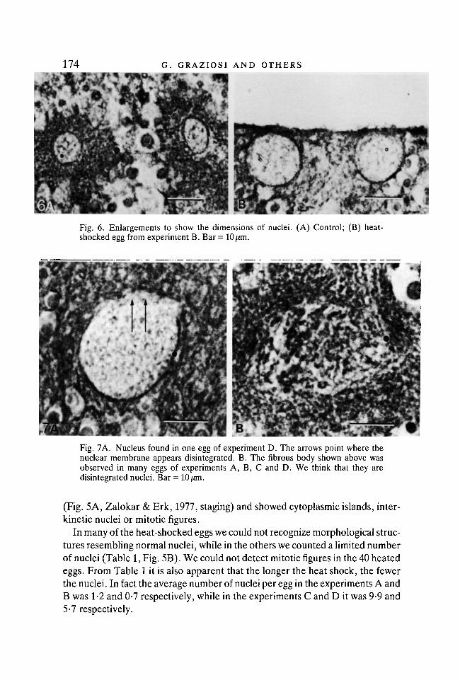
Fig. 6. Enlargements to show the dimensions of nuclei. (A) Control; (B) heat-shocked egg from experiment B. Bar = 10/im.
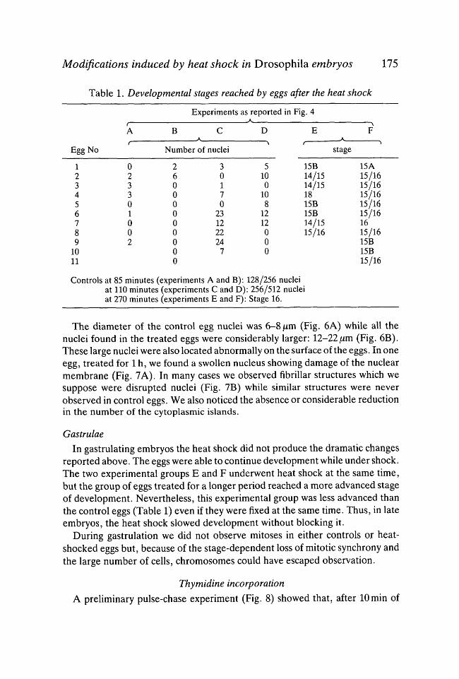
Fig. 7A. Nucleus found in one egg of experiment D. The arrows point where thenuclear membrane appears disintegrated. B. The fibrous body shown above wasobserved in many eggs of experiments A, B, C and D. We think that they aredisintegrated nuclei. Bar = 10/um.
(Fig. 5A, Zalokar & Erk, 1977, staging) and showed cytoplasmic islands, inter-kinetic nuclei or mitotic figures.
In many of the heat-shocked eggs we could not recognize morphological struc-tures resembling normal nuclei, while in the others we counted a limited numberof nuclei (Table 1, Fig. 5B). We could not detect mitotic figures in the 40 heatedeggs. From Table 1 it is also apparent that the longer the heat shock, the fewerthe nuclei. In fact the average number of nuclei per egg in the experiments A andB was 1-2 and 0-7 respectively, while in the experiments C and D it was 9-9 and5-7 respectively.
Modifications induced by heat shock in Drosophila embryos 175
Table 1. Developmental stages reached by eggs after the heat shock
Egg No
123456789
1011
f
A
r
023301002
Experiments as
B c
Number of nuclei
26000000000
30170
231222247
reportedA
D
•\
5100
108
1212000
in Fig. 4
E
15B14/1514/151815B15B14/1515/16
•\
F
>stage
15A15/1615/1615/1615/1615/161615/1615B15B15/16
Controls at 85 minutes (experiments A and B): 128/256 nucleiat 110 minutes (experiments C and D): 256/512 nucleiat 270 minutes (experiments E and F): Stage 16.
The diameter of the control egg nuclei was 6-8 jum (Fig. 6A) while all thenuclei found in the treated eggs were considerably larger: 12-22/im (Fig. 6B).These large nuclei were also located abnormally on the surface of the eggs. In oneegg, treated for 1 h, we found a swollen nucleus showing damage of the nuclearmembrane (Fig. 7A). In many cases we observed fibrillar structures which wesuppose were disrupted nuclei (Fig. 7B) while similar structures were neverobserved in control eggs. We also noticed the absence or considerable reductionin the number of the cytoplasmic islands.
Gastrulae
In gastrulating embryos the heat shock did not produce the dramatic changesreported above. The eggs were able to continue development while under shock.The two experimental groups E and F underwent heat shock at the same time,but the group of eggs treated for a longer period reached a more advanced stageof development. Nevertheless, this experimental group was less advanced thanthe control eggs (Table 1) even if they were fixed at the same time. Thus, in lateembryos, the heat shock slowed development without blocking it.
During gastrulation we did not observe mitoses in either controls or heat-shocked eggs but, because of the stage-dependent loss of mitotic synchrony andthe large number of cells, chromosomes could have escaped observation.
Thymidine incorporation
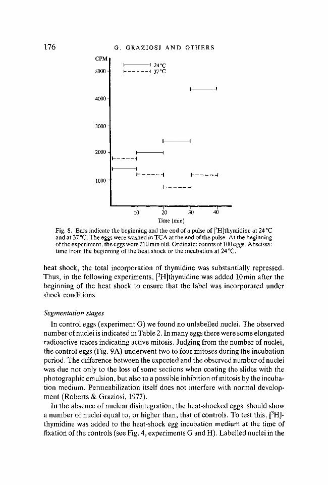
A preliminary pulse-chase experiment (Fig. 8) showed that, after lOmin of
176 G. GRAZIOSI AND OTHERSCPM
5000-
4000-
3000-
2000-
1000 -
H 24 °C\ 37°C
I A
I 1
I ,
10 20Time (min)
30 40
Fig. 8. Bars indicate the beginning and the end of a pulse of [3H]thymidine at 24 °Cand at 37 °C. The eggs were washed in TCA at the end of the pulse. At the beginningof the experiment, the eggs were 210 min old. Ordinate: counts of 100 eggs. Abscissa:time from the beginning of the heat shock or the incubation at 24 °C.
heat shock, the total incorporation of thymidine was substantially repressed.Thus, in the following experiments, [3H]thymidine was added 10min after thebeginning of the heat shock to ensure that the label was incorporated undershock conditions.
Segmentation stages
In control eggs (experiment G) we found no unlabelled nuclei. The observednumber of nuclei is indicated in Table 2. In many eggs there were some elongatedradioactive traces indicating active mitosis. Judging from the number of nuclei,the control eggs (Fig. 9A) underwent two to four mitoses during the incubationperiod. The difference between the expected and the observed number of nucleiwas due not only to the loss of some sections when coating the slides with thephotographic emulsion, but also to a possible inhibition of mitosis by the incuba-tion medium. Permeabilization itself does not interfere with normal develop-ment (Roberts & Graziosi, 1977).
In the absence of nuclear disintegration, the heat-shocked eggs should showa number of nuclei equal to, or higher than, that of controls. To test this, [3H]-thymidine was added to the heat-shock egg incubation medium at the time offixation of the controls (see Fig. 4, experiments G and H). Labelled nuclei in the
Modifications induced by heat shock in Drosophila embryos 111
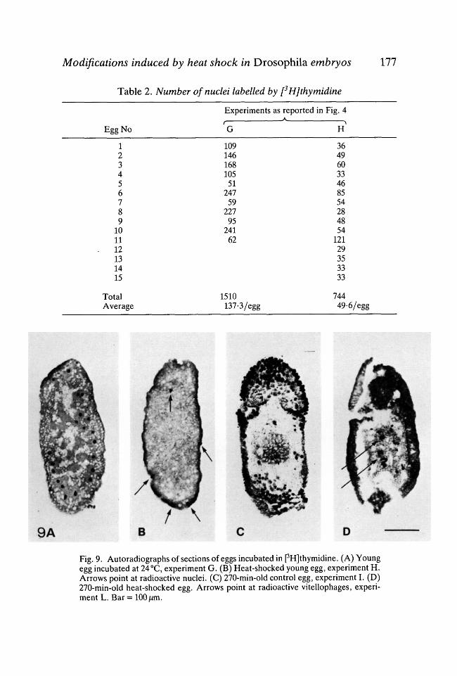
Table 2. Number of nuclei labelled by [3H]thymidine
Egg No
Experiments as reported in Fig. 4f
G
10914616810551247592279524162
1510137-3/egg
>H
3649603346855428485412129353333
74449-6/egg
123456789
101112131415
TotalAverage
9A
Fig. 9. Autoradiographs of sections of eggs incubated in pH]thymidine. (A) Youngegg incubated at 24°C, experiment G. (B) Heat-shocked young egg, experiment H.Arrows point at radioactive nuclei. (C) 270-min-old control egg, experiment I. (D)270-min-old heat-shocked egg. Arrows point at radioactive vitellophages, experi-ment L. Bar = 100 |Um.
178 G. GRAZIOSI AND OTHERS
heat-shocked eggs (experiment H) were fewer than those counted in controls(Table 2). In heat-shocked eggs we never observed elongated nuclear radioactivetraces. We found again large nuclei of 20 /im of diameter which were radioactive,and generally located at the surface of the egg (Fig. 9B). Only a few unlabellednuclei were found.
Gastrulae
In the fifteen control eggs examined (experiment I), a large number of nucleiwere radioactive. The majority of the radioactive nuclei was clustered in specificareas where the cells were invaginating: stomodeum, posterior midgut, germinalband and cephalic furrow (Fig. 9C). Few or no vitellophages were labelled.
Out of the 20 heat-shocked embryos (experiment L) only a few showedradioactive nuclei at the cephalic furrow and at the posterior midgut invagina-tion. The majority of eggs had radioactive vitellophages (Fig. 9D).
DISCUSSION
In quantitating the amount of [35S]methionine incorporated into the hsps wedid not intend to establish the absolute rate of synthesis of these proteins.Rather, our experimental procedure accounts only for the proportion of the labelwhich ended up in the heat-shock proteins. The synthesis of hsp 70 and 68 rosesteadily during development and did not show, on a logarithmic scale, any sharpincrease at blastoderm formation. Consequently it appears that these proteinscan be synthesized before blastoderm formation.
It has been reported that the bulk of the zygotic genome is activated at blasto-derm formation (Anderson & Lengyel, 1979; Lamb & Laird, 1976; McKnight &Miller, 1976; Zalokar, 1976). If this is also true for the heat-shock genes then hspsynthesis during early stages should be supported by maternal mRNAs. At leastas far as hsp 70 and, possibly, hsp 68 are concerned, we do not favour thishypothesis because: i) there was little or no synthesis in unfertilized or freshlylaid eggs, ii) their synthesis rose during the exponential phase of nuclearmultiplication and not at blastoderm formation and, iii) we did find mutant eggs(229 hat33) unable to synthesize hsp 70.
If hsps 70 and 68 are indeed translated from zygotic messages, their lowsynthesis or even absence in early stages is not surprising. If we compare thenumber of nuclei in the zygote with that of the egg at blastoderm formation wefind a ratio 1:6000. Hence, assuming equal transcriptional and translational ratesat all stages, the zygote is expected to produce far less hsps than the followingstages. Furthermore many nuclei disintegrated and, presumably, disintegratednuclei cannot be transcribed normally. This interpretation cannot be applied tohsp 83 whose synthesis was relatively high even in unfertilized eggs. Most prob-ably its synthesis is sustained by maternal mRNAs, at least during the earlystages.
Modifications induced by heat shock in Drosophila embryos 179
The morphology of the heat-shocked eggsIn many eggs, heat shocked during the nuclear multiplication stages, we found
no nuclei or just a few. It is obvious that eggs without nuclei or with damagednuclei cannot undergo normal development and, on the basis of the mortalitycurve, we think that such damage was caused only in eggs which did not reachthe stage of blastoderm formation. Our observations are very similar to thosereported by Zalokar & Erk (1976), who obtained swollen nuclei upon treatmentof eggs with dinitrophenol or anoxia. It is now known that both anoxia anddinitrophenol induce a 'heat-shock-like' response (Ashburner, 1970; Ellgaard,1972; Rensing, 1973).
Although our results can explain why the embryos did not survive the heatshock, they cannot explain the degeneration of nuclei. The unique morphologi-cal and physiological characteristics of the early embryos, as the absence ofplasmalemma and the fast mitotic cycles (Sonnenblick, 1950; Zalokar & Erk,1976), could account for the nuclear sensitivity to heat. But such characteristicscannot fully explain why some nuclei survive the exposure to the high tem-perature and the subsequent reduction of nuclei number as reported in Table 1.The unequal sensitivity of nuclei could be more convincingly explained by theunequal distribution of some protective agents either the hsps or any othercomponent of the egg. We know that the heat-shock proteins migrate into thenuclei (Arrigo, Fakan & Tissieres, 1980; Valazquez, Di Domenico & Lindquist,1980) and it is possible that their concentration in the egg was too low to exerta possible protective action on all nuclei. Furthermore, the early embryo is notcompartmentalized by plasmalemmas and substances could diffuse in nucleus-free areas.
Besides the disintegration of nuclei we found that the heat shock interferedwith the mitotic cycle of early embryos. Frequently in control eggs, we observedmitotic figures which were never seen in sections of heat-shocked eggs. Thus theheated nuclei were unable to enter mitoses. Furthermore, taking into accountthe synchrony of mitoses and that almost all nuclei of all the labelled eggs wereradioactive, it is apparent that the nuclei reached the S phase. Consequently theblock should occur between S and M, presumably in G2. Alternatively it ispossible that the nuclei did not reach M because they died just after the S phase.In any case DNA synthesis itself was not inhibited and the low incorporation ofthymidine during the heat shock was due to a low number of nuclei entering theS phase.
As far as gastrulating heat-shocked embryos are concerned, we found only adelay in development. Similar results were obtained in embryo by Dura (1981)and in pupae by Lindsley & Poodry (1977). Delayed development could becaused by a variety of reasons including the block of mitoses.
Finally, we must take into account the possibility that nuclear disintegrationas well as a slower development could be due to the perturbation of the normal
180 G. GRAZIOSI AND OTHERS
programme of embryonic gene expression and of coordinate maternal messagetranslation, as reported for tissue culture cells (McKenzie et al. 1975; Moran etal. 1978; Tissieres, Mitchell & Tracy, 1974). This hypothesis could be tested byan appropriate experimental design which could also show a way to usehyperthermia to discriminate between the contribution of the embryonicgenome and that of the maternal genome to development. Further investigationscould also indicate if the heat shock may be used as a physiological method forsynchronizing mitoses, which might have practical applications in a variety ofbiological systems.
We wish to thank Dr D. Ish-Horowicz for the kind gift of the mutant stock 229 kar31, andDr D. B. Roberts and C. R. Bebbington for critical reading and improvements to themanuscript. This research has been supported by the CNR grant No. 81 00488, 85-115, 2143and by the MPI grant 1981-82.
REFERENCESANDERSON, K. V. & LENGYEL, J. A. (1979). Rates of synthesis of major classes of RNA in
Drosophila embryos. Devi Biol. 70, 217-231.ARRIGO, A. P., FAKAN, S. & TISSIERES, A. (1980). Localization of the heat shock-induced
proteins in Drosophila melanogaster tissue culture cells. Devi Biol. 78, 86-103.ASHBURNER, M. (1970). Patterns of puffing activity in salivary gland chromosomes of
Drosophila. V. Responses to environmental treatment. Chromosoma 31, 356-376.ASHBURNER, M. & BONNER, J. J. (1979). The induction of gene activity in Drosophila by heat
shock. Cell 17, 241-254.BARNETT, T., ALTSCHULER, M., MCDANIEL, C. N. & MASCARENHAS, J. P. (1980). Heat shock
induced proteins in plant cells. Devi Genet. 1, 331-340.BONNER, J. J. & PARDUE, M. L. (1976). The effect of heat shock on RNA synthesis in
Drosophila tissues. Cell 8, 43-50.BONNER, W. M. & LASKEY, R. A. (1974). A film detection method for tritium labelled proteins
and nucleic acids in polyacrylamide gels. Eur. J. Biochem. 46, 83-88.CHOMYN, A., MOLLER, G. & MITCHELL, H. K. (1979). Patterns of protein synthesis following
heat shock in pupae of Drosophila melanogaster. Devi Genet. 1, 77-95.DURA, J. M. (1981). Stage dependent synthesis of heat shock induced proteins in early em-
bryos of Drosophila melanogaster. Mol. gen. Genet. 184, 381-385.ELLGAARD, E. G. (1972). Similarities in chromosomal puffing induced by temperature shocks
and dinitrophenol in Drosophila. Chromosoma 37, 417-422.FINK, K. & ZEUTHEN, E. (1970). Heat shock proteins in Tetrahymena studied under growth
conditions. Expl Cell Res. 128, 23-30.GIUDICE, G., ROCCHERI, M. C. & Di BERNARDO, M. G. (1980). Synthesis of 'heat shock'
proteins in sea urchin embryos. Cell Biol. Int. Re. 4, 69-74.GRAZIOSI, G., MICALI, F., MARZARI, R., DE CRISTINI, F. & SAVOINI, A. (1980). Variability of
response of early Drosophila embryos to heat shock. /. exp. Zool. 214, 141-145.HEDMAN, S. & KROGSTAD, B. (1963). Temperature effects upon egg development in
Drosophila melanogaster. Proc. Minn. Acad. Sci. 31, (1), 78-81.HENIKOFF, S. & MESELSON, M. (1977). Transcrition at two heat shock loci in Drosophila. Cell
12, 441-451.ISH-HOROWICZ, D., PINCHIN, S. M., GAUSZ, J., GYURKOVICS, H., BENCZE, G., GOLDSCHMIDT-
CLERMONT, M. & HOLDEN, J. J. (1979). Deletion mapping of two Drosophila melanogasterloci that code for the 70000 dalton heat-induced protein. Cell 17, 565-571.
KELLEY, P. M., ALIPERTI, G. & SCHLESINGER, M. J. (1980). In vitro synthesis of heat-shockproteins by mRNAs from chicken embryo fibroblasts. /. biol. Chem. 255, 3230-3233.
Modifications induced by heat shock in Drosophila embryos 181LAEMMLI, U. K. (1970). Cleavage of structural proteins during assembly of the head of bac-
teriophage T4. Nature 227, 680-685.LAMB,M. M. & LAIRD, C. D. (1976). Increase in nuclear poly(A)-containing RNA at syncytial
blastoderm in Drosophila melanogaster embryos. Devi Biol. 52, 31-42.LEWIS, M., HELMSING, P. J. & ASHBURNER, M. (1975). Parallel changes in puffing activity and
patterns of protein synthesis in salivary glands of Drosophila. Proc. natn.Acad. Sci., U.S.A.72, 3604-3608.
LIMBOURG, B. & ZALOKAR, M. (1973). Permeabilization of Drosophila egg. Devi Biol. 35,382-387.
LINDQUIST, S. (1980). Varying patterns of protein synthesis in Drosophila during heat shock:Implications for regulation. Devi Biol. 77, 463-479.
LINDQUIST, S. (1981). Regulation of protein synthesis during heat shock. Nature 293,311-314.LINDSLEY, D. E. & POODRY, C. A. (1977). A reversible temperature-induced developmental
arrest in Drosophila. Devi Biol. 56, 213-218.MCALISTER, L. & FINKELSTEIN, D. B. (1980). Alterations in translatable ribonucleic acid after
heat shock of Saccharomyces crevisiae. J. Bacteriol. 143, 603-612.MCKENZIE, S. L., HENIKOFF, S. & MESELSON, M. (1975). Localization of RNA from heat
induced polysomes at puff sites in Drosophila melanogaster. Proc. natn. Acad. Sci., U.S.A.72, 1117-1121.
MCKNIGHT, S. L. & MILLER, O. L. JR(1976). Ultrastructural patterns of RNA synthesis duringearly embryogenesis of Drosophila melanogaster. Cell 8, 305-319.
MIRAULT, M. E., GOLDSCHMIDT-CLERMON, M , MORAN, L., ARRIGO, A. P. & TISSIERES, A.(1978). The effect of heat shock on gene expression in Drosophila melanogaster. ColdSpring Harbor Symp. Quant. Biol. 42, 819-828.
MITCHELL, H. K. & LIPPS, L. S. (1978). Heat shock and phenocopy induction in Drosophila.Cell 15, 907-918.
MORAN, L., MIRAULT, M. E., ARRIGO, A. P., GOLDSCHMIDT-CLERMONT, M. & TISSIERES, A.(1978). Heat shock of Drosophila melanogaster induces the synthesis of new messengerRNAs and proteins. Phil. Trans. Roy. Soc. London B 283, 391-406.
O'FARREL, P. H. (1975). High resolution two-dimensional electrophoresis of proteins. J. biol.Chem. 250, 4007-4021.
PETERSON, N. S., MOLLER, G. & MITCHELL, H. H. (1979). Genetic mapping of the codingregions for three heat-shock proteins in Drosophila melanogaster. Genetics 92, 891^902.
POWSNER, L. (1935). The effects of temperature on the duration of the development stages ofDrosophila melanogaster. Physiol. Zool. 474-520.
RENSING, L. (1973). Effects of 2.4-dinitrophenol and dinactin and heat-sensitive andecdysone-specific puffs of Drosophila salivary gland chromosomes in vitro. Cell Differentia-tion 2, 221-228.
ROBERTS, D. B. & GRAZIOSI, G. (1977). Protein synthesis in the early Drosophila embryo:analysis of the protein species synthesized. J. Embryol. exp. Morph. 41, 101-110.
SANTAMARIA, P. (1979). Heat shock induced phenocopies of dominant mutants of the bithoraxcomplex in Drosophila melanogaster. Mol. gen. Genet. 172, 161-163.
SAVOINI, A., MICALI, F., MARZARI, R., DE CRISTINI, F. & GRAZIOSI, G. (1981). Low variabilityof the protein species synthesized by Drosophila melanogaster embryos. Wilhelm Roux'sArch, devl Biol. 190, 161-167.
SOKAL, R. R. & ROHLF, F. J. (1969). Biometry (San Francisco: W. H. Freeman and Com-pany).
SONNENBLICK, B. P. (1950). The early embryology of Drosophila melanogaster. In Biology ofDrosophila (ed. M. Demerec), pp. 62-163. New York: Hafner.
SPRADLING, A., PARDUE, M. L. & PENMAN, S. (1977). Messenger RNA in heat-shockedDrosophila cells. /. mol. Biol. 109, 559-587.
SPRADLING, A., PENMAN, S. & PARDUE, M. L. (1975). Analysis of Drosophila mRNA by in situhybridization: sequences transcribed in normal heat-shocked cultured cells. Cell 4,395-404.
TISSIERES, A., MITCHELL, H. K. & TRACY, U. M. (1974). Protein synthesis in salivary glandsof Drosophila melanogaster: relation to chromosome puffs. /. mol. Biol. 84, 389-398.

182 G. GRAZIOSI AND OTHERS
VELAZQUEZ, J. M., DI DOMENICO, B. J. & LINDQUIST, S. (1980). Intracellular localization ofheat shock proteins in Drosophila. Cell 20, 679-689.
ZALOKAR, M. (1976). Autoradiographic study of protein and RNA formation during earlydevelopment of Drosophila eggs. Devi Biol. 49, 425-437.
ZALOKAR, M. & ERK, I. (1976). Division and migration of nuclei during early embryogenesisof Drosophila melanogaster. J. Microscopie Biol. Cell. 25, 97-106.
ZALOKAR, M. & ERK, I. (1977). Phase-partition fixation staining of Drosophila eggs. StainTechnology. 52, 89-95.
{Accepted 27 April 1983)


