Morphologic and in myasthenia andin syndromes · cell processes facing reactive segments of the...
13
Journal of Neurology, Neurosurgery, and Psychiatry, 1980, 43, 577-589 Morphologic and immunopathologic findings in myasthenia gravis and in congenital myasthenic syndromes ANDREW G ENGEL From the Department of Neurology and Neuromuscular Research Laboratory, Mayo Clinic, Mayo Foundation and Mayo Medical School, Rochester, Minnesota SUMMARY Overwhelming evidence now supports Simpson's concept, originally proposed in 1960, that acquired myasthenia gravis (MG) is an autoimmune disease in which antibodies are directed against the nicotine postsynaptic acetylcholine receptor (AChR).1 An autoimmune pathogenesis of acquired MG implies that those myasthenic syndromes which occur in a con- genital and familial setting may have a different, non-autoimmune basis. This paper focuses on ultrastructural, immunoelectron microscopic and cytochemical aspects of acquired autoimmune MG and some recently recognised congenital myasthenic syndromes. Acquired autoimmune myasthenia gravis Morphometric reconstruction of the ultrastructure of the motor end-plate These studies were carried out between 1968 and 1970 in collaboration with Tetsuji Santa.2 External intercostal muscle motor end-plates were studied from MG patients who required thymectomy, from non-weak subjects who underwent thora- cotomy for intrathoracic disease and from patients with the Lambert-Eaton type of myasthenic syn- drome. Problems pertaining to finding the end-plates in the electron microscope and to quantitative analysis of end-plate morphology were noted early in the study. These were overcome by devising a method for readily finding end-plates in electron microscope3 and by developing a system for the morphometric analysis of the ultrastructure of the motor end-plate.2 In this way, we were able to study a large number of end-plates in a quan- titative manner and could assess the results statistically. Our initial hypothesis was based on the then current notion that the small amplitude of the miniature end-plate potential (mepp) in MG was due to a presynaptic defect in the synthesis of acetylcholine (ACh) or in the packaging of ACh Address for reprint requests: Dr AG Engel, Department of Neurology, Mayo Clinic, Rochester, Minnesota 55901, USA. into the synaptic vesicles. If this were the case, one might expect changes in the dimensions of the synaptic vesicles. Contrary to what we expected, the dimensions as well as the numerical density of the synaptic vesicles were normal. The mito- chondrial fraction of the nerve terminal was also normal. However, the nerve terminals were of smaller than normal size. The normal size of the synaptic vesicles provided evidence against a de- crease of the ACh content of the quantum, for when ACh synthesis is experimentally blocked by hemicholinium and the miniature end-plate poten- tial (mepp) amplitude decreases, the synaptic vesicles become smaller. Instead of corroborating a presynaptic defect in MG, the ultrastructural data pointed strongly to a postsynaptic abnormality. The postsynaptic regions showed degenerative changes with widen- ing of the primary and secondary synaptic clefts and accumulation of debris in the synaptic space. Many postsynaptic regions were highly simplified and at some regions the secondary synaptic clefts were totally absent. Morphometric analysis re- vealed a significant decrease of the length of postsynaptic membrane and of the length of the postsynaptic membrane per unit postsynaptic area (postsynaptic membrane density). In addition, we observed sprouting of nerve terminals near end- plates and encountered highly degenerated post- synaptic regions which were denuded of their 577 Protected by copyright. on August 22, 2019 by guest. http://jnnp.bmj.com/ J Neurol Neurosurg Psychiatry: first published as 10.1136/jnnp.43.7.577 on 1 July 1980. Downloaded from
Transcript of Morphologic and in myasthenia andin syndromes · cell processes facing reactive segments of the...

Journal of Neurology, Neurosurgery, and Psychiatry, 1980, 43, 577-589
Morphologic and immunopathologic findingsin myasthenia gravis and in congenital myasthenicsyndromesANDREW G ENGEL
From the Department of Neurology and Neuromuscular Research Laboratory, Mayo Clinic,Mayo Foundation and Mayo Medical School, Rochester, Minnesota
SUMMARY Overwhelming evidence now supports Simpson's concept, originally proposed in1960, that acquired myasthenia gravis (MG) is an autoimmune disease in which antibodies aredirected against the nicotine postsynaptic acetylcholine receptor (AChR).1 An autoimmunepathogenesis of acquired MG implies that those myasthenic syndromes which occur in a con-genital and familial setting may have a different, non-autoimmune basis. This paper focuses on
ultrastructural, immunoelectron microscopic and cytochemical aspects of acquired autoimmuneMG and some recently recognised congenital myasthenic syndromes.
Acquired autoimmune myasthenia gravis
Morphometric reconstruction of the ultrastructureof the motor end-plateThese studies were carried out between 1968 and1970 in collaboration with Tetsuji Santa.2 Externalintercostal muscle motor end-plates were studiedfrom MG patients who required thymectomy,from non-weak subjects who underwent thora-cotomy for intrathoracic disease and from patientswith the Lambert-Eaton type of myasthenic syn-drome. Problems pertaining to finding theend-plates in the electron microscope and toquantitative analysis of end-plate morphology werenoted early in the study. These were overcome bydevising a method for readily finding end-plates inelectron microscope3 and by developing a systemfor the morphometric analysis of the ultrastructureof the motor end-plate.2 In this way, we were ableto study a large number of end-plates in a quan-titative manner and could assess the resultsstatistically.Our initial hypothesis was based on the then
current notion that the small amplitude of theminiature end-plate potential (mepp) in MG wasdue to a presynaptic defect in the synthesis ofacetylcholine (ACh) or in the packaging of ACh
Address for reprint requests: DrAG Engel, Department ofNeurology,Mayo Clinic, Rochester, Minnesota 55901, USA.
into the synaptic vesicles. If this were the case,one might expect changes in the dimensions of thesynaptic vesicles. Contrary to what we expected,the dimensions as well as the numerical densityof the synaptic vesicles were normal. The mito-chondrial fraction of the nerve terminal was alsonormal. However, the nerve terminals were ofsmaller than normal size. The normal size of thesynaptic vesicles provided evidence against a de-crease of the ACh content of the quantum, forwhen ACh synthesis is experimentally blocked byhemicholinium and the miniature end-plate poten-tial (mepp) amplitude decreases, the synapticvesicles become smaller.
Instead of corroborating a presynaptic defectin MG, the ultrastructural data pointed stronglyto a postsynaptic abnormality. The postsynapticregions showed degenerative changes with widen-ing of the primary and secondary synaptic cleftsand accumulation of debris in the synaptic space.Many postsynaptic regions were highly simplifiedand at some regions the secondary synaptic cleftswere totally absent. Morphometric analysis re-vealed a significant decrease of the length ofpostsynaptic membrane and of the length of thepostsynaptic membrane per unit postsynaptic area(postsynaptic membrane density). In addition, weobserved sprouting of nerve terminals near end-plates and encountered highly degenerated post-synaptic regions which were denuded of their
577
Protected by copyright.
on August 22, 2019 by guest.
http://jnnp.bmj.com
/J N
eurol Neurosurg P
sychiatry: first published as 10.1136/jnnp.43.7.577 on 1 July 1980. Dow
nloaded from

Andrew G Engel
nerve terminal. These findings were indicative ofcycles of degeneration involving the postsynapticregions.
In the Lambert-Eaton myasthenic syndrome thepostsynaptic region was somewhat larger thannormal and the postsynaptic membrane lengthwas significantly increased relative to the presyn-aptic membrane length. There were no morpho-logic abnormalities of the nerve terminal and nopostsynaptic degenerative changes. Morphometricreconstruction of the end-plate in control subjects,in MG and in the Lambert-Eaton syndrome isshown in fig 1. In the light of current knowledge,the postsynaptic pathology in MG is a direct con-sequence of the autoimmune attack on the post-synaptic membrane.
Ultrastructural localisation and quantification ofAChR in MGRadiochemical evidence that a deficiency of AChRexisted in MG was obtained by Fambrough et alin 1973.4 My co-workers and I subsequently be-
*~~~~~~~~~~~~~~~~~~~~~~~~~~~~~~~~*
......~~~~ ~ ~ ~ ~ ~ ~ ~ ~~~~~~. ....~~~~~~~~~~~~~... Control
Membrane nlength5-83 )j/2I-a I1u
Myasthenia gravis
3.95u/,pI2 -
Fig I Morphometric reconstruction of the motorend-plate in controls, in MG and in the Lambert-Eaton syndrome. (Reproduced from ref 2, bypermission.)
came interested in the ultrastructural localisationof AChR at the MG end-plate.5 6 Nonweak sub-jects and patients with the Lambert-Eaton syn-drome were used for comparison. The study wasfacilitated by the development, in this laboratory,
<
::: .:* ... : . . ::::: .::..:: :::
::
::: .:
.: .. f .:
Si9 !U'. ... :.* ........ : .. , . . . . ... :.. X: i,;
BFig 2 Ultrastructural localisation of AChR at intercostal muscle end-plate from control subject (A) and inmoderately severe, generalised MG (B). In B, only short segments of simplified postsynaptic membranereact for AChR. Presynaptic staining, seen in A, is virtually absent in B. A, B X22 300. (Reproduced fromref 5, by permission.)
578
Protected by copyright.
on August 22, 2019 by guest.
http://jnnp.bmj.com
/J N
eurol Neurosurg P
sychiatry: first published as 10.1136/jnnp.43.7.577 on 1 July 1980. Dow
nloaded from

Morphology and immunopathology of acquired and congenital myasthenias
of a simple, one-stage procedure for localisationof AChR with peroxidase-labelled a-bungarotoxin.Labelled toxin was applied to fresh, oxygenatedmuscle strips, well rinsed before and after exposureto the reagent, followed by glutaraldehyde fixation.At normal end-plates, AChR was observed on
terminal expansions of the junctional folds andsometimes also on stalks of the folds (fig 2A).
Less intense reaction was detected on the pre-synaptic membrane and occasionally on Schwanncell processes facing reactive segments of thesynaptic folds. The distribution of AChR at end-plates in the Lambert-Eaton syndrome resembledthat seen at normal end-plates. There was de-ficiency of AChR at MG end-plates. In moderatelysevere, generalised MG the postsynaptic regions
A
BFig 3 AChR localization at intercostal end-plate in predominantly bulbar MG (A) and in moderatelysevere, generalised MG (B). Reaction for AChR is diminished in A and is undetectable in B. Markeddegeneration of junctional folds occurs in B. CompareB with schematised MG end-plate in fig 1.A, X 17900; B, X22 300. (Reproduced from ref 5, by permission.)
c
579
-,.-
-I.
Protected by copyright.
on August 22, 2019 by guest.
http://jnnp.bmj.com
/J N
eurol Neurosurg P
sychiatry: first published as 10.1136/jnnp.43.7.577 on 1 July 1980. Dow
nloaded from

580
usually showed a marked decrease (fig 2B), or evenabsence (fig 3B), of AChR. In less severe MG,AChR seemed normally abundant at some end-plate regions but was diminished at others (fig 3A).Usually, but not always, the simplest and mostdegenerated postsynaptic regions showed thegreatest decrease in toxin binding. As a rule, thepresynaptic staining was reduced or absent whenvis-a-vis junctional folds did not react for AChR(figs 2B and 3B). This suggested that the presyn-aptic staining was largely an artifact caused bydiffusion of reaction product from postsynapticsites.To quantify the observations, the length of the
postsynaptic mcmbrane reacting for AChR at agiven end-plate region was measured and thennormalised for the length of the presynaptic cleft.The value thus obtained was an index of therelative abundance of the postsynaptic AChR forthat region. In the MG patients the mean AChRindex ranged from 0-27 to 1 f62 (group mean+SEM: 0.85-fr0.14). In the nonweak controls thecorresponding values ranged from 3-06 to 3-29(3.21±0.05); and in the Lambert-Eaton syndromefrom 2 56 to 3-77 (3.05±0'16).
In collaboration with Dr EH Lambert, we at-tempted to correlate the morphometric data within vitro electrophysiologic observation.5 6 To thisend, the mean AChR indices obtained in eachsubject were compared with the correspondingmean mepp amplitudes. The comparison demon-strated a linear correlation between the two vari-ables (r=0-899; p 0 001).These observations in MG provided unambigu-
ous ultrastructural evidence for a deficiency ofpostsynaptic AChR and indicated that the de-ficiency of AChR determined the small mepp am-plitude. A linear relationship between the AChRcontent of muscle and the mepp amplitude wasalso noted in subsequent studies in which AChRwas radiochemically estimated.7 8
Ultrastructural localisation of IgG at theMG end-plateAlthough evidence for an autoimmune pathogene-sis of acquired MG had been accumulating since1973, no immunoglobulin was demonstrated at theMG end-plate before 1977. We succeeded inlocalising IgG at the MG end-plate with peroxi-dase-labelled staphylococcal protein A.9 Protein Abinds bivalently to Fc portions of human IgGsubclasses 1, 2 and 4 and has an increased affinityfor IgG molecules attached to antigenic sites.10The latter property enhances the specificity andsensitivity of the reagent and optimises the signal
Andrew G Engel
(reaction at the end-plate) to noise (backgroundstaining) ratio of the cytochemical procedure.As in the preceding study, the immunoreagent
was applied to fresh, intact muscle strips, wellrinsed before and after the application of theimmunoreagent. In less severe cases of MG, inwhich the postsynaptic folds were relatively wellpreserved, IgG was found over terminal expan-sions of the junctional folds where AChR isknown to be located (figs 4A and C). In moresevere cases, in which junctional folds were lesswell preserved, the postsynaptic membrane boundless IgG, but IgG was readily detected on degen-erated remnants of the junctional folds in thesynaptic space (fig 4B). Morphometric estimatesof the abundance of the postsynaptic membranebinding IgG were proportionate to the meppamplitude, but an exception to this relationshipwas noted in one of ten cases. This was a youngpatient who, despite having moderately severedisease, had well preserved junctional folds whichbound abundant IgG.From these findings we inferred that (1) the
amount of IgG bound to the postsynaptic mem-brane is proportionate to the amount of AChRwhich remains in the membrane; (2) IgG-coatedsegments of junctional folds are shed into thesynaptic space; (3) in most cases of MG the trans-mission defect is determined by the loss of AChRrather than by the fact that IgG is bound toAChR; (4) in most cases of MG, antibodies toAChR do not significantly block the responseof AChR to ACh, but in occasional patients anti-bodies do have a significant blocking effect.
Participation of the complement system in theautoimmune attack on the postsynaptic membraneWe sought evidence for antibody dependent com-plement mediated injury to the junctional folds bylocalising complement components at the MGend-plate.6 911 The third (C3) and ninth (C9)complement components were localised withmonospecific antibodies raised in rabbits. Theultrastructural localisation of C3 was identical tothat of IgG. Reaction product was detected on seg-ments of the postsynaptic membrane, on debris inthe synaptic space and on disintegrating junctionalfolds.9 The fact that the distributions of C3 andIgG at the end-plate are highly similar is consistentwith the assumption that anti-AChR antibodiescan fix complement and that the assembly phase ofthe complement reaction sequence (via Cl, C4,C2 and C3) has gone to completion. However, thepresence of C3 on the junctional folds does not initself establish that the attack (or lytic) phase of
Protected by copyright.
on August 22, 2019 by guest.
http://jnnp.bmj.com
/J N
eurol Neurosurg P
sychiatry: first published as 10.1136/jnnp.43.7.577 on 1 July 1980. Dow
nloaded from

Morphology and immunopathology of acquired and congenital myasthenias
::
.............
....AV..i ~~~~~~~~~~~~~~~~S;gji*'N;.%
* .r.
B~~~~~~~~~
A
is
CFig 4 Ultrastructural localisation of IgG at the MG end-plate. IgG is detected on terminal expansions ofwell preserved junctional folds (A), on degenerate material shed into synaptic space (arrows in B and C),and on short segments of the postsynaptic membrane (B and C). Note that in C presynaptic staining isreciprocal to postsynaptic reaction. A, X25 100; B, X29 700; C, X24 800. (Reproduced from ref 9 bypermission.)
581
W
Protected by copyright.
on August 22, 2019 by guest.
http://jnnp.bmj.com
/J N
eurol Neurosurg P
sychiatry: first published as 10.1136/jnnp.43.7.577 on 1 July 1980. Dow
nloaded from

582
the complement reaction has become activated.12The occurrence of complement mediated mem-
brane injury depends on the presence on the targetmembrane of C5b, C6, C7, C8 and C9, the consti-tuents of the membrane attack complex. Thecomplex, now known to be a dimer of C5b-9,induces focal and irreversible membrane lesionsand C9 is an absolute requirement for this.12 13Therefore, to obtain unambiguous evidence forcomplement mediated injury of the postsynapticmembrane, we decided to study the localisation ofC9 at the MG end-plate."At end-plates where the architecture of the post-
synaptic region was well preserved, C9 was local-ised on short segments of the junctional folds andon sparse debris in the synaptic space. At end-plates which showed degenerative changes, therewas intense reaction for C9 on degenerate materialin the synaptic space and this was most markedwhere the junctional folds were the most abnormal(fig 5A). Reaction for C9 also occurred nearexisting end-plates, on debris positioned betweenlayers of basal lamina. Finally, intense reactionfor C9 was found at highly degenerate postsyn-aptic regions denuded of their nerve terminal(fig 5B). Here the reaction was on debris whichoften was still arranged in the shape of the pre-existing junctional fold and was still surroundedby traces of basal lamina. On some muscle fibresdiscrete regions reacting for C9 were widely separ-ated. Some of these were postsynaptic regionscovered by nerve terminal; some consisted of de-generate folds and debris not covered by nerveterminal; and some consisted only of debris sand-wiched between layers of basal lamina (fig 5C). Incontrast with the localisation of IgG and C3, nodefinite relationship could be established betweenthe abundance of the reaction for C9 (most ofwhich was associated with debris) and the severityof the disease or the amplitude of the mepp.From these findings we inferred that (1) the
presence of IgG or C3 on the postsynaptic mem-brane does not in and of itself injure the junctionalfolds; (2) the complement reaction sequence, sub-ject to constraints of time and space, does not goto completion at all sites that had bound C3; (3)once formed, the C5b-9 dimer is more stable thanthe membrane which it attacks; (4) segments ofthe junctional folds attacked by the C5b-9 com-plex are shed into the synaptic space where theyfurther disintegrate; (5) debris labelled by theC5b-9 complex is tell-tale evidence of previous,repeated or ongoing complement attack on theend-plate; (6) remodelling of the end-plate andprogressive separation of end-plate regions occur
Andrew G Engelwhen the nerve terminal moves away from junc-tional folds destroyed by complement to an adja-cent site where a new end-plate region develops.Complement mediated lysis of the junctional
folds represents a plausible mechanism for loss ofAChR from the postsynaptic membrane in humanMG. Accelerated internalisation and intracellulardisposal of AChR molecules cross-linked by IgG(modulation) might also contribute to the receptordeficiency at the end-plate.14-'6 From the availableevidence, the relative significance of modulationversus shedding (internalisation versus external-isation) of AChR cannot be ascertained. Bothmechanisms may well play a role in causing AChRdeficiency. It should be noted, however, thatmodulation of AChR by antibody will not entailstructural damage to the postsynaptic membrane.Thus, if modulation alone were responsible for theAChR deficiency, one would not expect to seestructural changes at the MG end-plate. However,such changes do occur. Finally, there is as yet noimmunoelectron microscopic corroboration ofmodulation in human MG.Focal degeneration of the junctional folds can
also occur in Duchenne dystrophy and in certaincongenital myasthenic syndromes. However, wehave not detected immune complexes on junc-tional folds in these conditions. Thus, complementmediated lysis of the junctional folds is but one ofthe pathological mechanisms that can affect thestructural integrity of the postsynaptic region.
Congenital myasthenic syndromes
Congenital familial myasthenic syndromeattributed to a putative defect of ACh resynthesisThis disorder was studied in collaboration withZwi Hart, Ko Sahashi, EH Lambert and JonLindstrom.17 The clinical features of the familywe studied were similar to those described in"familial infantile myasthenia" in the literature.'8These included fluctuating ptosis since birth,feeding difficulty during infancy, easy fatigabilityon exertion and episodic apnea following crying,vomiting or febrile illnesses. No circulating anti-bodies to AChR were detected. This disease istransmitted by autosomal recessive inheritance.Electrophysiologic studies by EH Lambert re-vealed a decremental response at 2 Hz stimulationbut only after exercise. The mepp amplitude wasnormal in rested muscle but decreased to abnor-mally low values after 10 Hz stimulation for a fewminutes.
Electron microscopic examination of motor end-plates in rested muscle showed no qualitative ab-
Protected by copyright.
on August 22, 2019 by guest.
http://jnnp.bmj.com
/J N
eurol Neurosurg P
sychiatry: first published as 10.1136/jnnp.43.7.577 on 1 July 1980. Dow
nloaded from

:::
X.Z . .. \* lE . Sso
tr .z: ^.*: :
.:.
:
za
...
A
....
>
g.& .eB 4
<X<;1SuS e-z. s-XtX tv; ...... * .... . . .. 40~ ~ ~
'*4~~~~~~~~~~~~~~~~*
............
.4s.~ ~ ..ii
*~~ .\ S E
X2
p; *l ,
.s...~~ ~ ~ ~ ~ ~ ~ ~*4..+...~~~~~~~~~~~~~~~~~~~~~.......
*' ~~~~~~~~~~~~~~~...*~~~~~~~~~~~~~...'..: ..*::......... .. ....
Fig 5 Ultrastruct'ural localisation of C9 at the MG end-plate. In A, C9 positive debris isaccumulating in widened synaptic space on the right. Shortest folds are not covered bynerve terminal (asterisk). At the left, junctional folds are well preserved and only sparseC9-positive material appears between them. B shows highly degenerated postsynaptic regionwith disintegrating folds composed of C9-positive debris cove'red by basal lamina. Thispostsynaptic region is denuded of its nerve terminal. In C, reaction for C9 occurs on debriswhere an end-plate region previously existed (asterisk) and at the opposite, side of fibre neardegenerating junctional folds (arrow). Nerve terminal (n) is seen to right of folds.A, X 16 800; B, X 12 900; C, X5500. (Reproduced from ref I11, by permission.)
A., Protected by copyright.
on August 22, 2019 by guest.
http://jnnp.bmj.com
/J N
eurol Neurosurg P
sychiatry: first published as 10.1136/jnnp.43.7.577 on 1 July 1980. Dow
nloaded from

584
normality. Morphometric reconstruction of themotor end-plate was based on analysis of 122regions in 55 end-plates. The size of the nerveterminals was normal. Synaptic vesicles were ofnormal size but their numerical density was 60per cent greater than normal. The postsynapticarea of folds and clefts and the postsynaptic mem-brane length were normal. Ultrastructural local-isation of AChR showed a normal abundance anddistribution of the receptor on terminal expansionsof the junctional folds and a normal AChR index.Immunoelectron microscopic studies revealed noIgG or C3 at the end-plate. Acetylcholinesteraseactivity was normal by light microscopic criteria.
It is clear that the neuromuscular transmissiondefect in this disorder cannot be attributed to adeficiency of AChR. The fact that an abnormallysmall mepp amplitude was found only after pro-longed stimulation suggests a defect in ACh re-synthesis. This might be related to a defect in thefacilitated reuptake of choline by the nerve ter-minal, a deficiency of choline acetyltransferase, orpossibly an abnormality in the packaging of quantainto the synaptic vesicles. Additional ultrastruc-tural studies, including examination of the end-plate in stimulated muscle when neuromuscular
X
- t;' eSj*;!.. , ^
.A..........
..~ ~~~~~~~~~~~.......... .. . ........... ;:: :.............................~~~~~~......*. " ...~~~~~~~~~~~ ....
...:.....~~~~~~~~~~~~~~~~~~~.... .. ...... ......
.... .. .. ......
Andrew G Engel
transmission fails, and biochemical investigationswill be required to establish the nature of the basicabnormality.
Congenital myasthenic syndrome associated withend-plate acetylcholinesterase (AChE) deficiency,small nerve terminals and reduced ACh releaseThis disorder was investigated in collaborationwith EH Lambert and MR Gomez.18 The patientwas first observed by us at age 14 yr. His symptomsbegan soon after birth and included fluctuatingptosis, intermittent strabismus, delayed motor de-velopment, generalised weakness increased byexertion, easy fatigability, hyporeflexia and re-fractoriness to anticholinesterase drugs. No otherfamily members were similarly affected. Electro-physiologic studies by EH Lambert showed adecremental response at all frequencies of stimu-lation and a repetitive muscle action potentialresponse to single nerve stimulus. In three musclespecimens, mepp's were of normal amplitude butof decreased frequency. The mepp duration andhalf-decay times were markedly prolonged andprostigmine was without additional effect. Thequantum content of the end-plate potential (epp)at 1 Hz stimulation was decreased due to a re-
Fig 6 AChE localisation in patient with AChE deficiency (A), and in control subject (B). At thepatient's end-plate there is no reaction after I hour of incubation at room temperature. Controlend-plate is greatly overreacted after 30 minutes incubation at same temperature. A, X20 500;B, X8900. (Reproduced from ref 19, by permission.)
Protected by copyright.
on August 22, 2019 by guest.
http://jnnp.bmj.com
/J N
eurol Neurosurg P
sychiatry: first published as 10.1136/jnnp.43.7.577 on 1 July 1980. Dow
nloaded from
Morphology and immunopathology of acquired and congenital myasthenias
* .............
i * -\.
B... s.
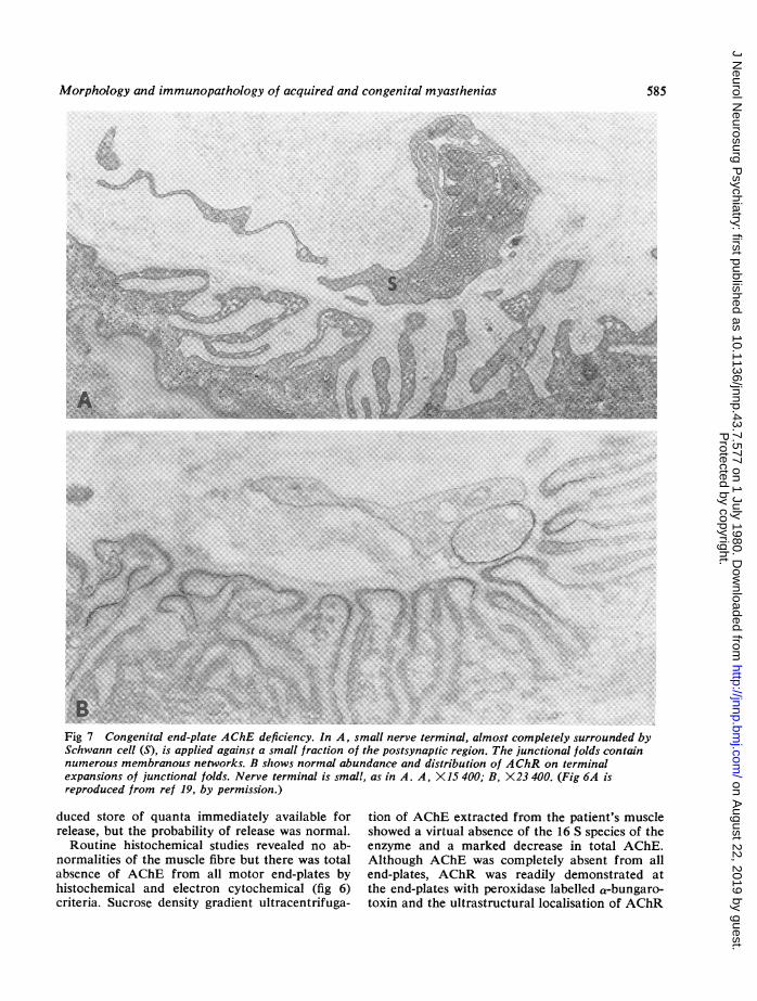
Fig 7 Congenital end-plate AChE deficiency. In A, small nerve terminal, almost completely surrounded bySchwann cell (S), is applied against a small fraction of the postsynaptic region. The junctional folds containnumerous membranous networks. B shows normal abundance and distribution of AChR on terminalexpansions of junctional folds. Nerve terminal is small, as in A. A, X15 400; B, X23 400. (Fig 6A isreproduced from ref 19, by permission.)
duced store of quanta immediately available forrelease, but the probability of release was normal.Routine histochemical studies revealed no ab-
normalities of the muscle fibre but there was totalabsence of AChE from all motor end-plates byhistochemical and electron cytochemical (fig 6)criteria. Sucrose density gradient ultracentrifuga-
tion of AChE extracted from the patient's muscleshowed a virtual absence of the 16 S species of theenzyme and a marked decrease in total AChE.Although AChE was completely absent from allend-plates, AChR was readily demonstrated atthe end-plates with peroxidase labelled a-bungaro-toxin and the ultrastructural localisation of AChR
585
Protected by copyright.
on August 22, 2019 by guest.
http://jnnp.bmj.com
/J N
eurol Neurosurg P
sychiatry: first published as 10.1136/jnnp.43.7.577 on 1 July 1980. Dow
nloaded from

586
was normal (fig 7B). The nerve terminals weregenerally small (fig 7) and quantitative electronmicroscopy revealed a three to fourfold decreaseof nerve terminal size and a significant increase inthe numerical density of synaptic vesicles. Somepostsynaptic folds showed focal degeneration and
Andrew G Engel
many were distended by labyrinthine membranousnetworks that communicated with the synapticspace (fig 7A).The absence of end-plate AChE readily explains
the patient's refractoriness to anticholinesterasedrugs, the repetitive muscle action potential to
.- - ,; . . . ... ... ~~~~~~~~~~~~~~~~~~~~~~~~~... &.'.... .:XS....................::..'.*. 7 ' r
*' AK\94< '-q^': '. 4 -f -;
'-'...
-.......
A
.... .. .. ..
<4+~~~~~~~~~~~~..... ..
... .. .. .. ..
B~~~~~~~ ~ ~ ~ ~ ~ ~ ~ ~ ~ ~ ~ ~ ~ ~ ~ ~ ~~~~~~~~~~ ......
Fig 8 Putative defect of ACh induced ion channel. In A, all junctional folds have degeneratedin end-plate region imaged at left. Numerous endocytotic vesicles appear in preservedjunctional folds on right. In B, junctional sarcoplasm and underlying fibre region containnumerous dilated vesicles of the sarcoplasmic reticulum. A, X15 500; B, X 19 000.
......
Protected by copyright.
on August 22, 2019 by guest.
http://jnnp.bmj.com
/J N
eurol Neurosurg P
sychiatry: first published as 10.1136/jnnp.43.7.577 on 1 July 1980. Dow
nloaded from

Morphology and immunopathology of acquired and congenital myasthenias
single nerve stimulus,20 the abnormal durationand prolonged half-decay time of the mepp,21 andthe failure of prostigmine to have any additionaleffect on the mepp in vitro. The decreased quan-tum content of the epp in the presence of a nor-mal probability of release is adequately accountedfor by the smallness of the nerve terminal.22 Thephysiological and clinical phenomena in this casecan be attributed to two findings: the end-plateAChE deficiency and the smallness of the nerveterminals. Of these, the enzyme deficiency is prob-ably primary and smallness of the nerve terminalcompensatory, or secondary. This assumption issupported by the fact that AChE was monoto-nously absent from all end-plates whereas the de-crease in nerve terminal size was statistical, withsome terminals of normal size. An alternativepossibility would be a primary, but yet undefined,presynaptic disturbance which resulted in smallnerve terminals and defective neural regulation ofend-plate AChE.
Familial congenital myasthenic syndromeatrributed to a putative defect of the ACh inducedion channelThis disorder was studied in collaboration withDrs EH Lambert, DW Mulder, K Sahashi, CFTorres, TE Bertorini and JN Whitaker.23 Thedisease presents in childhood or adult life, pro-
gresses slowly and is associated with variableweakness of ocular, facial, cervical and masti-catory muscles, and with selectively severe weak-ness and atrophy of scapular and forearm muscles.In the familial cases there was an autosomaldominant pattern of inheritance. Electrophysi-ologic studies by EH Lambert showed a repetitivemuscle action potential to single nerve stimulus inall muscles and a decremental response to 2 to3 Hz stimulation of clinically affected muscles.Microelectrode studies revealed markedly pro-longed epp's and mepp's which became even moreprolonged on addition of prostigmine, normalquantum content of the epp and a smaller thannormal or low normal mepp amplitude.
Light microscopic histochemical studies showedtype 1 fibre predominance, isolated or small groupsof atrophic fibres of either histochemical type,tubular aggregates and vacuoles in fibre regionsnear motor end-plates, abnormal variation infibre size, variable fibre splitting and, in somebiopsies, mild to moderate increase of perimysialconnective tissue. Abundant AChE activity waspresent at all motor end-plates. Immunoelectronmicroscopic studies revealed no IgG, C3 or C9 atthe motor end-plate. Quantitative electron micro-scopy of 132 end-plates in three cases demonstrated30 to 40 per cent decrease of nerve terminal size,an increased number of synaptic vesicles and re-
Fig 9 AChR localisation in patient with putative defect of the A Ch induced ion channel.AChR is lost where junctional folds have degenerated (asterisk). Circular, dense calcificdeposits occur in remnants of folds. X20 000.
587
Protected by copyright.
on August 22, 2019 by guest.
http://jnnp.bmj.com
/J N
eurol Neurosurg P
sychiatry: first published as 10.1136/jnnp.43.7.577 on 1 July 1980. Dow
nloaded from

588
duced postsynaptic membrane length. There was
focal degeneration of junctional folds (figs 8A and9) associated with loss of AChR (determined withperoxidase-labelled a-bungarotoxin), most markedin the case with the lowest mepp amplitude (fig 9).In addition, many postsynaptic regions had one or
more of the following abnormalities: myriad pino-cytotic vesicles and abundant labyrinthine mem-
branous networks in the junctional folds (figs 8Aand 9); dilated sarcoplasmic reticulum componentsin fibre regions near the end-plates (fig 8B); andfocal cytoplasmic degradation and autophagicvacuoles in the junctional region.The repetitive muscle action potential in this
syndrome is attributed to the prolonged epp. Thelatter could be due to a prolonged presence ofACh in the synaptic space, as when AChE is in-hibited,21 AChE deficiency (as in the previous
syndrome), or a prolonged open time of the AChinduced ion channel.24 The last explanation is themost likely because our patients were not exposedto anticholinesterase drugs and abundant AChEwas present at all end-plates. The ion channel ab-normality must be present in all muscles but thetransmission defect and weakness occur only inselected muscles. On the basis of the morphologicfindings, the transmission defect is attributed todegeneration of the junctional folds with con-
comitant loss of AChR. The degeneration may
be secondary to the abnormal subsynaptic ionicmilieu caused by the prolonged open time of thereceptor ion gate. Finally, the alterations inmuscle fibre architecture and innervation may
also be secondary to the abnormal interaction be-tween ACh and its receptor.
Other congenital myasthenic syndromesIt is entirely conceivable that additional my-
asthenic syndromes will be defined by combinedclinical, ultrastructural, electrophysiological andbiochemical studies. Hypothetically, there mightbe genetically determined abnormalities of theAChR macromolecule which could decrease itsaffinity for ACh, or result in a diminshed con-
ductance or open time of the ACh induced ionchannel. Mutations involving cytoskeletal proteinsmight cause faulty insertion of AChR into thepostsynaptic membrane or accelerated removalof AChR from the membrane. Further, any muta-tion affecting the structure of the synaptic vesicles,the presynaptic membrane or the junctional foldscould adversely affect the safety margin of neuro-muscular transmission. Thus, future studies of thecongenital myasthenic syndromes, as well as ofacquired autoimmune MG, may prove to be
Andrew G Engel
challenging and rewarding to clinicians and re-searchers alike.
Work in the author's laboratory was supported inpart by Research Grant NS6277 from the NationalInstitutes of Health and by a Research CentreGrant from the Muscular Dystrophy Association.
References
1 Simpson JA. Myasthenia gravis: A new hypoth-esis. Scot Med J 1960; 5:419-36.
2 Engel AG, Santa T. Histometric analysis of theultrastructure of the neuromuscular junction inmyasthenia gravis and in the myasthenic syn-drome. Ann NY Acad Sci 1971; 183:46-63.
3 Engel AG. Locating motor end-plates for electronmicroscopy. Mayo Clin Proc 1970; 45:450-4.
4 Fambrough DM, Drachman DB, Satyamurti S.Neuromuscular junction in myasthenia gravis:decreased acetylcholine receptors. Science 1973;182:293-5.
5 Engel AG, Lindstrom JM, Lambert EH, LennonVA. Ultrastructural localization of the acetyl-choline receptor in myasthenia gravis and in itsexperimental autoimmune model. Neurology(Minneap.) 1977; 27:307-15.
6 Engel AG, Sahashi K, Lambert EH, Howard FM.The ultrastructural localization of the acetylcho-line receptor, immunoglobulin G and the thirdand ninth complement coimponents at the motorend-plate and their implications for the patho-genesis of myasthenia gravis. In: Aguayo AJ,Karpati G, eds, Current Topics in Nerve andMuscle Research 1979; Amsterdam-Oxford: Ex-cerpta Medica, ICS 455:111-22.
7 Lindstrom J, Lambert EH. Content of acetylcho-line receptor and antibodies bound receptor inmyasthenia gravis, experimental autoimmune my-asthenia gravis, and Eaton-Lambert syndrome.Neucrology (Minneap.) 1978; 28:130-8.
8 Ito Y, Miledi R, Vincent A, Newsom-Davis J.Acetylcholine receptors and end-pla:e electro-physiology in myasthenia gravis. Brain 1978; 101:345-68.
9 Engel AG, Lambert EH, Howard FM. Immunecomplexes (IgG and C3) at the motor end-platein myasthenia gravis. Ultrastructural and lightmicroscopic localization and electrophysiologiccorrelations. Mayo Clin Proc 1977; 52:267-80.
10 Kessler SW. Cell membrane antigen isolation withthe staphylococcal protein-A antibody absorbent.J Immunol 1976; 117:1482-90.
11 Sahashi K, Engel AG, Lambert EH, Howard FM.Ultrastructural localization of the terminal andlytic ninth complement component (C9) at themotor end-plate in myasthenia gravis. J Neuro-pathol Exp Neurol 1980; In press.
12 Muller-Eberhard HJ. Complement. Annu RevBiochem 1975; 44:697-724.
Protected by copyright.
on August 22, 2019 by guest.
http://jnnp.bmj.com
/J N
eurol Neurosurg P
sychiatry: first published as 10.1136/jnnp.43.7.577 on 1 July 1980. Dow
nloaded from

Morphology and immunopathology of acquired and congenital myasthenias
13 Biesecker G, Podack ER, Halverson CA,. Muller-Eberhard HJ. C5b-9 dimer: isolation from com-
plement-dependent membrane lesions. J Exp Med1979; 149:448-58.
14 Reiness GC, Weinberg CB, Hall ZW. Antibodyto acetylcholine receptor increases degradation ofjunctional and extrajunctional receptor in adultmuscle. Nature 1978; 274:68-70.
15 Heinemann S, Merlie J, Lindstrom J. Modulationof acetylcholine receptor in rat diaphragm byantireceptor sera. Nature 1978; 274:65-8.
16 Stanley EF, Drachman DB. Effect of myasthenicimmunoglobulin an acetylcholine receptors ofintact mammalian neuromuscular junctions.Science 1978; 200:1285-7.
17 Hart Z, Sahashi K, Lambert EH, Engel AG,Lindstrom J. A congenital familial myasthenicsyndrome caused by a presynaptic defect of trans-mitter resynthesis or mobilization. Neurology(Minneap.) 1979; 29:559.
18 Robertson WC, Chun RWM, Kornguth SE.Familial infantile myasthenia. Arch Neurol 1980;37:117-9.
19 Engel AG, Lambert EH, Gomez MR. A new
myasthenic syndrome with end-plate acetylcho-linesterase deficiency, small nerve terminals andreduced acetylcholine release. Ann Neurol 1977;1:315-30.
20 Brown GL, Dale HH, Feldberg W. Reactionof the normal mammalian muscle to acetylchol-ine and eserine. J Physiol (Lond) 1936; 87:394-424.
21 Katz B, Miledi R. The binding of acetylcholineto receptors and its removal from the synapticcleft. J Physiol (Lond) 1973; 231:549-74.
22 Bennett MR, Florin TJ. A statistical analysis ofthe release of acetylcholine at newly formed syn-apses in striated muscle. J Physiol (Lond) 1974;238:93-107.
23 Engel AG, Lambert EH, Mulder DW, TorresCF, Sahashi K, Bertorini TE, Whitaker JN. In-vestigations of 3 cases of a newly recognizedfamilial, congenital myasthenic syndrome. AnnNeurol 1979; 6:146.
24 Anderson CR, Stevens CF. Voltage clamp ana-
lysis of acetylcholine produced end-plate currentfluctuations at frog neuromuscular junction JPhysiol (Lond) 1973; 235:655-91.
589
Protected by copyright.
on August 22, 2019 by guest.
http://jnnp.bmj.com
/J N
eurol Neurosurg P
sychiatry: first published as 10.1136/jnnp.43.7.577 on 1 July 1980. Dow
nloaded from