Monocyte activation in early onset rheumatoid...
7
Annals of the Rheumatic Diseases 1990; 49: 497-503 Monocyte activation in early onset rheumatoid arthritis I Fujii, M Shingu, M Nobunaga Abstract Monocytes from peripheral blood and synovial fluid of patients with definite and classic rheumatoid arthritis spontaneously produced significantly greater amounts of prostaglandin E2 (PGE2), leucotriene B4 (LTB4), and inter- leukin-l, (IL-10) than samples of peripheral blood from normal controls. Peripheral blood monocytes from patients with rheumatoid arthritis produced significantly greater amounts of PGE2 than control samples when stimulated with lipopolysaccharide. There were no significant differences in the spon- taneous release of superoxide or N-acetyl-i- D-glucosaminidase by peripheral blood mono- cytes between patients and healthy controls. Both stimulated and unstimulated peripheral blood monocytes from patients with definite or classic rheumatoid arthritis produced significantly greater amounts of PGE2 than samples from normal controls. This was true, regardless of the stage of disease and the presence or absence of roentgenological joint abnormalities. Amounts of N-acetyl-P-D- glucosaminidase released by peripheral blood monocytes from patients correlated positively with the erythrocyte sedimentation rate (ESR) and negatively with duration of disease. Amounts of IL-1lB and N-acetyl-13-D-gluco- saminidase released from the peripheral blood monocytes of patients who had had their disease for less than one year were significantly higher than those of normal controls. There were no significant correlations between the types of treatment and the amounts of PGE2, LTB4, IL-1l6 or N-acetyl-,3-D-glucosaminidase released by peripheral blood monocytes in patients with rheumatoid arthritis. The findings suggest that monocytes are activated in patients with rheumatoid arthritis both at the onset of disease and during its chronic phase, and that they produce large amounts of mediators which may have a role in the induction and extension of the inflam- matory process which leads to tissue damage. Department of CLinical Immunology, Medical Institute of Bioregulation, Kyushu University 69, Beppu, 874 Japan I Fujiii M Shingu M Nobunaga Correspondence to: Dr Fujii. Accepted for publication 18 August 1989 Rheumatoid arthritis affects peripheral joints, and the destruction of joint structures, demon- strable on x ray examination, is the most important feature for prognosis. The aetiology and pathogenesis of rheumatoid arthritis are poorly understood, but there are many indica- tions that immune mechanisms may be involved. Macrophages are prominent in the accumulation of inflammatory cells in synovial tissue and fluid. The presence of increased concentrations, of HLA-DR antigens on cells from peripheral blood, synovial fluid, and synovium of patients with rheumatoid arthritis suggests that these cells are in an activated state.' Changes in expression of certain surface antigens such as HLA-DR parallel the increased functional capacity of the cells. Thus cells originating from monocyte-macrophages are considered to have an important aetiological role in chronic inflam- mation2 3 by participating in the initiation and maintenance of the inflammatory process and subsequent tissue damage through the release of degrading lysosomal enzymes," superoxide anion (07),7 the prostaglandin E2 (PGE2)8 9 and interleukin-l (IL-1). 1012 The best characterised product of monocyte-macrophages is IL-1. It has been shown that IL-l plays an important part in tissue damage by inducing a variety of functional changes of pathogenetic importance in other cells.47 1-15 Stimulation of monocytes also leads to the release of lysosomal enzymes and activation of enzymes which produce medi- ators of inflammation. These include membrane bound nicotinamide-adenine dinucleotide phos- phate oxidase (NADPH oxidase), which gen- erates 02, and a 5-lipo-oxygenase, which cata- lyses the production of leucotrienes (LTB4). Lysosomal proteinases and 02 radicals are thought to be largely responsible for the result- ing tissue injury found during an inflammatory reaction. Monocytes in rheumatoid arthritis have been shown to possess enhanced Fc receptor binding activity 6 and enhanced meta- bolic and secretory activity.'7 Monocytes in rheumatoid arthritis also produce increased amounts of PGE218 and IL-l.'9 20 The aim of this study was to determine whether the release of PGE2, LTB4, IL-1, 02 and N-acetyl-4-D-glucosaminidase, a lysosomal proteinase, from monocytes is a specific marker of the early stage of rheumatoid arthritis and to correlate these mediators with inflammatory variables in patients with rheumatoid arthritis. Methods Forty four patients with rheumatoid arthritis (38 women, six men, mean age 55-7 years) satisfying the American Rheumatism Association (ARA) criteria2' for classic or definite rheuma- toid arthritis were investigated. The disease stage of joints in these patients was determined according to Steinbrocker et al.22 x Ray pictures of the joints were interpreted blind. The mean duration of rheumatoid arthritis was 7-7 years (range three months to 17-3 years). Disease activity was estimated by the Lans- bury activity index.23 Forty patients with definite and classic rheumatoid arthritis were 497 on 21 June 2018 by guest. Protected by copyright. http://ard.bmj.com/ Ann Rheum Dis: first published as 10.1136/ard.49.7.497 on 1 July 1990. Downloaded from
Transcript of Monocyte activation in early onset rheumatoid...

Annals ofthe Rheumatic Diseases 1990; 49: 497-503
Monocyte activation in early onset rheumatoidarthritis
I Fujii, M Shingu, M Nobunaga
AbstractMonocytes from peripheral blood and synovialfluid of patients with definite and classicrheumatoid arthritis spontaneously producedsignificantly greater amounts of prostaglandinE2 (PGE2), leucotriene B4 (LTB4), and inter-leukin-l, (IL-10) than samples of peripheralblood from normal controls. Peripheral bloodmonocytes from patients with rheumatoidarthritis produced significantly greateramounts of PGE2 than control samples whenstimulated with lipopolysaccharide. Therewere no significant differences in the spon-taneous release of superoxide or N-acetyl-i-D-glucosaminidase by peripheral blood mono-cytes between patients and healthy controls.Both stimulated and unstimulated peripheralblood monocytes from patients with definiteor classic rheumatoid arthritis producedsignificantly greater amounts of PGE2 thansamples from normal controls. This was true,regardless of the stage of disease and thepresence or absence of roentgenological jointabnormalities. Amounts of N-acetyl-P-D-glucosaminidase released by peripheral bloodmonocytes from patients correlated positivelywith the erythrocyte sedimentation rate (ESR)and negatively with duration of disease.Amounts of IL-1lB and N-acetyl-13-D-gluco-saminidase released from the peripheral bloodmonocytes of patients who had had theirdisease for less than one yearwere significantlyhigher than those of normal controls. Therewere no significant correlations between thetypes of treatment and the amounts of PGE2,LTB4, IL-1l6 or N-acetyl-,3-D-glucosaminidasereleased by peripheral blood monocytes inpatients with rheumatoid arthritis.The findings suggest that monocytes are
activated in patients with rheumatoid arthritisboth at the onset of disease and during itschronic phase, and that they produce largeamounts of mediators which may have a rolein the induction and extension of the inflam-matory process which leads to tissue damage.
Department of CLinicalImmunology,Medical Institute ofBioregulation,Kyushu University 69,Beppu, 874 JapanI FujiiiM ShinguM NobunagaCorrespondence to:Dr Fujii.Accepted for publication18 August 1989
Rheumatoid arthritis affects peripheral joints,and the destruction of joint structures, demon-strable on x ray examination, is the mostimportant feature for prognosis. The aetiologyand pathogenesis of rheumatoid arthritis are
poorly understood, but there are many indica-tions that immune mechanisms may be involved.Macrophages are prominent in the accumulationof inflammatory cells in synovial tissue andfluid. The presence of increased concentrations,of HLA-DR antigens on cells from peripheral
blood, synovial fluid, and synovium of patientswith rheumatoid arthritis suggests that thesecells are in an activated state.' Changes inexpression of certain surface antigens such asHLA-DR parallel the increased functionalcapacity of the cells. Thus cells originating frommonocyte-macrophages are considered to havean important aetiological role in chronic inflam-mation2 3 by participating in the initiation andmaintenance of the inflammatory process andsubsequent tissue damage through the release ofdegrading lysosomal enzymes," superoxideanion (07),7 the prostaglandin E2 (PGE2)8 9 andinterleukin-l (IL-1). 1012 The best characterisedproduct of monocyte-macrophages is IL-1. Ithas been shown that IL-l plays an importantpart in tissue damage by inducing a variety offunctional changes of pathogenetic importancein other cells.47 1-15 Stimulation of monocytesalso leads to the release of lysosomal enzymesand activation of enzymes which produce medi-ators of inflammation. These include membranebound nicotinamide-adenine dinucleotide phos-phate oxidase (NADPH oxidase), which gen-erates 02, and a 5-lipo-oxygenase, which cata-lyses the production of leucotrienes (LTB4).Lysosomal proteinases and 02 radicals arethought to be largely responsible for the result-ing tissue injury found during an inflammatoryreaction. Monocytes in rheumatoid arthritishave been shown to possess enhanced Fcreceptor binding activity 6 and enhanced meta-bolic and secretory activity.'7 Monocytes inrheumatoid arthritis also produce increasedamounts of PGE218 and IL-l.'9 20The aim of this study was to determine
whether the release of PGE2, LTB4, IL-1, 02and N-acetyl-4-D-glucosaminidase, a lysosomalproteinase, from monocytes is a specific markerof the early stage of rheumatoid arthritis and tocorrelate these mediators with inflammatoryvariables in patients with rheumatoid arthritis.
MethodsForty four patients with rheumatoid arthritis(38 women, six men, mean age 55-7 years)satisfying the American Rheumatism Association(ARA) criteria2' for classic or definite rheuma-toid arthritis were investigated. The diseasestage of joints in these patients was determinedaccording to Steinbrocker et al.22 x Ray picturesof the joints were interpreted blind. The meanduration of rheumatoid arthritis was 7-7 years(range three months to 17-3 years).
Disease activity was estimated by the Lans-bury activity index.23 Forty patients withdefinite and classic rheumatoid arthritis were
497
on 21 June 2018 by guest. Protected by copyright.
http://ard.bmj.com
/A
nn Rheum
Dis: first published as 10.1136/ard.49.7.497 on 1 July 1990. D
ownloaded from

Fujii, Shingu, Nobunaga
receiving non-steroidal anti-inflammatory drugs(NSAIDs); 23 patients were being treated withsteroids and seven with gold salts. The presenceofvasculitis in patients with rheumatoid arthritiswas evaluated either clinically or histologically,as previously described.24 Ten healthy femaleand six healthy male hospital employees, with amean age of 40-6 years (range 23-8 to 57 4 years)served as controls.Serum rheumatoid factor was measured by
laser nephelometry (Behring) using a rheumatoidarthritis test kit (Hoechst) (normal range below13 IU/ml). Total haemolytic complement con-centrations (CH50) were measured according tothe method of Kent and Fife.25 C3 and C4 weremeasured by single radial immunodiffusionusing commercial kits (Hoechst).
ISOLATION OF MONOCYTESTwenty millilitres of venous blood treated withheparin were collected, diluted with an equalvolume of phosphate buffered saline (PBS)(0-01 M phosphate, 0 138 M NaCl, pH 7 4),and the mononuclear cells were isolated byFicoll-Hypaque density gradient centrifugation(Pharmacia Fine Chemicals). Synovial fluid wasaspirated from the knee joints of patients withrheumatoid arthritis into polystyrene tubes con-taining sodium heparin, diluted 1/1 with PBS,and centrifuged at 200 g. Cells were resuspendedin PBS, layered on Ficoll-Hypaque, and centri-fuged. Cells at the resulting interface wereaspirated, washed twice in PBS, and suspendedat a concentration of 2 x 10⁶/ml in RPMI 1640medium, supplemented with 100 U/ml peni-cillin, 100 Zg/ml streptomycin, 0 25 ng/mlfungizone, 2 mmol/ml L-glutamine, and 10%heat inactivated fetal calf serum (FCS) (Gibco).These cells were then incubated in Petri dishes(Falcon, 3002) which had been coated with FCSand then incubated for one hour at 37°C in a 5%carbon dioxide incubator. Non-adherent cellswere gently poured from the dishes and dis-carded. The dishes were rinsed with medium,and adherent cells were released into RPMImedium by mechanical agitation. The resultantcell suspension was centrifuged and the pelletswere washed twice with medium. Cells wereresuspended in 10% FCS-RPMI 1640 at aconcentration of 1 x 10⁶/ml and constituted themonocyte preparation. The percentage ofmonocytes, as judged by Giemsa and esterasestaining, varied between 95% and 98%, and theviability, estimated by trypan blue dye exclu-sion, varied between 97% and 99%. In someexperiments monocytes were also suspended inHanks's buffered salt solution (HBSS).
CULTURE OF MONOCYTESMonocyte suspension (0-5 ml) was mixed with0 05 ml of 50 pg/ml lipopolysaccharide (Sigma)as a stimulating agent, in polystyrene tubes(Falcon). Cells incubated with PBS instead oflipopolysaccharide served as unstimulatedcontrols. Tubes were kept at 37°C in a 5%carbon dioxide incubator for 24 hours and thencentrifuged at 400 g for 10 minutes. Thesupernatants were collected and frozen at - 80°C
until use when they were assayed for PGE2,LTB4, N-acetyl-13-D-glucosaminidase, and IL-113. As a stimulating agent, heat aggregatedhuman IgG was used. This was prepared byheating native IgG (Cohn Fraction II, Sigma,10 mg/ml) at 37°C for 20 minutes, followed bycentrifugation at 3000 rpm for 20 minutes toremove large aggregates. The final concentrationof heat aggregated human IgG was 500 ptg/ml.A competitive binding radioimmunoassay
was used for PGE2 using a commercial kit(Amersham). Leucotriene B4 was also measuredby radioimmunoassay using a commercial kit(Amersham).
N-acetyl-f3-D-glucosaminidase was measuredas described previously.26 Briefly, 0-2 ml ofsample, diluted 1/1 with 0- 1 ml of 0-1 M citratebuffer (pH 4-5), was incubated in glass tubeswith 3-6 mM paranitrophenyl-2-acetamido-2-deoxy-13-D-glucopyranoside (Sigma) as substratefor four hours at 37°C. The reaction wasterminated with 0 3 ml of 1 M borate buffer(pH 9-8) and enzyme activity was determinedcolorimetrically at 405 nm as paranitrophenolwas released from the substrate. Values werecorrected for background levels in the medium.02 generation by monocytes was measured
as described previously.27 Monocytes (0-2 ml)suspended in HBSS were plated in 96-wellplates (Falcon), and ferricytochrome c (horseheart, type III, Sigma) was added at the finalconcentration of 55 iimol/l. In control wells 02dismutase (SOD, Sigma 3000 U/mg, 20 pg/ml)was added. Cells were then cultured for fourhours at 37°C. After incubation supernatantswere obtained, diluted in distilled water, andreduction of ferricytochrome c was measured byreading absorbance at 550 nm using a Beckmanmodel 35 spectrophotometer. The amount offerricytochrome c reduction inhibited by SODwas calculated as described previously.28 Ratesof ferricytochrome c reduction were linear evenat four hours.
IL-1,6 was measured by enzyme linkedimmunosorbent assay (ELISA) using a kitrecently developed by Otsuka PharmaceuticalCo Ltd.29 Ninety six well-plates were coatedovernight at room temperature with 0 1 ml ofantihuman mouse IL-1,B monoclonal antibody30diluted with PBS to a final concentration of 10[tg/ml. Plates were then washed three times withPBS. To avoid non-specific adsorption, 0 4 mlof 1% bovine serum albumin (Sigma) wasadded to each well. Plates were kept at roomtemperature for four hours. After the blockingsolution was removed each well was washedwith 0-4 ml of PBS and 0-05% Tween 20. Asolution of IL-1B containing 40 ng/ml of humanrecombinant IL-1,B was serially diluted withPBS and the dilutions used as standards.3'Samples or serially diluted standards (0-1 ml)were added to each well and incubated at 4°Covernight. Each well was washed three timeswith PBS after the samples or standards hadbeen removed. Rabbit antihuman IL-1,6 poly-clonal antibody (0-1 ml) was added to each welland incubated at room temperature for twohours. Wells were washed three times withPBS. Horseradish peroxidase-conjugated anti-rabbit IgG (0-1 ml) was added to each well.
498
on 21 June 2018 by guest. Protected by copyright.
http://ard.bmj.com
/A
nn Rheum
Dis: first published as 10.1136/ard.49.7.497 on 1 July 1990. D
ownloaded from

Monocyte activation in rheumatoid arthritis
After incubation for two hours at room tem-perature the wells were washed with PBS and0-1 ml of substrate solution (0-02% hydrogenperoxide, 1 mg/ml o-phenylenediamine dihydro-chloride in 01 M phosphate buffer, pH 5-5)was added. Plates were kept at room temperaturefor 10-15 minutes and the reaction was stoppedby adding 01 ml of 1 N H2SO4 solution.Absorbance was measured at 492 nm using an
ELISA reader (Dainippon-Sei-yaku Co Ltd).This ELISA assay detects 15-6-500 pg/ml ofIL-1,B.
Levels of significance were calculated byStudent's t test.
ResultsPeripheral blood monocytes or synovial fluidmonocytes from patients with classic or definiterheumatoid arthritis spontaneously producedsignificantly greater amounts of PGE2, LTB4,and IL-113 than peripheral blood monocytesfrom normal controls. Peripheral blood mono-
cytes from patients with rheumatoid arthritisproduced significantly greater amounts of PGE2compared with normal controls when stimulatedwith lipopolysaccharide. There were no signifi-cant differences in the spontaneous release ofN-acetyl-p3-D-glucosaminidase or 02 by peripheralblood monocytes between patients with rheu-matoid arthritis and healthy controls (table 1).When the stimulating effects of lipopolysac-charide and heat aggregated human IgG on
PGE2 release by peripheral blood monocytes inrheumatoid arthritis were compared, both lipo-polysaccharide and heat aggregated human IgGstimulated PGE2 production by peripheral bloodmonocytes in rheumatoid arthritis to the same
extent (data not shown).Both stimulated and unstimulated peripheral
blood monocytes from patients with definite orclassic rheumatoid arthritis produced signifi-cantly higher amounts of PGE2 than normalperipheral blood monocytes. Leucotriene B4production by peripheral blood monocytes frompatients with classic rheumatoid arthritis was
significantly higher than that by normal peri-pheral blood monocytes. Production of IL-11,02 or N-acetyl-p-D-glucosaminidase by peri-pheral blood monocytes from patients with anyof these diagnostic classifications was within thenormal range. Patients with definite and classicrheumatoid arthritis were classified by the stageof their joints determined by x ray, and amountsof mediators released from peripheral bloodmonocytes were compared with those releasedfrom normal peripheral blood monocytes.Peripheral blood monocytes from patients with
rheumatoid arthritis with no joint abnormalitiesspontaneously produced significantly higheramounts of PGE2 and IL-1,B than normalperipheral blood monocytes. Similarly, peri-pheral blood monocytes from patients withrheumatoid arthritis with stages I, III, IVchanges spontaneously produced significantlyhigher amounts of PGE2 than normal peripheralblood monocytes. Peripheral blood monocytesstimulated with lipopolysaccharide from patientswith rheumatoid arthritis with stages II, III, or
IV changes released significantly higher amountsof PGE2 than normal peripheral blood mono-
cytes. Leucotriene B4 production by peripheralblood monocytes from patients with rheumatoidarthritis with stages II, III, or IV changes was
significantly higher than that in normal peri-pheral blood monocytes. Only peripheral bloodmonocytes from patients with rheumatoidarthritis and stage II changes showed signifi-cantly increased 02 release compared withnormal peripheral blood monocytes; in theother groups the values were not increased.Release of N-acetyl-j3-D-glucosaminidase bymonocytes from patients with rheumatoidarthritis with stages I and III changes was
significantly increased compared with that ofnormal peripheral blood monocytes (table 2).The amounts ofN-acetyl-,3-D-glucosaminidase
released by peripheral blood monocytes frompatients with rheumatoid arthritis correlatedpositively with ESR and negatively with durationof disease. The amount of N-acetyl-P-D-gluco-saminidase released, however, did not correlatewith activity index (table 3). There were no
significant correlations between the amounts ofPGE2, LTB4, 02 or IL-1,6 and ESR, activityindex, or duration of disease. There were no
differences in the amounts of mediators releasedby peripheral blood monocytes in rheumatoidarthritis in the presence or absence of vasculitis(data not shown). When patients with rheuma-toid arthritis were classified according to diseaseduration of less or more than one year and theamounts of mediators released by unstimulatedperipheral blood monocytes were comparedwith those of healthy controls, the amounts ofIL-l and N-acetyl-,3-D-glucosaminidase pro-duced were significantly higher in patients withrheumatoid arthritis with disease duration lessthan one year. On the other hand, the produc-tion of PGE2 and LTB4 was not increased inpatients with rheumatoid arthritis with diseaseduration of less than one year, but was signifi-cantly increased in patients with rheumatoidarthritis with disease duration of more than one
year (table 4).
Table 1: Amounts of mediators (mean (SD)) released by peripheral or synovial fluid monocytes from patients and normalcontrols
Mediators released Peripheral blood monocytes Peripheral blood monocytes Synovial fluid monocytes(normal controls) (rheumatoid arthritis) (rheumatoid arthritis)
PGE2' (pg/106 cells) unstimulated 2741 (1649) (n=16) 5935 (3748)j (n=44) 3425 (2540) (n= 12)ttLPS" stimulated 4898 (3671) (n= 16) 9830 (5873)t (n=44) -
LTB4 (pg/10" cells) unstimulated 91-2 (48 7) (n=15) 146-4 (58-4)f (n=43) 113-8 (44-4) (n=ll)IL-l1W.: (nglO' cells) unstimulated 56 4 (58-5) (n=8) 122-9 (79.3)* (n=24) 47-9 (44 7) (n=8)¶NAG' ([Lmol) unstimulated 314-5 (16 6) (n=6) 331-9 (24 5) (n=35) -
02 (nmol) unstimulated 0 99 (0 25) (n=9) 1-16 (0 49) (n=20) -
Monocytes were cultured for 24 hours at 37'C and the supernatants were assayed for each mediator.kp<O-05; tp<0-02; fp<0-OOl (v normal), ttp<005, ¶p<0-02 (v peripheral blood monocytes in rheumatoid arthritis).IL-II'=interleukin-li-); LPS=lipopolysaccharide; NAG=N-acetyl---D-glucosaminidase; PGE2=prostaglandin E2.
499
on 21 June 2018 by guest. Protected by copyright.
http://ard.bmj.com
/A
nn Rheum
Dis: first published as 10.1136/ard.49.7.497 on 1 July 1990. D
ownloaded from

Fujii, Shingu, Nobunaga
Table 5 shows that steroids, gold salts, or blood monocytes. 07 release by peripheralNSAIDs did not affect PGE2 production by blood monocytes obtained from patients withperipheral blood monocytes in patients with rheumatoid arthritis taking gold salts was
rheumatoid arthritis. Similarly, these drugs did significantly higher than in patients not takingnot influence the production of LTB4, IL-1I,, or gold salts; other agents did not affect 02
N-acetyl-j3-D-glucosaminidase by peripheral release (table 5).
Table 2: Amounts (mean (SD)) of mediators released by peripheral blood monocytes from patients in relation to diagnostic criteria and stage at x ray
PGE2' (pg) LTB4' (pg) IL-I/3 (ng/ml) 02 (nmol) NAG* (jimolil)LPS* (-) LPS (+)
Normal controls 2741 (1649) 4898 (3671) 91 2 (48-7) 56 4 (58-5) 0-99 (0 25) 314-5 (16 6)(n= 16) (n= 16) (n= 15) (n =8) (n=9) (n=6)
Definite rheumatoid arthritis 6206 (4705)t 9945 (5961); 121-1 (67-6) 107-3 (72 5) 0 73 (0 43) 332 5 (19-5)(n=16) (n=16) (n=19) (n= 11) (n=7) (n=15)
Classic rheumatoid arthritis 5638 (2722)tt 10299 (6378)t 164-5 (56 4)tt 124-5 (88-0) 1-24 (0 52) 330-2 (27 4)(n=28) (n=28) (n=24) (n= 13) (n= 13) (n=20)
Stage 0 6438 (2063)t 8000 (1000) 119-1 (68 0) 150-0 (86 8)t 0-76 320-3 (18 5)(n=2) (n=2) (n=4) (n=3) (n= 1) (n =3)
Stage I 4338 (2448)Y 9194 (7438) 141-9 (97-5) 112-0 (84-4) 1-01 (0 58) 343-1 (18-5)t(n= 14) (n= 14) (n=8) (n=5) (n=6) (n=8)
Stage II 7036 (6074) 11251 (5630)t 145-6 (59 0)t 123-6 (78 7) 1-38 (0-37)t 329-9 (29-3)(n=8) (n=8) (n= 15) (n=8) (n=6) (n=9)
Stage III 7016 (2035)t 11062 (4329)i 165-3 (77-4)t 135-7 (111-1) 0-84 (0-31) 338-3 (11 0)t(n=8) (n=8) (n=7) (n=3) (n=3) (n =8)
Stage IV 5865 (3330)4 9427 (4667)'' 152-5 (56 8)t 108-8 (84-5) 1-22 (0-72) 319 6 (33-4)(n= 12) (n= 12) (n=9) (n=5) (n=4) (n=7)
Patients were categorised according to diagnostic criteria, and patients with definite or classic rheumatoid arthritis were classified by stage at x ray.*p<0.05; tp<0 02; tp<0 01 fp<0 001 (v normal).*For abbreviations see table 1.
Table 3: Relation between amounts of mediators released from peripheral blood monocytes and clinical variables in patients
Variables PGE2* LTB4t NAG* 0, IL-i/P
ESR r=0-081 r=-0-02 r=0-406* r=0-085 r=-0 181n=44 n=43 n=35 n=20 n=24
Serum rheumatoid factor r=-0077 r=0-082 - 0182n=44 n=43 n=24
Activity index r=0-008 r=0-012 r=0064 r=0-226 r= 0-183n=38 n=33 n=22 n= 16 n=21
Duration of disease r=0-078 r=0 171 r=-0 384*' r=-0 161 r=-0-036n=44 n=43 n=34 n=20 n=22
Data are expressed as correlation coefficients (r values). Patients include those with both definite and classic rheumatoid arthritis.,p<005.*For abbreviations see table 1.
Table 4: Comparison ofmediators released by unstimulated peripheral blood monocytes from normal controls andfrom patientswith disease duration of greater or less than one year
Mediators Normal controls Disease duration
<I year >I year
PGE2' (pg) 2741 (1649) (n= 16) 4688 (3714) (n= 10) 6247 (3738)4 (n=34)LTB4' (pg) 91-2 (48 7) (n=15) 117-1 (66-1) (n=11) 156 5 (67 3)t (n=32)IL-li"V (ng/ml) 56-4 (58-5) (n=8) 133-8 (63-2): (n=8) 117-5 (87-6) (n=16)02 (nmol) 0 99 (0 25) (n=9) 0 81 (0-38) (n=5) 1-27 (0 47) (n=15)NAG* (Omol/l) 314-5 (16.6) (n=6) 335 2 (16-6)' (n= 11) 331-3 (24-6) (n=24)
'p<0-05; tp<0-01; tp<O-OOl (v normal).*For abbreviations see table 1.
Table 5: Effect of treatment regimen on amounts of mediators released by peripheral blood monocytes from patients
Treatment PGE2 (Pg) LTB4 (pg) IL-If NAG 02 (nmol)(nglml) (pmol/l)
Steroid (±) 6304 (2952) 167-9 (89-3) 111-0 (82-8) 329-9 (27-6) 1-35 (0-47)(n=23) (n=24) (n= 15) (n= 19) (n= 10)
Steroid (-) 5620 (4345) 145-1 (73-6) 142-8 (73-2) 335-8 (21-7) 0 90 (0-48)(n=21) (n= 19) (n=9) (n= 16) (n= 10)
Gold (+)Steroid (-) 5938 (31%) 118-9 (71-7) 89-0 (72-5) 345-5 (26-5) 1-69 (0-12)'
(n=4) (n=7) (n=5) (n=4) (n=3)Gold (-)Steroid (-) 5563 (4471) 143-7 (67-7) 138-6 (80 3) 334-4 (20 4) 0-83 (0-45)
(n=24) (n= 15) (n=7) (n= 13) (n=9)NSAIDs (+) 6178 (3724) 143-2 (69-0) 119-6 (79 3) 332-7 (24 4) 1-12 (0 50)
(n=40) (n=40) (n=23) (n=29) (n= 15)
NSAIDs (-) 4963 (3884) 189-6 (50 3) 200 0 328-2 (27-5) 1-14 (0-61)(n=4) (n=3) (n= 1) (n=6) (n=5)
Data are expressed as mean (SD), *p<005 v gold (-) steroid (-).
500
on 21 June 2018 by guest. Protected by copyright.
http://ard.bmj.com
/A
nn Rheum
Dis: first published as 10.1136/ard.49.7.497 on 1 July 1990. D
ownloaded from
Monocyte activation in rheumatoid arthritis
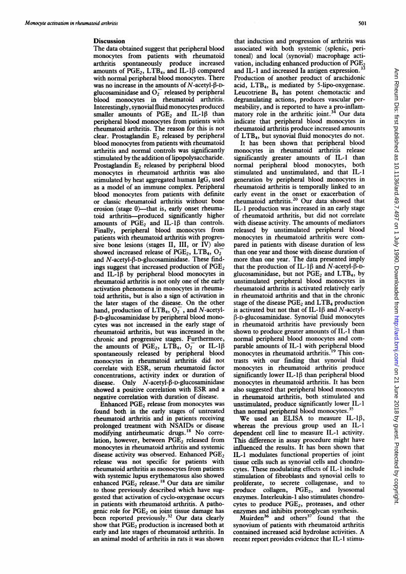
DiscussionThe data obtained suggest that peripheral bloodmonocytes from patients with rheumatoidarthritis spontaneously produce increasedamounts of PGE2, LTB4, and IL-1,3 comparedwith normal peripheral blood monocytes. Therewas no increase in the amounts of N-acetyl-4-D-glucosaminidase and 02 released by peripheralblood monocytes in rheumatoid arthritis.Interestingly, synovial fluid monocytes producedsmaller amounts of PGE2 and IL-1,13 thanperipheral blood monocytes from patients withrheumatoid arthritis. The reason for this is notclear. Prostaglandin E2 released by peripheralblood monocytes from patients with rheumatoidarthritis and normal controls was significantlystimulated by the addition oflipopolysaccharide.Prostaglandin E2 released by peripheral bloodmonocytes in rheumatoid arthritis was alsostimulated by heat aggregated human IgG, usedas a model of an immune complex. Peripheralblood monocytes from patients with definiteor classic rheumatoid arthritis without boneerosion (stage 0)-that is, early onset rheuma-toid arthritis-produced significantly higheramounts of PGE2 and IL-1,6 than controls.Finally, peripheral blood monocytes frompatients with rheumatoid arthritis with progres-sive bone lesions (stages II, III, or IV) alsoshowed increased release of PGE2, LTB4, 02and N-acetyl-p-D-glucosaminidase. These find-ings suggest that increased production of PGE2and IL-1,6 by peripheral blood monocytes inrheumatoid arthritis is not only one of the earlyactivation phenomena in monocytes in rheuma-toid arthritis, but is also a sign of activation inthe later stages of the disease. On the otherhand, production of LTB4, 02, and N-acetyl-,3-D-glucosaminidase by peripheral blood mono-cytes was not increased in the early stage ofrheumatoid arthritis, but was increased in thechronic and progressive stages. Furthermore,the amounts of PGE2, LTB4, 02 or IL-lospontaneously released by peripheral bloodmonocytes in rheumatoid arthritis did notcorrelate with ESR, serum rheumatoid factorconcentrations, activity index or duration ofdisease. Only N-acetyl-,3-D-glucosaminidaseshowed a positive correlation with ESR and anegative correlation with duration of disease.Enhanced PGE2 release from monocytes was
found both in the early stages of untreatedrheumatoid arthritis and in patients receivingprolonged treatment with NSAIDs or diseasemodifying antirheumatic drugs.'8 No corre-lation, however, between PGE2 released frommonocytes in rheumatoid arthritis and systemicdisease activity was observed. Enhanced PGE2release was not specific for patients withrheumatoid arthritis as monocytes from patientswith systemic lupus erythematosus also showedenhanced PGE2 release.'8 Our data are similarto those previously described which have sug-gested that activation of cyclo-oxygenase occursin patients with rheumatoid arthritis. A patho-genic role for PGE2 on joint tissue damage hasbeen reported previously.32 Our data clearlyshow that PGE2 production is increased both atearly and late stages of rheumatoid arthritis. Inan animal model of arthritis in rats it was shown
that induction and progression of arthritis wasassociated with both systemic (splenic, peri-toneal) and local (synovial) macrophage acti-vation, including enhanced production of PGE2and IL-1 and increased Ia antigen expression.33Production of another product of arachidonicacid, LTB4, is mediated by 5-lipo-oxygenase.Leucotriene B4 has potent chemotactic anddegranulating actions, produces vascular per-meability, and is reported to have a pro-inflam-matory role in the arthritic joint.34 Our dataindicate that peripheral blood monocytes inrheumatoid arthritis produce increased amountsof LTB4, but synovial fluid monocytes do not.
It has been shown that peripheral bloodmonocytes in rheumatoid arthritis releasesignificantly greater amounts of IL-1 thannormal peripheral blood monocytes, bothstimulated and unstimulated, and that IL-1generation by peripheral blood monocytes inrheumatoid arthritis is temporally linked to anearly event in the onset or exacerbation ofrheumatoid arthritis.20 Our data showed thatIL-1 production was increased in an early stageof rheumatoid arthritis, but did not correlatewith disease activity. The amounts of mediatorsreleased by unstimulated peripheral bloodmonocytes in rheumatoid arthritis were com-pared in patients with disease duration of lessthan one year and those with disease duration ofmore than one year. The data presented implythat the production of IL-1 B and N-acetyl-f3-D-glucosaminidase, but not PGE2 and LTB4, byunstimulated peripheral blood monocytes inrheumatoid arthritis is activated relatively earlyin rheumatoid arthritis and that in the chronicstage of the disease PGE2 and LTB4 productionis activated but not that of IL-1l6 and N-acetyl-P3-D-glucosaminidase. Synovial fluid monocytesin rheumatoid arthritis have previously beenshown to produce greater amounts of IL-1 thannormal peripheral blood monocytes and com-parable amounts of IL-1 with peripheral bloodmonocytes in rheumatoid arthritis.'9 This con-trasts with our finding that synovial fluidmonocytes in rheumatoid arthritis producesignificantly lower IL-1lB than peripheral bloodmonocytes in rheumatoid arthritis. It has beenalso suggested that peripheral blood monocytesin rheumatoid arthritis, both stimulated andunstimulated, produce significantly lower IL-1than normal peripheral blood monocytes.35We used an ELISA to measure IL-1,I
whereas the previous group used an IL-1dependent cell line to measure IL-1 activity.This difference in assay procedure might haveinfluenced the results. It has been shown thatIL-1 modulates functional properties of jointtissue cells such as synovial cells and chondro-cytes. These modulating effects of IL-l includestimulation of fibroblasts and synovial cells toproliferate, to secrete collagenase, and toproduce collagen, PGE2, and lysosomalenzymes. Interleukin-1 also stimulates chondro-cytes to produce PGE2, proteases, and otherenzymes and inhibits proteoglycan synthesis.
Muirden36 and others37 found that thesynovium of patients with rheumatoid arthritiscontained increased acid hydrolase activities. Arecent report provides evidence that IL-l stimu-
501
on 21 June 2018 by guest. Protected by copyright.
http://ard.bmj.com
/A
nn Rheum
Dis: first published as 10.1136/ard.49.7.497 on 1 July 1990. D
ownloaded from

Fujii, Shingu, Nobunaga
lates the N-acetyl-j3-D-glucosaminidase activityof synovialcells.5 At persistent or transient pHvalues low enough to promote clinically impor-tant activity of certain enzymes, such as mightoccur in the protected areas between synoviumand cartilage,38 N-acetyl-13-D-glucosaminidasemay have an important role in the lysis ofconnective tissue components or activation oflatent neutral proteases such as plasminogenactivator.39
It has been suggested that rheumatoid mono-cyte metabolic activity, as measured by hexosemonophosphate shunt activity, is stimulatedcompared with that in normal monocytes.Hurst et al showed that there was no significantdifference in unstimulated rates of monocyte07 production between normal controls andpatients with rheumatoid arthritis receivingNSAIDs alone, and patients with rheumatoidarthritis receiving second line treatment.' Thisfinding is very similar to our results.Most experiments on monocyte02 generation
have been done using an incubation period ofone hour; ours were performed using an incu-bation period of four hours. It has beensuggested that the differentiation of humanmonocytes into macrophages in vitro is accom-panied by an apparent reduction in the capacityto produce 07 and hydrogen peroxide, andthat the rates of production of 07 after threedays in culture decreased to half of that initiallycultured.4' These findings suggest that a fourhour culture does not cause clinically importantmonocyte differentiation and a large decrease of02 production.
It has been implied that monocyte metabolicactivity, in vitro and in vivo, is modulated byNSAIDs or disease modifying agents. Forexample, piroxicam, which inhibits cyclo-oxygenase, inhibits 07 production mediated byN-formyl-methionyl-leucyl-phenylalanine butnot LTB4 production by monocytes.42 Success-ful treatment with D-penicillamine or goldcompounds is associated with a significantincrease in monocyte 07 generation.43 It hasalso been shown that gold compounds inhibitIL-1 production by monocytes.44 Dexametha-sone, a potent phospholipase A2 inhibitor, wasshown to inhibit IL-1 production by humanmonocytes.45 Finally, auranofin, but not goldsalts, inhibited LTB4 production of normalperipheral blood monocytes. These observationsstrongly suggest that therapeutic agents inpatients with rheumatoid arthritis modify therates of production of various mediators bymonocytes. When the patients were groupedinto several categories according to the thera-peutic agents they were receiving in our studyand the rates of mediators released by peripheralblood monocytes were compared, there were nosignificant differences among the groups in therates of PGE2, LTB4, IL-113, and N-acetyl-P3-D-glucosaminidase released by peripheral bloodmonocytes in rheumatoid arthritis. 07- releasedby peripheral blood monocytes from patientsreceiving gold salts, however, was significantlygreater than that of peripheral blood monocytesfrom patients not receiving gold salts. This find-ing agreed with the findings of the previousreport.43
The data presented here suggest that mono-cytes are activated in patients with rheumatoidarthritis and that they produce increasedamounts of mediators which may have a role inthe induction and extension of the inflammatoryprocess. The mechanisms by which monocytesare metabolically activated in patients withrheumatoid arthritis are not yet fully under-stood, but immune complexes and activatedcomplement products may be responsible formonocyte activation. Monocytes from patientswith rheumatoid arthritis are known to haveincreased numbers of Fc receptors'6 and thismay also furnish a mechanism for monocyteactivation in these patients.
We thank Miss E Kohno and Miss N Ando for their excellenttechnical assistance and Dr Morris Ziff for helpful discussions.
l Firestein GS, Zvaifler N J. Peripheral blood and synovialfluid monocyte activation in inflammatory arthritis. ArthritisRheum 1987; 30: 857-63.
2Janossy G, Duke 0, Poulter L W, Panayi G, Bofill M,Goldstein G. Rheumatoid arthritis: a disease of T-lympho-cyte/macrophage immunoregulation. Lancet 1981; i: 839-42.
3 Ishikawa H, Ziff M. Electron microscopic studies ofimmunoreactive cells in the rheumatoid synovial membrane.Arthritis Rheum 1976; 19: 1-14.
4 Dayer J M, Stephenson M L, Schmidt E, Karge W, Krane SM. Purification of a factor from human blood monocyte-macrophages which stimulates the production of collagenaseand prostaglandin E2 by cells cultured from rheumatoidsynovial tissues. FEBS Lett 1981; 124: 253-6.
Schnyder J, Payne T, Dinarello C A. Human monocyte orrecombinant interleukinl'saFe specific for the secretion of ametallo-proteinase from chondrocytes. J Immunol 1987;138: 496-503.
6 Clarris J R E, Hamilton J A. Peripheral blood mononuclearcells stimulate N-acetyl-,3-glucosaminidase levels in humansynovial fibroblast-like cells. RheumatolInt 1985; 5: 55-60.
7 Matsubara T, Ziff M. Increased superoxide anion releasefrom human endotheial celis in response to cytokines. JImmunol 1986; 137: 3295-8.
8 Hamilton J A, Leizer T, Ligelbach S R. The stimulation ofhuman synovial fibroblast plasminogen activator activity:involvement of cyclic AMP and cyclooxygenase products.Biochim Biophys Acta 1986; 886: 195-202.
9 Mochan E, Uhl J, Newton R. Evidence that interleukin 1induction of synovial cell plasminogen activator is mediatedvia prostaglandin E2 and cAMP. Arthritis Rheum 1986; 29:1078-84.
10 Arend W P, Joslin F G, Massoni R J. Effects of immunecomplexes on production by human monocytes of interleukinI or an interleukin 1 inhibitor. J Immunol 1985; 134:3868-75.
11 Stimpson S A, Dalidorf F G, Otterness I G, Schwab J H.Exacerbation of arthritis by IL-I in rat joints previouslyinjured by peptidoglycan-polysaccharide. J Immunol 1988;140: 2964-9.
12 Ratcliffe A, Tyler J A, Hardingham T E. Articular cartilagecultured with interleukin 1. Increased release of linkprotein, hyaluronate-binding region and other proteoglycanfragments. BiochemJ1 1986; 238: 571-80.
13 Pohlman T H, Stanness K A, Beatty P G, Ochs H D, HarlanJ M. An endothelial cell surface factor(s) induced in vitro bylipopolysaccharide, interleukin 1, and tumor necrosisfactor-a increases neutrophil adherence by a CDwI8-dependent mechanism. J Immunol 1986; 136: 4548-53.
14 Cavender D E, Haskard DO, Joseph B, ZiffM. Interleukin 1increases the binding of human B and T lymphocytes toendothelial cell monolayers. J Immunol 1986; 136: 203-7.
15 Claris B J, Fraser J R E, Ash P, Leiser T, Hamilton J A.Interleukin-l, and interieukin-la stimulate the N-acetyl-,l-glucosaminidase activity of human synovial cells. RheumatolInt 1987; 7: 271-5.
16 MacKinnon S K, Starkebaum G. Monocyte Fc receptorfunction in rheumatoid arthritis. Enhanced cell-binding ofIgG induced by rheumatoid factors. Arthritis Rheum 1987;30: 498-506.
17 Kay N E, Douglas S D. Monocyte metabolic activation inpatients with rheumatoid arthritis (40541). Proc Exp BiolMed 1979; 161: 303-6.
18 Seiz M, Hunstein W. Enhanced prostaglandin release frommonocytes of patients with rheumatoid arthritis and systemiclupus erythematosus. Ann Rheum Dis 1985; 44: 438-45.
19 Danis V A, March L M, Nelson D S, Brooks P M.Interleukin-I secretion by peripheral blood monocytes andsynovial macrophages from patients with rheumatoidarthritis. IRhewmatol 1987; 14: 33-9.
20 Shore A, Jaglal S, Keystone E C. Enhanced interleukin 1generation by monocytes in vitro is temporally linked to anearly event in the onset or exacerbation of rheumatoidarthritis. Clin Exp Immunol 1986; 65: 293-302.
502
on 21 June 2018 by guest. Protected by copyright.
http://ard.bmj.com
/A
nn Rheum
Dis: first published as 10.1136/ard.49.7.497 on 1 July 1990. D
ownloaded from

Monocyte activation in rheumatoid arthritis
21 Ropes M W, Bennett G A, Cobb S, Jacox R, Jessar R A. 1958revision of diagnostic criteria for rheumatoid arthritis. BullRhewn Dis 1958; 9: 175-6.
22 Steinbrocker 0, Traeger C H, Batterman R C. Therapeuticcriteria in rheumatoid arthritis. JAMA 1949; 140: 659-62.
23 Lansbury J. Quantitation of activity of rheumatoid arthritis:method for summation of systemic indices of rheumatoidactivity. Am J Med Sci 1956; 232: 300-17.
24 Toyoda K, Saito S, Naito S, et al. HLA antigens in classicaland malignant rheumatoid arthritis in Japanese population.Tissue Antigens 1977; 10: 56-9.
25 Kent J F, Fife E H. Precise standardization of reagents forcomplement fixation. Am J Trop Med 1963; 12: 103-16.
26 LeMaeshall J, Fraser J R E, Muirden K D. Lysosomalactivation by neutral saccharides in cell cultures ofsynovium.Ann Rheum Dis 1977; 36: 130-8.
27 Pick E, Mizel D. Rapid microassays for the measurement ofsuperoxide and hydrogen peroxide production by macro-phages in culture using an automatic enzyme immunoassayreader. J Immunol Methods 1981; 46: 211-26.
28 Van Gelder B F, Slater E C. The extinction coefficient ofcytochrome C. Biochim Biophys Acta 1%2; 58: 593-5.
29 Tanaka K, Ishikawa E, Ohmoto Y, Hirai Y. In vitroproduction of human interleukin la and interleukin 1 byperipheral blood mononuclear cells examined by sensitivesandwich enzyme immunoassay. Eur j Immunol 1987; 17:1527-30.
30 Nishida T, Nishino N, Takano M, et al. cDNA clotting ofIL-hx and IL-16 from mRNA of U937 cell line. BiochemBiophys Res Commun 1987; 143: 345-52.
31 Hirai Y, Masui Y, Nakai S, Kikumoto Y, Hong Y-M.Interleukin-1: cDNA clotting, production and biologicalactivities of human interleukin-1. Gann monograph on cancerresearch: cellular and molecular mechanism of tumor immunity.Tokyo: Japan Scientific Societies Press, 1988: 156-66.
32 Dominguez J H, Mundy G R. Monocytes mediate osteoclasticbone resorption by prostaglandins production. Cakif TissueInt 1980; 31: 29-34.
33 Burmester G R, Dimitriu-Bona A, Waters S J, WinchesterR J. Identification of three major synovial lining cell popula-tions by monoclonal antibodies directed to la antigens andantigens associated with monocyte/macrophages and fibro-blasts. ScandJ7 Immunol 1983; 17: 69-82.
34 Klickstein L B, Shapleigh C, Goetzl E. Lipooxygenation ofarachidonic acid as a source of polymorphonuclear leukocyte
chemotactic factors in synovial fluid and tissue in rheuma-toid arthritis. J Clin Invest 1980; 66: 1166-70.
35 Whicher J T, Gilbert A M, Westacott C, Hutton C.Defective production of leucocyte mediator (interleukin 1)by peripheral blood leucocytes of patients with systemicsclerosis, systemic lupus erythematosus, rheumatoid arthritisand mixed connective tissue disease. Clin Exp Immunol1986; 65: 80-9.
36 Muirden K D. Lysosomal enzymes in synovial membrane inrheumatoid arthritis. Ann Rheum Dis 1972; 31: 265-71.
37 Wegelis 0, Klockars M, Vaino U. Acid phosphatase activityin rheumatoid synovium. Acta Med Scand 1968; 183:549-51.
38 Dingle J T. The secretion of enzymes into the pericellularenvironment. Philos Trans R Soc Lond [Bioll 1975; 271:315-24.
39 Eeckhout Y, Vaes G. Further studies on the activation ofprocollagenase, the latent precursor of bone collagenase.Effects of lysosomal cathepsin B, plasma and kallikrein, and.spontaneous activation. BiochemJ 1977; 166: 21-31.
40 Hurst N P, Bessac B, Nuki G. Monocyte superoxide anionproduction in rheuiatoid arthritis: preliminary evidence forenhanced rates of superoxide anion production by mono-cytes from patients receiving penicillamine, sodium auro-thiomalate and corticosteroids. Ann Rheum Dis 1984; 43:28-33.
41 Nakagawara A, Nathan C F, Cohn Z A. Hydrogen peroxidemetabolism in human monocytes during differentiation invitro. J Clin Invest 1981; 68: 1243-52.
42 French J K, Hurst N P, McColl S R, Cleland L. Effects ofpiroxicam on superoxide generation, phospholipid methyla-tion and leukotriene production by human blood mono-nuclear cells. J Rheumatol 1987; 14: 1018-21.
43 Hurst N P, Bell A L, Nuki G. Studies of the effect of D-penicillamine and sodium aurothiomalate therapy on super-oxide anion production by monocytes from patients withrheumatoid arthritis: evidence for in vivo stimulation ofmonocytes. Ann Rheum Dis 1986; 45: 37-43.
44 Haynes D R, Garrett I R, Whitehouse M W, Vernon-RobertsB. Do gold drugs inhibit interleukin- I? Evidence from an invitro lymphocyte activating factor assay. J Rheumatol 1988;15: 775-8.
45 Kern J A, Lamb R J, Reed J C, Daniele R P, Nowell P C.Dexamethasone inhibition of interleukin 1 beta productionby human monocytes. J Clin Invest 1988; 81: 237-44.
503
on 21 June 2018 by guest. Protected by copyright.
http://ard.bmj.com
/A
nn Rheum
Dis: first published as 10.1136/ard.49.7.497 on 1 July 1990. D
ownloaded from