
Monitoring of tumor radio frequency ablation using derivative spectroscopy · Monitoring of tumor...
9
Monitoring of tumor radio frequency ablation using derivative spectroscopy Jarich W. Spliethoff Erik Tanis Daniel J. Evers Benno H. W. Hendriks Warner Prevoo Theo J. M. Ruers Downloaded From: https://www.spiedigitallibrary.org/journals/Journal-of-Biomedical-Optics on 7/16/2018 Terms of Use: https://www.spiedigitallibrary.org/terms-of-use
Transcript of Monitoring of tumor radio frequency ablation using derivative spectroscopy · Monitoring of tumor...

Monitoring of tumor radio frequencyablation using derivative spectroscopy
Jarich W. SpliethoffErik TanisDaniel J. EversBenno H. W. HendriksWarner PrevooTheo J. M. Ruers
Downloaded From: https://www.spiedigitallibrary.org/journals/Journal-of-Biomedical-Optics on 7/16/2018Terms of Use: https://www.spiedigitallibrary.org/terms-of-use
Monitoring of tumor radio frequency ablation usingderivative spectroscopy
Jarich W. Spliethoff,a,* Erik Tanis,a Daniel J. Evers,a Benno H. W. Hendriks,b Warner Prevoo,c andTheo J. M. Ruersa,d
aThe Netherlands Cancer Institute, Department of Surgery, Plesmanlaan 121, Amsterdam 1066CX, The NetherlandsbMinimally Invasive Healthcare, Philips Research, High Tech Campus 34, Eindhoven 5656 AE, The NetherlandscThe Netherlands Cancer Institute, Department of Radiology, Plesmanlaan 121, Amsterdam 1066CX, The NetherlandsdUniversity of Twente, MIRA Institute, Building Zuidhorst P.O. Box 217, Enschede 7500 AE, The Netherlands
Abstract. Despite the widespread use of radio frequency (RF) ablation, an effective way to assess thermaltissue damage during and after the procedure is still lacking. We present a method for monitoring RF ablationefficacy based on thermally induced methemoglobin as a marker for full tissue ablation. Diffuse reflectance (DR)spectra were measured from human blood samples during gradual heating of the samples from 37 to 60, 70, and85°C. Additionally, reflectance spectra were recorded real-time during RF ablation of human liver tissue ex vivoand in vivo. Specific spectral characteristics of methemoglobin were extracted from the spectral slopes using acustom optical ablation ratio. Thermal coagulation of blood caused significant changes in the spectral slopes,which is thought to be caused by the formation of methemoglobin. The time course of these changes was clearlydependent on the heating temperature. RF ablation of liver tissue essentially led to similar spectral alterations.In vivo DR measurements confirmed that the method could be used to assess the degree of thermal damageduring RF ablation and long after the tissue cooled. © The Authors. Published by SPIE under a Creative Commons Attribution 3.0
Unported License. Distribution or reproduction of this work in whole or in part requires full attribution of the original publication, including its DOI. [DOI: 10
.1117/1.JBO.19.9.097004]
Keywords: radio frequency ablation; reflectance spectroscopy; treatment monitoring; liver.
Paper 140285RR received May 6, 2014; revised manuscript received Aug. 22, 2014; accepted for publication Aug. 22, 2014; pub-lished online Sep. 19, 2014.
1 IntroductionRadio frequency (RF) tumor ablation is a thermal ablation tech-nique using a needle-type electrode that is inserted into malig-nant tissue. The technique is widely used for the treatment ofmalignant lesions in the liver, kidneys, and lungs. To completelydestroy a tumor, the entire lesion must be heated to cytotoxictemperatures. However, lesion size and local blood flow oftencomplicate heating of the entire tumor volume, resulting inheterogeneity of heat deposition throughout a given lesion tobe treated. Determining whether a complete tumor ablation hasbeen achieved is difficult as there is no method to accuratelyevaluate the extent of the ablation zone. The local tumor recur-rence rates after liver RF ablation vary significantly betweenpublished series, with local tumor recurrence rates of 3.6%(Refs. 1 and 2) to 60%,3 where the latter is mainly due to incom-plete ablation of the tumor margin. Real-time monitoring couldcontribute to locally effective destruction of tumor tissue incombination with a preservation of healthy liver tissue.
Optical spectroscopy techniques, such as diffuse reflectance(DR) spectroscopy at the tip of a thin fiber-optic needle, mayenable real-time monitoring by measuring specific physiologicalinformation from the examined tissue. Unlike temperature mon-itoring with use of thermocouples, which measure the temporaryeffects of ablation, DR spectroscopy could be used to detectpersistent chemical and structural changes undergone by tissueduring thermal ablation. This may allow real-time monitoring ofthe progress of ablation and the adequacy well after the ablationhas been completed.
Various groups have successfully focused on spectroscopicdetection of thermal damage of liver tissue. It was shownthat an increase in reflectance intensity and a decrease in overallfluorescence intensity correlated with the histological degreeof thermal damage.4–6 However, focusing on absolute spectralintensities as an endpoint is prone to be affected by needlemovement, pooling of (coagulated) blood around the probetip, and differences in instrumentation or calibration. Insteadof evaluating the absolute magnitude of the spectrum, wepropose to use semiquantitative information extracted from thespectra that indicates irreversible tissue injury. Such a methodmay be more sensitive to subtle heat-induced changes and mayprovide advanced characterization of irreversible tissue damage.
Depending on the duration of heating and the tissue suscep-tibility for thermal damage, irreversible tissue damage occurs ata threshold temperature of ∼60°C.7 It is, therefore, importantto identify markers that indicate whether or not this thresholdtemperature has been reached. When tissue is subjected toincreasing heat, tissue proteins start a denaturation process andundergo irreversible structural changes. In addition, methemo-globin (metHb) is formed from hemoglobin at temperatures>60°C, making it a potential marker for irreversible liver tissuedamage.8
The group of Tromberg developed a broadband diffuseoptical spectroscopy (600 to 1000 nm) method to derive tissueconcentrations of metHb and four other chromophores.Chromophore concentrations could accurately be monitoredin vivo, despite significant overlapping spectral features.9
Formation of metHb following heat exposure has been reportedby a few other studies for a variety of clinical applications.Barton et al. found that changes in the optical properties of*Address all correspondence to: Jarich W. Spliethoff, E-mail: [email protected]
Journal of Biomedical Optics 097004-1 September 2014 • Vol. 19(9)
Journal of Biomedical Optics 19(9), 097004 (September 2014)
Downloaded From: https://www.spiedigitallibrary.org/journals/Journal-of-Biomedical-Optics on 7/16/2018Terms of Use: https://www.spiedigitallibrary.org/terms-of-use

hemoglobin in a skin model do occur during laser irradiationwith a 532 nm wavelength based on the detection of thermallyinduced metHb.10 More recently, Randeberg et al. showed thatmeasurements of the average metHb concentrations in port-winestains and telangiectasia veins immediately after laser exposuremay be used to verify that the blood temperature has been suf-ficiently high to induce thermal damage to the vessel wall.11
In the same way, high amounts of spectroscopically detectedmetHb appeared to be a good indicator of nonviability of ther-mal wounds.12
The aim of the present study is to investigate whether DRspectroscopy could be used to assess the efficacy of RFtumor ablation.
2 Materials and Methods
2.1 Spectroscopic System
Reflectance spectra were acquired using a portable spectro-scopic system as illustrated in Fig. 1. For illumination of thetissue, a white light halogen broadband light source (360 to2500 nm) with an internal shutter was used. The tissue wasprobed using a clinical-grade disposable 16 G fiber-embeddedneedle (INVIVO, Schwerin, Germany). The probe had onefiber (200 μm) connected to the light source and another fiber(200 μm) connected to a spectrometer (silicon detector; AndorTechnology, Belfast, United Kingdom, DU420A-BRDD), opti-mized for wavelengths between 400 and 1050 nmwith a spectralresolution of ∼4 nm. The center-to-center distance betweenthe emitting and collecting fibers was 0.34 mm. The probehad a polished angle tip of 72 deg to minimize tissue damageduring insertion, while the fiber ends were cut straight. Thespectroscopy needle was made from materials that are heat-resistant in the temperature range that was investigated. DRspectra were acquired with a 0.3 to 1.0 s integration time,depending on the signal intensity at the start of each experiment.The integration time was kept constant during each experiment.The system was controlled by a custom-made LabView softwareuser interface (National Instruments, Austin, Texas, USA).
2.2 Preparation and Heating of Blood Samples
Human venous blood (hematocrit ¼ 41%) was obtained froma healthy human donor. The blood was preserved in an ethyl-enediaminetetraacetic acid (EDTA) tube to prevent coagulation.In the visible wavelength range, the optical absorption of
hemoglobin in blood is dominant compared to the scatteringdue to red blood cells. To enhance diffuse reflectance of the sam-ples, Intralipid®-20% was added as a highly scattering medium.Saline was added to obtain a blood concentration representativefor liver tissue.13 The stock solution that was prepared contained10% blood, 70% saline, and 20% Intralipid®-20%.
To evaluate the effect of thermal coagulation, six samples ofthe stock solution were placed in 1.5 ml cuvettes and heatedfrom 37 to 60, 70, and 80°C, respectively. For this purpose, athermostat-controlled dry block heater (Techne, Staffordshire,United Kingdom DB-2D) was used. Once the set temperaturewas reached (after 3 to 5 min), the temperature was maintainedfor 15 min. The temperature of the samples was monitored byplacing a digital thermometer in a dummy sample. Reflectancespectra of the samples were acquired continuously with an inter-val of ∼60 s during the whole procedure.
2.3 Ex Vivo RF Ablation Monitoring
Prior to any human tissue experiments, it was confirmed that theobtained temperatures did not affect the spectral acquisitions inany way. This was done by heating Intralipid®-20% to thetypical maximum temperatures achieved during RF ablation[60 to 90°C (Ref. 14)].
Ex vivo reflectance spectra were acquired during RF ablationon two human liver resection specimens after partial hepaticresection for colorectal liver metastases. Within 10 min. afterpartial hepatic resection, the freshly excised tissue was grosslyinspected by the surgeon and released for the experimentalprocedure. Under ultrasound (US) guidance, an RF electrode(Cooltip™ RF ablation system) was placed in the tumor suchthat an ablation zone of 4 cm could be achieved, includingsurrounding normal liver parenchyma. Using US imaging, thespectroscopy needle was inserted into the liver parenchymadirectly outside the tumor, but within the expected zone of abla-tion. Reflectance spectra were continuously acquired (interval∼30 s) during the whole ablation procedure and continuedfor 5 min after the ablation was terminated in order to investigateany reversible spectroscopic changes. US was used to confirmthat the spectroscopy needle tip was located within the coagu-lated tissue.
2.4 In Vivo Study Procedures
To investigate heat-induced spectral changes in vivo, reflectancemeasurements were performed during an open RF procedure (4to 8 min) during a laparotomy in a patient with unresectablecolorectal liver metastases. This study was performed at TheNetherlands Cancer Institute under approval from the internalreview board committee (Dutch Trial Register NTR2557).
Just as in the ex vivo experiments, intraoperative US imagingwas used for accurate positioning of the RF electrode andthe spectroscopy needle. The RF electrode was inserted in thetumor, whereas the spectroscopy needle was placed just outsidethe tumor through a standard 14G guidance cannula (INVIVO)and was not further manipulated. Two sets of 20 reflectancespectra were acquired from exactly the same location beforeand after ablation. After performing the optical measurements,the spectroscopy needle was retracted and a 16 G core needlebiopsy from the measurement site was taken through the samecannula. The sample was stored on-site at −80°C to allow forhistopathological evaluation. Ablation was performed understandard operating procedures (according to the manufacturer’sFig. 1 Schematic overview of the spectroscopy setup.
Journal of Biomedical Optics 097004-2 September 2014 • Vol. 19(9)
Spliethoff et al.: Monitoring of tumor radio frequency ablation using derivative spectroscopy
Downloaded From: https://www.spiedigitallibrary.org/journals/Journal-of-Biomedical-Optics on 7/16/2018Terms of Use: https://www.spiedigitallibrary.org/terms-of-use
guidelines) over a 12-min period, using an internally cooledtripod RF electrode (Cooltip™ RF ablation system).
2.5 Histological Evaluations and Thermal DamageAssessment
At the pathology department, the tissue samples were processedusing a marker for cell metabolism nicotinamide adenine dinu-cleotide (NADH) diaphorase to determine the degree of celldeath. All slides were reviewed by a single pathologist blindedto the tissue treatment. Viable tissue (positive staining) wasdefined by a blue color on NADH staining. Nonviable tissueremained unstained (negative staining) and was typically pinkor yellow.
2.6 Spectral Data Processing and DerivativeAnalysis
The measured reflectance spectra were preprocessed using aButterworth second-order low-pass filter to reduce signal noise.To investigate subtle changes in the shape of the reflectancespectra, a first-order derivative analysis was applied. The follow-ing equation was used to calculate the spectral slope (first-orderderivative) from the filtered spectra:
R 0ðλiÞ ¼Rðλi þ 1Þ − RðλiÞ
λþ1 − λi; (1)
where λiþ1 and λi are the adjacent wavelengths. RðλiÞ and R 0ðλiÞare the original reflectance measurement and correspondingspectral slope at band λi, respectively. The reflectance measure-ments and corresponding spectral slope were evaluated forwavelengths ranging from 450 to 800 nm.
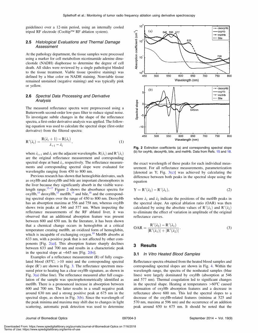
Previous research has shown that hemoglobin derivates, suchas oxyHb and deoxyHb and bile are important chromophores inthe liver because they significantly absorb in the visible wave-length range.15–17 Figure 2 shows the absorbance spectra foroxyHb,15 deoxyHb,15 metHb,15 and bile,18 and the correspond-ing spectral slopes over the range of 450 to 800 nm. DeoxyHbhas an absorption maxima at 556 and 758 nm, whereas oxyHbshows twin peaks at 546 and 577 nm. When inspecting thereflectance measurements of the RF ablated liver, it wasobserved that an additional absorption feature was presentbetween 600 and 650 nm. In the literature, it has been shownthat a chemical change occurs in hemoglobin at a criticaltemperature creating metHb, an oxidized form of hemoglobin,which is incapable of exchanging oxygen.10 MetHb absorbs at633 nm, with a positive peak that is not affected by other com-ponents [Fig. 2(a)]. This absorption feature sharply declinesbetween 633 and 700 nm and results in a characteristic peakin the spectral slope at ∼645 nm [Fig. 2(b)].
Examples of a reflectance measurement (R) of fully coagu-lated blood (85°C; >10 min) and the corresponding spectralslope (R’) are shown in Fig. 3. The reflectance spectrum mea-sured prior to heating has a clear oxyHb signature, as shown inFig. 3(a) (blue line). The reflectance measured after full coagu-lation of the sample was spectrophotometrically identified asmetHb. There is a pronounced increase in absorption between600 and 700 nm. The latter results in a small negative peakaround 630 nm and a strong positive peak at 675 nm in thespectral slope, as shown in Fig. 3(b). Since the wavelength ofthe peak minima and maxima may shift due to changes in lightscattering, automatic peak detection was used to determine
the exact wavelength of these peaks for each individual meas-urement. For all reflectance measurements, parameterization[denoted as Y; Fig. 3(c)] was achieved by calculating thedifference between both peaks in the spectral slope using theequation
Y ¼ R 0ðλ2Þ − R 0ðλ1Þ; (2)
where λ1 and λ2 indicate the positions of the metHb peaks inthe spectral slope. An optical ablation ratio (OAR) was thencalculated by using the absolute values of R 0ðλ1Þ and R 0ðλ2Þto eliminate the effect of variation in amplitude of the originalreflectance curves.
OAR ¼ R 0ðλ2Þ − R 0ðλ1ÞjR 0ðλ1Þj þ jR 0ðλ2Þj
: (3)
3 Results
3.1 In Vitro Heated Blood Samples
Reflectance spectra obtained from the heated blood samples andcorresponding spectral slopes are shown in Fig. 4. Within thewavelength range, the spectra of the nonheated samples (bluelines) were largely dominated by oxyHb (absorption at 546and 577 nm). Thermal coagulation led to significant changesin the spectral shape. Heating at temperatures >60°C causedattenuation of oxyHb absorption features and a decrease inreflectance above 600 nm. This led the spectral slopes to adecrease of the oxyHb-related features (minima at 525 and570 nm, maxima at 596 nm) and the occurrence of an additionpeak around 650 to 675 nm. It should be noted that the
Fig. 2 Extinction coefficients (a) and corresponding spectral slope(b) for oxyHb, deoxyHb, bile, and metHb. Data from Refs. 15 and 18.
Journal of Biomedical Optics 097004-3 September 2014 • Vol. 19(9)
Spliethoff et al.: Monitoring of tumor radio frequency ablation using derivative spectroscopy
Downloaded From: https://www.spiedigitallibrary.org/journals/Journal-of-Biomedical-Optics on 7/16/2018Terms of Use: https://www.spiedigitallibrary.org/terms-of-use

maximum in the spectral slopes that initially appeared around650 nm was red-shifted throughout the heating process.
The dynamics of the observed spectral changes were evalu-ated by calculating the OAR over time for each sample. Resultsare shown in Fig. 5. The rate of the observed alterations wasclearly dependent on the set temperature. At 60°C, a smallincrease in OAR was observed after 15 min of heating, whereasat temperatures of 70 and 80°C, a much larger change in OARwas seen within a few minutes of heating. The time course ofthe OAR could roughly be divided into three stages: a period ofnegligible change in the spectral shape, a steep increase inOAR, and a plateau phase. The third stage was not achievedfor the sample heated at a temperature of 60°C for more than15 min.
3.2 RF Ablation Monitoring
To investigate spectral changes occurring during RF ablation ofliver tissue, reflectance measurements (n ¼ 23 and n ¼ 29 spec-tra) were continuously acquired from two human liver resectionspecimens during RF ablation.
Figure 6 shows a typical example of the time course of thereflectance spectra [Fig. 6(a)] and corresponding spectral slopes[Fig. 6(b)], during a representative RF ablation experimentex vivo. The ablation was started at t ¼ 0 min and finishedafter 7.5 min. To facilitate changes in spectral shape, the reflec-tance measurements [Fig. 6(a)] were normalized using thereflectance intensity value at 800 nm. The maximal change inthe reflectance and spectral slopes was observed approximately
Fig. 3 Examples of reflectance spectra (R) of a human blood sample, measured before (blue lines) andafter (red lines) full coagulation. Intensities are given in arbitrary units. In (b), the corresponding spectralslopes (R’) are shown. The values of R’ are normalized to the maximal intensity between 450 and800 nm. (c) illustrates how peaks in the spectral slopes were used for quantification of metHb.
Fig. 4 Reflectance and corresponding spectral slopes obtained from blood samples heated to 60, 70,and 80°C. To facilitate comparison between samples, reflectance spectra were normalized using thereflectance intensity value at 800 nm. Note the increasing value for the positive peak at 650 to675 nm in the spectral slopes.
Journal of Biomedical Optics 097004-4 September 2014 • Vol. 19(9)
Spliethoff et al.: Monitoring of tumor radio frequency ablation using derivative spectroscopy
Downloaded From: https://www.spiedigitallibrary.org/journals/Journal-of-Biomedical-Optics on 7/16/2018Terms of Use: https://www.spiedigitallibrary.org/terms-of-use

2 min after the start of the ablation. No obvious changes in spec-tral shape occurred during the 5 min after the ablation was ter-minated, indicating that irreversible alterations were measured.The time course of the OAR during RF ablations [Fig. 6(c)]showed the same characteristic profile as that observed forthe heated blood samples. The changes in OAR consistentlycorresponded with the observed changes in the spectral shape.
To validate the previous findings in a clinical setting, reflec-tance spectra were measured in vivo before and after a full abla-tion of a liver metastasis of colorectal origin. The initial (i.e.,nonablated) and final (i.e., last recording) reflectance spectra,
spectral slopes, and corresponding histopathology images areshown in Fig. 7. Spectral changes were comparable to theones seen during the ex vivo ablation experiments. The OARmarkedly increased from 0.13 to 0.51. Macroscopic evaluationand histological analysis confirmed that the spectra acquiredafter RF ablation were performed in fully ablated liverparenchyma.
4 DiscussionDespite the widespread use of RF ablation, an effective way toassess thermal tissue damage during and after the procedure isstill lacking. To our knowledge, this report demonstrates the firstpublished results of first-derivative DR spectroscopy to assessthe efficacy of RF ablation in vivo.
In this study, reflectance measurements were performed dur-ing the heating of human blood samples to various temperaturesand during RF ablation of human liver tissue both ex vivo andin vivo. Thermal coagulation of blood samples caused signifi-cant changes in the spectral shape, which is attributed to thethermal conversion of hemoglobin to metHb. RF ablation ofliver tissue essentially led to similar spectral alterations.Specific spectral characteristics were extracted from the spectralslopes using an OAR. For the heated blood samples, the longi-tudinal changes in OAR were clearly dependent on the heatingtemperature and could be divided into three stages. In the firststage, minimal spectral change was observed. During the heat-ing of the (oxygenated) blood samples, deoxyHb occurred as atransient intermediate of oxyHb. We hypothesize that this is dueto hemoglobin’s decreased affinity for oxygen with an increasein temperature, a so-called right shift in the oxygen-hemoglobin
Fig. 5 Longitudinal change in optical ablation ratio for the bloodsamples heated to 60, 70, and 80°C.
Fig. 6 Radio frequency (RF) ablation monitoring ex vivo. Heat-induced spectral changes in reflectancespectra (a) and spectral slopes (b) during an RF ablation of human liver tissue ex vivo. Only 12 out of 23spectra are shown (sampled 1:2) to allow better visualization. (c) shows the time course of optical abla-tion ratio, as calculated from the spectral slopes.
Journal of Biomedical Optics 097004-5 September 2014 • Vol. 19(9)
Spliethoff et al.: Monitoring of tumor radio frequency ablation using derivative spectroscopy
Downloaded From: https://www.spiedigitallibrary.org/journals/Journal-of-Biomedical-Optics on 7/16/2018Terms of Use: https://www.spiedigitallibrary.org/terms-of-use

dissociation curve.19 When the threshold heat exposure that isneeded for permanent alterations of hemoglobin had beenreached, this resulted in a steep monotone increase in the OAR(defined as stage 2). In stage 3, a plateau was reached, at whichpoint no further spectral alterations occurred.
To investigate the thermal tissue damage occurring duringRF ablation, a series of ablations of human liver tissue was per-formed ex vivo. An important finding in our experiment was thatchanges in spectral characteristics achieved during ablation per-sisted after ablation was terminated and tissue was allowed tocool. As discussed, this is mainly attributed to thermally inducedmetHb formation. No significant chemical conversion of metHbis expected within a short period (hours) after cooling of thefully ablated tissue. Due to the coagulation of blood vessels,the ablated tissue has been isolated from perfusion and allenzymes are expected to be denatured.20 In this way, our methodwould allow evaluation of the ablation margins well after theablation has been completed.
The works of Ritz et al.21 and others10,11,22–26 provide a solidbasis for understanding the changes in the optical properties ofbiological tissues under the effect of heating to increasing tem-peratures. Formation of metHb following heat exposure hasbeen reported by several authors.8,10,11,27,28 In addition, at tem-peratures >60°C, there is rapid tissue coagulation, as proteinsdenature and undergo irreversible structural changes.7 The latterleads to an increase in the reduced scattering coefficient andassociated increase in reflectance.21,26 This is the reason whythermally coagulated tissue looks paler than normal tissue.This principle was exploited by Anderson et al.,5 who performedRF ablation on healthy animals and monitored reflectance mea-surements through a fiber-optic probe. Empirical methods wereused for analysis of reflectance spectra in which the spectralintensities at certain wavelengths were correlated with thedegree of thermal damage during RF ablation. They foundthat an increase in the absolute reflectance intensity correlatedwith the histological degree of thermal damage. Similar resultswere achieved by Hsu et al., who performed spectral measure-ments on both animal and human liver tissue.29 The time course
of spectral changes observed in Anderson are consistent withthe three characteristic stages of ablation observed in thepresent study.
In the study by Anderson, spectral changes occurred as theablation zone progressed past the spectroscopy probe. Duringthe first stage, spectra showed minimal deviation from the spec-tral shape of native liver parenchyma. Stage 2 changes occurredas the advancing hemorrhagic zone reached and passed thespectroscopic probe, whereas stage 3 changes occurred onlywhen the liver tissue was fully coagulated. However, the abso-lute magnitude of a reflectance spectrum is dependent on asubtle variation in probe-to-tissue coupling and pressure, mak-ing it difficult to obtain reproducible and reliable reflectancespectra from the measured tissue.30–32
This study differs from previous studies in various ways.First, using derivative spectroscopy, we mainly focused on thespectral characteristics of metHb, while eliminating the magni-tude difference and suppressing the background effects fromscattering and other substances (e.g., oxyHb, deoxyHb, andbile). We showed that calculating the slope of the reflectancespectra can be applied to follow subtle changes in the shape ofspectral bands. Second, by performing spectroscopy in vivoduring open RF ablation, we demonstrated that the identifiedspectral characteristics could be used to assess the degree ofthermal damage after RF ablation after the tissue temperaturehad normalized.
The feedback information provided by DR spectroscopy, asdeployed in a fiber-optic needle, can help the interventionist inmultiple ways. When the tumor being targeted is located nearvital structures that might be damaged by heating (e.g., gallbladder, major blood vessels, bowel), a spectroscopy needlecan be placed at a critical point away from the tumor. Real-time monitoring of tissue during an ablation procedure couldthen be used to determine when a particular level of tissuedamage has been reached and, therefore, reduce the chance oflocal recurrence, while preserving surrounding healthy tissue.Furthermore, DR spectroscopy could be used directly after RFablation to check focal areas of tissue that are suspected for
Fig. 7 Correlation of thermal tissue damage with spectral changes measured in vivo during open RFablation. The spectra shown in (a) and (b) were measured in native liver parenchyma prior to ablation,whereas (d) and (e) show corresponding spectral acquisitions after RF ablation. The blue color in (c) indi-cates viable liver parenchyma, whereas nonviable tissue remained unstained (f).
Journal of Biomedical Optics 097004-6 September 2014 • Vol. 19(9)
Spliethoff et al.: Monitoring of tumor radio frequency ablation using derivative spectroscopy
Downloaded From: https://www.spiedigitallibrary.org/journals/Journal-of-Biomedical-Optics on 7/16/2018Terms of Use: https://www.spiedigitallibrary.org/terms-of-use

inadequate ablation. This may improve procedure outcomeand disease-free survival. Further research is needed in whichthree-dimensional spectroscopic information is acquired at vari-ous distances from the ablation electrode during and after RFablation.
Although the results of the present study are of specificinterest for liver RF ablation, analogue results were observed inother fields, such as laser photocoagulation of vascular skinlesions10,11 and assessment of skin burns.12 Interestingly, similarresults were observed in the area of cardiac ablation monitoring,where methmyoglobin was found in ablated cardiac muscletissue, as described by Swartling et al.33 These results makeDR spectroscopy a promising diagnostic tool to verify irrevers-ible thermal damage for heat-based therapy in general.
It should be noted that the results presented here are subjectto some uncertainties. Although derivative analysis is insensitiveto slow changes in the measured reflectance curve, alterations inscattering slope may have influenced the calculated values forthe OAR to a certain extent. For example, in the spectral slopeshown in Fig. 6(c), it can be observed that the metHb peak at∼660 nm shifted toward higher wavelengths. This is expecteddue to an increase in the scattering within the tissue. To mini-mize the effect on the exact value for the OAR, automated peakdetection was used. Furthermore, during the heating of bloodsamples and tissue, the spectra of various tissue chromophores,including oxyHb and deoxyHb, may change shape and slightlyshift to red wavelengths (bathochromic shift). The influence ofthese dynamic optical property changes on the OAR were notfurther studied here. Another uncertainty is the exact tempera-ture and heating time needed to cause irreversible thermaldamage.
From a clinical point of view, an ideal marker for RF ablationshould provide a reliable, quantitative prediction as to whetheror not tissue has been adequately ablated. The presence of sucha marker should, therefore, be correlated with the extent of thethermal damage achieved. We have demonstrated that thermalcoagulation of liver tissue can be quantified by using specificspectral information, which is expected to be due to rapid thermalconversion of hemoglobin to metHb. A strategy using a combi-nation of the reflectance intensity, as mentioned earlier, and fea-tures extracted from the spectral shape may have the potential toimprove the overall sensitivity of a future instrument.
Furthermore in humans additional studies to evaluate theexact relation between the proposed spectral markers and extendof thermal tissue damage are needed. This will provide a meansto directly validate the quantitative physiological aspects of thistechnique in a clinical setting and may directly increase theclinical success rate of RF ablations of tumor lesions in liver,lung, and renal cancer.
5 ConclusionIn summary, this study shows the potential of real-time liverablation monitoring by reflectance measurements at the tip ofa needle. We have presented evidence that the thermal coagu-lation of liver tissue involves significant changes in the spectralslope, which is thought to be due to the thermal formation ofmetHb. This opens the potential to dynamically monitor theextent of irreversible thermal tissue damage based on thesespectral features. Currently, a more extensive in vivo humanstudy is being performed as a next step toward clinicalimplementation.
AcknowledgmentsThe authors would like to thank the members of the PhilipsResearch project for their technical support. We thank Jan-Nico Ridderbos and Joost van Ooij from the NKI pathologydepartment for their help with the tissue processing. We thankTorre Bydlon for reviewing the manuscript. This study wassupported by Philips Research, Eindhoven, the Netherlands.The author Theo Ruers was partly supported by the Centerfor Translational Medicine (CTMM), project VOLTA (Grant05T-201)
References1. S. A. Curley et al., “Radiofrequency ablation of unresectable primary
and metastatic hepatic malignancies: results in 123 patients,” Ann. Surg.230(1), 1–8 (1999).
2. T. M. Pawlik et al., “Combined resection and radiofrequency ablationfor advanced hepatic malignancies: results in 172 patients,” Ann. Surg.Oncol. 10(9), 1059–1069 (2003).
3. B. W. Kuvshinoff and D. M. Ota, “Radiofrequency ablation of livertumors: influence of technique and tumor size,” Surgery 132(4),605–611; discussion 611–602 (2002).
4. C. R. Buttemere et al., “In vivo assessment of thermal damage in theliver using optical spectroscopy,” J. Biomed. Opt. 9(5), 1018–1027(2004).
5. C. D. Anderson et al., “Real-time spectroscopic assessment of thermaldamage: implications for radiofrequency ablation,” J. Gastrointest.Surg. 8(6), 660–669 (2004).
6. C. D. Anderson et al., “Fluorescence spectroscopy accurately detectsirreversible cell damage during hepatic radiofrequency ablation,”Surgery 136(3), 524–531 (2004).
7. S. N. Goldberg, G. S. Gazelle, and P. R. Mueller, “Thermal ablationtherapy for focal malignancy: a unified approach to underlying princi-ples, techniques, and diagnostic imaging guidance,” AJR Am. J.Roentgenol. 174(2), 323–331 (2000).
8. L. L. Randeberg, A. J. Daae Hagen, and L. O. Svaasand, “Optical prop-erties of human blood as a function of temperature,” Proc. SPIE 4609,20–28 (2002).
9. J. Lee et al., “Noninvasive in vivo monitoring of methemoglobin for-mation and reduction with broadband diffuse optical spectroscopy,”J. Appl. Physiol. 100(2), 615–622 (2006).
10. J. K. Barton et al., “Cooperative phenomena in two-pulse, two-colorlaser photocoagulation of cutaneous blood vessels,” Photochem.Photobiol. 73(6), 642–650 (2001).
11. L. L. Randeberg et al., “Methemoglobin formation during laser inducedphotothermolysis of vascular skin lesions,” Lasers Surg. Med. 34(5),414–419 (2004).
12. K. M. Cross, “Assessment of tissue viability in acute thermal injuriesusing near infrared point spectroscopy,” PhD Thesis, University ofToronto (2010).
13. S. G. Schultz, “The gastrointestinal system, motility and circulation,”Section 6 in Handbook of Physiology, S. G. Schultz, Ed., p. 1519,American Physiological Society, Bethesda, MD (1989).
14. K. Hong and C. S. Georgiades, Percutaneous Tumor Ablation:Strategies and Techniques, ThiemeMedical Publishers, Inc., New York,NY (2011).
15. W. Zijlstra, A. Buursma, and O. v. Assendelft, Visible and Near InfraredAbsorption Spectra of Human and Animal Haemoglobin, 1st ed., VSPPublishing, Utrecht (2000).
16. A. Roggan et al., “Optical properties of circulating human blood inthe wavelength range 400–2500 nm,” J. Biomed. Opt. 4(1), 36–46(1999).
17. R. Nachabe et al., “Estimation of biological chromophores using diffuseoptical spectroscopy: benefit of extending the UV-VIS wavelengthrange to include 1000 to 1600 nm,” Biomed. Opt. Express 1(5),1432–1442 (2010).
18. R. Nachabe et al., “Effect of bile absorption coefficients on the estima-tion of liver tissue optical properties and related implications indiscriminating healthy and tumorous samples,” Biomed. Opt. Express2(3), 600–614 (2011).
Journal of Biomedical Optics 097004-7 September 2014 • Vol. 19(9)
Spliethoff et al.: Monitoring of tumor radio frequency ablation using derivative spectroscopy
Downloaded From: https://www.spiedigitallibrary.org/journals/Journal-of-Biomedical-Optics on 7/16/2018Terms of Use: https://www.spiedigitallibrary.org/terms-of-use
19. K. Schmidt-Nielsen, Animal Physiology—Adaptation and Environment,5th ed., Cambridge University Press, New York, NY (1997).
20. A. J. Welch and M. J. C. van Gemert, Optical-Thermal Response ofLaser-Irradiated Tissue, Springer, New York, NY (2011).
21. J. P. Ritz et al., “Optical properties of native and coagulated porcine livertissue between 400 and 2400 nm,” Lasers Surg. Med. 29(3), 205–212(2001).
22. J. P. Ritz et al., “Continuous changes in the optical properties of livertissue during laser-induced interstitial thermotherapy,” Lasers Surg.Med. 28(4), 307–312 (2001).
23. C. T. Germer et al., “Optical properties of native and coagulated humanliver tissue and liver metastases in the near infrared range,” Lasers Surg.Med. 23(4), 194–203 (1998).
24. A. M. Nilsson et al., “Changes in spectral shape of tissue optical proper-ties in conjunction with laser-induced thermotherapy,” Appl. Opt. 37(7),1256–1267 (1998).
25. J. F. Black and J. K. Barton, “Chemical and structural changes inblood undergoing laser photocoagulation,” Photochem. Photobiol.80(1), 89–97 (2004).
26. J. W. Pickering, P. Posthumus, and M. J. van Gemert, “Continuousmeasurement of the heat-induced changes in the optical properties (at1,064 nm) of rat liver,” Lasers Surg. Med. 15(2), 200–205 (1994).
27. G. G. Fechner and D. J. Gee, “Study on the effects of heat on bloodand on the post-mortem estimation of carboxyhaemoglobin and met-haemoglobin,” Forensic Sci. Int. 40(1), 63–67 (1989).
28. K. Farahani et al., “MRI of thermally denatured blood: methemoglobinformation and relaxation effects,” Magn. Reson. Imaging 17(10),1489–1494 (1999).
29. C. P. Hsu et al., “Liver tumor gross margin identification and ablationmonitoring during liver radiofrequency treatment,” J. Vasc. Interv.Radiol. 16(11), 1473–1478 (2005).
30. B. Yu et al., “Diffuse reflectance spectroscopy of epithelial tissue with asmart fiber-optic probe,” Biomed. Opt. Express 5(3), 675–689 (2014).
31. E. K. Chan et al., “Effects of compression on soft tissue optical proper-ties,” IEEE J. Quantum Electron. 2(4), 943–950 (1996).
32. Y. Ti andW. C. Lin, “Effects of probe contact pressure on in vivo opticalspectroscopy,” Opt. Express 16(6), 4250–4262 (2008).
33. J. Swartling et al., “Changes in tissue optical properties due to radio-frequency ablation of myocardium,” Med. Biol. Eng. Comput. 41(4),403–409 (2003).
Jarich W. Spliethoff is a PhD candidate at the Netherlands CancerInstitute, the Netherlands. He received his BS and MS degrees intechnical medicine from the University of Twente in 2007 and 2011,respectively. His current research focuses on human tissue sensingbased on optical spectroscopy. Incorporation of this technique intoexisting medical devices could improve the accuracy and reliabilityof these devices.
Biographies of the other authors are not available.
Journal of Biomedical Optics 097004-8 September 2014 • Vol. 19(9)
Spliethoff et al.: Monitoring of tumor radio frequency ablation using derivative spectroscopy
Downloaded From: https://www.spiedigitallibrary.org/journals/Journal-of-Biomedical-Optics on 7/16/2018Terms of Use: https://www.spiedigitallibrary.org/terms-of-use


