Monitoring multiple myeloma patients treated with ... multiple myeloma patients treated with...
10
Clin Chem Lab Med 2016; aop a Christopher McCudden and Amy E. Axel contributed equally. *Corresponding author: A. Kate Sasser, Janssen Research & Development, LLC, Spring House, PA, USA, E-mail: [email protected] Christopher McCudden: Department of Pathology and Laboratory Medicine, The Ottawa Hospital, University of Ottawa, Ottawa, ON, Canada Amy E. Axel, Pamela L. Clemens, Jaime Bald and Tahamtan Ahmadi: Janssen Research & Development, LLC, Spring House, PA, USA Dominique Slaets: BARC, a division of CERBA European Lab, Ghent, Belgium Thomas Dejoie: Biochemistry Laboratory, Hospital of Nantes, Nantes, France Sandy Frans: Janssen Research & Development, Beerse, Belgium Torben Plesner: Vejle Hospital and University of Southern Denmark, Vejle, Denmark Joannes F.M. Jacobs: Department of Laboratory Medicine, RadboudUMC, Nijmegen, The Netherlands Niels W.C.J. van de Donk: Department of Hematology, VU University Medical Center, Amsterdam, The Netherlands Philippe Moreau: University Hospital of Nantes, Nantes, France Jordan M. Schecter: Janssen Research & Development, LLC, Raritan, NJ, USA Open Access Christopher McCudden a , Amy E. Axel a , Dominique Slaets, Thomas Dejoie, Pamela L. Clemens, Sandy Frans, Jaime Bald, Torben Plesner, Joannes F.M. Jacobs, Niels W.C.J. van de Donk, Philippe Moreau, Jordan M. Schecter, Tahamtan Ahmadi and A. Kate Sasser* Monitoring multiple myeloma patients treated with daratumumab: teasing out monoclonal antibody interference DOI 10.1515/cclm-2015-1031 Received October 21, 2015; accepted February 10, 2016 Abstract Background: Monoclonal antibodies are promising anti- myeloma treatments. As immunoglobulins, monoclonal antibodies have the potential to be identified by serum protein electrophoresis (SPE) and immunofixation elec- trophoresis (IFE). Therapeutic antibody interference with standard clinical SPE and IFE can confound the use of these tests for response assessment in clinical trials and disease monitoring. Methods: To discriminate between endogenous myeloma protein and daratumumab, a daratumumab-specific immunofixation electrophoresis reflex assay (DIRA) was developed using a mouse anti-daratumumab antibody. To evaluate whether anti-daratumumab bound to and shifted the migration pattern of daratumumab, it was spiked into daratumumab-containing serum and resolved by IFE/SPE. The presence (DIRA positive) or absence (DIRA negative) of residual M-protein in daratumumab-treated patient samples was evaluated using predetermined assessment criteria. DIRA was evaluated for specificity, limit of sensi- tivity, and reproducibility. Results: In all of the tested samples, DIRA distinguished between daratumumab and residual M-protein in com- mercial serum samples spiked with daratumumab and in daratumumab-treated patient samples. The DIRA limit of sensitivity was 0.2 g/L daratumumab, using spiking experiments. Results from DIRA were repro- ducible over multiple days, operators, and assays. The anti-daratumumab antibody was highly specific for daratumumab and did not shift endogenous M-protein. Conclusions: As the treatment of myeloma evolves to incorporate novel monoclonal antibodies, additional solutions will be needed for clinical monitoring of patient responses to therapeutic regimens. In the interim, assays such as DIRA can inform clinical outcomes by distinguish- ing daratumumab from endogenous M-protein by IFE. Keywords: complete response; daratumumab; immuno- fixation electrophoresis; monoclonal antibody; multiple myeloma. Introduction Multiple myeloma (MM) is an incurable disease character- ized by the presence of malignant plasma cells that secrete high levels of a monoclonal immunoglobulin protein (M-protein) [1, 2]. The International Myeloma Working Group (IMWG) has established criteria for clinical response ©2016, A. Kate Sasser et al., published by De Gruyter. This work is licensed under the Creative Commons Attribution-NonCommercial-NoDerivatives 3.0 License. Unauthenticated Download Date | 4/12/16 11:46 AM
-
Upload
trinhkhanh -
Category
Documents
-
view
217 -
download
2
Transcript of Monitoring multiple myeloma patients treated with ... multiple myeloma patients treated with...
Clin Chem Lab Med 2016; aop
aChristopher McCudden and Amy E. Axel contributed equally.*Corresponding author: A. Kate Sasser, Janssen Research & Development, LLC, Spring House, PA, USA, E-mail: [email protected] McCudden: Department of Pathology and Laboratory Medicine, The Ottawa Hospital, University of Ottawa, Ottawa, ON, CanadaAmy E. Axel, Pamela L. Clemens, Jaime Bald and Tahamtan Ahmadi: Janssen Research & Development, LLC, Spring House, PA, USADominique Slaets: BARC, a division of CERBA European Lab, Ghent, BelgiumThomas Dejoie: Biochemistry Laboratory, Hospital of Nantes, Nantes, FranceSandy Frans: Janssen Research & Development, Beerse, BelgiumTorben Plesner: Vejle Hospital and University of Southern Denmark, Vejle, DenmarkJoannes F.M. Jacobs: Department of Laboratory Medicine, RadboudUMC, Nijmegen, The NetherlandsNiels W.C.J. van de Donk: Department of Hematology, VU University Medical Center, Amsterdam, The NetherlandsPhilippe Moreau: University Hospital of Nantes, Nantes, FranceJordan M. Schecter: Janssen Research & Development, LLC, Raritan, NJ, USA
Open Access
Christopher McCuddena, Amy E. Axela, Dominique Slaets, Thomas Dejoie, Pamela L. Clemens, Sandy Frans, Jaime Bald, Torben Plesner, Joannes F.M. Jacobs, Niels W.C.J. van de Donk, Philippe Moreau, Jordan M. Schecter, Tahamtan Ahmadi and A. Kate Sasser*
Monitoring multiple myeloma patients treated with daratumumab: teasing out monoclonal antibody interference
DOI 10.1515/cclm-2015-1031Received October 21, 2015; accepted February 10, 2016
Abstract
Background: Monoclonal antibodies are promising anti-myeloma treatments. As immunoglobulins, monoclonal antibodies have the potential to be identified by serum protein electrophoresis (SPE) and immunofixation elec-trophoresis (IFE). Therapeutic antibody interference with standard clinical SPE and IFE can confound the use of these tests for response assessment in clinical trials and disease monitoring.Methods: To discriminate between endogenous myeloma protein and daratumumab, a daratumumab-specific immunofixation electrophoresis reflex assay (DIRA) was
developed using a mouse anti-daratumumab antibody. To evaluate whether anti-daratumumab bound to and shifted the migration pattern of daratumumab, it was spiked into daratumumab-containing serum and resolved by IFE/SPE. The presence (DIRA positive) or absence (DIRA negative) of residual M-protein in daratumumab-treated patient samples was evaluated using predetermined assessment criteria. DIRA was evaluated for specificity, limit of sensi-tivity, and reproducibility.Results: In all of the tested samples, DIRA distinguished between daratumumab and residual M-protein in com-mercial serum samples spiked with daratumumab and in daratumumab-treated patient samples. The DIRA limit of sensitivity was 0.2 g/L daratumumab, using spiking experiments. Results from DIRA were repro-ducible over multiple days, operators, and assays. The anti- daratumumab antibody was highly specific for daratumumab and did not shift endogenous M-protein.Conclusions: As the treatment of myeloma evolves to incorporate novel monoclonal antibodies, additional solutions will be needed for clinical monitoring of patient responses to therapeutic regimens. In the interim, assays such as DIRA can inform clinical outcomes by distinguish-ing daratumumab from endogenous M-protein by IFE.
Keywords: complete response; daratumumab; immuno-fixation electrophoresis; monoclonal antibody; multiple myeloma.
IntroductionMultiple myeloma (MM) is an incurable disease character-ized by the presence of malignant plasma cells that secrete high levels of a monoclonal immunoglobulin protein (M-protein) [1, 2]. The International Myeloma Working Group (IMWG) has established criteria for clinical response
©2016, A. Kate Sasser et al., published by De Gruyter. This work is licensed under the Creative Commons Attribution-NonCommercial-NoDerivatives 3.0 License.
UnauthenticatedDownload Date | 4/12/16 11:46 AM

2 McCudden et al.: Mitigation of antibody interference with IFE
to treatment in MM, which include changes in serum/urine M-protein levels by serum protein electrophoresis (SPE) and immunofixation electrophoresis (IFE), percentage of bone marrow plasma cells, and free light chain (FLC) ratios [3–5]. For a patient to be classified as having a complete response (CR) by IMWG criteria, the serum and urine must be negative for M-protein, as determined by IFE and SPE, and bone marrow plasma cells must be ≤ 5%. In serum FLC-only patients, CR is defined as a normal FLC ratio in addition to the other criteria required to classify a CR [4]. For the more robust, deeper classification of stringent complete response (sCR), all of the criteria for CR must be met, along with a normal FLC ratio and absence of clonal plasma cells in the bone marrow, as measured by 2- to 4-color flow cytometry or immunohistochemistry.
The treatment of MM is evolving with the introduc-tion of therapeutic monoclonal antibodies (mAbs) [6–8]. Since SPE and IFE are used to quantify and characterize the clonal nature of immunoglobulins, respectively, these assays are subject to interference from therapeutic mAbs [9, 10]. Experiments with spiked samples demonstrated that all mAbs evaluated could be detected by SPE and IFE, down to 0.1 g/L [10]. Interference on serum IFE from treated patients has been reported with several mAbs, including siltuximab, ofatumumab, and daratumumab [1, 9, 10], and similar interference has been observed with elo-tuzumab [7, 11]. The IMWG criteria for achieving CR specify no detectable M-protein by IFE and SPE [3]; thus, antibody interference can have a clinically important impact on the assessment of response to treatment and may result in underestimation of CR rates for mAb therapies. As thera-peutic mAbs become utilized in myeloma, methods are needed to assess clinical responses, particularly CR/sCR, in light of this potential interference.
Daratumumab, a human IgG1κ mAb, binds with high affinity to a unique CD38 epitope, inducing tumor cell death through a variety of mechanisms, including complement-dependent cytotoxicity, antibody-dependent cell-mediated cytotoxicity, antibody-dependent cellular phagocytosis, and induction of apoptosis [12–15]. Addi-tionally, subpopulations of regulatory T cells, regulatory B cells, and myeloid-derived suppressor cells with high CD38 expression are sensitive to daratumumab [16]. Cyto-toxic T cell activation, expansion, and increased T cell clonality have been observed after monotherapy treat-ment in relapsed or refractory disease, suggesting a possi-ble immunomodulatory role for daratumumab in MM [16].
In GEN501, a phase 1/2 study of patients with relapsed or refractory MM, daratumumab monotherapy was well tolerated, and 36% of patients receiving daratumumab at 16 mg/kg achieved at least a partial response (PR) or better
[6]. SIRIUS, a phase 2 study, examined daratumumab in patients with at least three lines of prior therapy or double refractory MM [8]. Overall response rate (ORR) was 29% and responses deepened with continued treatment; median overall survival was 17.5 months (95% confidence interval, 13.7–not estimable) in these heavily pretreated patients (median of 5 prior lines of treatment) [8]. On the basis of these studies, daratumumab was recently approved in the United States for the treatment of patients with MM who have received 3 or more lines of prior therapy including a proteasome inhibitor (PI) and immunomodulatory drug (IMiD), or are double refractory to a PI and an IMiD [17]. Daratumumab is also being investigated in phase 3 clini-cal studies in combination with other therapeutic agents in patients with MM.
At the recommended dosing schedule (16 mg/kg weekly for 8 weeks, then every 2 weeks for 16 weeks, and every 4 weeks thereafter), daratumumab reaches peak serum concentrations of approximately 915 μg/mL (0.915 g/L) at the end of the weekly dosing period [18], making it readily detectable on most SPE/IFE assays [1]. As a human IgGκ immunoglobulin, daratumumab may be detected by IFE and can thus be misinterpreted as a myeloma-associated M-protein, thereby interfering with the response criteria [19].
To help distinguish daratumumab from endogenous M-protein in serum IFE, the daratumumab-specific immu-nofixation electrophoresis reflex assay (DIRA) was devel-oped to confirm suspected daratumumab interference and to allow separation of daratumumab bands from residual endogenous M-protein. DIRA relies on the use of an anti-daratumumab antibody that binds daratumumab and alters its migration on IFE. The present study describes the validation of DIRA for clinical trial testing, which included determination of the assay’s limit of sensitivity, specific-ity, and reproducibility. This assay is currently being uti-lized in clinical trials to distinguish daratumumab from endogenous M-protein by IFE and has triggered additional clinical response assessments to confirm CRs in myeloma patients treated with daratumumab.
Materials and methodsSerum sample collection
Human serum samples from patients with MM or healthy donors were acquired from a commercial source (Bioreclamation, Westbury, NY, USA) or from daratumumab-treated patients (n = 33). Serum sam-ples from clinical trials of daratumumab as monotherapy (GEN501 and SIRIUS) or as combination therapy with lenalidomide in an
UnauthenticatedDownload Date | 4/12/16 11:46 AM

McCudden et al.: Mitigation of antibody interference with IFE 3
ongoing study (GEN503; ClinicalTrials.gov Identifier: NCT01615029) were collected in 2.5 or 8.5 mL serum separator tubes (Becton Dickinson, Franklin Lakes, NJ, USA) and centrifuged at 1300–2000 × g for 10–15 min, after 30 min at room temperature, to allow complete blood clotting/cooling. Serum samples were collected and shipped (frozen) to a central laboratory (BARC, Ghent, Belgium) for SPE and IFE or subsequent DIRA testing. Patients with low-level ( < 5 g/L) or negative SPE but repeated positive IgGκ IFE were flagged as having potential daratumumab interference, and were utilized for valida-tion and DIRA testing. Samples were based on suspected interference rather than predefined time points. Clinical trials were approved by the independent Institutional Review Boards at study sites in accord-ance with the Declaration of Helsinki and consistent with Good Clini-cal Practices. All patients provided written informed consent.
Anti-daratumumab antibody
A murine anti-daratumumab antibody clone (5–3–9–4) (John-son & Johnson, New Brunswick, NJ, USA) was produced from a hybridoma cell line (Genmab, Utrecht, The Netherlands). Superna-tants from cultured cells were concentrated using tangential flow filtration ( Millipore, Billerica, MA, USA), purified by MabSelect-Sure (GE Healthcare, Marlborough, MA, USA), and dialyzed into Dulbecco’s phosphate-buffered saline, pH 7.2 (Life Technologies, Grand Island, NY, USA).
IFE and SPE
Immunofixations were performed on semi-automatic Hydrasys or Hydrasys 2 using Maxikit Hydragel 4IF or 9IF (Sebia, Norcross, GA, USA). SPE was performed on Capillarys using the Capillarys Protein 6 kit (both from Sebia). Both IFE and SPE were performed according to the manufacturer’s specifications.
DIRA
For DIRA, anti-daratumumab or saline was spiked into baseline or daratumumab-treated patient serum, incubated at room temperature for 15 min, and separated by electrophoresis according to the stand-ard IFE methods described previously. One lane of each baseline and daratumumab-treated patient serum was fixed as a reference and anti-human, anti IgG, or κ (Sebia) antisera were applied to detect heavy and light chains. Upon completion of electrophoresis and staining, gels were assessed for (1) migration of control daratumumab with anti-dara-tumumab, (2) lack of migration of baseline M-protein with the addi-tion of anti-daratumumab, (3) a shift in the migration pattern of the putative daratumumab band relative to the daratumumab control in daratumumab-treated serum samples, and (4) the presence or absence of a non-daratumumab M-protein band. The absence of remaining dis-ease M-protein was defined as a DIRA-negative result. The presence of remaining disease M-protein qualified as a DIRA-positive result.
Limit of sensitivity
Ten commercial MM samples were spiked with 0.25, 0.5, and 1.0 g/L daratumumab with and without anti-daratumumab in a 1:1 ratio to
determine the effectiveness and reproducibility of anti-daratumumab to shift daratumumab bands. Ten additional MM serum samples and 10 normal human serum (NHS) samples were spiked with a wider range of clinically relevant concentrations of daratumumab (0, 0.1, 0.2, 0.25, and 0.5 g/L) with and without anti-daratumumab in a 1:1 ratio. Two independent reviewers evaluated the results.
The limit of sensitivity was defined as the lowest level of dara-tumumab detectable by at least one parameter (daratumumab IgG, daratumumab + anti-daratumumab complex IgG, daratumumab κ, or daratumumab + anti-daratumumab κ by IFE; daratumumab or daratumumab + anti-daratumumab by SPE) in all samples tested.
Specificity
To demonstrate that the anti-daratumumab antibody did not shift endogenous M-protein migration, commercially available serum sam-ples from patients with MM (n = 51) were spiked with daratumumab, anti-daratumumab, or daratumumab + anti-daratumumab (0.5 g/L and 1 g/L; 1:1 ratio) and were analyzed by IFE. Additionally, a subset (n = 35) evaluated fixed concentrations of 1 g/L anti-daratumumab and 0.5 g/L daratumumab. Gels were assessed by determining whether there was a shift in daratumumab, with no corresponding shift in M-protein with anti-daratumumab alone. In addition, in each DIRA assay, control serum samples from patients prior to treatment with daratumumab were spiked with anti-daratumumab and evalu-ated for a shift of endogenous M-protein on IFE.
Reproducibility
Three independent runs of 10 commercial samples spiked with 0.25, 0.5, and 1 g/L daratumumab and 10 samples from daratumumab-treated patients with M-protein ≤ 5 g/L, by SPE, were performed using DIRA. The results were assessed for reproducibility by two independent reviewers. The reviewers’ evaluations were standard-ized using predefined assessment criteria. These criteria, as well as the reviewers’ responses to a single sample, are shown in Table 1. Inter-day and inter-operator reproducibility was evaluated using three commercial MM samples on three separate days by two opera-tors, and interpreted by two independent reviewers.
Results
Daratumumab can be shifted with anti-daratumumab
To determine whether a shift in daratumumab could be detected by SPE and IFE, spiking experiments were per-formed, with varying concentrations of daratumumab with or without anti-daratumumab added to myeloma serum or NHS and analyzed by SPE or IFE. Daratumumab was effectively detected and shifted with anti-daratumumab in all samples tested. (Figure 1A and data not shown). To evaluate the amount of anti-daratumumab needed to
UnauthenticatedDownload Date | 4/12/16 11:46 AM

4 McCudden et al.: Mitigation of antibody interference with IFE
Figure 1: Daratumumab can be identified on IFE/SPE and can be shifted with anti-daratumumab.Daratumumab can be detected by IFE (A), anti-daratumumab antibody can bind and shift daratumumab migration pattern on IFE (B); 1:1 ratios of daratumumab:anti-daratumumab are enough to completely shift daratumumab on IFE. Similarly, on SPE, a 1:1 ratio of daratumumab:anti-daratumumab was able to completely shift daratumumab (C). Daratumumab and daratumumab:anti-daratumumab complex are indicated by the blue and green arrows, respectively. IFE, immunofixation electrophoresis; SPE, serum protein electrophoresis; Dara, daratumumab.
Table 1: Concordance of reviewer assessments of the same sample across multiple experiments based on predefined acceptance criteria.
Lane Run 1 Run 2 Run 3
Reviewer 1 Migration of Dara + anti-Dara in control? 4 vs. 3 Y Y Y Migration of endogenous M-protein at baseline? 6 and 10 N N N Migration of Dara in ≥ PR due to the disappearance of Dara (DD)
or the appearance of Dara + anti-Dara complex (AC)? 8 vs. 7 and 12 vs. 11 Y
DD+AC Y
DD+AC Y
DD+AC Presence of M-protein after migration of Dara? 8 and 12 N N N M-protein (M) or Dara (D)? D D D Conclusion Negative Negative Negative
Reviewer 2 Migration of Dara + anti-Dara in control? 4 vs. 3 Y Y Y Migration of endogenous M-protein at baseline? 6 and 10 N N N Migration of Dara in ≥ PR due to the disappearance of Dara (DD)
or the appearance of Dara + anti-Dara complex (AC)? 8 vs. 7 and 12 vs. 11 Y
DD+AC Y
DD+AC Y
DD+AC Presence of M-protein after migration of Dara? 8 and 12 N N N M-protein (M) or Dara (D)? D D D Conclusion Negative Negative Negative
Dara, daratumumab; Y, yes; N, no; PR, partial response.
completely shift daratumumab on IFE and SPE, varying ratios of anti-daratumumab were spiked into serum con-taining 1 g/L daratumumab, the maximum predicted
concentration in patient serum after weekly dosing [15]. A 1:1 ratio of daratumumab:anti-daratumumab or excess anti-daratumumab completely shifted daratumumab on
UnauthenticatedDownload Date | 4/12/16 11:46 AM

McCudden et al.: Mitigation of antibody interference with IFE 5
IFE (Figure 1B). Excess mouse anti-daratumumab was not detected by human antiserum. Densitometry of SPE lanes showed a 1:1 ratio of daratumumab:anti-daratumumab was necessary to completely shift daratumumab; excess daratumumab or anti-daratumumab were also visualized as protein peaks since SPE does not discern total protein from human or mouse antibodies (Figure 1C). Because of its specificity for human antibody and its greater sensitiv-ity, IFE was used to develop DIRA.
DIRA distinguishes daratumumab from endogenous M-protein
Among patients enrolled in daratumumab clinical studies, a residual IgGκ band or a faint IgGκ band that appeared over time was often observed on IFE. Daratu-mumab interference was suspected and had the potential to mask CRs. DIRA was developed to distinguish daratu-mumab from endogenous M-protein in patients with low measurable M-protein by SPE ( ≤ 5 g/L) and IgGκ band by IFE. Exploratory analyses utilized samples with a higher range of SPE to help refine criteria for implementation and validation of the assay. Figure 2 shows a schematic that outlines the samples, controls, and loading in a typical DIRA.
DIRA evaluates patient samples prior to (baseline) and after treatment when daratumumab interference is sus-pected. DIRA requires 12 sample lanes and uses a protein fixative and 2 antisera (IgG and κ; Figure 2A). Lanes 1 and 2 comprise baseline and post-treatment samples with total protein fixative and display the migration patterns of all serum proteins at baseline and post-treatment. Lanes 3 and 4 are controls containing daratumumab and dara-tumumab + anti-daratumumab in saline, respectively. Lanes 5 and 6 (with anti-IgG antisera) include the base-line sample alone and with anti-daratumumab, respec-tively, to characterize endogenous M-protein migration and to demonstrate that anti-daratumumab alone has no effect on endogenous M-protein. Lanes 7 and 8 (with anti-IgG antisera) include the post-treatment sample alone and with anti-daratumumab, respectively, to char-acterize daratumumab and to determine whether disease M-protein remains. If the entire remaining band shifts with the addition of anti-daratumumab, indicating that endog-enous M-protein is absent and that only daratumumab remains, the result is determined to be DIRA negative (similar to a standard IFE-negative result; Figure 2B). If the band only shifts partially, indicating that endogenous M-protein remains, the result is determined to be DIRA positive (similar to a typical IFE-positive result; Figure 2B).
Lanes 9 through 12 contain the same samples as lanes 5 through 8, but are probed with anti-κ antisera.
Validation of DIRA
For clinical validation, the sensitivity, specificity, and reproducibility of DIRA, in both commercial and daratu-mumab-treated myeloma serum samples, were evaluated. Sensitivity was determined by evaluating 10 myeloma and 10 NHS samples spiked with a range of daratumumab ± anti-daratumumab by SPE and IFE. Due to the potential for daratumumab or the daratumumab-anti- daratumumab complex to comigrate with M-protein with either IgG or κ antisera, sensitivity was defined by detection by at least one parameter (daratumumab or daratumumab + anti-daratumumab complex with IgG or κ by IFE; daratumumab or daratumumab + anti-daratumumab complex by SPE). The sensitivity per sample was defined by the lowest level of daratumumab that could be detected by any of these parameters. In myeloma serum samples, the sensitivity of DIRA was determined to be 90% for 0.1 g/L daratumumab and 100% for 0.2 g/L by IFE. In NHS, the sensitivity of DIRA by IFE was 80% for 0.1 g/L daratumumab and 100% for 0.2 g/L. By SPE, sensitivity was determined to be 30% for 0.1 g/L and 100% for 0.2 g/L in MM serum and 100% at 0.2 g/L in NHS. Therefore, the sensitivity of DIRA for dara-tumumab is ~0.2 g/L. Typically MM patients are immuno-suppressed, such that background polyclonal interference has not been an issue to date. In spiked NHS samples, it was not possible to consistently identify residual daratu-mumab below 0.2 g/L. While IFE and thus DIRA is not a quantitative assay, determining the lower range of sensi-tivity demonstrated daratumumab can be detected and DIRA is functional within the range of predicted serum concentrations in treated patients.
The specificity of DIRA relies on the specificity of the anti-daratumumab. Accordingly, DIRA includes control lanes containing baseline serum samples that have been spiked with or without anti-daratumumab (Figure 2, Lanes 5 and 6). In commercial samples spiked with 0.5 or 1 g/L daratumumab, the antibody was shifted by anti- daratumumab at both concentrations in all samples (51 of 51 [100%]). No shift in M-protein occurred with the addi-tion of anti-daratumumab alone in any of the samples. When only anti-daratumumab was spiked into the serum, a weak polyclonal smear appeared in the lanes with IgG antisera in four of 51 (8%) samples. However, this did not interfere with the interpretation of DIRA, as the band cor-responding to daratumumab:anti-daratumumab complex was distinctly visible and the smear was not observed
UnauthenticatedDownload Date | 4/12/16 11:46 AM

6 McCudden et al.: Mitigation of antibody interference with IFE
when daratumumab was present. While experienced reviewers consistently identify the faint residual band in the DIRA assays, it may be difficult to identify in Figure 2B. This is a known issue with faint bands on agarose gels;
scanned gels do not have the same resolution or detail as the physical version [20]. Therefore, anti- daratumumab appears highly specific for daratumumab. Specificity of the anti-daratumumab antibody, along with false-negative
Figure 2: Daratumumab-specific IFE reflex assay.Baseline (prior to treatment) serum samples are run ± anti-daratumumab next to serum samples from a post-treatment time point with suspected daratumumab interference, ± anti-daratumumab, to determine whether the remaining M-protein band shifts completely with anti-daratumumab. Both IgG and κ antisera are used for staining and fixation (A). DIRA positive, similar to IFE positive, indicates that endogenous M-protein (in red, and indicated by a red arrow in lane 1) remains. DIRA negative, similar to IFE negative, indicates that only daratumumab (in blue, and indicated by a blue arrow in lane 3) is remaining and endogenous M-protein is no longer detected (A). The DIRA template utilized daratumumab ± anti-daratumumab as controls for migration of the therapeutic antibody and the daratumumab-anti- daratumumab shifted complexes (in green, and indicated by a green arrow in lane 4). In patient samples, baseline and post-treatment serum ± anti-daratumumab were compared to determine whether M-protein remained after shifting daratumumab (B). DIRA-positive results showed M-protein, whereas DIRA-negative results showed only a shift in daratumumab but no remaining M-protein (lanes 8 and 12). IFE, immunofixation electrophoresis; M-protein, monoclonal immunoglobulin protein; DIRA, daratumumab-specific immunofixation electrophoresis reflex assay; Dara, daratumumab; PBS, phosphate buffered saline; SP, total serum protein fix; G, IgG antisera; κ, kappa antisera.
UnauthenticatedDownload Date | 4/12/16 11:46 AM
McCudden et al.: Mitigation of antibody interference with IFE 7
and false-positive rates of DIRA, will be evaluated further in a randomized, phase 3 clinical study of daratumumab versus control.
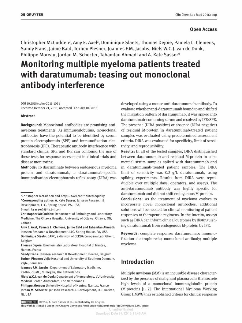
The reproducibility of DIRA was assessed by perform-ing the assay on daratumumab-treated patient samples in triplicate. In all daratumumab-treated patient samples (10/10), results were consistent across three independ-ent experiments. To determine inter-operator and inter-day reproducibility, DIRA was performed on commercial samples spiked with daratumumab ± anti-daratumumab, by two operators on three separate days. Further, results were evaluated by two independent reviewers. Concord-ance among reviews was demonstrated in 100% of assays. Reviewers’ responses to a set of predetermined assess-ment criteria are shown for a single patient sample over three separate experiments (Table 1).
DIRA Plus
To ensure 1 g/L anti-daratumumab was sufficient to shift daratumumab in patient serum samples for which dara-tumumab concentration data were not available or SPE measurements were higher than the average range of daratumumab concentrations, samples from 14 dara-tumumab-treated patients were tested using increased concentrations of anti-daratumumab (a modification known as “DIRA Plus”; Figure 3). For these assays,
Figure 3: DIRA Plus for the evaluation of patients with serum con-centrations of daratumumab above the normal range.One gram per liter of anti-daratumumab was sufficient to migrate daratumumab in all samples (A). Higher concentrations of anti- daratumumab added to baseline serum (1 g/L and 4 g/L) resulted in the appearance of a faint, polyclonal smear (arrow) with IgG antisera. DIRA, daratumumab-specific immunofixation electrophoresis reflex assay; Dara, daratumumab; SP, total serum protein fix; G, IgG antisera; κ, kappa antisera.
anti-daratumumab concentrations of 1–4 g/L were used. In all cases (14 of 14 samples), 1 g/L of anti-daratumumab was sufficient to interpret DIRA. Anti-daratumumab con-centrations of ≥ 1 g/L caused a weak, polyclonal smear to appear with no other change in the assay result versus the standard concentration of 1 g/L. Thus, the use of concen-trations of anti-daratumumab > 1 g/L is neither warranted nor recommended.
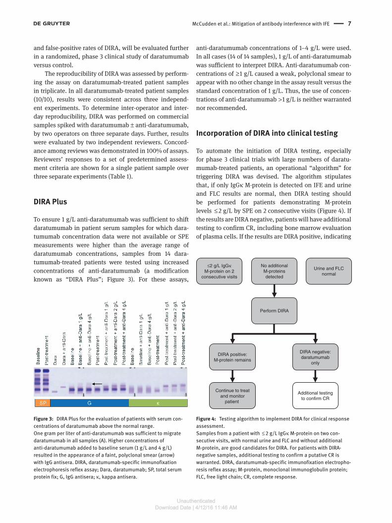
Incorporation of DIRA into clinical testing
To automate the initiation of DIRA testing, especially for phase 3 clinical trials with large numbers of daratu-mumab-treated patients, an operational “ algorithm” for triggering DIRA was devised. The algorithm stipulates that, if only IgGκ M-protein is detected on IFE and urine and FLC results are normal, then DIRA testing should be performed for patients demonstrating M-protein levels ≤ 2 g/L by SPE on 2 consecutive visits (Figure 4). If the results are DIRA negative, patients will have additional testing to confirm CR, including bone marrow evaluation of plasma cells. If the results are DIRA positive, indicating
≤2 g/L IgGκM-protein on 2
consecutive visits
No additionalM-proteinsdetected
Urine and FLCnormal
Perform DIRA
Additional testingto confirm CR
Continue to treatand monitor
patient
DIRA negative:daratumumab
only
DIRA positive:M-protein remains
Figure 4: Testing algorithm to implement DIRA for clinical response assessment.Samples from a patient with ≤ 2 g/L IgGκ M-protein on two con-secutive visits, with normal urine and FLC and without additional M-protein, are good candidates for DIRA. For patients with DIRA-negative samples, additional testing to confirm a putative CR is warranted. DIRA, daratumumab-specific immunofixation electropho-resis reflex assay; M-protein, monoclonal immunoglobulin protein; FLC, free light chain; CR, complete response.
UnauthenticatedDownload Date | 4/12/16 11:46 AM
8 McCudden et al.: Mitigation of antibody interference with IFE
remaining disease M-protein, then no additional testing is warranted and disease monitoring will be continued (Figure 4).
DiscussionDaratumumab, a human anti-CD38 mAb, has demon-strated robust clinical efficacy in relapsed and refractory myeloma, including CRs in some patients. However, as a monoclonal immunoglobulin, daratumumab is detect-able on the SPE and IFE assays that are used to monitor and characterize endogenous immunoglobulin protein. At the recommended 16 mg/kg dose and schedule, the mean (±standard deviation) maximum trough daratu-mumab concentrations was 0.573±0.331 g/L, a concen-tration which can interfere with interpretation of the SPE and IFE assays (data on file). Current IMWG criteria for a CR include negative serum and urine protein electropho-resis and IFE, which is not possible when daratumumab is present at concentrations that fall within the therapeu-tic range. Therefore, DIRA was developed, validated, and implemented to distinguish daratumumab from myeloma M-protein.
DIRA utilizes a highly specific anti-daratumumab antibody to bind daratumumab and shift its migration on IFE gels. Patients with a single IgGκ band that is shifted completely by DIRA are considered to have no remaining M-protein (DIRA negative) and, thus, are candidates for additional IMWG-required confirmatory testing, including bone marrow assessment for plasma cells, to determine whether criteria for CR/sCR (as defined by the IMWG) are met. Patients with remaining endogenous M-protein on DIRA are considered to be DIRA positive, and disease monitoring is continued.
DIRA was highly specific, sensitive, and reproducible both in commercial samples spiked with daratumumab and in clinical samples from daratumumab-treated patients. The presence, or even excess, of anti- daratumumab did not affect the detection or migration of endogenous M-proteins. In the absence of daratumumab, a weak poly-clonal smear was observed in IgG antisera lanes in four of 51 samples when anti-daratumumab was added, but the daratumumab:anti-daratumumab complex was still easily distinguishable by visual inspection and it did not inter-fere with the interpretation of DIRA. DIRA was always able to detect daratumumab by at least one parameter. The limit of sensitivity of DIRA was determined to be 0.2 g/L in serum from patients with myeloma. At this concentration and above, daratumumab interference with M-protein is pre-dicted. Trough daratumumab concentrations throughout
the weekly and every 2 weeks dosing periods are typically above the DIRA sensitivity and may result in daratumumab detection by IFE. However, daratumumab trough concen-trations during every 4 weeks dosing may fall below the DIRA sensitivity and may not interfere with M-protein monitoring during this time. Further, DIRA could be modified for patients with higher than average serum con-centrations (DIRA Plus) by increasing the amount of anti-daratumumab used, although assay reliability decreased with increasing anti-daratumumab concentrations > 1 g/L.
Reproducibility was assessed several different ways. Two independent reviewers scored all DIRA tests, and their assessments were always in agreement; a third reviewer was never required. Reproducibility tests were performed with 10 samples, and results for individual samples were consistent across multiple repetitions; similar results were obtained. Taken together, these findings indicate that DIRA is a robust test with high sensitivity, specificity, and reproducibility.
Despite these advantages, DIRA also has limitations. First, DIRA is not quantitative and interpretation by a trained operator is required. Although rare in myeloma, high polyclonal background signals may make it diffi-cult to assess responses in some patients, leading to false interpretations. Second, DIRA is highly specific to daratu-mumab. Responses in patients receiving other antibodies cannot be resolved using DIRA. Other potential methods to address antibody interference, such as mass spectrom-etry, will be needed for patients receiving combinations of antibodies or patients requiring quantitative testing.
DIRA is important for determining response in dara-tumumab clinical studies, particularly for patients with IgGκ M-protein. Patients with non-IgGκ endogenous serum M-proteins (i.e. urine, FLC, IgA κ or λ, or IgGλ M-proteins) that were positive for IgGκ by IFE were readily detected using DIRA but, to meet current IMWG criteria, they also had to be evaluated to demonstrate that only daratumumab remained.
In phase 2 studies, an IgGκ band often appeared in SPE/IFE over the course of daratumumab treatment in patients originally classified as having non-IgGκ myeloma (IgA, IgM, IgE myeloma, or light-chain–only myeloma). It is likely that this band is indicative of daratumumab interference rather than a new plasma cell clone secreting an IgG monoclonal M-protein. In cases where the origi-nal myeloma clone was IgA or light chain only, reported as approximately 24% and 11% of patients in the general myeloma population, respectively [21], daratumumab was easily identified with DIRA and a lack of endogenous M-protein could be confirmed. However, 60% of patients with MM have IgG M-protein [21], and for those with IgGκ
UnauthenticatedDownload Date | 4/12/16 11:46 AM

McCudden et al.: Mitigation of antibody interference with IFE 9
the distinction between daratumumab and endogenous M-protein can be difficult. The most difficult cases to inter-pret were those in which the migration of daratumumab completely overlapped with M-protein and the entire band did not shift with anti-daratumumab. Waiting until M-protein measurements on SPE were lower, in accord-ance with the operational algorithm, reduced the number of these cases. IMWG has recently released a clarification to address antibody interference. In these updated guide-lines, only the original myeloma clone(s) are required to be undetectable by SPE/IFE [22]. However, DIRA can still distinguish IgGκ clones from daratumumab.
The development and validation of DIRA offers a solution to mitigate daratumumab interference in IFE and improve clinical response assessment in daratumumab-treated patients. Until recently, the modest success of mAbs for the treatment of MM in the clinic did not neces-sitate a solution for mAb assay interference. Phase 1 and 2 studies of daratumumab as a monotherapy have yielded deep responses, including CRs and sCRs [6, 8], making it essential to establish a reliable method for distinguish-ing M-protein from daratumumab. As myeloma therapy evolves to incorporate additional mAb therapeutics, other methods to mitigate antibody interference on SPE/IFE will be needed. These alternative methods could include the incorporation of minimal residual disease (MRD) detec-tion into formal clinical criteria for CR and sCR. MRD detection by 8- to 10-color flow cytometry could be sus-ceptible to antibody interference as well, but standardized approaches using noncompeting antibodies may offer a solution. Utilization of methods such as polymerase chain reaction/next-generation sequencing could also be evaluated.
ConclusionsDIRA is an effective test with high sensitivity, specificity, and reproducibility to distinguish endogenous M-pro-tein from daratumumab. DIRA is currently employed in daratumumab clinical trials to determine if patients with outcomes of very good PR should undergo con-firmatory assessments for characterization of CR. These studies will provide functional validation of DIRA as a clinical tool.
Acknowledgments: This study was sponsored by Janssen Research & Development, LLC. Medical writing and edi-torial support were provided by Erica Chevalier-Larsen, PhD, of MedErgy, and was funded by Janssen Global
Services, LLC. Anti-daratumumab was originally gener-ated and produced by Paul Parren, PhD, and Jeroen Lam-merts van Bueren, PhD, at Genmab.Author contributions: All the authors have accepted responsibility for the entire content of this submitted manuscript and approved submission.Research funding: CM received research support for this project from Janssen Pharmaceuticals. NWCJvdD has received research support from Janssen Pharmaceuticals, Amgen, and Celgene.Employment or leadership: NWCJvdD serves on advisory boards for Janssen Pharmaceuticals, Amgen, and Celgene. AEA, PLC, SF, JB, JMS, TA, and AKS are employees of Janssen Research & Development, LLC. All other authors report no conflicts.Honorarium: None declared.Competing interests: This study was sponsored by Janssen Research & Development, LLC.
References1. Axel AE, McCudden CR, Xie H, Hall BM, Sasser AK. Development
of clinical assay to mitigate daratumumab, an IgG1k monoclonal antibody, interference with serum immunofixation (IFE) and clinical assessment of M-protein response in multiple myeloma. Cancer Res 2014;74(Suppl. 19). Abstract 2563.
2. Zent CS, Wilson CS, Tricot G, Jagannath S, Siegel D, Desikan KR, et al. Oligoclonal protein bands and Ig isotype switching in multiple myeloma treated with high-dose therapy and hematopoietic cell transplantation. Blood 1998;91:3518–23.
3. Durie BG, Harousseau JL, Miguel JS, Blade J, Barlogie B, Anderson K, et al. International uniform response criteria for multiple myeloma. Leukemia 2006;20:1467–73.
4. Rajkumar SV, Harousseau JL, Durie B, Anderson KC, Dimopoulos M, Kyle R, et al. Consensus recommendations for the uniform reporting of clinical trials: report of the International Myeloma Workshop Consensus Panel 1. Blood 2011;117:4691–5.
5. Kyle RA, Rajkumar SV. Criteria for diagnosis, staging, risk stratification and response assessment of multiple myeloma. Leukemia 2009;23:3–9.
6. Lokhorst HM, Plesner T, Laubach JP, Nahi H, Gimsing P, Hansson M, et al. Targeting CD38 with daratumumab monotherapy in multiple myeloma. N Engl J Med 2015;373:1207–19.
7. Lonial S, Dimopoulos M, Palumbo A, White D, Grosicki S, Spicka I, et al. Elotuzumab therapy for relapsed or refractory multiple myeloma. N Engl J Med 2015;373:621–31.
8. Lonial S, Weiss B, Usmani S, Singhal S, Chari A, Bahlis N, et al. Daratumumab monotherapy in patients with treatment-refractory multiple myeloma (SIRIUS): an open-label, randomised, phase 2 trial. Lancet. 2016 Jan 6. pii: S0140-6736(15)01120-4. doi: 10.1016/S0140-6736(15)01120-4. [Epub ahead of print].
9. Genzen JR, Kawaguchi KR, Furman RR. Detection of a monoclonal antibody therapy (ofatumumab) by serum protein and immunofixation electrophoresis. Br J Haematol 2011;155:123–5.
UnauthenticatedDownload Date | 4/12/16 11:46 AM

10 McCudden et al.: Mitigation of antibody interference with IFE
10. McCudden CR, Voorhees PM, Hainsworth SA, Whinna HC, Chapman JF, Hammett-Stabler CA, et al. Interference of monoclonal antibody therapies with serum protein electrophoresis tests. Clin Chem 2010;56:1897–9.
11. Dimopoulos M, Lonial S, Casado LF, Golightly M, Doyen C, Shelat S, et al. Elotuzumab: serum protein electrophoresis and immunofixation interference with clinical assessment of M-protein response in relapsed/refractory multiple myeloma (RRMM). Poster presented at: 15th International Myeloma Work-shop; September 23–26, 2015; Rome, Italy.
12. de Weers M, Tai YT, van der Veer MS, Bakker JM, Vink T, Jacobs DC, et al. Daratumumab, a novel therapeutic human CD38 monoclonal antibody, induces killing of multiple myeloma and other hematological tumors. J Immunol 2011;186:1840–8.
13. Jansen JH, Boross P, Overdijk MB, Lammerts van Bueren JJ, Parren PW, Leusen HH. Daratumumab, a human CD38 antibody induces apoptosis of myeloma tumor cells via Fc receptor-mediated crosslinking. Blood 2012;120. Abstract 2974.
14. Lammerts van Bueren J, Jakobs D, Kaldenhoven N, Roza M, Hiddingh S, Meesters J, et al. Direct in vitro comparison of daratumumab with surrogate analogs of CD38 antibodies MOR03087, SAR650984 and Ab79. Abstract presented at: 56th ASH Annual Meeting and Exposition; December 6–9, 2014; San Francisco, CA. Abstract 3474.
15. Overdijk MB, Verploegen S, Bogels M, van Egmond M, Lammerts van Bueren JJ, Mutis T, et al. Antibody-mediated phagocytosis contributes to the anti-tumor activity of the
therapeutic antibody daratumumab in lymphoma and multiple myeloma. MAbs 2015;7:311–21.
16. Krejcik J, Casneuf T, Nijhof I, Verbist B, Bald J, Plesner T, et al. Immunomodulatory effects and adaptive immune response to daratumumab in multiple myeloma. Blood 2015;126. Abstract 3037.
17. McKeage K. Daratumumab: first global approval. Drugs 2016;76:275–81.
18. McCudden C, Axel A, Slaets D, Frans S, Bald J, Schecter JM, et al. Assessing clinical response in multiple myeloma (MM) patients treated with monoclonal antibodies (mAbs): validation of a daratumumab IFE reflex assay (DIRA) to distinguish malignant M-protein from therapeutic antibody. J Clin Oncol 2015;33. Abstract 8590.
19. Rajkumar SV, Dimopoulos MA, Palumbo A, Blade J, Merlini G, Mateos MV, et al. International Myeloma Working Group updated criteria for the diagnosis of multiple myeloma. Lancet Oncol 2014;15:e538–48.
20. Bender LM, Cotten SW, Fedoriw Y, Willis MS, McCudden CR. Evaluation of digital images for identification and characterization of monoclonal immunoglobulins by immunofixation. Clin Biochem 2013;46:255–8.
21. Greipp PR, San Miguel J, Durie BG, Crowley JJ, Barlogie B, Blade J, et al. International staging system for multiple myeloma. J Clin Oncol 2005;23:3412–20.
22. Durie BG, Miguel JF, Blade J, Rajkumar SV. Clarification of the definition of complete response in multiple myeloma. Leukemia 2015;29:2416–7.
UnauthenticatedDownload Date | 4/12/16 11:46 AM