![Current Biology Reviewflygen.org/pdfs/currbiol.2018.rabreview.pdf · Rab GTPases in Wild-Type Neurons ... [54,55]. Rab7 is a key regulator of multivesicular body maturation from early](https://static.fdocuments.net/doc/165x107/5cefaec388c993eb6f8d7d69/current-biology-rab-gtpases-in-wild-type-neurons-5455-rab7-is-a-key.jpg)
Mon1 Ccz1 activates Rab7 only on late endosomes and...
12
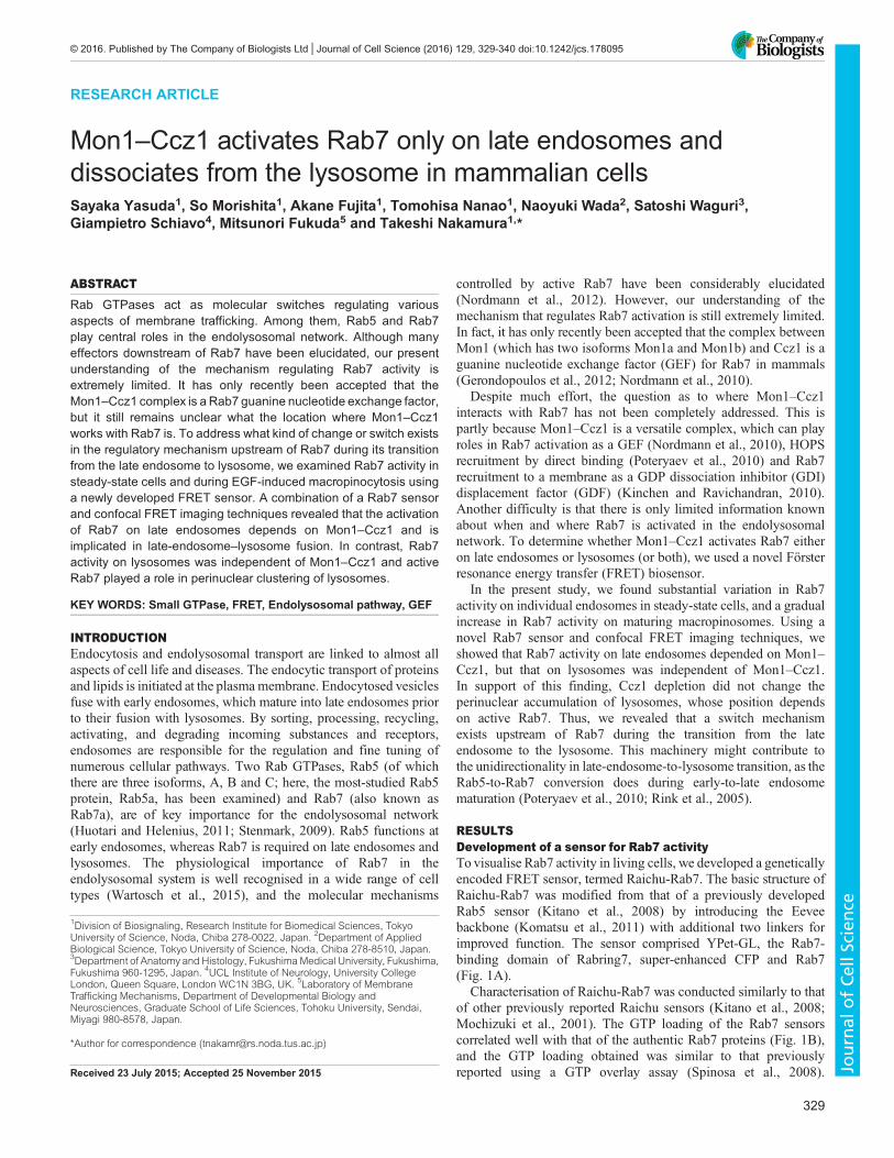
RESEARCH ARTICLE Mon1–Ccz1 activates Rab7 only on late endosomes and dissociates from the lysosome in mammalian cells Sayaka Yasuda 1 , So Morishita 1 , Akane Fujita 1 , Tomohisa Nanao 1 , Naoyuki Wada 2 , Satoshi Waguri 3 , Giampietro Schiavo 4 , Mitsunori Fukuda 5 and Takeshi Nakamura 1, * ABSTRACT Rab GTPases act as molecular switches regulating various aspects of membrane trafficking. Among them, Rab5 and Rab7 play central roles in the endolysosomal network. Although many effectors downstream of Rab7 have been elucidated, our present understanding of the mechanism regulating Rab7 activity is extremely limited. It has only recently been accepted that the Mon1–Ccz1 complex is a Rab7 guanine nucleotide exchange factor, but it still remains unclear what the location where Mon1–Ccz1 works with Rab7 is. To address what kind of change or switch exists in the regulatory mechanism upstream of Rab7 during its transition from the late endosome to lysosome, we examined Rab7 activity in steady-state cells and during EGF-induced macropinocytosis using a newly developed FRET sensor. A combination of a Rab7 sensor and confocal FRET imaging techniques revealed that the activation of Rab7 on late endosomes depends on Mon1–Ccz1 and is implicated in late-endosome–lysosome fusion. In contrast, Rab7 activity on lysosomes was independent of Mon1–Ccz1 and active Rab7 played a role in perinuclear clustering of lysosomes. KEY WORDS: Small GTPase, FRET, Endolysosomal pathway, GEF INTRODUCTION Endocytosis and endolysosomal transport are linked to almost all aspects of cell life and diseases. The endocytic transport of proteins and lipids is initiated at the plasma membrane. Endocytosed vesicles fuse with early endosomes, which mature into late endosomes prior to their fusion with lysosomes. By sorting, processing, recycling, activating, and degrading incoming substances and receptors, endosomes are responsible for the regulation and fine tuning of numerous cellular pathways. Two Rab GTPases, Rab5 (of which there are three isoforms, A, B and C; here, the most-studied Rab5 protein, Rab5a, has been examined) and Rab7 (also known as Rab7a), are of key importance for the endolysosomal network (Huotari and Helenius, 2011; Stenmark, 2009). Rab5 functions at early endosomes, whereas Rab7 is required on late endosomes and lysosomes. The physiological importance of Rab7 in the endolysosomal system is well recognised in a wide range of cell types (Wartosch et al., 2015), and the molecular mechanisms controlled by active Rab7 have been considerably elucidated (Nordmann et al., 2012). However, our understanding of the mechanism that regulates Rab7 activation is still extremely limited. In fact, it has only recently been accepted that the complex between Mon1 (which has two isoforms Mon1a and Mon1b) and Ccz1 is a guanine nucleotide exchange factor (GEF) for Rab7 in mammals (Gerondopoulos et al., 2012; Nordmann et al., 2010). Despite much effort, the question as to where Mon1–Ccz1 interacts with Rab7 has not been completely addressed. This is partly because Mon1–Ccz1 is a versatile complex, which can play roles in Rab7 activation as a GEF (Nordmann et al., 2010), HOPS recruitment by direct binding (Poteryaev et al., 2010) and Rab7 recruitment to a membrane as a GDP dissociation inhibitor (GDI) displacement factor (GDF) (Kinchen and Ravichandran, 2010). Another difficulty is that there is only limited information known about when and where Rab7 is activated in the endolysosomal network. To determine whether Mon1–Ccz1 activates Rab7 either on late endosomes or lysosomes (or both), we used a novel Förster resonance energy transfer (FRET) biosensor. In the present study, we found substantial variation in Rab7 activity on individual endosomes in steady-state cells, and a gradual increase in Rab7 activity on maturing macropinosomes. Using a novel Rab7 sensor and confocal FRET imaging techniques, we showed that Rab7 activity on late endosomes depended on Mon1– Ccz1, but that on lysosomes was independent of Mon1–Ccz1. In support of this finding, Ccz1 depletion did not change the perinuclear accumulation of lysosomes, whose position depends on active Rab7. Thus, we revealed that a switch mechanism exists upstream of Rab7 during the transition from the late endosome to the lysosome. This machinery might contribute to the unidirectionality in late-endosome-to-lysosome transition, as the Rab5-to-Rab7 conversion does during early-to-late endosome maturation (Poteryaev et al., 2010; Rink et al., 2005). RESULTS Development of a sensor for Rab7 activity To visualise Rab7 activity in living cells, we developed a genetically encoded FRET sensor, termed Raichu-Rab7. The basic structure of Raichu-Rab7 was modified from that of a previously developed Rab5 sensor (Kitano et al., 2008) by introducing the Eevee backbone (Komatsu et al., 2011) with additional two linkers for improved function. The sensor comprised YPet-GL, the Rab7- binding domain of Rabring7, super-enhanced CFP and Rab7 (Fig. 1A). Characterisation of Raichu-Rab7 was conducted similarly to that of other previously reported Raichu sensors (Kitano et al., 2008; Mochizuki et al., 2001). The GTP loading of the Rab7 sensors correlated well with that of the authentic Rab7 proteins (Fig. 1B), and the GTP loading obtained was similar to that previously reported using a GTP overlay assay (Spinosa et al., 2008). Received 23 July 2015; Accepted 25 November 2015 1 Division of Biosignaling, Research Institute for Biomedical Sciences, Tokyo University of Science, Noda, Chiba 278-0022, Japan. 2 Department of Applied Biological Science, Tokyo University of Science, Noda, Chiba 278-8510, Japan. 3 Department of Anatomy and Histology, Fukushima Medical University, Fukushima, Fukushima 960-1295, Japan. 4 UCL Institute of Neurology, University College London, Queen Square, London WC1N 3BG, UK. 5 Laboratory of Membrane Trafficking Mechanisms, Department of Developmental Biology and Neurosciences, Graduate School of Life Sciences, Tohoku University, Sendai, Miyagi 980-8578, Japan. *Author for correspondence ([email protected]) 329 © 2016. Published by The Company of Biologists Ltd | Journal of Cell Science (2016) 129, 329-340 doi:10.1242/jcs.178095 Journal of Cell Science
Transcript of Mon1 Ccz1 activates Rab7 only on late endosomes and...
RESEARCH ARTICLE
Mon1–Ccz1 activates Rab7 only on late endosomes anddissociates from the lysosome in mammalian cellsSayaka Yasuda1, So Morishita1, Akane Fujita1, Tomohisa Nanao1, Naoyuki Wada2, Satoshi Waguri3,Giampietro Schiavo4, Mitsunori Fukuda5 and Takeshi Nakamura1,*
ABSTRACTRab GTPases act as molecular switches regulating variousaspects of membrane trafficking. Among them, Rab5 and Rab7play central roles in the endolysosomal network. Although manyeffectors downstream of Rab7 have been elucidated, our presentunderstanding of the mechanism regulating Rab7 activity isextremely limited. It has only recently been accepted that theMon1–Ccz1 complex is a Rab7 guanine nucleotide exchange factor,but it still remains unclear what the location where Mon1–Ccz1works with Rab7 is. To address what kind of change or switch existsin the regulatory mechanism upstream of Rab7 during its transitionfrom the late endosome to lysosome, we examined Rab7 activity insteady-state cells and during EGF-induced macropinocytosis usinga newly developed FRET sensor. A combination of a Rab7 sensorand confocal FRET imaging techniques revealed that the activationof Rab7 on late endosomes depends on Mon1–Ccz1 and isimplicated in late-endosome–lysosome fusion. In contrast, Rab7activity on lysosomes was independent of Mon1–Ccz1 and activeRab7 played a role in perinuclear clustering of lysosomes.
KEY WORDS: Small GTPase, FRET, Endolysosomal pathway, GEF
INTRODUCTIONEndocytosis and endolysosomal transport are linked to almost allaspects of cell life and diseases. The endocytic transport of proteinsand lipids is initiated at the plasmamembrane. Endocytosed vesiclesfuse with early endosomes, which mature into late endosomes priorto their fusion with lysosomes. By sorting, processing, recycling,activating, and degrading incoming substances and receptors,endosomes are responsible for the regulation and fine tuning ofnumerous cellular pathways. Two Rab GTPases, Rab5 (of whichthere are three isoforms, A, B and C; here, the most-studied Rab5protein, Rab5a, has been examined) and Rab7 (also known asRab7a), are of key importance for the endolysosomal network(Huotari and Helenius, 2011; Stenmark, 2009). Rab5 functions atearly endosomes, whereas Rab7 is required on late endosomes andlysosomes. The physiological importance of Rab7 in theendolysosomal system is well recognised in a wide range of celltypes (Wartosch et al., 2015), and the molecular mechanisms
controlled by active Rab7 have been considerably elucidated(Nordmann et al., 2012). However, our understanding of themechanism that regulates Rab7 activation is still extremely limited.In fact, it has only recently been accepted that the complex betweenMon1 (which has two isoforms Mon1a and Mon1b) and Ccz1 is aguanine nucleotide exchange factor (GEF) for Rab7 in mammals(Gerondopoulos et al., 2012; Nordmann et al., 2010).
Despite much effort, the question as to where Mon1–Ccz1interacts with Rab7 has not been completely addressed. This ispartly because Mon1–Ccz1 is a versatile complex, which can playroles in Rab7 activation as a GEF (Nordmann et al., 2010), HOPSrecruitment by direct binding (Poteryaev et al., 2010) and Rab7recruitment to a membrane as a GDP dissociation inhibitor (GDI)displacement factor (GDF) (Kinchen and Ravichandran, 2010).Another difficulty is that there is only limited information knownabout when and where Rab7 is activated in the endolysosomalnetwork. To determine whether Mon1–Ccz1 activates Rab7 eitheron late endosomes or lysosomes (or both), we used a novel Försterresonance energy transfer (FRET) biosensor.
In the present study, we found substantial variation in Rab7activity on individual endosomes in steady-state cells, and a gradualincrease in Rab7 activity on maturing macropinosomes. Using anovel Rab7 sensor and confocal FRET imaging techniques, weshowed that Rab7 activity on late endosomes depended on Mon1–Ccz1, but that on lysosomes was independent of Mon1–Ccz1.In support of this finding, Ccz1 depletion did not change theperinuclear accumulation of lysosomes, whose position dependson active Rab7. Thus, we revealed that a switch mechanismexists upstream of Rab7 during the transition from the lateendosome to the lysosome. This machinery might contribute tothe unidirectionality in late-endosome-to-lysosome transition, as theRab5-to-Rab7 conversion does during early-to-late endosomematuration (Poteryaev et al., 2010; Rink et al., 2005).
RESULTSDevelopment of a sensor for Rab7 activityTo visualise Rab7 activity in living cells, we developed a geneticallyencoded FRET sensor, termed Raichu-Rab7. The basic structure ofRaichu-Rab7 was modified from that of a previously developedRab5 sensor (Kitano et al., 2008) by introducing the Eeveebackbone (Komatsu et al., 2011) with additional two linkers forimproved function. The sensor comprised YPet-GL, the Rab7-binding domain of Rabring7, super-enhanced CFP and Rab7(Fig. 1A).
Characterisation of Raichu-Rab7 was conducted similarly to thatof other previously reported Raichu sensors (Kitano et al., 2008;Mochizuki et al., 2001). The GTP loading of the Rab7 sensorscorrelated well with that of the authentic Rab7 proteins (Fig. 1B),and the GTP loading obtained was similar to that previouslyreported using a GTP overlay assay (Spinosa et al., 2008).Received 23 July 2015; Accepted 25 November 2015
1Division of Biosignaling, Research Institute for Biomedical Sciences, TokyoUniversity of Science, Noda, Chiba 278-0022, Japan. 2Department of AppliedBiological Science, Tokyo University of Science, Noda, Chiba 278-8510, Japan.3Department of Anatomy and Histology, FukushimaMedical University, Fukushima,Fukushima 960-1295, Japan. 4UCL Institute of Neurology, University CollegeLondon, Queen Square, London WC1N 3BG, UK. 5Laboratory of MembraneTrafficking Mechanisms, Department of Developmental Biology andNeurosciences, Graduate School of Life Sciences, Tohoku University, Sendai,Miyagi 980-8578, Japan.
*Author for correspondence ([email protected])
329
© 2016. Published by The Company of Biologists Ltd | Journal of Cell Science (2016) 129, 329-340 doi:10.1242/jcs.178095
Journal
ofCe
llScience
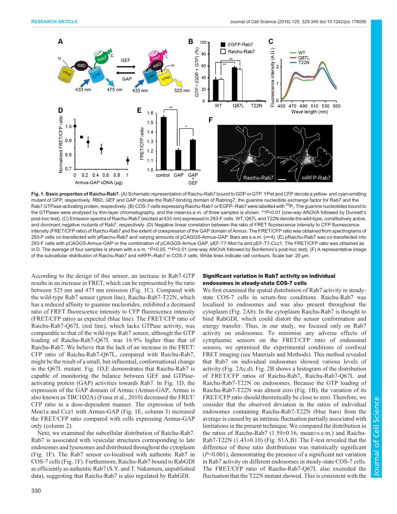
According to the design of this sensor, an increase in Rab7-GTPresults in an increase in FRET, which can be represented by the ratiobetween 525 nm and 475 nm emission (Fig. 1C). Compared withthe wild-type Rab7 sensor (green line), Raichu-Rab7-T22N, whichhas a reduced affinity to guanine nucleotides, exhibited a decreasedratio of FRET fluorescence intensity to CFP fluorescence intensity(FRET/CFP ratio) as expected (blue line). The FRET/CFP ratio ofRaichu-Rab7-Q67L (red line), which lacks GTPase activity, wascomparable to that of the wild-type Rab7 sensor, although the GTPloading of Raichu-Rab7-Q67L was 16.9% higher than that ofRaichu-Rab7. We believe that the lack of an increase in the FRET/CFP ratio of Raichu-Rab7-Q67L, compared with Raichu-Rab7,might be the result of a small, but influential, conformational changein the Q67L mutant. Fig. 1D,E demonstrates that Raichu-Rab7 iscapable of monitoring the balance between GEF and GTPase-activating protein (GAP) activities towards Rab7. In Fig. 1D, theexpression of the GAP domain of Armus (Armus-GAP; Armus isalso known as TBC1D2A) (Frasa et al., 2010) decreased the FRET/CFP ratio in a dose-dependent manner. The expression of bothMon1a and Ccz1 with Armus-GAP (Fig. 1E, column 3) increasedthe FRET/CFP ratio compared with cells expressing Armus-GAPonly (column 2).Next, we examined the subcellular distribution of Raichu-Rab7.
Rab7 is associated with vesicular structures corresponding to lateendosomes and lysosomes and distributed throughout the cytoplasm(Fig. 1F). The Rab7 sensor co-localised with authentic Rab7 inCOS-7 cells (Fig. 1F). Furthermore, Raichu-Rab7 bound to RabGDIas efficiently as authentic Rab7 (S.Y. and T. Nakamura, unpublisheddata), suggesting that Raichu-Rab7 is also regulated by RabGDI.
Significant variation in Rab7 activity on individualendosomes in steady-state COS-7 cellsWe first examined the spatial distribution of Rab7 activity in steady-state COS-7 cells in serum-free conditions. Raichu-Rab7 waslocalised to endosomes and was also present throughout thecytoplasm (Fig. 2Ab). In the cytoplasm Raichu-Rab7 is thought tobind RabGDI, which could distort the sensor conformation andenergy transfer. Thus, in our study, we focused only on Rab7activity on endosomes. To minimise any adverse effects ofcytoplasmic sensors on the FRET/CFP ratio of endosomalsensors, we optimised the experimental conditions of confocalFRET imaging (see Materials and Methods). This method revealedthat Rab7 on individual endosomes showed various levels ofactivity (Fig. 2Ac,d). Fig. 2B shows a histogram of the distributionof FRET/CFP ratios of Raichu-Rab7, Raichu-Rab7-Q67L andRaichu-Rab7-T22N on endosomes. Because the GTP loading ofRaichu-Rab7-T22N was almost zero (Fig. 1B), the variation of itsFRET/CFP ratio should theoretically be close to zero. Therefore, weconsider that the observed deviation in the ratios of individualendosomes containing Raichu-Rab7-T22N (blue bars) from theaverage is caused by an intrinsic fluctuation partially associated withlimitations in the present technique.We compared the distribution inthe ratios of Raichu-Rab7 (1.59±0.16, mean±s.e.m.) and Raichu-Rab7-T22N (1.43±0.10) (Fig. S1A,B). The F-test revealed that thedifference of these ratio distributions was statistically significant(P<0.001), demonstrating the presence of a significant net variationin Rab7 activity on different endosomes in steady-state COS-7 cells.The FRET/CFP ratio of Raichu-Rab7-Q67L also exceeded thefluctuation that the T22Nmutant showed. This is consistent with the
Fig. 1. Basic properties of Raichu-Rab7. (A) Schematic representation of Raichu-Rab7 bound toGDPorGTP.YPet andCFPdenote ayellow- and cyan-emittingmutant of GFP, respectively. RBD, GEF and GAP indicate the Rab7-binding domain of Rabring7, the guanine nucleotide exchange factor for Rab7 and theRab7GTPase-activating protein, respectively. (B) COS-7 cells expressingRaichu-Rab7 or EGFP–Rab7were labelledwith 32Pi. The guanine nucleotides bound tothe GTPases were analysed by thin-layer chromatography, and the mean±s.e.m. of three samples is shown. **P<0.01 (one-way ANOVA followed by Dunnett’spost-hoc test). (C) Emission spectra of Raichu-Rab7 (excited at 433 nm) expressed in 293-F cells. WT, Q67L and T22N denote thewild-type, constitutively active,and dominant negative mutants of Rab7, respectively. (D) Negative linear correlation between the ratio of FRET fluorescence intensity to CFP fluorescenceintensity (FRET/CFP ratio) of Raichu-Rab7 and the extent of coexpression of the GAP domain of Armus. The FRET/CFP ratio was obtained from spectrograms of293-F cells co-transfected with pRaichu-Rab7 and varying amounts of pCAGGS-Armus-GAP. Bars are s.e.m. (n=4). (E) pRaichu-Rab7 was co-transfected into293-F cells with pCAGGS-Armus-GAP or the combination of pCAGGS-Armus-GAP, pEF-T7-Mon1a and pEF-T7-Ccz1. The FRET/CFP ratio was obtained asin D. The average of four samples is shown with s.e.m. *P<0.05, **P<0.01 (one-way ANOVA followed by Bonferroni’s post-hoc test). (F) A representative imageof the subcellular distribution of Raichu-Rab7 and mRFP–Rab7 in COS-7 cells. White lines indicate cell contours. Scale bar: 20 μm.
330
RESEARCH ARTICLE Journal of Cell Science (2016) 129, 329-340 doi:10.1242/jcs.178095
Journal
ofCe
llScience
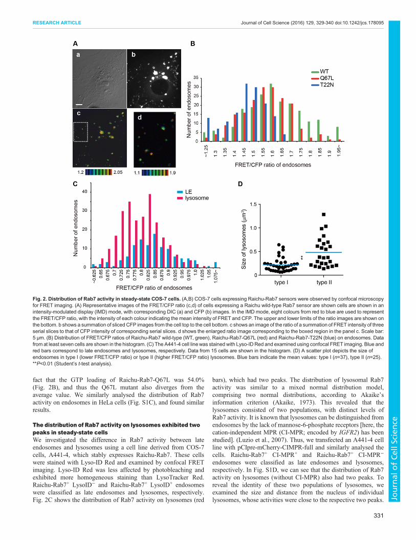
fact that the GTP loading of Raichu-Rab7-Q67L was 54.0%(Fig. 2B), and thus the Q67L mutant also diverges from theaverage value. We similarly analysed the distribution of Rab7activity on endosomes in HeLa cells (Fig. S1C), and found similarresults.
The distribution of Rab7 activity on lysosomes exhibited twopeaks in steady-state cellsWe investigated the difference in Rab7 activity between lateendosomes and lysosomes using a cell line derived from COS-7cells, A441-4, which stably expresses Raichu-Rab7. These cellswere stained with Lyso-ID Red and examined by confocal FRETimaging. Lyso-ID Red was less affected by photobleaching andexhibited more homogeneous staining than LysoTracker Red.Raichu-Rab7+ LysoID− and Raichu-Rab7+ LysoID+ endosomeswere classified as late endosomes and lysosomes, respectively.Fig. 2C shows the distribution of Rab7 activity on lysosomes (red
bars), which had two peaks. The distribution of lysosomal Rab7activity was similar to a mixed normal distribution model,comprising two normal distributions, according to Akaike’sinformation criterion (Akaike, 1973). This revealed that thelysosomes consisted of two populations, with distinct levels ofRab7 activity. It is known that lysosomes can be distinguished fromendosomes by the lack of mannose-6-phosphate receptors [here, thecation-independent MPR (CI-MPR; encoded by IGFR2) has beenstudied]. (Luzio et al., 2007). Thus, we transfected an A441-4 cellline with pCIpre-mCherry-CIMPR-full and similarly analysed thecells. Raichu-Rab7+ CI-MPR+ and Raichu-Rab7+ CI-MPR−
endosomes were classified as late endosomes and lysosomes,respectively. In Fig. S1D, we can see that the distribution of Rab7activity on lysosomes (without CI-MPR) also had two peaks. Toreveal the identity of these two populations of lysosomes, weexamined the size and distance from the nucleus of individuallysosomes, whose activities were close to the respective two peaks.
Fig. 2. Distribution of Rab7 activity in steady-state COS-7 cells. (A,B) COS-7 cells expressing Raichu-Rab7 sensors were observed by confocal microscopyfor FRET imaging. (A) Representative images of the FRET/CFP ratio (c,d) of cells expressing a Raichu wild-type Rab7 sensor are shown cells are shown in anintensity-modulated display (IMD) mode, with corresponding DIC (a) and CFP (b) images. In the IMD mode, eight colours from red to blue are used to representthe FRET/CFP ratio, with the intensity of each colour indicating the mean intensity of FRET and CFP. The upper and lower limits of the ratio images are shown onthe bottom. b shows a summation of sliced CFP images from the cell top to the cell bottom. c shows an image of the ratio of a summation of FRET intensity of threeserial slices to that of CFP intensity of corresponding serial slices. d shows the enlarged ratio image corresponding to the boxed region in the panel c. Scale bar:5 μm. (B) Distribution of FRET/CFP ratios of Raichu-Rab7 wild-type (WT, green), Raichu-Rab7-Q67L (red) and Raichu-Rab7-T22N (blue) on endosomes. Datafrom at least seven cells are shown in the histogram. (C) The A441-4 cell linewas stained with Lyso-ID Red and examined using confocal FRET imaging. Blue andred bars correspond to late endosomes and lysosomes, respectively. Data from 15 cells are shown in the histogram. (D) A scatter plot depicts the size ofendosomes in type I (lower FRET/CFP ratio) or type II (higher FRET/CFP ratio) lysosomes. Blue bars indicate the mean values: type I (n=37), type II (n=25).**P<0.01 (Student’s t-test analysis).
331
RESEARCH ARTICLE Journal of Cell Science (2016) 129, 329-340 doi:10.1242/jcs.178095
Journal
ofCe
llScience
Lysosomes with higher Rab7 activity (type II) tended to belarger (0.48±0.06 µm2) than those having lower Rab7 activity (typeI; 0.22±0.02 µm2, mean±s.e.m.) (Fig. 2D). There was no significantdifference in the distance from the nucleus between the twolysosomal populations (Fig. S1E). Rab7 activity on late endosomesthat were Lyso-ID Red-negative (Fig. 2C) or CI-MPR-positive(Fig. S1E) showed a single-peak distribution. The peak in thedistribution of the FRET/CFP ratio on late endosomes was similar tothe higher peak of lysosomes.
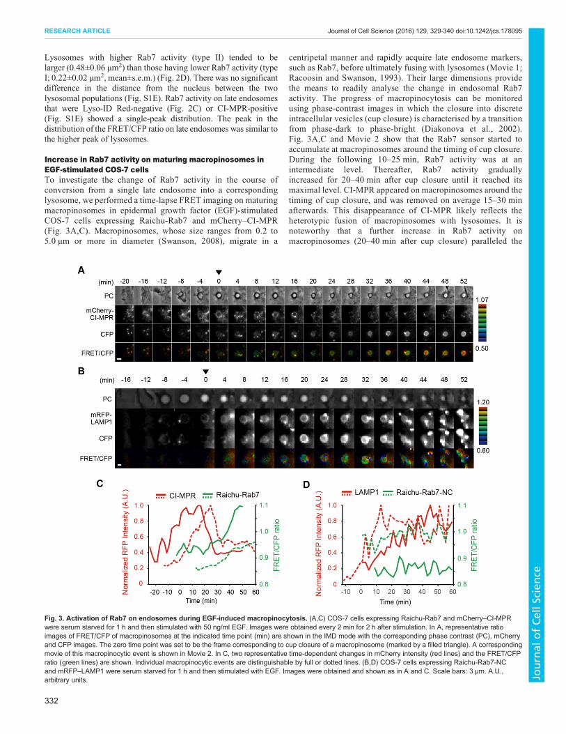
Increase in Rab7 activity on maturing macropinosomes inEGF-stimulated COS-7 cellsTo investigate the change of Rab7 activity in the course ofconversion from a single late endosome into a correspondinglysosome, we performed a time-lapse FRET imaging on maturingmacropinosomes in epidermal growth factor (EGF)-stimulatedCOS-7 cells expressing Raichu-Rab7 and mCherry–CI-MPR(Fig. 3A,C). Macropinosomes, whose size ranges from 0.2 to5.0 µm or more in diameter (Swanson, 2008), migrate in a
centripetal manner and rapidly acquire late endosome markers,such as Rab7, before ultimately fusing with lysosomes (Movie 1;Racoosin and Swanson, 1993). Their large dimensions providethe means to readily analyse the change in endosomal Rab7activity. The progress of macropinocytosis can be monitoredusing phase-contrast images in which the closure into discreteintracellular vesicles (cup closure) is characterised by a transitionfrom phase-dark to phase-bright (Diakonova et al., 2002).Fig. 3A,C and Movie 2 show that the Rab7 sensor started toaccumulate at macropinosomes around the timing of cup closure.During the following 10–25 min, Rab7 activity was at anintermediate level. Thereafter, Rab7 activity graduallyincreased for 20–40 min after cup closure until it reached itsmaximal level. CI-MPR appeared on macropinosomes around thetiming of cup closure, and was removed on average 15–30 minafterwards. This disappearance of CI-MPR likely reflects theheterotypic fusion of macropinosomes with lysosomes. It isnoteworthy that a further increase in Rab7 activity onmacropinosomes (20–40 min after cup closure) paralleled the
Fig. 3. Activation of Rab7 on endosomes during EGF-induced macropinocytosis. (A,C) COS-7 cells expressing Raichu-Rab7 and mCherry–CI-MPRwere serum starved for 1 h and then stimulated with 50 ng/ml EGF. Images were obtained every 2 min for 2 h after stimulation. In A, representative ratioimages of FRET/CFP of macropinosomes at the indicated time point (min) are shown in the IMD mode with the corresponding phase contrast (PC), mCherryand CFP images. The zero time point was set to be the frame corresponding to cup closure of a macropinosome (marked by a filled triangle). A correspondingmovie of this macropinocytic event is shown in Movie 2. In C, two representative time-dependent changes in mCherry intensity (red lines) and the FRET/CFPratio (green lines) are shown. Individual macropinocytic events are distinguishable by full or dotted lines. (B,D) COS-7 cells expressing Raichu-Rab7-NCand mRFP–LAMP1 were serum starved for 1 h and then stimulated with EGF. Images were obtained and shown as in A and C. Scale bars: 3 µm. A.U.,arbitrary units.
332
RESEARCH ARTICLE Journal of Cell Science (2016) 129, 329-340 doi:10.1242/jcs.178095
Journal
ofCe
llScience
decrease in mCherry–CI-MPR (15–30 min after cup closure)(Fig. S2A). We examined the specificity of the increase in Rab7activity on maturing macropinosomes by a similar time-lapseexperiment using Raichu-Rab7-NC as a negative control sensor,because the dominant-negative form, Racihu-Rab7-T22N,inhibits macropinosome formation and thus cannot be used. InRaichu-Rab7-NC, the Rab7-binding domain of Rabring7 wasreplaced with the Rab11-binding domain of GRAB (alsoknown as RAB3IL1) (Horgan et al., 2013). Validation of thisnegative control was shown by the fact that the expression ofArmus-GAP did not decrease the FRET/CFP ratio of Racihu-Rab7-NC (Fig. S2B). When Raichu-Rab7-NC was used in thisexperiment, the FRET/CFP ratio did not change duringmacropinocytosis (Fig. 3B,D; Fig. S2C,D). As shown inFig. 3A, the level of Raichu-Rab7 on the early endosome stateof macropinosomes was low, and thus we cannot obtain a reliableratio image of Raichu-Rab7 there due to this technical limitation.To acquire information about the Rab7 activity on the earlyendosome state of macropinosomes, we examined the recruitmentof EGFP–Mon1 to macropinosomes in COS-7 cells expressingRFP–APPL1, one of Rab5 effectors (Erdmann et al., 2007). Asshown in Fig. S2E,F, the Mon1 level began to increase behindthe rise in the level of APPL1 and reached a plateau at aroundthe timing of the Rab5-to-Rab7 switch, roughly corresponding tocup closure (S.M. and T. Nakamura, unpublished data).Therefore, we presume that a large fraction of Rab7 on theearly endosome state of macropinosomes is in a GDP-boundinactive state.
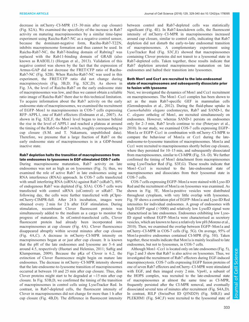
Rab7 depletion halts the transition of macropinosomes fromlate endosomes to lysosomes in EGF-stimulated COS-7 cellsDuring macropinosome maturation, Rab7 activity on lateendosomes was not as high as on lysosomes (Fig. 3); thus, weexamined the role of active Rab7 in late endosomes using anRNA interference (RNAi) approach. In COS-7 cells transfectedwith small interfering RNA (siRNA) against Rab7 (siRab7), 80%of endogenous Rab7 was depleted (Fig. S3A). COS-7 cells weretransfected with control siRNA (siControl) or siRab7. Thefollowing day, the cells were further transfected with pCIpre-mCherry-CIMPR-full. After 24 h incubation, images wereobtained every 2 min for 2 h after EGF stimulation. Duringstimulation, recombinant Clover fluorescent protein wassimultaneously added to the medium as a cargo to monitor theprogress of maturation. In siControl-transfected cells, Cloverfluorescent protein had already been incorporated intomacropinosomes at cup closure (Fig. 4A). Clover fluorescencedisappeared abruptly within several minutes after cup closure(Fig. 4A,C). A decrease in mCherry–CI-MPR intensity onmacropinosomes began at or just after cup closure. It is knownthat the pH of the late endosomes and lysosome are 5–6 andaround 4.5, respectively (Huotari and Helenius, 2011; Saftig andKlumperman, 2009). Because the pKa of Clover is 6.2, theextinction of Clover fluorescence might begin on mature lateendosomes. The decrease in mCherry–CI-MPR intensity showedthat the late-endosome-to-lysosome transition of macropinosomesoccurred at between 10 and 25 min after cup closure. Thus, dimClover proteins might start to be degraded at ∼15 min after cupclosure. In Fig. S3B,D, we confirmed the timing of the transitionof macropinosomes in control cells using LysoTracker Red. Incontrast, in Rab7-depleted cells, the fluorescent intensity ofClover in macropinosomes did not change for more than 1 h aftercup closure (Fig. 4B,D). The difference in fluorescent intensity
between control and Rab7-depleted cells was statisticallysignificant (Fig. 4E). In Rab7-knockdown cells, the fluorescentintensity of mCherry–CI-MPR in macropinosomes increasedaround cup closure, similar to control cells, indicating that Rab7depletion did not disturb the early-to-late endosomal transitionof macropinosomes. A complementary experiment usingLysoTracker Red (Fig. S3C,E) showed that macropinosomescontaining Clover proteins did not transit to a lysosomal state inRab7-depleted cells. Taken together, these results indicate thatRab7 depletion arrested macropinosome maturation on lateendosomes and halted their transition into lysosomes.
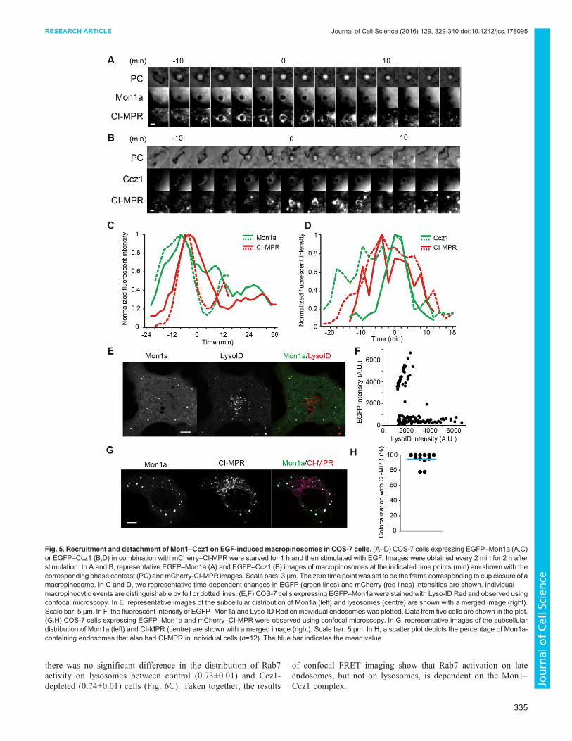
Both Mon1 and Ccz1 are recruited to the late-endosomalstate of macropinosomes and subsequently dissociate priorto fusion with lysosomeNext, we investigated the dynamics of Mon1 and Ccz1 recruitmenton macropinosomes. The Mon1–Ccz1 complex has been shown toact as the main Rab7-specific GEF in mammalian cells(Gerondopoulos et al., 2012). During the fluid-phase uptake inCaenorhabditis elegans coelomocytes, Rab7 and SAND-1, theC. elegans ortholog of Mon1, are recruited simultaneously onendosomes. However, whereas SAND-1 persists on endosomesonly for 2–3 min, Rab7 levels continue to rise (Poteryaev et al.,2010). In our study, we examined COS-7 cells expressing EGFP–Mon1a or EGFP–Ccz1 in combination with mCherry–CI-MPR tocompare the behaviour of Mon1a or Ccz1 during the late-endosome-to-lysosome transition of macropinosomes. Mon1a andCcz1 were recruited to macropinosomes shortly before cup closure,where they persisted for 10–15 min, and subsequently dissociatedfrom macropinosomes, similar to CI-MPR (Fig. 5A–D). We furtherconfirmed the timing of Mon1 detachment from macropinosomesusing LysoTracker Red (Fig. S3F,G). These results indicate thatMon1–Ccz1 is recruited to the late-endosomal state ofmacropinosomes and dissociates from their lysosomal state inCOS-7 cells.
COS-7 cells expressing EGFP–Mon1awere stained with Lyso-IDRed and the recruitment of Mon1a on lysosomes was examined. Asshown in Fig. 5E, Mon1a-positive vesicles were distributedthroughout cytoplasm, with very little overlap with lysosomes.Fig. 5F shows a correlation plot of EGFP–Mon1a and Lyso-ID Redintensities for individual endosomes. A group of endosomes withhigh EGFP signal (>3000) and relatively low LysoID signal werecharacterised as late endosomes. Endosomes exhibiting low Lyso-ID signal without EGFP–Mon1a were characterised as secretoryvesicles, which are known to have a relatively low pH (Moreno et al.,2010). Then, we examined the overlap between EGFP–Mon1a andmCherry–CI-MPR in COS-7 cells (Fig. 5G). On average, 95% ofMon1a-positive endosomes contained CI-MPR (Fig. 5H). Takentogether, these results indicate that Mon1a is mainly localised to lateendosomes, but not to lysosomes, in COS-7 cells.
AlthoughMon1–Ccz1 is located only on late endosomes (Fig. 5),Figs 2 and 3 show that Rab7 is also active on lysosomes. Thus, weinvestigated the recruitment of Rab7 effectors during EGF-inducedmacropinocytosis. COS-7 cells expressing EGFP fusion proteins ofone of seven Rab7 effectors and mCherry–CI-MPR were stimulatedwith EGF, and then imaged every 2 min. Vps41, a subunit ofthe HOPS complex, was recruited to the late-endosomal stateof macropinosomes at almost the same time as CI-MPR,frequently persisted after the CI-MPR removal, and eventuallydissociated several tens of minutes after recruitment (Fig. S4A,D).In contrast, RILP (SwissProt ID Q5ND29) (Fig. S4B,E) andPLEKHM1 (Fig. S4C,F) were recruited to the lysosomal state of
333
RESEARCH ARTICLE Journal of Cell Science (2016) 129, 329-340 doi:10.1242/jcs.178095
Journal
ofCe
llScience
macropinosomes in place of CI-MPR. The behaviour of RILP andPLEKHM1 supports the idea that active Rab7 plays roles onlysosome as well as on late endosomes.
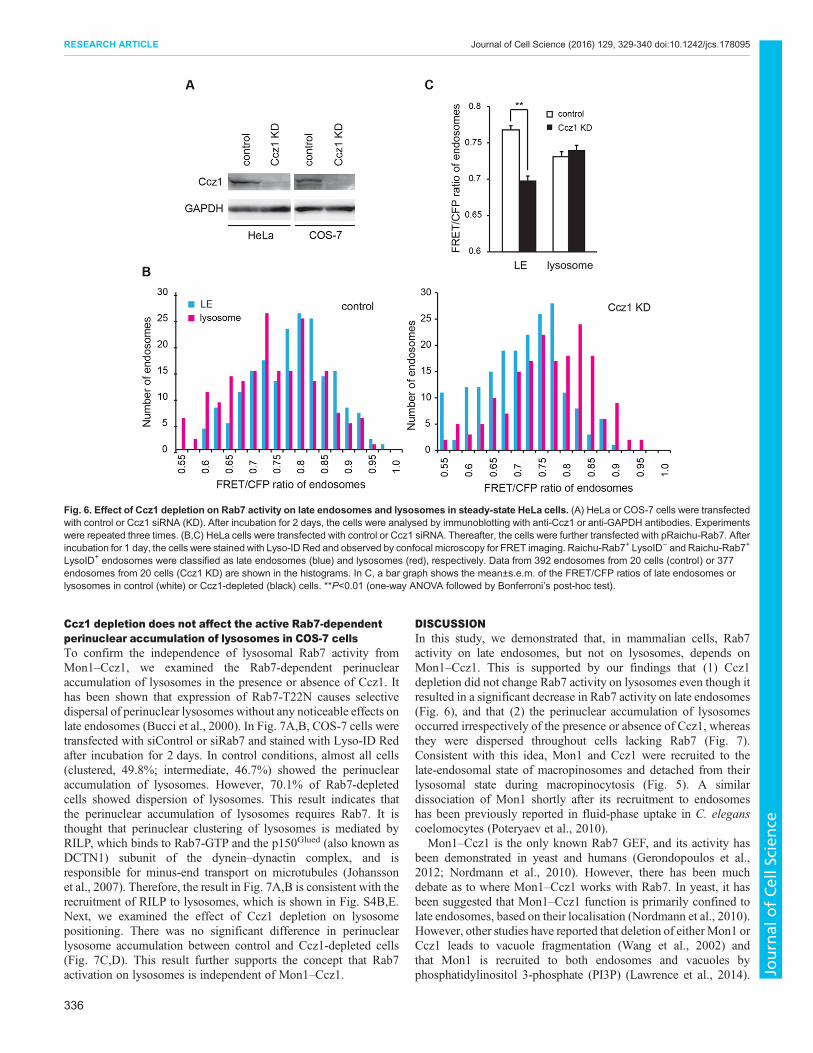
Rab7 activation on lysosomes is independent of Mon1–Ccz1in HeLa cellsThe finding that lysosomes are devoid of Mon1–Ccz1 raises thepossibility that Rab7 activity on lysosomes, shown in Figs 2 and3, is independent of Mon1–Ccz1. To explore this possibility, weexamined the effect of Ccz1 depletion on Rab7 activity on lateendosomes and lysosomes. Because mammals have two Mon1isoforms, it might be difficult to completely deplete both Mon1proteins. As shown in Fig. 6A, Ccz1 siRNA (siCcz1) efficientlyreduced the amount of endogenous Ccz1 in HeLa (85%
depletion) and COS-7 cells (80% depletion). One day aftertransfection with siControl or siCcz1, HeLa cells were transfectedwith Raichu-Rab7. After 24 h incubation, the cells were stainedwith Lyso-ID Red and examined by confocal FRET imaging.The distribution of Rab7 activity on late endosomes andlysosomes in siControl-transfected cells (Fig. 6B, left) wasessentially the same as that shown in Fig. 2C. In Ccz1-depletedcells (Fig. 6B, right), the distribution of Rab7 activity on lateendosomes (blue bars) shifted towards a lower level than that incontrol cells, as expected from the residence of Mon1–Ccz1 onlate endosomes. The averages of the FRET/CFP ratios on lateendosomes in control and Ccz1-depleted cells were 0.77±0.01and 0.70±0.01 (mean±s.e.m.), respectively (Fig. 6C). Thisdifference was statistically significant (P<0.01). In contrast,
Fig. 4. Effect of Rab7 depletion on EGF-induced macropinocytosis. COS-7 cells were transfected with control (A,C) or Rab7 siRNA (KD) (B,D). After 24 hincubation, the cells were further transfected with pCIpre-mCherry-CIMPR-full. After serum starvation for 1 h, images were obtained every 2 min for 2 h afteraddition of EGF and 5 µg/ml of recombinant Clover. After pausing of the imaging at an appropriate time, the medium containing Clover was replaced by freshimaging medium and imaging was resumed. The zero time point was set to be the frame corresponding to cup closure of a macropinosome. In A and B,representative phase contrast (PC), Clover and mCherry (CI-MPR) images of macropinosomes in the control (A) or Rab7-depleted (B) cells at the indicated timepoint (min) are shown. Scale bars: 3 µm. In C and D, two representative time-dependent changes in Clover intensity (green lines) and mCherry–CI-MPR intensity(red lines) of macropinosomes in the control (C) or Rab7-depleted (D) cells are shown. Individual macropinocytic events are distinguishable by full or dotted lines.(E) Themean±s.e.m. of normalised Clover intensity of macropinosomes at the indicated time-points in the control or Rab7-depleted cells are shown. The numberof experiments was control (n=4) and Rab7-depleted (n=4). *P<0.05; **P<0.01 (Student’s t-test).
334
RESEARCH ARTICLE Journal of Cell Science (2016) 129, 329-340 doi:10.1242/jcs.178095
Journal
ofCe
llScience
there was no significant difference in the distribution of Rab7activity on lysosomes between control (0.73±0.01) and Ccz1-depleted (0.74±0.01) cells (Fig. 6C). Taken together, the results
of confocal FRET imaging show that Rab7 activation on lateendosomes, but not on lysosomes, is dependent on the Mon1–Ccz1 complex.
Fig. 5. Recruitment and detachment of Mon1–Ccz1 on EGF-induced macropinosomes in COS-7 cells. (A–D) COS-7 cells expressing EGFP–Mon1a (A,C)or EGFP–Ccz1 (B,D) in combination with mCherry–CI-MPR were starved for 1 h and then stimulated with EGF. Images were obtained every 2 min for 2 h afterstimulation. In A and B, representative EGFP–Mon1a (A) and EGFP–Ccz1 (B) images of macropinosomes at the indicated time points (min) are shown with thecorresponding phase contrast (PC) andmCherry-CI-MPR images. Scale bars: 3 µm. The zero time point was set to be the frame corresponding to cup closure of amacropinosome. In C and D, two representative time-dependent changes in EGFP (green lines) and mCherry (red lines) intensities are shown. Individualmacropinocytic events are distinguishable by full or dotted lines. (E,F) COS-7 cells expressing EGFP–Mon1a were stained with Lyso-ID Red and observed usingconfocal microscopy. In E, representative images of the subcellular distribution of Mon1a (left) and lysosomes (centre) are shown with a merged image (right).Scale bar: 5 µm. In F, the fluorescent intensity of EGFP–Mon1a and Lyso-ID Red on individual endosomes was plotted. Data from five cells are shown in the plot.(G,H) COS-7 cells expressing EGFP–Mon1a and mCherry–CI-MPR were observed using confocal microscopy. In G, representative images of the subcellulardistribution of Mon1a (left) and CI-MPR (centre) are shown with a merged image (right). Scale bar: 5 µm. In H, a scatter plot depicts the percentage of Mon1a-containing endosomes that also had CI-MPR in individual cells (n=12). The blue bar indicates the mean value.
335
RESEARCH ARTICLE Journal of Cell Science (2016) 129, 329-340 doi:10.1242/jcs.178095
Journal
ofCe
llScience
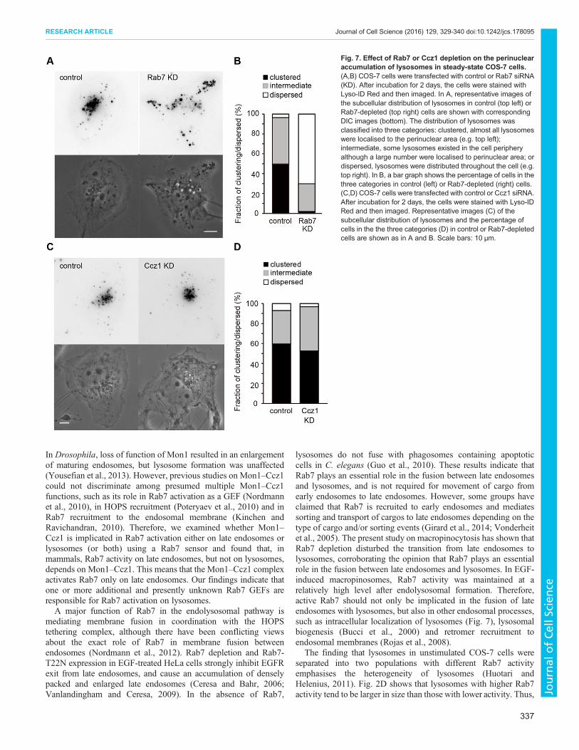
Ccz1 depletion does not affect the active Rab7-dependentperinuclear accumulation of lysosomes in COS-7 cellsTo confirm the independence of lysosomal Rab7 activity fromMon1–Ccz1, we examined the Rab7-dependent perinuclearaccumulation of lysosomes in the presence or absence of Ccz1. Ithas been shown that expression of Rab7-T22N causes selectivedispersal of perinuclear lysosomes without any noticeable effects onlate endosomes (Bucci et al., 2000). In Fig. 7A,B, COS-7 cells weretransfected with siControl or siRab7 and stained with Lyso-ID Redafter incubation for 2 days. In control conditions, almost all cells(clustered, 49.8%; intermediate, 46.7%) showed the perinuclearaccumulation of lysosomes. However, 70.1% of Rab7-depletedcells showed dispersion of lysosomes. This result indicates thatthe perinuclear accumulation of lysosomes requires Rab7. It isthought that perinuclear clustering of lysosomes is mediated byRILP, which binds to Rab7-GTP and the p150Glued (also known asDCTN1) subunit of the dynein–dynactin complex, and isresponsible for minus-end transport on microtubules (Johanssonet al., 2007). Therefore, the result in Fig. 7A,B is consistent with therecruitment of RILP to lysosomes, which is shown in Fig. S4B,E.Next, we examined the effect of Ccz1 depletion on lysosomepositioning. There was no significant difference in perinuclearlysosome accumulation between control and Ccz1-depleted cells(Fig. 7C,D). This result further supports the concept that Rab7activation on lysosomes is independent of Mon1–Ccz1.
DISCUSSIONIn this study, we demonstrated that, in mammalian cells, Rab7activity on late endosomes, but not on lysosomes, depends onMon1–Ccz1. This is supported by our findings that (1) Ccz1depletion did not change Rab7 activity on lysosomes even though itresulted in a significant decrease in Rab7 activity on late endosomes(Fig. 6), and that (2) the perinuclear accumulation of lysosomesoccurred irrespectively of the presence or absence of Ccz1, whereasthey were dispersed throughout cells lacking Rab7 (Fig. 7).Consistent with this idea, Mon1 and Ccz1 were recruited to thelate-endosomal state of macropinosomes and detached from theirlysosomal state during macropinocytosis (Fig. 5). A similardissociation of Mon1 shortly after its recruitment to endosomeshas been previously reported in fluid-phase uptake in C. eleganscoelomocytes (Poteryaev et al., 2010).
Mon1–Ccz1 is the only known Rab7 GEF, and its activity hasbeen demonstrated in yeast and humans (Gerondopoulos et al.,2012; Nordmann et al., 2010). However, there has been muchdebate as to where Mon1–Ccz1 works with Rab7. In yeast, it hasbeen suggested that Mon1–Ccz1 function is primarily confined tolate endosomes, based on their localisation (Nordmann et al., 2010).However, other studies have reported that deletion of either Mon1 orCcz1 leads to vacuole fragmentation (Wang et al., 2002) andthat Mon1 is recruited to both endosomes and vacuoles byphosphatidylinositol 3-phosphate (PI3P) (Lawrence et al., 2014).
Fig. 6. Effect of Ccz1 depletion on Rab7 activity on late endosomes and lysosomes in steady-state HeLa cells. (A) HeLa or COS-7 cells were transfectedwith control or Ccz1 siRNA (KD). After incubation for 2 days, the cells were analysed by immunoblotting with anti-Ccz1 or anti-GAPDH antibodies. Experimentswere repeated three times. (B,C) HeLa cells were transfected with control or Ccz1 siRNA. Thereafter, the cells were further transfected with pRaichu-Rab7. Afterincubation for 1 day, the cells were stained with Lyso-ID Red and observed by confocal microscopy for FRET imaging. Raichu-Rab7+ LysoID− and Raichu-Rab7+
LysoID+ endosomes were classified as late endosomes (blue) and lysosomes (red), respectively. Data from 392 endosomes from 20 cells (control) or 377endosomes from 20 cells (Ccz1 KD) are shown in the histograms. In C, a bar graph shows the mean±s.e.m. of the FRET/CFP ratios of late endosomes orlysosomes in control (white) or Ccz1-depleted (black) cells. **P<0.01 (one-way ANOVA followed by Bonferroni’s post-hoc test).
336
RESEARCH ARTICLE Journal of Cell Science (2016) 129, 329-340 doi:10.1242/jcs.178095
Journal
ofCe
llScience
In Drosophila, loss of function of Mon1 resulted in an enlargementof maturing endosomes, but lysosome formation was unaffected(Yousefian et al., 2013). However, previous studies on Mon1–Ccz1could not discriminate among presumed multiple Mon1–Ccz1functions, such as its role in Rab7 activation as a GEF (Nordmannet al., 2010), in HOPS recruitment (Poteryaev et al., 2010) and inRab7 recruitment to the endosomal membrane (Kinchen andRavichandran, 2010). Therefore, we examined whether Mon1–Ccz1 is implicated in Rab7 activation either on late endosomes orlysosomes (or both) using a Rab7 sensor and found that, inmammals, Rab7 activity on late endosomes, but not on lysosomes,depends on Mon1–Ccz1. This means that the Mon1–Ccz1 complexactivates Rab7 only on late endosomes. Our findings indicate thatone or more additional and presently unknown Rab7 GEFs areresponsible for Rab7 activation on lysosomes.A major function of Rab7 in the endolysosomal pathway is
mediating membrane fusion in coordination with the HOPStethering complex, although there have been conflicting viewsabout the exact role of Rab7 in membrane fusion betweenendosomes (Nordmann et al., 2012). Rab7 depletion and Rab7-T22N expression in EGF-treated HeLa cells strongly inhibit EGFRexit from late endosomes, and cause an accumulation of denselypacked and enlarged late endosomes (Ceresa and Bahr, 2006;Vanlandingham and Ceresa, 2009). In the absence of Rab7,
lysosomes do not fuse with phagosomes containing apoptoticcells in C. elegans (Guo et al., 2010). These results indicate thatRab7 plays an essential role in the fusion between late endosomesand lysosomes, and is not required for movement of cargo fromearly endosomes to late endosomes. However, some groups haveclaimed that Rab7 is recruited to early endosomes and mediatessorting and transport of cargos to late endosomes depending on thetype of cargo and/or sorting events (Girard et al., 2014; Vonderheitet al., 2005). The present study on macropinocytosis has shown thatRab7 depletion disturbed the transition from late endosomes tolysosomes, corroborating the opinion that Rab7 plays an essentialrole in the fusion between late endosomes and lysosomes. In EGF-induced macropinosomes, Rab7 activity was maintained at arelatively high level after endolysosomal formation. Therefore,active Rab7 should not only be implicated in the fusion of lateendosomes with lysosomes, but also in other endosomal processes,such as intracellular localization of lysosomes (Fig. 7), lysosomalbiogenesis (Bucci et al., 2000) and retromer recruitment toendosomal membranes (Rojas et al., 2008).
The finding that lysosomes in unstimulated COS-7 cells wereseparated into two populations with different Rab7 activityemphasises the heterogeneity of lysosomes (Huotari andHelenius, 2011). Fig. 2D shows that lysosomes with higher Rab7activity tend to be larger in size than those with lower activity. Thus,
Fig. 7. Effect of Rab7 or Ccz1 depletion on the perinuclearaccumulation of lysosomes in steady-state COS-7 cells.(A,B) COS-7 cells were transfected with control or Rab7 siRNA(KD). After incubation for 2 days, the cells were stained withLyso-ID Red and then imaged. In A, representative images ofthe subcellular distribution of lysosomes in control (top left) orRab7-depleted (top right) cells are shown with correspondingDIC images (bottom). The distribution of lysosomes wasclassified into three categories: clustered, almost all lysosomeswere localised to the perinuclear area (e.g. top left);intermediate, some lysosomes existed in the cell peripheryalthough a large number were localised to perinuclear area; ordispersed, lysosomes were distributed throughout the cell (e.g.top right). In B, a bar graph shows the percentage of cells in thethree categories in control (left) or Rab7-depleted (right) cells.(C,D) COS-7 cells were transfected with control or Ccz1 siRNA.After incubation for 2 days, the cells were stained with Lyso-IDRed and then imaged. Representative images (C) of thesubcellular distribution of lysosomes and the percentage ofcells in the the three categories (D) in control or Rab7-depletedcells are shown as in A and B. Scale bars: 10 µm.
337
RESEARCH ARTICLE Journal of Cell Science (2016) 129, 329-340 doi:10.1242/jcs.178095
Journal
ofCe
llScience
the population with higher Rab7 activity might have higher fusingcapabilities, which allows them to continue to fuse with lateendosomes and other lysosomes, resulting in larger vesicles. Ingeneral, the fusion of late endosomes with lysosomes generates atransient hybrid organelle, called an endolysosome, in which activedegradation takes place (Bright et al., 1997; Mullock et al., 1998).This endolysosome might correspond to the population with higherRab7 activity. The endolysosome is converted into a maturelysosome, which constitutes a storage organelle for lysosomalhydrolases and other degrading enzymes (Huotari and Helenius,2011). The puncta with lower Rab7 activity in Fig. 2C might indeedcorrespond to mature lysosomes. Because lysosomes have beenreported to have divergent functions, such as secretion, plasmamembrane repair and energy metabolism in addition to theirdegradative function (Settembre et al., 2013), these organellesshould be regarded as a heterogeneous population (Huotari andHelenius, 2011). What kind of functional divergence exits betweenlysosomal pools with different Rab7 activity is an attractive subjectfor future research.The present study demonstrated that Rab7-GDP was localised on
endosomal membranes, and, in EGF-stimulated COS-7 cells, Rab7was initially recruited to macropinosomes in a GDP-bound inactiveform and subsequently became activated during maturation,eventually leading to fusion with lysosomes. These results indicatethat an unknown factor exists, which can deliver Rab7-GDP ontoendosomal membranes. Rab GEFs display the minimal targetingmachinery for targeting Rabs from the cytosol to the correctcompartment (Blumer et al., 2013). For Rab1, Legionellapneumophila DrrA has remarkably efficient GEF activity, and itsGEF activity is sufficient to displace the GDI from the Rab1–GDIcomplex (Schoebel et al., 2009). Rab GEFs vary tremendouslyin their catalytic efficiency, and the endpoint of the GDI displacementis determined by the relative affinities of Rab-GDP and Rab-GTPto the GDI and the ratio between the GTP and GDP concentration(Schoebel et al., 2009). The efficiency of Mon1–Ccz1 GEFactivity is moderate (2000 M−1 s−1) and slower than DrrA andTRAPP (Cai et al., 2008; Nordmann et al., 2010; Schoebel et al.,2009). Thus, we believe that Mon1–Ccz1 cannot facilitate thedisplacement of GDI from Rab7-GDP, but that an unidentified GDFdissociates the Rab7–GDI complex for delivery of Rab7 to theendosomalmembrane. For Rab9, the integral membrane protein Yip3acts catalytically to dissociate complexes of Rab9 bound to GDI, andto deliver it onto membranes (Sivars et al., 2003). Lgl1 interactsdirectly with Rab10-GDP and acts to release GDI from Rab10(Wang et al., 2011). Future studies aiming at the identification ofnovel GDFs acting on Rab7 are essential for uncovering themechanism controlling the localisation and activity of Rab7 in theendolysosomal network.
MATERIALS AND METHODSFRET biosensorspRaichu-Rab7/A441, derived from the pCAGGS expression vector,encoded a FRET biosensor based on the Eevee backbone (a gift fromMichiyuki Matsuda, Kyoto University, Japan; Komatsu et al., 2011),designated Raichu-Rab7, which comprised YPet-GL (Nguyen andDaugherty, 2005), the Rab7-binding domain of mouse Rabring7 (aminoacids 10–145; Mizuno et al., 2003), SECFP (a brighter version of CFPdeveloped by Atsushi Miyawaki, RIKEN Brain Research Institute, Japan),and canine Rab7 (a gift from Yoshimi Takai, Kobe University, Japan). Theflexible linkers, collectively designated MeV (modified evading),consisted of the 15-amino-acid peptide (GGSGG)3, between YPet-GLand the Rab7-binding domain of Rabring7, the EV linker of 116 aminoacids (Komatsu et al., 2011) between the RBD domain of Rabring7 and
SECFP, and the 15-amino-acid peptide, (GGSGG)3, between SECFP andRab7. If necessary, wild-type Rab7 was replaced with Rab7-Q67L orRab7-T22N. In pRaichu-Rab7-NC, the Rab7-binding domain of Rabring7was replaced with the Rab11-binding domain of GRAB (Horgan et al.,2013).
PlasmidsThe cDNAs for wild-type Rab7, Rab7-Q67L and Rab7-T22N weresubcloned into pCAGGS-EGFP and pCXN2-mRFP (Ohba et al., 2003).The cDNA for a GAP domain of Armus (a gift from Vania M. Braga,Imperial College, London, UK; Frasa et al., 2010) was subcloned into thepCAGGS expression vector. The cDNAs for mouse Mon1a and Ccz1 wereobtained by conventional PCR techniques (Fukuda et al., 1999) andsubcloned into pEGFP-C1 (Clontech, Mountain View, CA). The RILPcDNA was subcloned into pCAGGS-EGFP. pEGFP-C1-PLEKHM1(Tabata et al., 2010) and pmRFP-N1-Lamp1 (Kimura et al., 2007) wereprovided by Tamotsu Yoshimori (Osaka University, Japan). pcDNA3-RFP-APPL1 (Erdmann et al., 2007) was provided by Pietro De Camilli (YaleUniversity, New Haven, CT). The cDNA for CI-MPR (CI-MPR is alsoknown as IGF2R) (Waguri et al., 2006) was used to prepare pCIpre-mCherry-CIMPR-full. The cDNA for mouse Vps41 was amplified by PCRand subcloned into pmStr-C1 (Ohbayashi et al., 2012). The piggyBactransposon-based expression vector (Yusa et al., 2009) was provided byAllan Bradley (Wellcome Trust Sanger Institute, UK).
Cells, reagents and antibodies293-F cells (Life Technologies, Carlsbad, CA) were maintained in FreeStyle293 expression medium (Life Technologies). COS-7 and HeLa cells weremaintained in Dulbecco’s modified Eagle’s medium (DMEM) containing10% fetal bovine serum. A cell line derived from COS-7 cells, A441-4,which stably expresses Raichu-Rab7, was established as describedpreviously (Yusa et al., 2009). EGF was purchased from Calbiochem (LaJolla, CA). Lyso-ID Red was obtained from Enzo Life Sciences(Farmingdale, NY). The following primary antibodies were used: rabbitpolyclonal antibodies to Rab7 (1:300 dilution, R4779, Sigma-Aldrich,St Louis, MO); goat polyclonal antibodies to Ccz1 (1:500 dilution, T-19,Santa Cruz Biotechnology, Santa Cruz, CA); and rabbit polyclonalantibodies to GAPDH (1:1000 dilution, FL-335, Santa CruzBiotechnology). Anti-GFP polyclonal antibody was a gift from NaokiMochizuki (NCVC Research Institute, Japan), and 2 μl of antibody wasused for immunoprecipitation.
RNA interferenceRab7 Stealth siRNA and Ccz1 Silencer Select siRNA were obtained fromLife Technologies. The 25mer nucleotide sequence (sense) to target humanand monkey Rab7 mRNA was 5′-CAUUCAUGAACCAGUAUGUGAA-UAA-3′. The nucleotide sequence (sense) to target human and monkeyCcz1 mRNA was 5′-GCUCAUCUGGAGUGGAUUAGAACAA-3′.COS-7 or HeLa cells were transfected with 20 nM siRNA usingLipofectamine RNAiMAX (Life Technologies) according to themanufacturer’s instructions and used after 48 h incubation.
In vitro spectrofluorometryAt 48 h after transfection, 293-F cells were transferred into a quartz cuvetteand fluorescence spectra were obtained using an FP-750 spectrometer(JASCO, Hachioji, Japan) at an excitation wavelength of 433 nm.
Analysis of guanine nucleotides bound to Rab7Guanine nucleotides bound to Raichu sensors or EGFP–Rab7 wereanalysed as previously described (Kawase et al., 2006). Briefly, COS-7cells were transfected with pRaichu-Rab7 or pCAGGS-EGFP-Rab7. At 36h after transfection, cells were labelled with 32Pi in phosphate-freemodified Eagle’s medium (Life Technologies) for 4 h. Raichu-Rab7 andEGFP–Rab7 were immunoprecipitated with anti-GFP antibodies. Theimmunoprecipitates were analysed by thin layer chromatography. GTP andGDP bound to Rab7 were quantified using a FLA-7000 image analyser(Fuji Film, Tokyo, Japan).
338
RESEARCH ARTICLE Journal of Cell Science (2016) 129, 329-340 doi:10.1242/jcs.178095
Journal
ofCe
llScience
Time-lapse FRET imagingCOS-7 cells expressing FRET sensors were serum-starved for 1 h withFluoroBrite DMEM medium (Life Technologies) containing 0.1% BSAand 4 mM L-glutamine prior to stimulation with 50 ng/ml EGF. Themedium was covered with mineral oil (Sigma-Aldrich) to preventevaporation. Cells were imaged at 37°C with an IX81 invertedmicroscope (Olympus, Tokyo, Japan) equipped with a Cool SNAP-HQcooled charge-coupled device camera (Roper Scientific, Trenton, NJ), alaser auto-focusing system (IX2-ZDC, Olympus) and an automaticallyprogrammable XY stage (MD-XY30100T-Meta, SIGMA KOKI, Tokyo,Japan), which allowed time-lapse images of several fields of view to beobtained in a single experiment. The following filters were used for dual-emission imaging: an FF01-438/24-25 excitation filter (Semrock,Rochester, NY); an XF2034 (455DRLP) dichroic mirror (OmegaOptical, Brattleboro, VT); and two emission filters (FF01-483/32-25for CFP and FF01-542/27-25 for FRET, Semrock). Cells wereilluminated with a 75-W Xenon lamp through a 6% neutral densityfilter or an X-Cite 120LED (Lumen Dynamics, Mississauga, Canada),and viewed through a Plan Apochromat 60× oil objective lens (NA 1.4,Olympus). The exposure times for 4×4 binning were 200 ms for CFP andFRET images and 100 ms for phase contrast images. After backgroundsubtraction, FRET/CFP ratio images were created with MetaMorphsoftware (Universal Imaging, West Chester, PA), and the images wereused to represent FRET efficiency according to Kawase et al. (2006) withsome modifications. Single macropinocytic events were selectedmanually, and the macropinosome was centred in the image. The localbackground was determined as the average fluorescence of the regionsurrounding the target macropinosome, with a three-pixel width,corresponding to 0.225 µm.
Confocal microscopyCells were fixed with 3.7% formaldehyde. After washing with PBS, thesamples were imaged with an IX81 inverted microscope equipped with anFV-1000 confocal imaging system (Olympus) using a Plan Apochromat60× oil objective lens (NA 1.4, Olympus).
For confocal FRET imaging, HeLa or COS-7 cells expressing FRETsensors were cultured with FluoroBrite DMEM medium containing0.1% BSA and 4 mM L-glutamine and then imaged with the IX81inverted microscope equipped with the FV-1000 confocal imagingsystem and a PlanApoN 60× oil objective lens (NA 1.4, Olympus) at37°C. The excitation laser and fluorescence filter settings were asfollows: excitation laser, 440 nm; excitation dichroic mirror, DM405–440; CFP channel PMT dichroic mirror, SDM 510; CFP channel PMTfilter, BA460–490; FRET channel PMT filter, BA515–615. For Lyso-ID-Red-stained cells, the wavelength of the excitation laser was 559 nmand the filter settings for red fluorescence were as follows: excitationdichroic mirror, BS20/80; RFP channel PMT filter, BA575–675. ForRab7 sensors, images were obtained in a fixed condition (laser power2%; scan speed 20 µs/pixel; confocal aperture, 100 µm; slice width,0.42 µm; 2× zoom). Image processing was performed with MetaMorphsoftware. A summation image of three serial planes where theendosomes were clearly visible was created and subjected tobackground subtraction. Using Top-hat morphological filtering,individual endosomes, whose areas were 10–500 pixels andfluorescent intensities in CFP and FRET channels were ≥500, wereselected. Endosomes with aberrant morphologies were eliminated bysight. Thereafter, CFP and FRET fluorescent intensities were obtainedfor individual endosomes and FRET/CFP ratios were calculated. Ifnecessary, RFP intensities from Lyso-ID Red were similarly obtained.According to their red fluorescent intensities, endosomes were classifiedas late endosomes (<1000) or lysosomes (≥1000).
Preparation of recombinant Clover proteinClover (a gift fromMichael Lin, Stanford University, CA; Lam et al., 2012)was produced using the pGEX-6P expression vector (GE Healthcare, LittleChalfont, UK). GST–Clover proteins were purified using glutathione–Sepharose and cleaved with Precision protease (GE Healthcare). The purityof the eluted Clover was checked by SDS-PAGE.
Statistical analysisOne-way ANOVA followed by Dunnett’s or Bonferroni’s post-hoc testswere performed using GraphPad PRISM (version 5.04, GraphPad Software,San Diego, CA). Evaluation of the fitting of the distribution of Rab7 activityon lysosomes with a mixed normal distribution according to Akaike’sinformation criterion (Akaike, 1973; Burnham and Anderson, 1998) wasimplemented by MATLAB software (2015a, MathWorks, Natick, MA).
AcknowledgementsThe authors are grateful to Michiyuki Matsuda for generous support and valuableadvice, Yuichi Sakumura for providing aMATLAB script to execute optimal fitting to amixed normal distribution, and Yuuki Onishi and Mr Kazuto Kawasaki for theirworthwhile discussions about the experimental design. The authors thank KimikoNakamura for technical assistance and members of the Nakamura laboratory fortheir input.
Competing interestsThe authors declare no competing or financial interests.
Author contributionsT. Nakamura conceived the idea. S.Y., N.W., S.W., G.S., M.F. and T. Nakamuradesigned the experiments. S.Y., S.M., A.F., T. Nanao and T. Nakamura performedthe experiments and analyzed the data. G.S. and T. Nakamura wrote themanuscript.
FundingThis work was supported by Grants-in-Aid for Scientific Research from the JapanSociety for the Promotion of Science [grant numbers 23300134 and 24657094]; andgrants from the Yamada Science Foundation: the Naito Foundation; and theHamaguchi Foundation for the Advancement of Biochemistry to T. Nakamura.
Supplementary informationSupplementary information available online athttp://jcs.biologists.org/lookup/suppl/doi:10.1242/jcs.178095/-/DC1
ReferencesAkaike, H. (1973). Information theory and an extension of the maximum likelihood
principle. In Proceedings of the 2nd International Symposium (ed. B. N. Petrovand F. Csaki), pp. 267-281. Budapest: Akademiai Kiado.
Blumer, J., Rey, J., Dehmelt, L., Mazel, T., Wu, Y.-W., Bastiaens, P., Goody, R. S.and Itzen, A. (2013). RabGEFs are a major determinant for specific Rabmembrane targeting. J. Cell Biol. 200, 287-300.
Bright, N. A., Reaves, B. J., Mullock, B. M. and Luzio, J. P. (1997). Dense corelysosomes can fuse with late endosomes and are re-formed from the resultanthybrid organelles. J. Cell Sci. 110, 2027-2040.
Bucci, C., Thomsen, P., Nicoziani, P., McCarthy, J. and van Deurs, B. (2000).Rab7: a key to lysosome biogenesis. Mol. Biol. Cell 11, 467-480.
Burnham, K. P. and Anderson, D. R. (1998). Model Selection and Inference: APractical Information-Theoretical Approach. New York: Springer-Verlag.
Cai, Y., Chin, H. F., Lazarova, D., Menon, S., Fu, C., Cai, H., Sclafani, A.,Rodgers, D. W., De La Cruz, E. M., Ferro-Novick, S. et al. (2008). The structuralbasis for activation of the Rab Ypt1p by the TRAPP membrane-tetheringcomplexes. Cell 133, 1202-1213.
Ceresa, B. P. and Bahr, S. J. (2006). rab7 activity affects epidermal growth factor:epidermal growth factor receptor degradation by regulating endocytic traffickingfrom the late endosome. J. Biol. Chem. 281, 1099-1106.
Diakonova, M., Bokoch, G. and Swanson, J. A. (2002). Dynamics of cytoskeletalproteins during Fcγ receptor-mediated phagocytosis in macrophages. Mol. Biol.Cell 13, 402-411.
Erdmann, K. S., Mao, Y., McCrea, H. J., Zoncu, R., Lee, S., Paradise, S.,Modregger, J., Biemesderfer, D., Toomre, D. and De Camilli, P. (2007). A roleof the Lowe syndrome protein OCRL in early steps of the endocytic pathway. Dev.Cell 13, 377-390.
Frasa, M. A. M., Maximiano, F. C., Smolarczyk, K., Francis, R. E., Betson, M. E.,Lozano, E., Goldenring, J., Seabra, M. C., Rak, A., Ahmadian, M. R. et al.(2010). Armus is a Rac1 effector that inactivates Rab7 and regulates E-cadherindegradation. Curr. Biol. 20, 198-208.
Fukuda, M., Kanno, E. and Mikoshiba, K. (1999). Conserved N-terminal cysteinemotif is essential for homo- and heterodimer formation of synaptotagmins III, V, VI,and X. J. Biol. Chem. 274, 31421-31427.
Gerondopoulos, A., Langemeyer, L., Liang, J.-R., Linford, A. and Barr, F. A.(2012). BLOC-3 mutated in Hermansky-Pudlak syndrome is a Rab32/38 guaninenucleotide exchange factor. Curr. Biol. 22, 2135-2139.
Girard, E., Chmiest, D., Fournier, N., Johannes, L., Paul, J.-L., Vedie, B. andLamaze, C. (2014). Rab7 is functionally required for selective cargo sorting at theearly endosome. Traffic 15, 309-326.
339
RESEARCH ARTICLE Journal of Cell Science (2016) 129, 329-340 doi:10.1242/jcs.178095
Journal
ofCe
llScience
Guo, P., Hu, T., Zhang, J., Jiang, S. and Wang, X. (2010). Sequential action ofCaenorhabditis elegans Rab GTPases regulates phagolysosome formationduring apoptotic cell degradation. Proc. Natl. Acad. Sci. USA 107, 18016-18021.
Horgan, C. P., Hanscom, S. R. and McCaffrey, M. W. (2013). GRAB is a bindingpartner for the Rab11a and Rab11b GTPases. Biochem. Biophys. Res. Commun.441, 214-219.
Huotari, J. and Helenius, A. (2011). Endosome maturation. EMBO J. 30,3481-3500.
Johansson, M., Rocha, N., Zwart, W., Jordens, I., Janssen, L., Kuijl, C.,Olkkonen, V. M. and Neefjes, J. (2007). Activation of endosomal dynein motorsby stepwise assembly of Rab7-RILP-p150Glued, ORP1L, and the receptor βlllspectrin. J. Cell Biol. 176, 459-471.
Kawase, K., Nakamura, T., Takaya, A., Aoki, K., Namikawa, K., Kiyama, H.,Inagaki, S., Takemoto, H., Saltiel, A. R. and Matsuda, M. (2006). GTPhydrolysis by the Rho family GTPase TC10 promotes exocytic vesicle fusion.Dev.Cell 11, 411-421.
Kimura, S., Noda, T. and Yoshimori, T. (2007). Dissection of the autophagosomematuration process by a novel reporter protein, tandem fluorescent-tagged LC3.Autophagy 3, 452-460.
Kinchen, J. M. and Ravichandran, K. S. (2010). Identification of two evolutionarilyconserved genes regulating processing of engulfed apoptotic cells. Nature 464,778-782.
Kitano, M., Nakaya, M., Nakamura, T., Nagata, S. and Matsuda, M. (2008).Imaging of Rab5 activity identifies essential regulators for phagosomematuration.Nature 453, 241-245.
Komatsu, N., Aoki, K., Yamada, M., Yukinaga, H., Fujita, Y., Kamioka, Y. andMatsuda, M. (2011). Development of an optimized backbone of FRET biosensorsfor kinases and GTPases. Mol. Biol. Cell 22, 4647-4656.
Lam, A. J., St-Pierre, F., Gong, Y., Marshall, J. D., Cranfill, P. J., Baird, M. A.,McKeown, M. R., Wiedenmann, J., Davidson, M. W., Schnitzer, M. J. et al.(2012). Improving FRET dynamic range with bright green and red fluorescentproteins. Nat. Methods 9, 1005-1012.
Lawrence, G., Brown, C. C., Flood, B. A., Karunakaran, S., Cabrera, M.,Nordmann, M., Ungermann, C. and Fratti, R. A. (2014). Dynamic association ofthe PI3P-interacting Mon1-Ccz1 GEF with vacuoles is controlled through itsphosphorylation by the type 1 casein kinase Yck3. Mol. Biol. Cell 25, 1608-1619.
Luzio, J. P., Pryor, P. R. and Bright, N. A. (2007). Lysosomes: fusion and function.Nat. Rev. Mol. Cell Biol. 8, 622-632.
Mizuno, K., Kitamura, A. and Sasaki, T. (2003). Rabring7, a novel Rab7 targetprotein with a RING finger motif. Mol. Biol. Cell 14, 3741-3752.
Mochizuki, N., Yamashita, S., Kurokawa, K., Ohba, Y., Nagai, T., Miyawaki, A.and Matsuda, M. (2001). Spatio-temporal images of growth-factor-inducedactivation of Ras and Rap1. Nature 411, 1065-1068.
Moreno, A., SantoDomingo, J., Fonteriz, R. I., Lobaton, C. D., Montero, M. andAlvarez, J. (2010). A confocal study on the visualization of chromaffin cellsecretory vesicles with fluorescent targeted probes and acidic dyes. J. Struct. Biol.172, 261-269.
Mullock, B. M., Bright, N. A., Fearon, C. W., Gray, S. R. and Luzio, J. P. (1998).Fusion of lysosomes with late endosomes produces a hybrid organelle ofintermediate density and is NSF dependent. J. Cell Biol. 140, 591-601.
Nguyen, A. W. and Daugherty, P. S. (2005). Evolutionary optimization offluorescent proteins for intracellular FRET. Nat. Biotechnol. 23, 355-360.
Nordmann, M., Cabrera, M., Perz, A., Brocker, C., Ostrowicz, C., Engelbrecht-Vandre, S. and Ungermann, C. (2010). The Mon1-Ccz1 complex is the GEF ofthe late endosomal Rab7 homolog Ypt7. Curr. Biol. 20, 1654-1659.
Nordmann, M., Ungermann, C. and Cabrera, M. (2012). Role of Rab7/Ypt7 inorganizaing membrane trafficking at the late emdosome. In Rab GTPases andMembrane Trafficking (ed. G. Li and N. Seqev), pp. 132-143. Sharjah: BenthamScience Publishers.
Ohba, Y., Kurokawa, K. and Matsuda, M. (2003). Mechanism of the spatio-temporal regulation of Ras and Rap1. EMBO J. 22, 859-869.
Ohbayashi, N., Maruta, Y., Ishida, M. and Fukuda, M. (2012). Melanoregulinregulates retrograde melanosome transport through interaction with the RILP-p150Glued complex in melanocytes. J. Cell Sci. 125, 1508-1518.
Poteryaev, D., Datta, S., Ackema, K., Zerial, M. and Spang, A. (2010).Identification of the switch in early-to-late endosome transition.Cell 141, 497-508.
Racoosin, E. L. and Swanson, J. A. (1993). Macropinosomematuration and fusionwith tubular lysosomes in macrophages. J. Cell Biol. 121, 1011-1020.
Rink, J., Ghigo, E., Kalaidzidis, Y. and Zerial, M. (2005). Rab conversion as amechanism of progression from early to late endosomes. Cell 122, 735-749.
Rojas, R., van Vlijmen, T., Mardones, G. A., Prabhu, Y., Rojas, A. L.,Mohammed, S., Heck, A. J. R., Raposo, G., van der Sluijs, P. andBonifacino, J. S. (2008). Regulation of retromer recruitment to endosomes bysequential action of Rab5 and Rab7. J. Cell Biol. 183, 513-526.
Saftig, P. and Klumperman, J. (2009). Lysosome biogenesis and lysosomalmembrane proteins: trafficking meets function. Nat. Rev. Mol. Cell Biol. 10,623-635.
Schoebel, S., Oesterlin, L. K., Blankenfeldt, W., Goody, R. S. and Itzen, A.(2009). RabGDI displacement by DrrA from Legionella is a consequence of itsguanine nucleotide exchange activity. Mol. Cell 36, 1060-1072.
Settembre, C., Fraldi, A., Medina, D. L. and Ballabio, A. (2013). Signals from thelysosome: a control centre for cellular clearance and energy metabolism. Nat.Rev. Mol. Cell Biol. 14, 283-296.
Sivars, U., Aivazian, D. and Pfeffer, S. R. (2003). Yip3 catalyses the dissociation ofendosomal Rab–GDI complexes. Nature 425, 856-859.
Spinosa, M. R., Progida, C., De Luca, A., Colucci, A. M. R., Alifano, P. andBucci,C. (2008). Functional characterization of Rab7 mutant proteins associated withCharcot-Marie-Tooth type 2B disease. J. Neurosci. 28, 1640-1648.
Stenmark, H. (2009). Rab GTPases as coordinators of vesicle traffic.Nat. Rev. Mol.Cell Biol. 10, 513-525.
Swanson, J. A. (2008). Shaping cups into phagosomes and macropinosomes. Nat.Rev. Mol. Cell Biol. 9, 639-649.
Tabata, K., Matsunaga, K., Sakane, A., Sasaki, T., Noda, T. and Yoshimori, T.(2010). Rubicon and PLEKHM1 negatively regulate the endocytic/autophagicpathway via a novel Rab7-binding domain. Mol. Biol. Cell 21, 4162-4172.
Vanlandingham, P. A. and Ceresa, B. P. (2009). Rab7 regulates late endocytictrafficking downstream of multivesicular body biogenesis and cargosequestration. J. Biol. Chem. 284, 12110-12124.
Vonderheit, A., Helenius, A. and Simons, K. L. (2005). Rab7 associates with earlyendosomes to mediate sorting and transport of Semliki forest virus to lateendosomes. PLoS. Biol. 3, e233.
Waguri, S., Tomiyama, Y., Ikeda, H., Hida, T., Sakai, N., Taniike, M., Ebisu, S.and Uchiyama, Y. (2006). The luminal domain participates in the endosomaltrafficking of the cation-independent mannose 6-phosphate receptor. Exp. CellRes. 312, 4090-4107.
Wang, C.-W., Stromhaug, P. E., Shima, J. and Klionsky, D. J. (2002). The Ccz1-Mon1 protein complex is required for the late step of multiple vacuole deliverypathways. J. Biol. Chem. 277, 47917-47927.
Wang, T., Liu, Y., Xu, X.-H., Deng, C.-Y., Wu, K.-Y., Zhu, J., Fu, X.-Q., He, M. andLuo, Z.-G. (2011). Lgl1 activation of rab10 promotes axonal membrane traffickingunderlying neuronal polarization. Dev. Cell 21, 431-444.
Wartosch, L., Bright, N. A. and Luzio, J. P. (2015). Lysosomes. Curr. Biol. 25,R315-R316.
Yousefian, J., Troost, T., Grawe, F., Sasamura, T., Fortini, M. and Klein, T.(2013). Dmon1 controls recruitment of Rab7 to maturing endosomes inDrosophila. J. Cell Sci. 126, 1583-1594.
Yusa, K., Rad, R., Takeda, J. and Bradley, A. (2009). Generation of transgene-freeinduced pluripotent mouse stem cells by the piggyBac transposon. Nat. Methods6, 363-369.
340
RESEARCH ARTICLE Journal of Cell Science (2016) 129, 329-340 doi:10.1242/jcs.178095
Journal
ofCe
llScience