Molluscan Research: Edgbastonia alanwillsi n. gen & n. sp.
18
89 Molluscan Research 28(2): 89–106 Magnolia Press http://www.mapress.com/mr/ ISSN 1323-5818 COPYRIGHT © 2008 MALACOLOGICAL SOCIETY OF AUSTRALASIA Edgbastonia alanwillsi n. gen & n. sp. (Tateinae: Hydrobiidae s.l.: Rissooidea: Caenogastropoda); a snail from an artesian spring group in western Queensland, Australia, convergent with some Asian Amnicolidae W. F. PONDER 1 , T. WILKE 2 , W.-H. ZHANG 3 , R. E. GOLDING 4 , H. FUKUDA 1,5 , & R. A. B. MASON 1 1 Australian Museum, 6 College Street, Sydney, NSW 2010, Australia 2 Animal Ecology and Systematics, Justus Liebig University Giessen Heinrich-Buff-Ring 26-32 (IFZ), D-35392 Giessen, Germany 3 College of Life Sciences and Technology, Xinjiang University, Urumqi, 830046, China 4 Department of Anatomy and Histology, University of Sydney, NSW 2006, Australia 5 Conservation of Aquatic Biodiversity, Faculty of Agriculture, Okayama University, Tsushima-naka 1-1-1, Okayama 700-8530, Japan Abstract Edgbastonia alanwillsi n. gen. & n. sp. is restricted to a small group of springs on Edgbaston Station near Aramac in western Queensland where it is assumed to be relictual. It has close similarities in shell morphology to some species of Amnicolidae found in China and India and the female reproductive system has unique characters that separate it from any other described taxon attributed to the Hydrobiidae or any related family. Sequence data from COI, 16S and 18S indicate that this species is closely allied to taxa included in Jardinella Iredale & Whitley, 1938, the only genus of Hydrobiidae (sensu lato) that has been previously recognised as inhabiting the Queensland artesian discharge springs. However, several important morphological characters separate this taxon from species included in the possibly para- or polyphyletic Jardinella. Based on our molecular results, the Australasian ‘hydrobiid’ taxa fall within a distinct clade (Tateinae) within a broad concept of Hydrobiidae. Key words: anatomy, Great Artesian Basin, relict, molecular phylogeny, COI, 16S, Pomatiopsidae, Amnicolidae, Edgbaston spring group Introduction The species described below was found in a few of the 50 artesian springs on Edgbaston Station, near Aramac in western Queensland, Australia. These springs, fed by water from the Great Artesian Basin, form part of the Barcaldine Supergroup of springs (Ponder 1986, 2004; Fensham and Fairfax 2003). The springs on Edgbaston Station are also home to seven previously described endemic gastropods and an undescribed planorbid, as well as a number of other endemic invertebrates and two fishes (Ponder 2004). Most of the gastropod species confined to these springs are currently included in the genus Jardinella Iredale & Whitley, 1938, which, like many other Australasian taxa, have been treated as members of the Hydrobiidae. The use of this family name for Australasian taxa is currently under review and is used herein in the broad sense (see also Discussion). The species that is the subject of this paper was recorded and figured by Ponder and Clark (1990) as Jardinella sp. and was known at that time from a single empty shell. Additional material found recently has, on anatomical examination, shown that this taxon is anatomically distinct from Jardinella and other ‘hydrobiid’ taxa. Materials and methods Material was collected by scooping sediment and vegetation using a fine mesh hand sieve in each spring at Edgbaston Station in western Queensland (for location see Fig. 1). Spring numbers follow the system used by Wager (1995). The samples were fixed unsorted in either 95% ethanol or 10% formalin, and sorted subsequently using a dissecting microscope. Dissection was undertaken (mainly by W.-H. Z and H. F.) using a Leica MZ8 stereoscopic microscope, and drawings were done of the dissected structures using a drawing apparatus. Six females and five males were dissected. FIGURE 1. Map of Queensland, showing location of Edgbaston Station and some towns and cities.
Transcript of Molluscan Research: Edgbastonia alanwillsi n. gen & n. sp.

89
Molluscan Research 28(2): 89–106
Magnolia Press http://www.mapress.com/mr/ISSN 1323-5818
COPYRIGHT © 2008 MALACOLOGICAL SOCIETY OF AUSTRALASIA
Edgbastonia alanwillsi n. gen & n. sp. (Tateinae: Hydrobiidae s.l.: Rissooidea: Caenogastropoda); a snail from an artesian spring group in western Queensland,
Australia, convergent with some Asian Amnicolidae
W. F. PONDER1, T. WILKE2, W.-H. ZHANG3, R. E. GOLDING4, H. FUKUDA1,5, & R. A. B. MASON1
1Australian Museum, 6 College Street, Sydney, NSW 2010, Australia2 Animal Ecology and Systematics, Justus Liebig University Giessen
Heinrich-Buff-Ring 26-32 (IFZ), D-35392 Giessen, Germany3College of Life Sciences and Technology, Xinjiang University, Urumqi, 830046, China
4Department of Anatomy and Histology, University of Sydney, NSW 2006, Australia5Conservation of Aquatic Biodiversity, Faculty of Agriculture, Okayama University, Tsushima-naka 1-1-1, Okayama 700-8530, Japan
Abstract
Edgbastonia alanwillsi n. gen. & n. sp. is restricted to a small group of springs on Edgbaston Station near Aramac in westernQueensland where it is assumed to be relictual. It has close similarities in shell morphology to some species of Amnicolidaefound in China and India and the female reproductive system has unique characters that separate it from any other describedtaxon attributed to the Hydrobiidae or any related family. Sequence data from COI, 16S and 18S indicate that this species isclosely allied to taxa included in Jardinella Iredale & Whitley, 1938, the only genus of Hydrobiidae (sensu lato) that has beenpreviously recognised as inhabiting the Queensland artesian discharge springs. However, several important morphologicalcharacters separate this taxon from species included in the possibly para- or polyphyletic Jardinella. Based on our molecularresults, the Australasian ‘hydrobiid’ taxa fall within a distinct clade (Tateinae) within a broad concept of Hydrobiidae.
Key words: anatomy, Great Artesian Basin, relict, molecular phylogeny, COI, 16S, Pomatiopsidae, Amnicolidae, Edgbastonspring group
Introduction
The species described below was found in a few of the 50artesian springs on Edgbaston Station, near Aramac inwestern Queensland, Australia. These springs, fed by waterfrom the Great Artesian Basin, form part of the BarcaldineSupergroup of springs (Ponder 1986, 2004; Fensham andFairfax 2003). The springs on Edgbaston Station are alsohome to seven previously described endemic gastropods andan undescribed planorbid, as well as a number of otherendemic invertebrates and two fishes (Ponder 2004). Most ofthe gastropod species confined to these springs are currentlyincluded in the genus Jardinella Iredale & Whitley, 1938,which, like many other Australasian taxa, have been treatedas members of the Hydrobiidae. The use of this family namefor Australasian taxa is currently under review and is usedherein in the broad sense (see also Discussion).
The species that is the subject of this paper wasrecorded and figured by Ponder and Clark (1990) asJardinella sp. and was known at that time from a singleempty shell. Additional material found recently has, onanatomical examination, shown that this taxon isanatomically distinct from Jardinella and other ‘hydrobiid’taxa.
Materials and methods
Material was collected by scooping sediment and vegetationusing a fine mesh hand sieve in each spring at EdgbastonStation in western Queensland (for location see Fig. 1).Spring numbers follow the system used by Wager (1995).
The samples were fixed unsorted in either 95% ethanol or10% formalin, and sorted subsequently using a dissectingmicroscope. Dissection was undertaken (mainly by W.-H. Zand H. F.) using a Leica MZ8 stereoscopic microscope, anddrawings were done of the dissected structures using adrawing apparatus. Six females and five males weredissected.
FIGURE 1. Map of Queensland, showing location of EdgbastonStation and some towns and cities.

PONDER ET AL. (2008) MOLLUSCAN RESEARCH, VOL. 2890
Shells and radulae were examined using a Leo 435 VPScanning Electron Microscope and mounted using standardtechniques.
Four specimens (one male and three females) werepost-fixed in Bouin’s fixative and sectioned in paraffin withsections cut at 6 microns and stained with haematoxylin andCason’s trichrome.
Measurements were made using a digitizing padattached to a computer. This method and the nature of themeasurements are similar to those described in detail byPonder et al. (1989).
Molecular methodsDNA isolation and sequencing. DNA was isolated from
individual snails with a DNeasy Blood and Tissue kit(QIAGEN) or by the method described in Wilke et al.(2006). A fragment of the COI gene was amplified withprimers LCO1490 and HCO2198 (Folmer et al. 1994), LSUrRNA (16S) with primers 16Sar-L and 16Sbr-H (Palumbi etal. 1991) and SSU rRNA (18S) with the universal metazoanprimers of Holland et al. (1991). For Edgbastonia, 16S andCOI were amplified following the thermal cycling programof Perez et al. (2005) using 30μg Bovine Serum Albumin perPCR reaction, and with the 16Sar-L primer modified from Tto A at base 11. Sequencing (in forward and reversedirections) used BigDye Version 3.1 Dye Terminatorchemistry (Applied Biosystems, Foster City, CA), or usedthe LI-COR (Lincoln, NE) DNA sequencer Long ReadIR4200 with the Thermo Sequenase Fluorescent LabeledPrimer Cycle Sequencing kit (Amersham PharmaciaBiotech, Piscataway, NJ). Pre-existing sequences for manytaxa were obtained from Genbank (see Appendix for details).
The protein-coding COI sequences wereunambiguously aligned in BioEdit 7.0.4.1 (Hall 1999). Thefirst ten base pairs (bp) behind the 3’ end of each primerwere uniformly cut off, leaving a 638 bp-long overlappingfragment for the COI gene.
The alignment of the two rRNA fragments from allsources was carried out using the default settings inClustalW (Thompson et al. 1994) resulting in fragmentlengths of 514 bp and 611 bp for the 16S and 18S genes,respectively. The 16S and 18S fragments both contained twohighly variable loop regions that were difficult to align (16S:positions 244–266 and 328–333; 18S: positions 146-201,268–301). These regions were excluded from subsequentanalyses. All sequences are available from GenBank (seeAppendix).
Phylogenetic analysisPhylogenetic analyses were performed on the molecular
dataset using Bayesian inference (BI) as implemented inMrBayes 3.1.2 (Ronquist and Huelsenbeck 2003). Treeswere reconstructed with two parallel runs each of 2,000,000generations, the current tree saved at intervals of tengenerations and the general model suggested by Modeltest3.6 (Posada and Crandall 1998) optimized on the fly for eachdata partition separately. At the end of the run, the standarddeviation of split frequencies reached < 0.01 and the trees
were summarized using the SUMT comment with aBURNIN value that corresponded to 25% of the samples.
Institutional abbreviationsAMS—Australian Museum, Sydney; NMNZ—
Museum of New Zealand Te Papa Tongarewa, Wellington;QM—Queensland Museum, Brisbane.
Results
Taxonomy and morphological results
Family Hydrobiidae sensu lato.
As noted in the Introduction and in the Discussion, the statusof this family is currently under review. Hence we refer to itas ‘Hydrobiidae’ below.
Subfamily Tateinae Thiele, 1925
Tateinae was introduced by Thiele (1925: 80) for theestuarine genus Tatea Tenison-Woods, 1879 (see Ponder etal. 1991 for a detailed description of this genus and itstaxonomy) but he later (Thiele 1929) included the genus inHemistomiinae in Rissoidae. Iredale and McMichael (1962:43) raised the name to family status but most authors haveignored this taxon and used Hydrobiidae withoutqualification. Ponder and Warén (1988) and Bouchet andRocroi (2005) in their synopses of gastropod families useTateinae as a subfamily of Hydrobiidae. Ioganzen andStarobogatov (1982) elevated the name to superfamily rank,a status not adopted by later authors. Synonyms of Tateinaeare Potamopyrgidae F. C. Baker, 1928 and HemistomiinaeThiele, 1929.
Although aspects of the systematics of the
‘hydrobioid’1 family-group taxa remain in a state of flux, wetentatively maintain the rank of subfamily for Tateinae in abroad concept of Hydrobiidae (see also molecular resultsbelow).
The anatomy of the female reproductive system of thetaxon described below is atypical of the ‘Hydrobiidae’.However, the results of the molecular analysis (see below)convincingly group it with three taxa included in Jardinellain Tateinae suggesting that these morphological states arederived.
Edgbastonia Ponder, new genus
Type species: Edgbastonia alanwillsi n. sp.
1. The informal group ‘hydrobioid’ is used which encom-passes a number of families related to, or similar to, theHydrobiidae (e.g., Hershler and Ponder 1998). This doesnot imply that we support a superfamily concept separatefrom the Rissooidea at this time.

EDGBASTONIA ALANWILLSI N. GEN & N. SP. FROM AUSTRALIA 91
EtymologyNamed for Edgbaston Station, near Aramac, Western
Queensland. Gender feminine.
DiagnosisDistinguished from other tateine genera by the
following combination of characters: Shell ovate-conic,protoconch of about 1.1 whorls; teleoconch with fine spiralthreads, aperture with short columellar keel-like fold. Centralteeth with two pairs of basal cusps, outer pair much smallerthan inner. Male with narrow pallial vas deferens havingseveral coils, penis long, simple, tapering to point, penialduct narrow throughout. Female with posterior bursacopulatrix and seminal receptacle; albumen gland shorterthan capsule gland; common duct opens to large ventralchamber open to posterior capsule gland; chamber narrowsanteriorly to form tube separate from anterior half of capsulegland, and extends anterior to gland.
Further details provided in species description.
RemarksThe relationships of this genus, based on our molecular
results (see below), are with Jardinella s.l.. The shell isdistinctive in having a columellar fold similar to that seen ina few Australasian ‘hydrobiid’ taxa (e.g., Hemistomiaflexicolumella Ponder, 1982 from Lord Howe Island; Ponder1982, fig. 87 and a few species of Beddomeia from TasmaniaPonder et al. 1993). A similar fold is also seen in some Asianamnicolid species, namely Erhaia (Davis et al. 1992, figs25A, D-F, 26, 138B; Davis and Kang 1995, figs 13I–L, 23I-K) and Chencuia chinensis Liu & Zhang, 1979 (Davis et al.1992, figs 17C,D, 18) but the fold in these taxa is muchlonger than in Edgbastonia, extending back about a whorl.Erhaia wufungensis (Kang, 1983) has two folds on thecolumella (Davis and Kang 1995, fig. 23I–K). Columellarfolds are also seen in some Asian pomatiopsids, includingWuconchona Kang, 1983 (Davis and Kang 1990, fig. 2)where the keel-like fold is located well inside the aperture sothat it is not visible in apertural view, and LacunopsisDeshayes, 1876 (Davis 1979, fig. 22e,f).
The female system differs markedly from othermembers of the Tateinae and also other members of‘Hydrobiidae’ in lacking a ventral channel throughout thecapsule gland. The swollen, largely separated ventral duct,which is presumably derived in large part from the ventralchannel (see Discussion) apparently shares both sperm andegg transport (see also Discussion). The expanded posteriorregion of this duct appears to function as a copulatory bursaas it contains unorientated sperm in some specimens. Itopens to the capsule gland for a short distance (where it is atleast analogous to a ventral channel) and, as confirmed insections, there is no apparent additional opening to thecapsule gland other than where it communicates with thealbumen gland. It appears that fertilized eggs must traveldown the common duct, into the expanded region at theposterior end of the capsule gland where they are moved intothe albumen gland and then back into the capsule gland.Once encapsulated, the eggs then presumably move back
into the expanded region and anteriorly through theseparated duct (see below). If this interpretation is correct,this arrangement is unique in rissooidean gastropods.
Edgbastonia alanwillsi Ponder, new species.Figures 2–9.
Jardinella sp. Ponder and Clark 1990: 333, fig. 26.
EtymologyNamed for Alan Wills, owner of Edgbaston Station.
Type materialHolotype, Queensland, Edgbaston Station near Aramac,
Spinifex Ridge Spring, SE60, 22° 45.871’S, 145° 25.817’E,15 Sept. 2006, W. Ponder, R. Fairfax & R. Fensham (AMS,C. 460841) and 21 paratypes, same data (AMS, C.457754, 1paratype), same data (AMS, C.457895); 2 paratypes, samedata (QM, MO78865).
Material examinedHolotype and paratypes.Queensland, Edgbaston Station, near Aramac: Deep
Pool Raised Spring, SE 20, medium small flowing spring, inoutflow, 22° 45.32' S 145° 26.28' E, 14 Sept. 2006, W.Ponder, R. Fairfax & R. Fensham (AMS, C.460970, 6); samelocality, 1997, R. Wager (AMS, C.460973, 2); ‘spring 6’,Sept. 1991, P. Unmack (AMS, C.460972,1); Short Spring,NE50, medium-sized spring, 22° 43.259’S, 145° 26.283’E,15 Sept. 2006, W. Ponder, R. Fairfax & R. Fensham (AMS,C.457883, 1); SW65, small spring, 22° 45.156’S, 145°25.558’E, 15 Sept. 2006, W. Ponder, R. Fairfax & R.Fensham (AMS, C.457896, 5); Goby Spring (?=SpinifexRidge Spring, SE 60), Sept. 1991, P. Unmack (AMS,C.460978, 50+); Little Frog Spring, NW80, small spring, 22°43.189’S, 145° 25.682’E, 14 Sept. 2006, W. Ponder, R.Fairfax & R. Fensham (AMS, C.457897, 1), same locality,‘spring 16’, collector unknown, 10 Jan 1992 (AMS,C.460977, 6); Spring 28 (?=SE72, 22° 43.110’ S, 145°25.596 E), 10 Jan. 1992, unknown collector (AMS,C.460976, 50+); Spring 316 (=Long Spring, NE 40, 22°43.248 S, 145° 26.235 E) 10 Jan 1992, unknown collector(AMS, C.460974, 3); Big Spring, stn QMS 11B (= SW 70),20° 45.083’S 145° 25.600’E, W. Ponder & C. Lydeard 5 May2001, in sandy narrow channel, (AMS C.460975, 1); samelocality, 26 Sept. 1984, W. Ponder & P. Colman (AMS,C.157167, 1).
DescriptionShell (Figs 2, 3): ovate-conic, solid, semitransparent,
yellow-brown, 2.0–2.4 mm in length, 1.5–2.0 mm in width,of about 3.9–4.5 whorls, narrowly umbilicate. Protoconch(Fig. 3—one uneroded specimen examined) dome-shaped, of1.1 whorls; initial portion with fine, irregular granulatemicrosculpture (Fig. 3C, E), edge marked by fine line (Fig.3A, D, E, ep) followed by short section showing regular lowaxial folds (Fig. 3E) terminated by one or two stronger folds.Next 0.5 whorl (Fig. 3A, between 1 and 2) with irregular

PONDER ET AL. (2008) MOLLUSCAN RESEARCH, VOL. 2892
growth lines and similar granulate microsculpture (Fig. 3D).Both areas probably represent larval growth whileencapsulated and comprise part of the larval shell. Lattersection marked with clear transition to teleoconch (Fig. 3A,2). Teleoconch with convex whorls, sutures incised,narrowly shouldered below; sculpture of irregular collabralgrowth lines and ridges (Figs 2D, 3B), variably developed indifferent specimens. Whole surface covered with very fine,closely-spaced spiral threads. Base evenly convex, axialsculpture extending across base, slightly weakening near
umbilicus. Aperture ovate, slightly narrower posteriorly thananteriorly, inner lip narrowly separated from parietal wall;outer lip evenly convex, sharp, slightly thickened within.Columella narrow, with prominent fold just posterior to mid-point (Fig. 2A, B, C, E, fd) that curves around columella forabout fifth of whorl (Fig. 2C, fd); columella, but not fold,covered with microsculpture of protruding crystals (Fig. 2E-G).
Dimensions: See Table 1.
Operculum (Fig. 4) pyriform, paucispiral, thin, flat,reddish, semi-transparent, inner side with pyriform musclescar, otherwise simple; nucleus markedly eccentric (at about
third of total length); exterior with weak growth-lines only.Retracts far into aperture (Fig. 2C, op).
FIGURE 2. Shell of Edgbastonia alanwillsi n. gen. & n. sp. A, B. Holotype (A), paratype (B). C. back view of shell with lastpart of last whorl removed to show columellar fold (fd) and retracted operculum (op). D. Detail of shell sculpture on lastwhorl. E. Shell aperture tilted to show columellar fold. F. Detail of columellar fold showing full extent. G. Detail ofmicrosculpture on columella. Scales: A, B, C—0.5 mm; D, F—50 µm; E – 0.2 mm; G—5 µm.
TABLE 1. Shell dimensions (in mm) and total whorl count. Measurements for paratypes: minimum–maximum (mean ±standard deviation).
Shell length Shell width Aperture length Aperture width Length of last whorl Number of whorls
Holotype 2.25 1.53 0.93 1.03 1.84 4.3
Figured paratype 2.42 1.72 0.95 1.08 2.21 4.5
Paratypes (20, C.457754)
1.97–2.42(2.15 ± 0.15)
1.45–1.97(0.97 ± 0.13)
0.86–1.17 (0.97 ± 0.08)
0.89–1.25 (1.01 ± 0.09)
1.57–2.21 (1.78 ± 0.18)
3.85–4.50 (4.21 ± 0.14)

EDGBASTONIA ALANWILLSI N. GEN & N. SP. FROM AUSTRALIA 93
FIGURE 4. Operculum of Edgbastonia alanwillsi n. gen. &n. sp. A. Outer side. B. Inner side. Scales: 200 µm
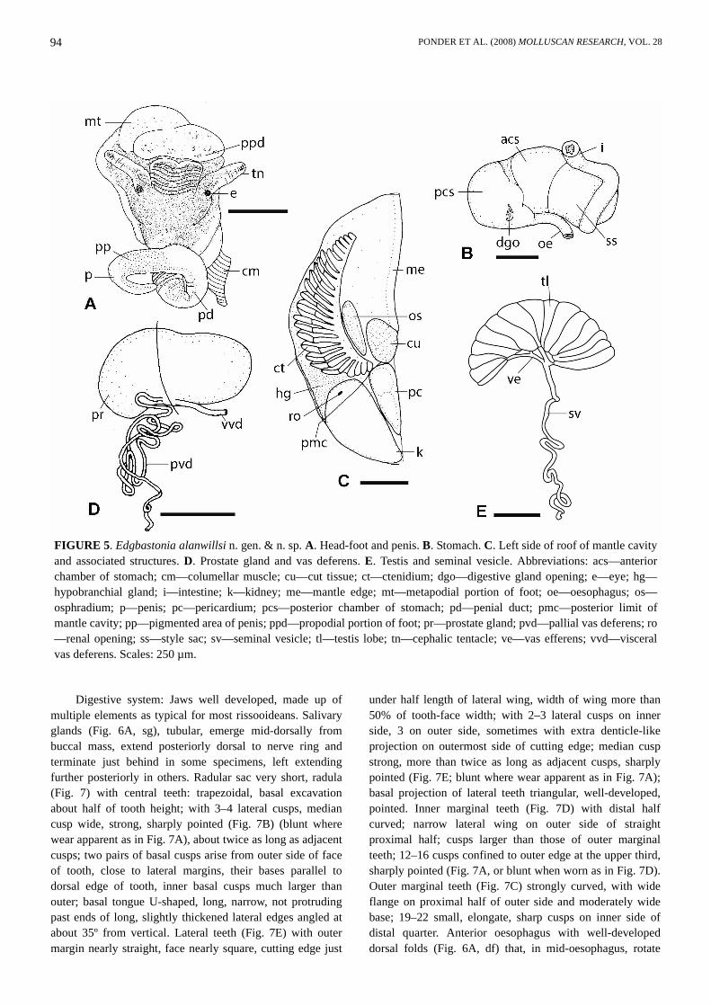
Head-foot (Fig. 5A) with short, bilobed snout; cephalictentacles of moderate length; foot apparently short (allmaterial contracted); snout and dorsal foot (includingopercular lobes) black, neck dark grey, cephalic tentacleswith black to grey dorsal strip, sometimes confined toproximal half to two thirds, and around eyes, otherwiseunpigmented; eyes in weak swellings at outer bases oftentacles; sole largely unpigmented, some black pigment
cells visible anteriorly. Anterior third of foot partiallyconstricted from metapodial area; propodial fold indistinct;anterior mucous gland extends across anterior edge, made upof about 20 short groups of gland cells. Sole with scatteredgrey to black subepithelial pigment cells. Available materialcontracted but sides of foot apparently lacking omniphoricgroove and suprapedal fold. Mantle roof and visceral coilblack, clearly visible through shell.
Mantle cavity (Fig. 5C) with well-developed ctenidium(Fig. 5C, ct) comprising 23–25 broadly-triangular filaments(n=6), apex towards right side. Osphradium (Fig. 5C, os)narrowly-oval, encircled by narrow ciliated ridge; located atmiddle to slightly posterior to middle of ctenidium,ctenidium 2.5–2.9 times longer. Hypobranchial gland (Fig.5C, hg) thick, mainly located in posterior end of mantlecavity between ctenidium and rectum/pallial genital duct.Rectum “straight” (i.e., curved following right side of thepallial genital duct and, in the case of the male, the rightmantle wall), anus opens well back from mantle edge (about0.2–0.3 length of mantle cavity). Kidney and pericardium lieimmediately behind posterior wall of mantle cavity. Renalopening (Fig. 5C, ro) small, simple, on middle of posteriorpallial wall. Dorsal wall of renal organ with thick, densely-staining renal gland.
FIGURE 3. Protoconch and shell of Edgbastonia alanwillsi n. gen. & n. sp. A. Dorsal view of spire, including the protoconch.b, c, d, e indicate regions shown in B-E on this figure. B. Detail of adult shell (area marked b on A). C. Detail of initial part ofprotoconch (marked c on A). D. Detail of last part of protoconch (marked d on A). E. Detail of second part of protoconch(marked e on A). The numbers 1, 2 (with arrows) indicate lines showing the two boundaries of the protoconch; ep – linemarking end of granular sculpture. Scales: A—100 µm; B, D, E—30 µm; C—20 µm.
PONDER ET AL. (2008) MOLLUSCAN RESEARCH, VOL. 2894
Digestive system: Jaws well developed, made up ofmultiple elements as typical for most rissooideans. Salivaryglands (Fig. 6A, sg), tubular, emerge mid-dorsally frombuccal mass, extend posteriorly dorsal to nerve ring andterminate just behind in some specimens, left extendingfurther posteriorly in others. Radular sac very short, radula(Fig. 7) with central teeth: trapezoidal, basal excavationabout half of tooth height; with 3–4 lateral cusps, mediancusp wide, strong, sharply pointed (Fig. 7B) (blunt wherewear apparent as in Fig. 7A), about twice as long as adjacentcusps; two pairs of basal cusps arise from outer side of faceof tooth, close to lateral margins, their bases parallel todorsal edge of tooth, inner basal cusps much larger thanouter; basal tongue U-shaped, long, narrow, not protrudingpast ends of long, slightly thickened lateral edges angled atabout 35º from vertical. Lateral teeth (Fig. 7E) with outermargin nearly straight, face nearly square, cutting edge just
under half length of lateral wing, width of wing more than50% of tooth-face width; with 2–3 lateral cusps on innerside, 3 on outer side, sometimes with extra denticle-likeprojection on outermost side of cutting edge; median cuspstrong, more than twice as long as adjacent cusps, sharplypointed (Fig. 7E; blunt where wear apparent as in Fig. 7A);basal projection of lateral teeth triangular, well-developed,pointed. Inner marginal teeth (Fig. 7D) with distal halfcurved; narrow lateral wing on outer side of straightproximal half; cusps larger than those of outer marginalteeth; 12–16 cusps confined to outer edge at the upper third,sharply pointed (Fig. 7A, or blunt when worn as in Fig. 7D).Outer marginal teeth (Fig. 7C) strongly curved, with wideflange on proximal half of outer side and moderately widebase; 19–22 small, elongate, sharp cusps on inner side ofdistal quarter. Anterior oesophagus with well-developeddorsal folds (Fig. 6A, df) that, in mid-oesophagus, rotate
FIGURE 5. Edgbastonia alanwillsi n. gen. & n. sp. A. Head-foot and penis. B. Stomach. C. Left side of roof of mantle cavityand associated structures. D. Prostate gland and vas deferens. E. Testis and seminal vesicle. Abbreviations: acs—anteriorchamber of stomach; cm—columellar muscle; cu—cut tissue; ct—ctenidium; dgo—digestive gland opening; e—eye; hg—hypobranchial gland; i—intestine; k—kidney; me—mantle edge; mt—metapodial portion of foot; oe—oesophagus; os—osphradium; p—penis; pc—pericardium; pcs—posterior chamber of stomach; pd—penial duct; pmc—posterior limit ofmantle cavity; pp—pigmented area of penis; ppd—propodial portion of foot; pr—prostate gland; pvd—pallial vas deferens; ro—renal opening; ss—style sac; sv—seminal vesicle; tl—testis lobe; tn—cephalic tentacle; ve—vas efferens; vvd—visceralvas deferens. Scales: 250 µm.

EDGBASTONIA ALANWILLSI N. GEN & N. SP. FROM AUSTRALIA 95
ventrally through and behind nerve ring. Initially, ventralpart of mid-oesophagus with thick lining of ciliatedcolumnar cells (Fig. 6A, ve) which may be partly glandular(not an oesophageal gland). Thickened epithelium persiststhrough loop of oesophagus (in contracted state) behindwhich cells shorten, marking beginning of posterioroesophagus. Stomach (Fig. 5B) short, weakly divided intoanterior (acs) and posterior (pcs) chambers; no posteriorcaecum; single opening to digestive gland. Style sac (Fig.5B, ss) slightly shorter than rest of stomach; intestine withinitial vertical portion posterior to anterior end of style sac.
Rectum curved along right pallial genital duct/mantle roof.Genital system: Male: Testis (Fig. 5E) with several
simple lobes emptying to few vasa efferentia (ve) joiningbelow middle of testis as vas deferens; vas deferens formslong, coiled, narrow seminal vesicle (sv). Prostate gland(Fig. 5D) pyriform, narrow anteriorly. Pallial vas deferensnarrow, with several tight coils at anterior end of prostategland; anterior part with undulations and/or one or two smallloops before entering base of penis. Penis (Fig. 5A) large,near middle of head about same distance behind eyes asbetween eyes; non-glandular, long and gradually tapering tosimple distal point (no stylet); unpigmented apart fromdorsal strip of grey in distal half. Penial duct (Figs 5A, 6B,pd) very narrow, with thin muscle layer and very narrowlumen, coiled in basal part, straightening distally.
Female: Ovary (Fig. 8C) composed of a few largelobes, with large yolky eggs. Upper oviduct opens to thick,shining, muscular inverted U-shaped renal oviduct (Figs 8A,9E, ro) which continues anteriorly as S-shaped duct toanterior end of albumen gland where it joins bursal duct.
Gono-pericardial duct not confirmed. Seminal receptacle(Figs 8A, B, D, 9D, sr) ovoid, with short duct, lies on leftside of lower part of bursa, opens to posterior renal oviduct;contains orientated sperm. Bursa copulatrix (Figs 8A, B, D,9D, E, b) ovoid, immediately posterior to albumen gland;epithelium tall columnar cells and unorientated sperm inlumen. Bursal duct (Figs 8A, D, 9D, E, bd) partly embeddedin left side of albumen gland, moderately long, with U-shaped bend in anterior half; opens to ventro-anterior end ofbursa. Common duct (oviduct proximal to junction of bursalduct; Figs 8A, B, D, 9, cd) narrow initially (Figs 8D, 9A),expanding before opening to capsule gland by way of shortventral slit (Figs 8D, 9B, ocg); anterior to this point, ductwider (Figs 8D, 9C) and runs along left ventral wall ofcapsule gland without any further connection. Expandedportion of common duct walls made up of thick layer ofmainly circular muscle fibres; lumen lined with ciliated,columnar epithelium arranged in two major longitudinalridges (Fig. 9A–C) that probably serve to separate passage ofeggs and sperm. Albumen gland (Fig. 8A, B, D, ag) shorterthan capsule gland, cells staining clear to pale blue, dorsal toanterior half of bursa and common oviduct. Capsule gland(Figs 8A, B, D, 9, cg) large, cells staining dense red, abouthalf as high as long, almost entirely within pallial roof;lumen dorso-ventral slit (Fig. 9A–C, lcg), simple grooveventrally except where open to common duct. Ventralmuscular common duct runs anteriorly well beyond anteriorend of capsule gland as free papilla-like ‘vestibule’ (Fig. 8A,B, D, v); opening small, near terminal, ventral (Fig. 8A, D,co).
FIGURE 6. Histological sections showing some details of the anterior gut and penis of Edgbastonia alanwillsi n. gen. & n. sp.A. Anterior oesophagus and nerve ring. B. Penis. Abbreviations: ce—cerebral ganglion; df—dorsal fold in oesophagus; p—penis; pb—base of penis; pd—penial duct; sg—salivary glands; ve—ventral epithelium of oesophagus. Scales: 100 µm.

PONDER ET AL. (2008) MOLLUSCAN RESEARCH, VOL. 2896
Nervous system epiathroid, with cerebral gangliaseparated by long commissure (about third length ofganglion); optic and tentacle nerves arise from dorso-anteriorganglion, latter nerve bifurcates in tentacle; 4 labial nervesalso arise from anterior part of ganglion. Supraoesophagealganglion abuts right pleural ganglion; suboesophagealganglion fused to left pleural, distinguished by constriction.Pedal ganglia with short commissure; no metapodialcommissure. Metapodial ganglia globular, about third lengthof pedal ganglia. Propodial ganglia slender, difficult todistinguish from nerve. Buccal ganglia large, as wide ascerebral and about third of length. Visceral ganglion notobserved. Nerve ring slightly asymmetrical with rightcerebro-pedal connective slightly longer than left. Statocystslocated at anterior end of pedal ganglia; with single statolith.
RemarksThe shape of the shell of the new species somewhat
resembles one of the endemic ‘hydrobiids’, Jardinellaedgbastonensis Ponder & Clark, 1990, found in the springsat Edgbaston Station. Edgbastonia differs in its shell being alittle smaller and narrower, and having a distinctivecolumellar tooth. In addition, the operculum retracts only asfar as the outer part of the aperture in the species currentlyincluded in Jardinella, whereas in Edgbastonia it retracts outof sight deep within the aperture (Fig. 2C), exposing thecolumellar tooth.
The reduced number of protoconch whorls (about 1.1—using the counting method illustrated by Hershler andPonder 1998, fig. 17M) seen in the one suitable specimen ofEdgbastonia (others examined being too eroded) is unusualin ‘Hydrobiidae’. The larval shell is apparently composed oftwo parts with the initial protoconch and a short sectionrepresenting shell accretion during larval development priorto hatching, although these observations requireconfirmation with additional specimens.
FIGURE 7. Radula of Edgbastonia alanwillsi n. gen. & n. sp. A. Six rows of radula. B. Detail of central teeth and cuttingedges of lateral and inner marginal teeth. C. Detail of isolated outer marginal teeth, base of front tooth damaged. D. Detail ofinner marginal tooth. E. Detail of lateral tooth. Scales: A—10 µm, B–E—5 µm.

EDGBASTONIA ALANWILLSI N. GEN & N. SP. FROM AUSTRALIA 97
The new species is uncommon, mostly being found invery small numbers (usually only one or a few even insamples that can contain hundreds of ‘hydrobiids’) and hasbeen found in ten of the 50 active springs at EdgbastonStation. The type locality is only one of four springs in whichmore than five specimens of Edgbastonia have been found.Given its general similarity to one of the most abundant ofthe Edgbaston ‘hydrobiids’, and its rarity, it has not been
observed in the field so its preferred microhabitat isunknown. It is possible that Edgbastonia is amphibious and,while the relatively large ctenidium, well developedhypobranchial gland and reasonably long tentacles allsuggest that it is an aquatic species, these characteristics arealso found in amphibious pomatiopsines (Davis 1967), alongwith the bipartite foot that may be present in Edgbastonia.
FIGURE 8. Female reproductive system of Edgbastonia alanwillsi n. gen. & n. sp. A, B. Female genital system from left andright sides; A, left, B, right. C. Ovary. D, E. Diagrammatic representations of female genital systems of Edgbastonia (D),Jardinella (E) and Amnicola (F). The arrows show the probable passage of eggs (straight arrows) and sperm (wavy arrows)through the system. Structure of E based on Hershler and Thompson (1988, fig. 8). Abbreviations: ag – albumen gland; b –bursa copulatrix; bd—bursal duct; cd—common duct; cg—capsule gland; co—common opening; o—oviduct; ocg—openingof capsule gland to common duct; oo—opening of oviduct to albumen gland; ro—renal oviduct; sd—sperm duct(‘spermathecal duct’); sr—seminal receptacle; v—vestibule. Scales: 250 µm.

PONDER ET AL. (2008) MOLLUSCAN RESEARCH, VOL. 2898
FIGURE 9. Histological sections of the female genital system of Edgbastonia alanwillsi n. gen. & n. sp. A, B, C. Transversesections of the pallial oviduct through the capsule gland; A, posterior part; B, about posterior third; C, anterior part. D, E.Longitudinal sections of posterior female genital system showing bursa copulatrix, common duct and capsule gland.Abbreviations: b—bursa copulatrix; bd—bursal duct; cd—common duct; cg—capsule gland; dg—digestive gland; i—intestine; k—kidney; lcg—lumen of capsule gland; mc—mantle cavity; ocg —opening of capsule gland to common duct; os—oesophagus; r—rectum; ro—renal oviduct; sr—seminal receptacle; st—stomach. Scales: 100 µm.

EDGBASTONIA ALANWILLSI N. GEN & N. SP. FROM AUSTRALIA 99
Molecular resultsThe Bayesian analysis produced the tree shown in
Figure 10, with a littorinid as the outgroup. The freshwaterrissooideans are differentiated into several distinct cladessome of which correspond to family groups (Amnicolidae +Bythinella + Emmericiidae; Hydrobiidae sensu lato(including in our tree Cochliopidae); Moitessieriidae;Pomatiopsidae). The Australasian ‘hydrobiids’ (Tateinae) aresister to Cochliopidae (mainly Americas), while the claderepresenting the restricted concept of Hydrobiidae (whichincludes members of Hydrobiinae, as well as other putativetaxa often treated as subfamilies or, in some cases, families-Pyrgulinae, Pseudamnicolinae, Islamiinae, Horatiinae andNymphophylinae) (Europe + N. America) is sister to theTateinae + Cochliopidae clade. The four Queensland taxaincluded in the analysis form a clade with Edgbastoniarendering Jardinella paraphyletic.
Discussion
Morphological comparisonsBased on morphology, Edgbastonia does not appear to
be closely related to any extant Australian taxon but themolecular results place it with Jardinella (Hydrobiidae s.l.,Tateinae). In shell characters, Edgbastonia resembles severalAsian amnicolids as detailed above. Apart from a species ofErhaia recorded from northern India (Davis and Rao 1997),amnicolids are unknown from Gondwanan fragments. TheIndian taxon is likely derived from China where aconsiderable number of similar taxa are known (e.g., Daviset al. 1992).
Morphologically, Edgbastonia is certainly a member ofthe so called ‘hydrobioid’ group of families which includes,among others, the Hydrobiidae, Amnicolidae andPomatiopsidae. There are few clear-cut morphologicaldifferences between these three families and they are mostlyfocused on the female reproductive system. Molecular datadoes, however, separate them (e.g., Wilke et al. 2001; andmolecular results presented below).
While the molecular data clearly supports a tateinerelationship, the morphological data is not so clear cut.Consequently, we have broadened the discussion below to
encompass remarks on some amnicolid1 and pomatiopsidtaxa in relation to some of the morphological attributes ofEdgbastonia. Any relationship based on shell similarity in‘hydrobioid’ taxa is tenuous at best and, given theinconclusiveness of the morphological data we rely primarilyon the results from the molecular data (see also below), andwe are confident in our conclusion that Edgbastonia is aderived member of Tateinae.
The configuration of the head and foot in Edgbastoniaappears to be similar to that of ‘Hydrobiidae’ and
Amnicolidae except for the apparent subdivision of theanterior part of the foot (which needs to be confirmed inliving material). If this observation is correct, it is not theplesiomorphic condition for that group of family-group taxa(‘Hydrobioidea’) and is a condition otherwise seen in theamphibious Pomatiopsinae and Truncatellidae. The aquatictriculine pomatiopsids, hydrobiids and amnicolids includingerhaiines, have a simple foot.
The male reproductive system is unusual in having atightly coiled pallial vas deferens, a feature also seen in a fewspecies of Jardinella from Queensland artesian springs(Ponder and Clark 1990, fig. 4E) as well as a few otherTateinae. The penis of Edgbastonia is simple and resemblesthose of the majority of Tateinae as well as many other‘hydrobiids’.
In particular, it is the female genital system ofEdgbastonia that is unique in that it has an expanded,muscular duct that opens to a short portion of the anteriorregion of the capsule gland and then continues as a separate,expanded, muscular, vestibule-like combined egg + spermduct anteriorly. The pallial oviduct of Edgbastonia could bederived from a normal ‘hydrobiid’ system by the ventralchannel of the capsule gland closing over along most of itslength.
The female system of the Asian amnicolid Erhaia (andrelated taxa) is generally similar in its configuration to‘Hydrobiidae’, it differs in lacking a ventral channel and inhaving a narrow ‘spermathecal duct’ which lies below thecapsule gland and opens anteriorly separately from thefemale opening (Davis et al. 1985, 1992).
It has been hypothesised that the sperm tube(‘spermathecal duct’) is derived from the closure of thesperm groove in the ventral channel and has developedindependently in several rissooidean families (Ponder 1988;Hershler and Ponder 1998; Wilke et al. 2001), includingPomatiopsidae and Amnicolidae. The sperm ducts(‘spermathecal ducts’) of amnicolids and pomatiopsinesdiffer markedly from the condition seen in Edgbastonia inbeing narrow and separate from the capsule gland and, ifthey join with the oviduct anteriorly, it is with a smallvestibular region and not the capsule gland. Davis (1979)regarded the spermathecal duct as a pomatiopsine character
and led to the placement of Erhaia 2 in the Pomatiopsinae byDavis et al. (1985). However, while arguing for apomatiopsine relationship, Davis et al. (1985: 69–70) alsostated that the “classification is debatable because
1. Our concept of the composition of Amnicolidae followsWilke et al. (2000, 2001), and includes Erhaia Davis &Kuo, 1985 (and related taxa) previously placed in thePomatiopsidae.
2. In analyses of 16S, 18S and COI sequences, Wilke et al.(2000, 2001) showed that Erhaia was a member of a cladewith Amnicola Gould & Halderman, 1840 (USA) and Mar-stoniopsis van Regteren Altena, 1936 (Europe), withBythinella Moquin-Tandon, 1855 (Europe) a likely sister tothat clade. The family-group name Erhaiini Davis & Kuo,1985 (=Pseudobythinellini Davis & Chen, 1992—typegenus Pseudobythinella Lu & Zhang, 1979, a junior hom-onym of Pseudobythinella Melville, 1956—see Kabat andHershler 1993 for discussion) is available for the Asiantaxa if subdivision of the family is necessary.

PONDER ET AL. (2008) MOLLUSCAN RESEARCH, VOL. 28100
phenetically, Erhaia closely resembles Bythinella” and “isclosely linked to the Amnicolidae”. These authors rejected arelationship of Erhaia with Bythinella and Amnicola largelybecause of the simple penis (amnicolids as then recognisedhad an accessory penial appendage and duct). A penialappendage with associated duct and gland is found inAmnicola (Hershler and Thompson 1988) and Marstoniopsis(e.g., Falniowski 1987) as well as Bythinella. While agreeingthat Amnicolidae is an appropriate location for Erhaia andrelated taxa, given the penial differences, recognition of themas a separate subgroup (Erhaiini) may be warranted. It isworthy of note that a very similar penial configuration to thatseen in Amnicola also occurs in Bithyniidae which isunrelated to Amnicolidae in all but the COI tree in Wilke etal. (2001). A recently described bithyniid genus,Pseudobithynia Gloeer & Pesic, 2006, has a simple penis(Gloeer and Pesic 2006).
To derive an amnicolid (Fig. 8E) or pomatiopsid femalesystem from a common ancestor with Edgbastonia, thesperm tube would have to be severed from the capsule gland,for which a separate anterior opening would be needed, andthe connection of the oviduct with the glandular oviductwould have to be moved a little posteriorly to the albumengland. The probable passage of eggs and sperm in bothJardinella, Edgbastonia and Amnicola is shown in Figure8D-F. Both are physically separated in Amnicola (Fig. 8F)and are functionally separated in Jardinella (and other‘hydrobiids’) by folds in separating the ventral channel fromthe lumen of the capsule gland. A similar functionalseparation is achieved in Edgbastonia by the pair of largelongitudinal ridges that effectively divide the common ductinto a dorsal and ventral channel (Fig. 9B, C). These ridgesare presumably the homologues of the folds of the ventralchannel of ‘hydrobiids’. We hypothesize that the conditionseen in Edgbastonia arose from an ancestral conditionsimilar to (but presumably convergent with) that seen insome Jardinella species such as J. colmani Ponder & Clark,1990 (Ponder and Clark 1990, fig. 5D) where the anteriorvestibule is separated from the anterior part of the capsulegland. The “vestibule” in some taxa is very large (e.g., J.corrugata Ponder & Clark, 1990 (Ponder and Clark 1990,fig. 12C) and J. jesswiseae Ponder & Clark, 1990; Ponderand Clark 1990, fig. 22C) but is in fact an expanded ventralchannel which remains open to the capsule gland over all butthe anterior-most part (Ponder and Clark 1990, fig. 5E).
While there is currently no evidence that there are anyAustralasian amnicolid taxa, Climo (1977) placed two NewZealand ‘hydrobiids’ that he had originally (Climo 1974)included in Kuschelita Climo, 1974, in the Japanese
amnicolid genus Saganoa Kuroda, Habe & Tamu, 19581, adecision that he argued was justified because of their similarshells, radula, operculum and penis, as well as their phreatichabit. However, it is difficult to see how this conclusion wasreached as only details of the radula, opercula and head-footof the Japanese taxa have been published (Kuroda and Habe1958). Haase (2008) provided some details of the femalegenital system of Kuschelita, and noted the absence of a
seminal receptacle, bursa copulatrix and ventral channel butdid not give any details of the configuration of the lumen ofthe capsule gland or specifically reject the possibility of asperm tube. Unfortunately, the three specimens available tous for sectioning [NMNZ M174041 (2), M32474 (1)] wereall males. The morphology of the central tooth of the radula(Climo 1974, fig 15E) is similar to that of Erhaia and relatedtaxa although there is only one basal denticle. Climo (1974)reported that the ctenidium and osphradium were lacking inKuschelita which he argued was highly modified for itssubterranean habitat. Haase (2008) noted that K. mica lackeda ctenidium but had a well developed osphradium andhypobranchial gland and this was confirmed in our sectionedmales. Clearly, determination of the relationships ofKuschelita must await molecular data and further anatomicalinformation.
Molecular analysisThe tree presented in Figure 10 differs from that
produced by the maximum likelihood analysis of Wilke et al.(2001, fig. 3) using COI and 18S sequences. Their tree hadCochliopidae and Amnicolidae + Bythinella as sister taxaand Moitessieriidae + Bithyniidae + Hydrobiidae formed atricotomy. Australasian ‘hydrobiids’ were not included in theWilke et al. (2001) analysis. It should be noted, however, thatboth in the analysis of Wilke et al. (2001) and in the presentstudy, deeper nodes are not well supported resulting inuncertainties relative to the relationships among higher taxawithin the Rissooidea. Therefore, the resolution of the‘hydrobioid’ family-level taxa will be subject to a muchmore comprehensive analysis (Wilke et al. in prep.). In themeantime, the result of our analysis raises the issue of thefamily status of the Australasian ‘hydrobiids’ referred toTateinae herein. One resolution would be to treat thesubclade comprising the more restricted concept ofHydrobiidae, together with the subclade Tateinae +Cochliopidae, as a very broad concept of Hydrobiidae (i.e.,as a family-rank taxon), with the subclades given subfamilialrank (i.e., Tateinae and Cochliopinae). Alternatively, on thebasis of our tree, and because Cochliopidae is now generally
1. Liu et al. (1982) included a Chinese species in the Japa-nese Akiyoshia Kuroda & Habe, 1954 (in the subgenusSaganoa) and this placement was tentatively maintained byDavis et al. (1992, 1994) who pointed out that the Chinesetaxon at least is related to Erhaia. Information on the anat-omy of the Japanese taxa has not yet been published so thatthis relationship remains untested. Wilke et al. 2000 (p.455) noted that Moria Kuroda & Habe, 1958, another Japa-nese taxon, has a penis like that of Bythinella and Amnicolaand this observation has been confirmed by one of us (HF),but details have not been published. Moria falls in Amni-colidae in our molecular analysis. Davis and Rao (1997)created a new genus, Chencuia Davis, 1997 for the ChineseBythinella chinensis Liu & Zhang, 1979, a species previ-ously included in Erhaia by Davis et al. (1985) anddescribed in full by Davis et al. (1992) as Pseudo-bythinella. As noted above, all Erhaiine taxa were trans-ferred to Amnicolidae on the basis of molecular evidence.

EDGBASTONIA ALANWILLSI N. GEN & N. SP. FROM AUSTRALIA 101
regarded as a distinct family group, Tateinae could be raisedto family level. However, given the continuing confusionsurrounding the familial status of these taxa, the lack ofinclusion of some key taxa in our analysis, and the lack ofany clear-cut distinguishing morphological synapomorphies,we prefer to adopt a conservative approach and continue totreat Tateinae as a subfamilial group within a broad conceptof Hydrobiidae pending the results of the morecomprehensive analysis referred to above. By adopting this
approach, we are not suggesting that the rank ofCochliopidae should be affected in the meantime.
The four Queensland taxa included in the analysis forma clade with Edgbastonia rendering Jardinella paraphyletic.This result is not surprising given the considerablemorphological (Ponder and Clark 1990; Ponder 1991) andmolecular (Perez et al. 2005) diversity within Jardinella. Amajor re-evaluation of the genus is currently in progress.
FIGURE 10. Bayesian tree based on three gene fragments for representatives of selected rissooidean families. As outgrouptaxon, Littorina obtusata (family Littorinidae) was used as the outgroup taxon. Note the secure position of Edgbastoniaalanwillsi n. gen. & n. sp. within the subfamily Tateinae. Bayesian posterior probabilities (when ≥ 0.95) are provided belowthe nodes. The scale bar indicates the expected number of substitutions per site according to the model of sequence evolutionapplied.

PONDER ET AL. (2008) MOLLUSCAN RESEARCH, VOL. 28102
General discussionThe antiquity of the various ‘hydrobioid’ taxa is still an
open question. Members of Tateinae occur in Australia, NewZealand, New Guinea, New Caledonia and a few Pacificislands including Fiji (e.g., Haase et al. 2006), suggestive ofan original eastern Gondwana distribution. Davis (1979,1981) postulated that the Pomatiopsidae divided into twomain groups (Pomatiopsinae and Triculinae) about 150 mya(in the Jurassic) prior to the break-up of Gondwana, whilethe Amnicolidae (as formulated in Wilke et al. 2001) hasmembers in Europe, North America and Asia.
Whether Edgbastonia is a relictual or recently highlyderived taxon is a question that can only be answered withthe analysis of more Australasian taxa. The existence of arelictual species in the artesian springs of the AustralianGreat Artesian Basin (GAB) would, however, be of no greatsurprise as some other examples have been documented inCrustacea (e.g., Wilson and Keable 2004) andPlatyhelminthes (e.g., Sluys 1986). These taxa have beenable to survive in permanent spring habitats during the onsetof aridity that occurred during the late Pliocene andcontinued periodically through the Pleistocene. This aridityresulted in widespread extinctions of aquatic biotapresumably occurred in what is now arid and semi-aridAustralia, including western Queensland (e.g., DeDeckker1986). The group of springs on Edgbaston Station in whichEdgbastonia occurs has several other endemic animals andplants, including eight other gastropods (Ponder 2004) and isundoubtedly the most significant spring group inQueensland. These springs form a subgroup of springswithin the Barcaldine Supergroup located in westernQueensland along with a further seven spring supergroups(Fensham and Fairfax 2003). Many of the Queenslandsprings have become extinct or severely degraded in the last100 years—mainly due to drawdown resulting from waterextraction for pastoral and other purposes from the GAB(Ponder 1986, 2004; Fairfax and Fensham 2003; Fenshamand Fairfax 2003). Currently only two of the springs onEdgbaston Station are protected by fences which wereerected in an attempt to protect endemic fish (Wager 1995).
Acknowledgements
We thank the Department of Environment and Heritage (nowDept of Environment and Water Resources) and theAustralian Biological Resources Study (ABRS) for fundingthat assisted in supporting this study. Elizabeth Jefferyssorted many of the samples containing the new species andRod Fensham and Russell Fairfax assisted with some of thefield work. R. Wager and P. Unmack provided some earlysamples from Edgbaston Springs. We particularly thank Alanand Fay Wills for their help and allowing access to thesprings on Edgbaston Station on a number of occasions. Wealso thank two reviewers for their useful comments.
References
Bouchet, P. & Rocroi, J.-P. (eds.) (2005) A nomenclator andclassification of gastropod family-group names. Withclassification by J. Frýda, J., B. Hausdorf, W. Ponder, A. Valdesand A. Warén. Malacologia 47, 1–397.
Climo, F.M. (1974) Description and affinity of the subterraneanmolluscan fauna of New Zealand. New Zealand Journal ofZoology 1, 247–284.
Climo, F.M. (1977) Notes on the New Zealand hydrobiid fauna(Mollusca: Gastropoda: Hydrobiidae). Journal of the RoyalSociety of New Zealand 7, 67–77.
Davis, G.M. (1967) The systematic relationship of Pomatiopsislapidaria and Oncomelania hupensis formosana(Prosobranchia: Hydrobiidae). Malacologia 6, 1–143.
Davis, G.M. (1979) The origin and evolution of the gastropodfamily Pomatiopsidae with emphasis on the Mekong RiverTriculinae. Monographs of the Academy of Natural Sciences ofPhiladelphia 20, 1–120.
Davis, G.M. (1981) Different modes of evolution and adaptiveradiation in the Pomatiopsidae (Prosobranchia:Mesogastropoda). Malacologia 21, 209–262.
Davis, G.M., Chen, C.-E., Chun, W., Kuang, T.-F., Xing, X.-G., LiL., Liu, W.-J.& Yan, Y.-L. (1992) The Pomatiopsidae of Hunan,China (Gastropoda: Rissoacea). Malacologia 34, 143–342.
Davis, G.M., Chen, C.-E., Kang, Z.-B. & Liu, Y.-Y. (1994) Snailhosts of Paragonimus in Asia and the Americas. Biomedicaland Environmental Sciences 7, 369–382.
Davis, G.M. & Kang, Z.-B. (1990) The genus Wuconchona of China(Gastropoda: Pomatiopsidae: Triculinae): anatomy, systematics,cladistics, and transmission of Schistosoma. Proceedings of theAcademy of Natural Sciences of Philadelphia 142, 119–142.
Davis, G.M. & Kang, Z.-B. (1995) Advances in the systematics ofErhaia (Gastropoda: Pomatiopsidae) from the People's Republicof China. Proceedings of the Academy of Natural Sciences ofPhiladelphia 146, 391–427.
Davis, G.M., Kuo, Y.H., Hoagland, K.E., Chen, P.-L., Yang, H.-M.& Chen, D.-J. (1985) Erhaia, a new genus and new species ofPomatiopsidae from China (Gastropoda: Rissoacea).Proceedings of the Academy of Natural Sciences ofPhiladelphia 137, 48–78.
Davis, G.M. & Rao, S. (1997) Discovery of Erhaia (Gastropoda:Pomatiopsidae) in northern India with description of a newgenus of Erhaiini from China. Proceedings of the Academy ofNatural Sciences of Philadelphia 148, 273–299.
DeDeckker, P. (1986) What happened to the Australian biota 18 000years ago? In: De Deckker, P. & Williams, W. D. (Eds.),Limnology in Australia. CSIRO, Melbourne and Dr W. JunkPublishers, Dortrecht, pp. 487–496.
Fairfax, R.J. & Fensham, R. J. (2003) Great Artesian Basin springsin southern Queensland 1911–2000. Memoirs of the QueenslandMuseum 49, 285–293.
Falniowski, A. (1987) Hydrobioidea of Poland (Prosobranchia:Gastropoda). Folia Malacologica 1, 1–122.
Fensham, R.J. & Fairfax, R.J. (2003) Spring wetlands of the GreatArtesian Basin, Queensland, Australia. Wetland Ecology andManagement 11, 343–362.
Folmer, O.M., Black, W., Hoeh, R., Lutz, R. & Vrijenhoek, R.(1994) DNA primers for amplification of mitochondrialcytochrome c oxidase subunit I from diverse metazoaninvertebrates. Molecular Marine Biology and Biotechnology 3,294–299.
Gloeer, P. & Pesic, V. (2006) On the identity of Bithynia graecaWesterlund, 1879 with the description of three newPseudobithynia n. gen. species from Iran and Greece (Gastro-poda : Bithyniidae). Malakologische Abhandlungen 24, 29–36.
Haase, M. (2005) Rapid and convergent evolution of parental carein hydrobiid gastropods from New Zealand. Journal of

EDGBASTONIA ALANWILLSI N. GEN & N. SP. FROM AUSTRALIA 103
Evolutionary Biology 18, 1076–1086.Haase, M. (2008) The radiation of hydrobiid gastropods in New
Zealand: a revision including the description of new speciesbased on morphology and mtDNA sequence information.Systematics and Biodiversity 6, 99–159.
Haase, M., Ponder, W.F. & Bouchet, P. (2006) The genusFluviopupa Pilsbry, 1911 from Fiji (Caenogastropoda,Rissooidea). Journal of Molluscan Studies 72, 119–136.
Hall, T.A. (1999) BioEdit: a user-friendly biological sequencealignment editor and analysis program for Windows 95/98/NT.Nucleic Acids Symposium Series 41, 95–98.
Hershler, R. & Ponder, W.F. (1998) A review of morphologicalcharacters of hydrobioid snails. Smithsonian Contributions toZoology 600, 1–55.
Hershler R. & Thompson F.G. (1988) Notes on morphology ofAmnicola limosa (Say, 1817) (Gastropoda: Hydrobiidae) withcomments on status of the subfamily Amnicolinae.Malacological Review 21, 81–92.
Holland, P.W.H., Hacker, A.M., & Williams, N.A. (1991) Amolecular analysis of the phylogenetic affinities of Saccoglossucambrensis Brambell & Cole (Hemichordata). PhilosophicalTransactions of the Royal Society of London B, 332, 185–189.
Ioganzen, B.G. & Starobogatov, Ya. I. (1982) O nakhodke v Sibiripresnovodnogo molliuska semeistva Triculidae (Gastropoda,Prosobranchia). Zoologicheskii Zhurnal 61, 1141–1147.
Iredale, T. & McMichael, D.F. (1962) A reference list of the marineMollusca of New South Wales. Australian Museum Memoir 11,185 pp.
Kabat, A.R. & Hershler, R. (1993) The prosobranch snail familyHydrobiidae (Gastropoda: Rissooidea): Review of classificationand supraspecific taxa. Smithsonian Contributions to Zoology547, 1–94.
Kuroda, T. & Habe, T. (1958) Troglobiontic aquatic snails fromJapan. Venus 19, 183–196.
Liu, H.-P., Hershler, R. & Thompson, F.G. (2001) Phylogeneticrelationships of the Cochliopinae (Rissooidea: Hydrobiidae): anenigmatic group of aquatic gastropods. MolecularPhylogenetics and Evolution 21, 17–25.
Liu, Y.-Y., Zhang, W.-Z., Wang, Y.-X., Chen, C.-E. & Chen, S.-Z.(1982) Discovery of Akiyoshia Kuroda et Habe (Hydrobiidae:Mollusca) from China with descriptions of two new species.Acta Zootaxonomica Sinica 7, 364–367.
Palumbi, S., Martin, A., Romano, S., McMillian, W.O., Stice, L. &Grabowski, G. (1991) The simple fool's guide to PCR.University of Hawaii, Honolulu.
Perez, K.E., Ponder, W.F., Colgan, D.J., Clark, S.A. & Lydeard, C.(2005) Molecular phylogeny and biogeography of spring-associated hydrobiid snails of the Great Artesian Basin,Australia. Molecular Phylogenetics and Evolution 34, 545–556.
Ponder, W.F. (1982) Hydrobiidae of Lord Howe Island (Mollusca:Gastropoda: Prosobranchia). Australian Journal of Marine andFreshwater Research 33, 89–159.
Ponder, W.F. (1986) Mound springs of the great artesian basin. In:De Deckker, P. & Williams, W.D. (Eds.), Limnology inAustralia. CSIRO, Melbourne and Dr W. Junk Publishers,Dortrecht, pp. 403–420.
Ponder, W.F. (1988) The trucatelloidean (=Rissoacean) radiation – apreliminary phylogeny. In: Ponder, W.F. (Ed.) Prosobranchphylogeny. Malacological Review, supplement 4, pp 129–166.
Ponder, W.F. (1991) The eastern seaboard species of Jardinella(Mollusca, Gastropoda, Hydrobiidae), Queensland rainforest-inhabiting freshwater snails derived from the west. Records ofthe Australian Museum 43, 275–289.
Ponder, W.F. (2004) Endemic aquatic macroinvertebrates ofartesian springs of the Great Artesian Basin – progress andfuture directions. Records of the South Australian Museum 7,101–110.
Ponder, W.F. & Clark, G.A. (1990) A radiation of hydrobiid snailsin threatened artesian springs in Western Queensland. Recordsof the Australian Museum 42, 301–363.
Ponder, W.F., Clark, G.A., Miller, A. & Toluzzi, A. (1993) On amajor radiation of freshwater snails in Tasmania and easternVictoria - a preliminary overview of the Beddomeia group(Mollusca: Gastropoda: Hydrobiidae). Invertebrate Taxonomy7, 501–750.
Ponder, W.F., Colgan, D.J. & Clark, G.A. (1991) The morphology,taxonomy and genetic structure of Tatea (Mollusca:Gastropoda: Hydrobiidae), estuarine snails from temperateAustralia. Australian Journal of Zoology 39, 447–497.
Ponder, W.F., Hershler, R. & Jenkins, B. (1989) An endemicradiation of Hydrobiidae from artesian springs in northernSouth Australia: their taxonomy, physiology, distribution andanatomy. Malacologia 31, 1–140.
Ponder, W.F. & Warén, A. (1988) Classification of theCaenogastropoda and Heterostropha - a list of the family-groupnames and higher taxa. In: Ponder, W.F. (Ed.) ProsobranchPhylogeny. Malacological Review, Supplement 4, pp. 288–328.
Posada, D. & Crandall, K.A. (1998) Modeltest: testing the model ofDNA substitution. Bioinformatics 14, 817–818.
Reid, D.G., Rumbak, E. & Thomas, R.H. (1996) DNA, morphologyand fossils: phylogeny and evolutionary rates of the gastropodgenus Littorina. Philosophical Transactions of the RoyalSociety of London, Series B: Biological Sciences 351, 877–895.
Ronquist, F. & Huelsenbeck, J.P. (2003) MrBayes 3: Bayesianphylogenetic inference under mixed models. Bioinformatics 19,1572–1574.
Sluys, R. (1986) First representative of the Order Macrostomida inAustralia (Platyhelminthes, Macrostomidae). Records of theSouth Australian Museum 19, 400–404.
Thiele, J. (1925) Handbuch der Zoologie 5(1) De Gruyter, Berlin &Leipzig.
Thiele, J. (1929–1931) Handbuch der SystematischenWeichtierkunde. Vol. 1. Gustav Fischer Verlag, Jena.
Thompson, F. G. (1968) The aquatic snails of the familyHydrobiidae of Peninsular Florida. University of Florida Press,Gainsville.
Thompson, J.D., Gibson, T.J., Plewniak, F., Jeanmougin, F. &Higgins, D.G. (1997) The CLUSTAL X windows interface:flexible strategies for multiple sequence alignment aided byquality analysis tools. Nucleic Acids Research 24, 4876–4882.
Wager, R. (1995) Recovery plan for Queensland artesian springfishes. Project No. 417. Australian Nature ConservationAgency: Canberra.
Wilke, T., Davis, G.M., Falniowski, A., Giusti, F., Bodon, M. &Szarowska, M. (2001) Molecular systematics of Hydrobiidae(Mollusca: Gastropoda: Rissooidea): Testing monophyly andphylogenetic relationships. Proceedings of the Academy ofNatural Sciences of Philadelphia 151, 1–21.
Wilke, T., Davis, G.M., Gong, X. & Liu, H.-X. (2000) Erhaia(Gastropoda: Rissooidea): Phylogenetic relationships and thequestion of Paragonimus coevolution in Asia. AmericanJournal of Tropical Medicine and Hygiene 62, 453–459.
Wilke, T., Davis, G.M., Qiu, D. & Spear, R.C. (2006) Extrememitochondrial sequence diversity in the intermediateschistosomiasis host Oncomelania hupensis robertsoni: anothercase of ancestral polymorphism? Malacologia 48, 143–157.
Williams, S.T. & Reid, D.G. (2004) Speciation and diversity ontropical rocky shores: a global phylogeny of snails of the genusEchinolittorina. Evolution 58, 2227–2251.
Wilson, G.D.F. & Keable, S.J. (2004) A new family and genus ofPhreatoicidea (Crustacea, Isopoda) from artesian springs insouthwestern Queensland, Australia. Memoirs of theQueensland Museum 49, 741–759.

PONDER ET AL. (2008) MOLLUSCAN RESEARCH, VOL. 28104
Appendix. Family assignment, locality information, and GenBank accession numbers for the taxa studied. GenBank sequences that weregenerated by workers not belonging to our groups are marked with asterisks (* Liu et al. 2001; ** Haase 2005; *** Williamsand Reid 2004; **** Reid et al. 1996).
GenBank #
Family/subfamily Species Location COI 16S 18S
Littorinidae (outgroup)
Littorina obtusata (Linna-eus, 1758)
United Kingdom, Isles of Scilly (COI, 18S); United Kingdom, Pembroke Dock (16S)
AJ622947*** U46812**** AJ488715***
Amnicolidae Amnicola limosa (Say, 1817)
USA, Michigan, Washtenaw County, Blind Lake
AF213348 AF212903 AF212916
Erhaia jianouensis (Liu and Zhang, 1979)
China, Fujian, Nanping, Tianxi AF367652 EU573984 AF367688
Marstoniopsis insubrica (Küster, 1853)
Germany, Rostock, Warnow River near the feeder of the Rostock waterworks
AF322408 AY341257 AF367676
Moria kikuchii (Habe, 1961)
Japan, Fukuoka-ken, Tagawa-gun, Soeda-machi, Mt Hikosan, Hiko-san Shrine
AF213350 AF212905 AF2122918
Antroselates spiralis Hubricht, 1963
USA, Harrison Cave Spring, Harrison Co.
AF354758* - -
‘Bythinellidae’ Bythinella austriaca (Frauenfeld, 1856)
Austria, Steiermark, National Park “Kalkalpen”
AF213349 AF212904 AF212917
Bythinella pannonica (Frauenfeld, 1865)
Slovakia, Hrhov, Slovensky Kras AY222650 AY222660 EU573994
Emmericiidae Emmericia expansilabris Bourguignat, 1880
Croatia, Izvor River - EU573985 EU573995
Hydrobiidae Cincinnatia winkleyi (Pil-sbry, 1912)
USA, Maine, Cumberland County, Spurwink River
AF118370 AF212901 AF212914
Mercuria similis (Drapar-naud, 1805)
Italy, Friuli-Venetia-Julia, Udine, Aquileia, Canale Panigai near Panigai
AF367646 AF478393 AF367682
Pseudamnicola lucensis (Issel, 1866)
Italy, Tuscany, Lucca, Bagni di Lucca, Bagni Caldi, thermal spring
AF367651 AF478394 AF367687
Adrioinsulana conovula (Frauenfeld, 1863)
Croatia, Pag Island, Zubovici AF367628 EU573986 AF367656
Dianella thiesseana (Kobelt, 1878)
Greece, Lake Trichonida at Lou-tres Mirtias
AY676127 AY676121 AY676125
Pyrgula annulata (Lin-naeus, 1767)
Italy, Brescia, Lake Garda, Desen-zano del Garda
AY341258 AY676122 AY676124
Adriohydrobia gagati-nella (Küster, 1852)
Croatia, Krka River near Skradin AF317857 EU573987 AF367657
Hydrobia acuta (Drapar-naud, 1805)
France, Hérault, Etang du Prévost AF278808 AY222659 AF367680
Ventrosia ventrosa (Mon-tagu, 1803)
United Kingdom, Norfolk, The Wash, Snettisham lagoon RSPB bird reserve
AF118335 AF478402 AF367681
Avenionia brevis beren-guieri (Bourguignat, 1882)
France, Gard, spring of the fountain of St.-Victor-La Coste
AF367638 - AF367670
Fissuria boui Boeters, 1981
France, Alpes Maritimes, Peymei-nade, spring near La Prouveresse
AF367654 - AF367690
.....continued

EDGBASTONIA ALANWILLSI N. GEN & N. SP. FROM AUSTRALIA 105
APPENDIX (continued)
GenBank #
Family/subfamily Species Location COI 16S 18S
Islamia piristoma Bodon and Cianfanelli, 2001
Italy, Liguria, La Spezia, Arcola, spring at Magra River
AF367639 - AF367671
Graziana alpestris (Frauenfeld, 1863)
Italy, Liguria, Savona, Molino, spring at the Porra River
AF367641 AY676123 AF367673
Hauffenia tellinii (Pollo-nera, 1898)
Italy, Friuli-Venetia Julia, Gorizia, Isonzo River near Sagrado, spring
AF367640 EU573988 AF367672
Horatia klecakiana Bourguignat, 1887
Croatia, spring of Vrana River, between Vrana and Radosinovci
AF367637 AY222656 AF367669
Orientalina callosa (Paulucci, 1881)
Italy, Abruzzo, Pescara, Carama-nico Terme
AF367649 - AF367685
Sadleriana fluminensis (Küster, 1853)
Slovenia, Mocilnik near Vrhnika, main spring of Ljubljanica River
AY273996 AY222657 EU573996
Tateinae Edgbastonia alanwillsi n.sp.
Australia, Queensland, Edgbaston Stn (AMS, C. 457896)
EU580440 EU580438 EU580439
Potamopyrgus antipoda-rum (Gray, 1843)
Great Britain, London, West India Dock
EU573983 EU573989 EU573997
Potamopyrgus oppidanus Haase, 2008
New Zealand, Wadestown AY631112** AY634090** -
Jardinella tumorosa Ponder, 1991
Australia, Queensland, Little Mul-grave River
AY622455 AY622385 -
Jardinella jesswiseae Ponder & Clark, 1990
Australia, Queensland, Edgbaston Stn, Big Spring
AY622437 - -
Jardinella pallida Ponder & Clark, 1990
Australia, Queensland, Edgbaston Stn, Big Spring (AMS, C.400140) and for 18S spring SE 20 (AMS, C.458211)
AY622436 AY622418 EU580437
Fonscochlea zeidleri Pon-der et al., 1989
Australia, South Australia, Stran-gways Springs
AY622460 AY622390 -
Trochidrobia punicea Pon-der et al., 1989
Australia, South Australia, Finniss Springs West
AY622459 AY622389 -
Leptopyrgus manneringi Climo, 1974
New Zealand, Waikaretu AY622389** AY634052** -
Meridiopyrgus murihiku Haase, 2008
New Zealand, Dunedin AY631083** AY634061** -
Opacuincola delira Haase, 2008
New Zealand, Crazy Paving Cave AY634068** AY631090** -
Sororipyrgus kutukutu Haase, 2008
New Zealand, N of Awakino Gorge
AY631109** AY634087** -
Cochliopidae Heleobops carrikeri Davis and McKee, 1989
USA, Maryland, Dorchester Co., Little Choptank River at the end of Ragged Point Road
AF213347 AF212902 AF212915
Onobops jacksoni (Bartsch, 1953)
USA, Maryland, Dorchester Co., Town Point at the end of Town Point Road
AF367645 EU573990 AF367678
Spurwinkia salsa (Pilsbry, 1905)
USA, Maryland, Dorchester Co., Town Point at the end of Town Point Road
AF367633 EU573991 AF367663
Moitessieridae Moitessieria cf. puteana Coutagne, 1883
France, Alpes Maritimes, Peymei-nade, spring near La Prouveresse
AF367635 EU573992 AF367665
.....continued

PONDER ET AL. (2008) MOLLUSCAN RESEARCH, VOL. 28106
APPENDIX (continued)
GenBank #
Family/subfamily Species Location COI 16S 18S
Bythiospeum cf. diapha-num (Michaud 1831)
France, Gard, Lirac, Source de la Nizon
AF367634 - AF367664
Pomatiopsidae Pomatiopsis lapidaria (Say, 1817)
USA, Michigan, Washtenaw County, Bridgewater Township, near Raisin River at Allen Road
AF367636 AY676118 AF367666
Oncomelania h. hupensis Gredler, 1881
China, Anhui, Dalin AF254547 DQ212859 AF367667
AF254547 DQ212859 AF367667
Tricula sp. China, Sichuan, Chengdu, Huang Ba
AF253071 AF212895 AF411141
Gammatricula chinensis Davis, Liu and Chen, 1990
China, Zhejiang Province, Kaiwa Co, Tong Cun Town, Bai Keng Village
AF253067 EU573993 AF367668