MolecularAnalysis ofthe P2 Haemophilus - Infection...
8
Vol. 60, No. 12 INFECTION AND IMMUNITY, Dec. 1992, p. 5204-5211 0019-9567/92/125204-08$02.00/0 Copyright C 1992, American Society for Microbiology Molecular Analysis of the P2 Porin Protein of Nontypeable Haemophilus influenzae DANIEL J. SIKKEMA* AND TIMOTHY F. MURPHY Division of Infectious Diseases, Department of Medicine, and Department of Microbiology, State University of New York at Buffalo, and Veterans Affairs Medical Center, Medical Research 151, 3495 Bailey Avenue, Buffalo, New York 14215 Received 29 June 1992/Accepted 8 September 1992 The P2 porin protein is the most abundant outer membrane protein (OMP) of nontypeable Haemophilus influenzae (NTHI) and shows extensive antigenic heterogeneity among strains. To study the molecular basis of this heterogeneity, the DNA sequences of the genes encoding the P2 proteins of three unrelated strains of NTHI were determined, and restriction fragment length polymorphisms around the P2 genes of 35 strains were analyzed. The deduced amino acid sequences of the P2 genes from the three strains of NTHI revealed four major (12 to 35 amino acids long) and several smaller (2 to 7 amino acids) hypervariable regions in each protein. The major variations occurred in identical portions of the genes, and these regions showed a high antigenic index and surface exposure probability in computer modeling analysis. Differences in the molecular mass of the P2 protein correlate with differences in the size of the variable region in each strain. Oligonucleotide primers suitable for amplification of the P2 genes by polymerase chain reaction were developed. Restriction fragment length polymorphism analysis showed marked heterogeneity in and around the ompP2 locus of 35 NTHI strains. These results contrast with the high degree of conservation of the P2 genes in H. influenzae type b strains. We conclude that the molecular mass and antigenic heterogeneity of the P2 molecule of NTHI is due to variations in gene sequence that are clustered primarily in four large hypervariable regions of the gene. Nontypeable Haemophilus influenzae (NTHI) strains are an important cause of infection in patients with chronic obstructive pulmonary disease and are a common cause of acute otitis media in children (3, 4, 30). Recurrent infections are usually caused by different strains of NTHI in which the major outer membrane protein (OMP) profile is altered in sodium dodecyl sulfate-polyacrylamide gel electrophoresis (SDS-PAGE) gels (2, 16, 34). Differences in immunoreactiv- ity of certain OMPs to polyclonal and monoclonal antibodies are observed in these new isolates as well (16, 17, 31, 49). When the same strain persists in a patient with chronic obstructive pulmonary disease, immunochemical changes in the surface epitopes of OMP P2 are seen (49). OMP P2, the most predominant OMP, functions as a porin (45, 47, 48) and has a molecular mass that varies among strains from 36,000 to 42,000 Da (1, 22, 35). This variation in molecular mass among P2 molecules provides the basis for a subtyping system for NTHI (34, 35). The P2 molecule of nontypeable strains expresses highly strain-specific, immu- nodominant epitopes on the bacterial surface (17). P2 is also a target for human serum bactericidal antibody (32). The antigenic differences in the P2 molecules among strains of NTHI may induce a strain-specific host immune response and allow other strains to cause recurrent infection. The goal of this study is to determine the molecular basis of the antigenic heterogeneity of P2 molecules among strains. The sequences of the P2 gene of three strains of NTHI were determined. Restriction fragment length poly- morphism analysis of the ompP2 locus of 35 strains was used to chart relationships among a diverse collection of strains. * Corresponding author. MATERIALS AND METHODS Bacterial strains, plasmids, and phages. A total of 35 strains of NTHI was used in this study. Strains were chosen so that a diverse collection of isolates could be studied. The strains represent a variety of OMP subtypes (34, 35), OMP serotypes (29), and lipooligosaccharide serotypes (6). The isolates were recovered from a variety of clinical samples and from several different cities in the United States (Table 1). One strain of H. influenzae biogroup aegyptius was recovered from a patient with Brazilian purpuric fever (5). Escherichia coli DH5aF' and bacteriophages M13mpl8 and M13mpl9 were obtained from Bethesda Research Laborato- ries, Inc. (Gaithersburg, Md.). Plasmid pGEM7ZF- was obtained from Promega Corp. (Madison, Wis.). Molecular cloning and sequencing. To clone the P2 gene from NTHI strain 5657, a family of 16 oligonucleotide probes corresponding to the reverse-translated N-terminal amino acid sequence of the mature protein was synthesized (39). A Agtll genomic library of this strain containing inserts of 2 to 8 kb was screened by hybridization with oligonucleotides end labeled with [-y- 2P]ATP (New England Nuclear) by using T4 polynucleotide kinase (Bethesda Research Labora- tories, Inc.) (42). Hybridization was carried out overnight at 37°C, and washes were performed at 52°C in the presence of tetramethylammonium chloride (51). One clone (no. 18A), determined by restriction endonuclease analysis to contain the entire P2 gene, was subcloned into bacteriophage M13 for sequence determination (18). Amplification of the P2 gene by PCR. Polymerase chain reaction (PCR) was used to isolate the P2 genes from chromosomal DNA of NTHI strains 1479 and 2019. Chro- mosomal template DNA was purified by the method of Silhavy et al. (44); 1 ,ug was incubated with 100 ng of oligonucleotides, and the P2 gene was amplified in overlap- ping halves by 30 rounds of denaturation and polymeriza- tion. PCRs were performed with a 1-min annealing time at 5204 on July 1, 2018 by guest http://iai.asm.org/ Downloaded from
Transcript of MolecularAnalysis ofthe P2 Haemophilus - Infection...

Vol. 60, No. 12INFECTION AND IMMUNITY, Dec. 1992, p. 5204-52110019-9567/92/125204-08$02.00/0Copyright C 1992, American Society for Microbiology
Molecular Analysis of the P2 Porin Protein of NontypeableHaemophilus influenzae
DANIEL J. SIKKEMA* AND TIMOTHY F. MURPHYDivision of Infectious Diseases, Department ofMedicine, and Department ofMicrobiology, State University
ofNew York at Buffalo, and Veterans Affairs Medical Center, Medical Research 151,3495 Bailey Avenue, Buffalo, New York 14215
Received 29 June 1992/Accepted 8 September 1992
The P2 porin protein is the most abundant outer membrane protein (OMP) of nontypeable Haemophilusinfluenzae (NTHI) and shows extensive antigenic heterogeneity among strains. To study the molecular basis ofthis heterogeneity, the DNA sequences of the genes encoding the P2 proteins of three unrelated strains of NTHIwere determined, and restriction fragment length polymorphisms around the P2 genes of 35 strains were
analyzed. The deduced amino acid sequences of the P2 genes from the three strains of NTHI revealed fourmajor (12 to 35 amino acids long) and several smaller (2 to 7 amino acids) hypervariable regions in eachprotein. The major variations occurred in identical portions of the genes, and these regions showed a highantigenic index and surface exposure probability in computer modeling analysis. Differences in the molecularmass of the P2 protein correlate with differences in the size of the variable region in each strain. Oligonucleotideprimers suitable for amplification of the P2 genes by polymerase chain reaction were developed. Restrictionfragment length polymorphism analysis showed marked heterogeneity in and around the ompP2 locus of 35NTHI strains. These results contrast with the high degree of conservation of the P2 genes in H. influenzae typeb strains. We conclude that the molecular mass and antigenic heterogeneity of the P2 molecule of NTHI is dueto variations in gene sequence that are clustered primarily in four large hypervariable regions of the gene.
Nontypeable Haemophilus influenzae (NTHI) strains arean important cause of infection in patients with chronicobstructive pulmonary disease and are a common cause ofacute otitis media in children (3, 4, 30). Recurrent infectionsare usually caused by different strains of NTHI in which themajor outer membrane protein (OMP) profile is altered insodium dodecyl sulfate-polyacrylamide gel electrophoresis(SDS-PAGE) gels (2, 16, 34). Differences in immunoreactiv-ity of certain OMPs to polyclonal and monoclonal antibodiesare observed in these new isolates as well (16, 17, 31, 49).When the same strain persists in a patient with chronicobstructive pulmonary disease, immunochemical changes inthe surface epitopes of OMP P2 are seen (49).OMP P2, the most predominant OMP, functions as a porin
(45, 47, 48) and has a molecular mass that varies amongstrains from 36,000 to 42,000 Da (1, 22, 35). This variation inmolecular mass among P2 molecules provides the basis for asubtyping system for NTHI (34, 35). The P2 molecule ofnontypeable strains expresses highly strain-specific, immu-nodominant epitopes on the bacterial surface (17). P2 is alsoa target for human serum bactericidal antibody (32). Theantigenic differences in the P2 molecules among strains ofNTHI may induce a strain-specific host immune responseand allow other strains to cause recurrent infection.The goal of this study is to determine the molecular basis
of the antigenic heterogeneity of P2 molecules amongstrains. The sequences of the P2 gene of three strains ofNTHI were determined. Restriction fragment length poly-morphism analysis of the ompP2 locus of 35 strains was usedto chart relationships among a diverse collection of strains.
* Corresponding author.
MATERIALS AND METHODSBacterial strains, plasmids, and phages. A total of 35
strains of NTHI was used in this study. Strains were chosenso that a diverse collection of isolates could be studied. Thestrains represent a variety of OMP subtypes (34, 35), OMPserotypes (29), and lipooligosaccharide serotypes (6). Theisolates were recovered from a variety of clinical samplesand from several different cities in the United States (Table1). One strain of H. influenzae biogroup aegyptius wasrecovered from a patient with Brazilian purpuric fever (5).Escherichia coli DH5aF' and bacteriophages M13mpl8 andM13mpl9 were obtained from Bethesda Research Laborato-ries, Inc. (Gaithersburg, Md.). Plasmid pGEM7ZF- wasobtained from Promega Corp. (Madison, Wis.).
Molecular cloning and sequencing. To clone the P2 genefrom NTHI strain 5657, a family of 16 oligonucleotide probescorresponding to the reverse-translated N-terminal aminoacid sequence of the mature protein was synthesized (39). AAgtll genomic library of this strain containing inserts of 2 to8 kb was screened by hybridization with oligonucleotidesend labeled with [-y- 2P]ATP (New England Nuclear) byusing T4 polynucleotide kinase (Bethesda Research Labora-tories, Inc.) (42). Hybridization was carried out overnight at37°C, and washes were performed at 52°C in the presence oftetramethylammonium chloride (51). One clone (no. 18A),determined by restriction endonuclease analysis to containthe entire P2 gene, was subcloned into bacteriophage M13for sequence determination (18).
Amplification of the P2 gene by PCR. Polymerase chainreaction (PCR) was used to isolate the P2 genes fromchromosomal DNA of NTHI strains 1479 and 2019. Chro-mosomal template DNA was purified by the method ofSilhavy et al. (44); 1 ,ug was incubated with 100 ng ofoligonucleotides, and the P2 gene was amplified in overlap-ping halves by 30 rounds of denaturation and polymeriza-tion. PCRs were performed with a 1-min annealing time at
5204
on July 1, 2018 by guesthttp://iai.asm
.org/D
ownloaded from
P2 PROTEIN OF NONTYPEABLE H. INFLUENZAE 5205
TABLE 1. Sources of NTHI strains
Clinical sourceGeographic Cerebro-
source Sputum EeMiddle ear Blo pnl Naso-Sptm Eye fluid/otitisa Blood slpinial hr1Xfluid/titis'fluid pharynx
Boston 2 1Brazil 1Buffalo 10 4 1 1 1Dallas 3Houston 6 1St. Louis 3 1
a Middle ear fluid obtained by tympanocentesis.
50°C, an extension time of 2 min at 72°C, and a denaturationtime of 1 min at 95°C. PCR fragments of approximately 600bp each were purified from 1.2% agarose gels with Gene-Clean (Bio 101, La Jolla, Calif.) and cloned by using the TACloning Kit (Invitrogen Corp., San Diego, Calif.) as de-scribed by the manufacturer. Taq DNA polymerase andother PCR reagents were purchased from Cetus (Norwalk,Conn.). Other enzymes were purchased from BethesdaResearch Laboratories, Inc., or Promega Corp. and used inaccordance with the manufacturers' instructions.
Determination of DNA sequence. Dideoxy sequencing wasperformed on M13 and pCr2000 clones with Sequenase (USBiochemicals, Cleveland, Ohio) according to the manufac-turer's instructions. Two independent clones were se-quenced from each PCR-derived ligation. [35S]deoxyadenos-ine 5'-(a-thio)-triphosphate was purchased from NewEngland Nuclear Corp.DNA sequence and peptide structural analysis. DNA and
amino acid sequences were analyzed with the GeneticsComputer Group Sequence Analysis Software package (8).Amino acid sequences were analyzed for antigenic index,surface exposure probability, and hydrophilicity. Antigenicindex is measured by summing several weighted measures ofsecondary structure as described by Modrow and Wolf (24).Probability of surface exposure is calculated by the formulaof Emini et al. (10), and hydrophilicity was calculated by thealgorithm of Hopp and Woods (21).
Restriction fragment length polymorphism analysis. Chro-mosomal DNA from 35 strains of NTHI was isolated asdescribed by Silhavy et al. (44). Genomic DNA (5 pg) wasdigested to completion with EcoRI or PvuII. Fragmentswere electrophoresed on 0.8% agarose gels and transferredto Nytran (Schleicher & Schuell, Inc., Keene, N.H.) inaccordance with the manufacturer's recommendations.
Oligonucleotides were purchased from Biosynthesis, Inc.(Denton, Tex.) and end labeled with [32P]ATP (New EnglandNuclear Corp.) by using T4 polynucleotide kinase. Hybrid-ization and washes were performed at 5 to 7°C below thetemperature of dissociation for the probes (51), which was 48to 52°C for all probes. Autoradiography was performedovernight at -70°C.
Nucleotide sequence accession numbers. The P2 gene se-quences of strains 1479, 2019, and 5657 have been assignedGenBank accession numbers M93268, M93269, and M93270,respectively.
RESULTS
Molecular cloning and sequencing. The Agtll genomiclibrary of NTHI strain 5657 was immunoscreened with
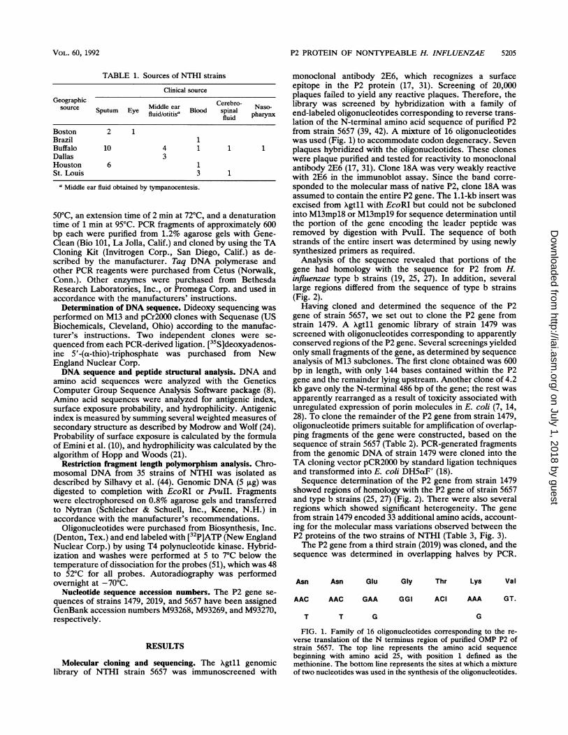
monoclonal antibody 2E6, which recognizes a surfaceepitope in the P2 protein (17, 31). Screening of 20,000plaques failed to yield any reactive plaques. Therefore, thelibrary was screened by hybridization with a family ofend-labeled oligonucleotides corresponding to reverse trans-lation of the N-terminal amino acid sequence of purified P2from strain 5657 (39, 42). A mixture of 16 oligonucleotideswas used (Fig. 1) to accommodate codon degeneracy. Sevenplaques hybridized with the oligonucleotides. These cloneswere plaque purified and tested for reactivity to monoclonalantibody 2E6 (17, 31). Clone 18A was very weakly reactivewith 2E6 in the immunoblot assay. Since the band corre-sponded to the molecular mass of native P2, clone 18A wasassumed to contain the entire P2 gene. The 1.1-kb insert wasexcised from Xgtll with EcoRI but could not be subclonedinto M13mpl8 or M13mpl9 for sequence determination untilthe portion of the gene encoding the leader peptide wasremoved by digestion with PvuII. The sequence of bothstrands of the entire insert was determined by using newlysynthesized primers as required.
Analysis of the sequence revealed that portions of thegene had homology with the sequence for P2 from H.influenzae type b strains (19, 25, 27). In addition, severallarge regions differed from the sequence of type b strains(Fig. 2).Having cloned and determined the sequence of the P2
gene of strain 5657, we set out to clone the P2 gene fromstrain 1479. A Agtll genomic library of strain 1479 wasscreened with oligonucleotides corresponding to apparentlyconserved regions of the P2 gene. Several screenings yieldedonly small fragments of the gene, as determined by sequenceanalysis of M13 subclones. The first clone obtained was 600bp in length, with only 144 bases contained within the P2gene and the remainder lying upstream. Another clone of 4.2kb gave only the N-terminal 486 bp of the gene; the rest wasapparently rearranged as a result of toxicity associated withunregulated expression of porin molecules in E. coli (7, 14,28). To clone the remainder of the P2 gene from strain 1479,oligonucleotide primers suitable for amplification of overlap-ping fragments of the gene were constructed, based on thesequence of strain 5657 (Table 2). PCR-generated fragmentsfrom the genomic DNA of strain 1479 were cloned into theTA cloning vector pCR2000 by standard ligation techniquesand transformed into E. coli DH5otF' (18).Sequence determination of the P2 gene from strain 1479
showed regions of homology with the P2 gene of strain 5657and type b strains (25, 27) (Fig. 2). There were also severalregions which showed significant heterogeneity. The genefrom strain 1479 encoded 33 additional amino acids, account-ing for the molecular mass variations observed between theP2 proteins of the two strains of NTHI (Table 3, Fig. 3).The P2 gene from a third strain (2019) was cloned, and the
sequence was determined in overlapping halves by PCR.
Asn Asn Glu Gly Thr Lys Val
AAC AAC GAA GGI ACI AAA GT.
T T G G
FIG. 1. Family of 16 oligonucleotides corresponding to the re-verse translation of the N terminus region of purified OMP P2 ofstrain 5657. The top line represents the amino acid sequencebeginning with amino acid 25, with position 1 defined as themethionine. The bottom line represents the sites at which a mixtureof two nucleotides was used in the synthesis of the oligonucleotides.
VOL. 60, 1992
on July 1, 2018 by guesthttp://iai.asm
.org/D
ownloaded from

5206 SIKKEMA AND MURPHY INFECT. IMMUN.
1479 ATGAAAAAAACACTTGCAGCATTAATCGTTGTTGCATTCGCAGCTTCAGCAGCAAACGCAGCTGTTGTTTATAACAACGAAGGGACTAAA 90MetLysLysThrLeuAlaAlaLeuI leValValAlaPheAlaAlaSerAlaAlaAsnAlaAlaValValTyrAsnAsnGluGlyThrLys 30
2019 .. . . . . . . . . . . . . . . . . .C
1479 GTAGAATTAGGCGGTCGTTTAAGCGTTATTGCGGAACAAAGCAGCAGCACTGAAGATAATCAAGAACAGCAACACGGTGCATTACGTAAT 180ValGluLeuGlyGlyArgLeuSerValIleAiaGluGlnSerSerSerThrGluAspAsnGinGluGlnGlnHisGlyAlaLeuArgAsn 60
2019 .......T.......A .C..C.A.....T.AT....AT.A.GG....A............C...............Ilie.........Asn....IleLysAsp. .. .Lys.............
5657 ..............CGA. .C..C.A.....T..T.A....CTG....G....A............C...............ThrIle...........Asn .. .Leu. .. .Asp .. .Lys.............
1479 CAGGGTTCACGTTTCCACATTAAAGCAACGCATAACTTCGGTGATGGTTTCTATGCACAAGGTTATTTAGAAACTCGTTTTGTTTCAAAA 270GlnGlySerArgPheHisIlieLysAlaThrHisAsnPheGlyAspGlyPheTyrAlaGinGlyTyrLeuGluThrArgPheValSerLys 90
2019 ..~AA.......................................A....GCT 270...Ser.....................................Leu....Ala 90
5657 ...A...........................................--267.......................... .... .... .... ..... .... .... .... ..... .... .... ... 89
1479 GCCTCTAAAGAAAAAGCAGATCAATTCGCTGATATTGTAAACAAATATGCTTATCTTACTTTAGGAAATAACACATTCGGTGAAGTAAAA 360AlaSerLysGiuLysAlaAspGlnPheAlaAsplieValAsnLysTyrAlaTyrLeuThrLeuGlyAsnAsnThrPheGlyGluValLys 120
2019 CAA. .. .GG.ACCG.GT....A.C. .... G.C...A... C........G.C.......TT.G. .AAG.......360Gin....GiyThrGiuSer....An....GlyHis... IleThr........Vai.......PheAlaLys....... 120
5657 AAG.A....C.TT.....C.... A.AG .... AC. .C........G.........AG.....G....... 357LysTyr....AspAsn....His....AspSer....ThrThr.......Vai.......LysAiaLeu....... 119
1479 CTTGGTCGCGCAAAAACTATTGCTGATGAAATTACAACCGCAGAAGATAAAGAATATGGTCTTCTCAACTCTAAAAAATATATCCCTACT 450LeuGiyArgAiaLysThrIleAiaAspGiuIlieThrThrAlaGiuAspLysGiuTyrGiyLeuLeuAsnSerLysLysTyrleProThr 150
2019..T....T.G.........GC. .A.T. .G............G.....AA. .GC.....T....450...............Giy... IieSer.............Vai....AsnSer......... 150
5657..T....T.G.........GC..A. ... .G............G.A....A..GC.....T.... 447............. .Giy....Ser.............Vai....AsnSer......... 149
1479 AATGGTAACACCGTTGGCTATACTTTTAATGGTATTGATGGTTTAGTATTAGGCGCTAATTATTTATTAGCACAAGAGCGTGATTTACGG 540AsnGiyAsnThrVaiGiyTyrThrPheAsnGlyleAspGiyLeuVaiLeuGiyAiaAsnlTyrLeuLeuAiaGilnGiuArgAspLeuArg 180
2019 ......T.......C.....A...C..........T............C....C..AAGTAT 540...............Lys.........................Gin....HisLysTyr 180
5657 ......T.......C....G.A....C..........T............C.A...A-----531..... .........Giu.........................Gin....Asn----177
1479 ACT----------CTTGATTCTAGAACTAATCCTACGAAAAGTGGTGAAGTAACTGTAGGTGAAGTCAGTAACGGAATTCAA 612Thr----------LeuAspSerArgThrAsnProThrLysSerGiyGiuVaiThrValGiyGiuVaiSerAsnGlGylieGin 204
2019 ....GCTGCTGCTGCTGCTGCTGC. .C.G. .G.TGG.GC. .G.G.TGTTGCA. .A....TTA.CC.CAAA..A. .....C..T. .CG. .....G 630...AiaAlaAiaAiaAiaAiaAiaAlaAlaGlyGiyAiaArgAiaVaiAia......TyrProGinLysIle......Vai ... 210
5657------------------G. .C. .GG. .GT.CGGCA. .A....TGT. .C.CA.T.A... ..C..T..CG.....G 588-----------------AiaHisGiySerThrAia......ValAlaGlnValIle......Vai... 196
1479 GTTGGTGCAAAATATGATGCTAACAACATTATTGTAGCTATTGCTTACGGTCGTACAAATTATAAAGACAGTAATCATAGTTATACGCAA 702ValGiyAiaLysTyrAspAlaAsnAsnIlieIleValAlaIlieAlaTyrGiyArgThrAsnTyrLysAspSerAsnHisSerTyrThrGiln 234
2019............A.C......C.G......T.........G....AGA. .T.ACA.TAACAC.AGC. 720..................AlaGly..............ArgGiuAspIleThrIleThrProAia 240
5657............A.C......C.G......T.........G....AGA.CT.-----GCAG.A... 672.................AlaGiy..............ArgGluAspLeu----AlaAia... 224
1479 AAAATCCCCAAAGCCAACGCCGCCGACGCCGACACCGACACCACCATAATTTACCCCCATCACGGTAAAA~AACAAGAAGTAAATGGTGCT 792LysIleProLysAiaAsnAiaAiaAspAiaAspThrAspThrThrIieIieTyrProHisHisGlyLysLysGilnGiuVaiAsnGiyAia 264
2019 G.TT.---------------------------A.GTTA.......C...C......756Asp----------------------------LysLeu.......Gin........252
5657 GG.-----------------------------G. .TCA.A......C.....C.... 708Giy----------------------------AspSerAsp......Gin........236
1479 TTAGCTAGTTTAGGTTACCGTTTTAGTGATTTAGGCTTATTAGTCTCTCTAGATAGTGGCTATGCAAAAACTAAAAACTATAAAGCTAAA 882LeuAlaSerLeuGlyTyrArgPheSerAspLeuGiyLeuLeuVaiSerLeuAspSerGlyTyrAiaLysThrLysAsnTyrLysAiaLys 294
2019 C.TT.A.C....C.T.T.....................................846...SerThr........................................... 282
5657 C.TT.A.C.C...C.T..............A.....................A ...G 798... SerThr........................................Asp.. 266
FIG. 2. Complete nucleotide and derived amino acid sequence of the P2 genes from NTHI strains 1479, 2019, and 5657. Regions of identityare denoted as periods, and gaps are depicted as dashes. The amino acid and nucleotide position are displayed on the right-hand side of eachrow, while the strain is listed on the left-hand side.
on July 1, 2018 by guesthttp://iai.asm
.org/D
ownloaded from

P2 PROTEIN OF NONTYPEABLE H. INFLUENZAE 5207
CACGAAAAAAGCTATTTCGTATCTCCAGGTTTCCAATATGAATTAATGGAAGATACTAATGTCTATGGCAACTTCAAATATGAACGTAATHisGluLysSerTyrPheValSerProGlyPheGlnTyrGluLeuMetGluAspThrAsnValTyrGlyAsnPheLysTyrGluArgAsn.................................................................... T ................. C ...................................
..C.....C.T.C...
.........Arg.
TCAGTAGATCAAGGTGAGAAAGAACGTGAACAAGCACTGTTATTCGGTATAGATCATAAACTTCACAAACAAGTATTAACCTATATTGAASerValAspGlnGlyGluLysGluArgGluGlnAlaLeuLeuPheGlyIleAspHisLysLeuHisLysGlnValLeuThrTyrIleGlu................................... ......................................... TG ...........................a.............. ...........................................Val..T A.A.........C.. G.A. G................ Lys . Ala. His.. Val.........Val.......................................
GGTGCTTACTCTAGAACTAGAACAACTTCTGTAGGTGATAAGCAAGTTGCTTCAAAAGTAAAAACTGAAAAAGAAAAATCAGTGGGTGTAGlyAlaTyrSerArgThrArgThrThrSerValGlyAspLysGlnValAlaSerLysValLysThrGluLysGluLysSerValGlyVal........................... AG............AACGAA............................ Thr. Ser . ThrAsn..G .........A ....A----.C-.....-MA...............................
.........Ala............AsnAspLys.. Lys---------------ThrGlu.
GGTTTACGCGTTTACTTC 1170GlyLeuArgValTyrPhe 390.................. ..1134.................. . ...378.................. TAATCATTCGTTAGAAATACATTATTAAAGGCA 1204.................. . ...357
FIG. 2-Continued
Sequence data again showed marked heterogeneity amongstrains; however, as before, long stretches of conservedsequences occurred between the variable portions. Aminoacid identity among strains ranged from 79 to 85% with thepercent similarity being slightly higher (Table 3).The sequence of the region corresponding to the carboxy-
terminal primer used for amplification of the P2 genes wasdetermined as follows. The carboxy-terminal portion of theP2 gene of strains 1479 and 2019 was amplified by PCR witha primer downstream of the P2 gene and a primer in thecoding region of the gene (Table 2). The resulting fragment ofstrain 1479 was sequenced directly following gel purificationof the -600-bp EcoRI-digested PCR product. The analogousfragment from strain 2019 was similarly sequenced followingdigestion with XbaI. The sequences of these regions of theP2 gene were identical for all three strains.
Analysis of sequences of the P2 genes. Variations in themolecular mass of P2 among strains of NTHI form the basisof a subtyping system (34, 35). The gene sequences pre-sented here encode proteins with calculated molecularmasses of 40,915 Da (strain 1479), 39,072 Da (strain 2019),and 37,271 Da (strain 5657). These data correlate well withestimates from SDS-PAGE (Fig. 3, Table 3).
Regions of dissimilarity could be broken down into fourmajor hypervariable regions: Vl, amino acids 85 to 115; V2,amino acids 175 to 209; V3, amino acids 226 to 255; and V4,
TABLE 2. Oligonucleotides used in PCR and restriction fragmentlength polymorphism analysis
Oligonucleotide (5'- 3') Strand Nucleotidese
TTCGCAGCIT CAGCAGCb Gene 37-53GCATCATATTTGCACC Opposite 591-607TATTAGCACAACAGCGT Gene 512-528GAAGTAAACGCGTAACCb Opposite 1054-1071TAATAATGTA1TTGTAAC Opposite 1081-1099
aRelates to the nucleotide positions in Fig. 2 for strain 5657."Probes used in restriction fragment length polymorphism analysis (Fig. 6
and 7).
amino acids 363 to 374. These numbers vary among strainsdue to differences in the lengths of the variable regionsamong strains, yet all the differences occur in the samerelative region of the protein molecule (Fig. 4).The variable regions of each gene were analyzed for
surface exposure and antigenic index (10, 24). All of thesevariable regions contain potentially surface-exposed regionswhich vary in their surface exposure probability and hydro-philicity (10, 21, 24). These data were incorporated into thenew model for the porin molecule of H. influenzae type bdeveloped by Srikumar et al. (46) (Fig. 5).With regard to previously reported P2 sequences in H.
influenzae type b, amino acids 181 to 188 in OMP subtypes2L and 6U varied from those in the P2 of strain Minn A(OMP subtype 1H) (25). These differences coincide with V2in NTHI (Fig. 4 and 5) or hydrophilic loop number 4 in thetype b topology model (46). An additional amino acid vari-ation at position 340 in OMP subtype 6U maps to V4 ofNTHI and hydrophilic loop number 8 of the type b model(46). All other amino acid variations in P2 molecules ofOMPsubtype 6U isolates map to proposed transmembrane betaregions in the type b model (25, 46).
Restriction frament length polymorphism. To furthercharacterize the ompP2 locus at the nucleotide level, ge-
TABLE 3. Properties of the P2 gene products of three NTHIstrains and one type b strain
Molecular No. of % Identity withStrain mass" amino strain 1479
(Da) acids' sequence
NTHI1479 40,915 3702019 39,072 358 80.05657 37,271 337 80.7
H. influenzae type b Minn AC 37,782 341 78.8
aCalculated from predicted amino acid sequence."Amino acid chain length of the mature P2 peptide.c From reference 27.
147 9
2019
5657
1479
2019
5657
1479
2019
5657
1479
2019
5657
972324936312888296
10623541026342978326
115238411163721053351
VOL. 60, 1992
on July 1, 2018 by guesthttp://iai.asm
.org/D
ownloaded from

5208 SIKKEMA AND MURPHY
DISCUSSION
94 -.-
6 7 _1
43 qA
30qA B C
FIG. 3. SDS-polyacrylamide gel stained with Coomassie blue.Molecular mass standards are shown on the left (in kilodaltons).Purified P2 was obtained from the following NTHI strains: A, 1479;B, 2019; C, 5657.
nomic DNA from 35 strains of NTHI was digested withEcoRI or PvuII and subjected to restriction fragment lengthpolymorphism analysis. Digested genomic DNA was sub-jected to agarose gel electrophoresis and transferred to anylon membrane. The resulting Southern blot was incubatedwith two end-labeled oligonucleotide probes correspondingto regions of the genes where DNA sequences are the sameamong strains 1479, 2019, and 5657 (Fig. 6 and 7). One probecorresponded to sequence near the amino terminus, and theother corresponded to the carboxy terminus (Table 2). Theprobes hybridized to one or two bands in each of the 35strains, depending on the presence of restriction endonucle-ase sites within the gene (Fig. 6 and 7).While several strains showed similar banding patterns
(Fig. 6 and 7), a great deal of heterogeneity in restrictionfragment size was observed with both EcoRI- and PvuII-restricted DNA. Fourteen of 35 strains contained a centralEcoRI site, contrasting to 100% of type b strains examined(25, 27). The P2 gene of 13 of 35 strains contained PvuIIsites, again distinguishing the nontypeable strains from thetype b isolates, the P2 genes of which all contain a singlePvuII site at the N terminus of the mature protein (25, 27).No correlation between restriction fragment length patternsand the geographic origin or body site of isolation'of thestrains was noted.
Together, these data indicate that antigenic differences inthe P2 protein are due to heterogeneity in the P2 genes ofNTHI strains and that most of the heterogeneity is present infour hypervariable regions of the P2 gene.
The data reported in this study indicate that the antigenicheterogeneity of P2 molecules among NTHI strains is due togene sequence variation in defined regions of the P2 gene.Molecular mass variation among P2 proteins is due todifferences in the size of these variable regions. Four majorhypervariable regions (affecting 12 to 35 amino acids) wereidentified in each of the three P2 genes sequenced; alloccurred in identical regions of the gene. Several smallervariable regions were also noted (affecting two to sevenamino acids).
Restriction fragment length polymorphism analysis ofDNA from 35 NTHI strains demonstrated significant heter-ogeneity in and around the ompP2 locus. While some strainsshowed similar-sized fragments on Southern blot analysis,no correlation was noted between restriction fragment lengthpatterns and biotype, clinical source, or geographic origin.The P2 proteins of nontypeable strains of H. influenzae
differ from the P2 proteins of type b strains in molecularmass in SDS-PAGE and antigenic heterogeneity (28, 29, 35).Studies by Munson et al. (25) have shown a high degree ofconservation of the P2 gene among type b strains. Further-more, the DNA sequence of the P2 genes of two epidemio-logically unrelated type b strains showed 100% homology(19, 27). The sequence of P2 genes from NTHI has not beenreported previously. The P2 proteins of nontypeable strainsare quite different from each other, while the P2's of type bstrains are conserved (17, 19, 20, 25). The present studyestablishes that the differences among P2 proteins of non-typeable strains are due to marked variability at specificlocations within the P2 gene. These observations are consis-tent with alloenzyme electrophoretic typing studies showingthat type b strains are basically clonal, whereas nontypeablestrains are genetically diverse (36, 37, 40, 41).
Previous studies have shown that strain-specific epitopeson the P2 proteins of nontypeable strains appear to beimmunodominant epitopes. Groeneveld et al. (16) showedthat immunization of rabbits with intact bacteria resulted inthe production of anti-P2 antibodies that were protectiveonly against the immunizing strain. Studies in our laboratoryidentified the presence of a strain-specific immunodominantepitope by analysis with monoclonal antibodies to P2 (17).Sequence data obtained in this study reveal several poten-tially heterogeneous epitopes. Computer modeling indicatesthat these variable regions are likely surface exposed andpossess a high antigenic index. Epitope mapping experi-ments are under way to identify which variable portion(s) ofthe P2 gene encodes the major immunodominant surface-exposed epitope(s) in these bacteria.
1-20 45-521-20 45-521-20 45-52
L
85-115 130-3 144-585-115 130-3 144-585-115 130-3 144-5
175-203175-209175-195
218-219 226-255222-223 232-243207-209 218-228
363-374 390351-362 378
Met 335-341 357- .II II El~~~~~~~~~~~~~~~~~~~~~~~~~~~~~~~~~~~~~~~~~~~~~~~~~~~~~~~~~~~~~~~~~VI V2 V3 I V4
CnBrFIG. 4. Diagrammatic representation of the P2 genes of NTHI strains 1479, 2019, and 5657. Regions of sequence variability are shown as
solid rectangles. Amino acid positions affected by the variable regions of each gene are shown. The total number of amino acids present ineach P2 is shown in bold at the far right. L, leader peptide; Vl through V4, four major variable regions of the P2 gene; CnBr, cyanogenbromide.
Strain147920195657
INFECT. IMMUN.
on July 1, 2018 by guesthttp://iai.asm
.org/D
ownloaded from

P2 PROTEIN OF NONTYPEABLE H. INFLUENZAE 5209
30 0 G® A@ L@ 40
@) G
A@01 F
G0,
G (L 0O F
V G
I©I r0D
©4(
VI
0 A1
A V
V YF A
0GO
@0
A Fy G
F ©G v
L @0L Y
120G l80
Y P
@ ®130
® G
@ 0A1 F
I IAX00GG
A
G CJ Li6100
V3
220® AA®YGAP ©
@
@0
G VD
0 A21®y)
170 ~) P
0 0
@ L
@ G
I0G V
A GA A
LI A
I ®~
®D L
IA -Y
@190
FIG. 5. Predicted topological structure of NTHI porin (strain 1479) as it conforms to the model developed by Srikumar et al. (46). Sixteenbeta strands nearly identical to those ofH. influenzae type b are shown as rectangles. For each beta strand, the right side shows residues thatare proposed to face the water-filled lining of the pore, and the left side shows residues that are hypothesized to interact with the outermembrane bilayer. Some amino acid positions of the mature peptide are shown numerically throughout the diagram. Residues of significanthydrophilicity are circled; variable portions of the P2 molecule outside the beta regions are enclosed. Vl, V2, V3, and V4 correspond to theregions depicted in Fig. 4.
High levels of homology among the P2 proteins suggest acommon ancestry, structure, and function for these mole-cules. The four variable regions, differing in length amongstrains, do not appear likely to contribute to the overalltertiary structure of the molecule but may form extramem-branous loops outside the cell surface, where they interactwith the host. Analysis of the amino acid sequence of the P2proteins reveals 16 beta-pleated sheet domains within thevarious conserved regions of each gene (Fig. 5). This issimilar to the porin proteins of E. coli. These OMPs repre-sent a "beta barrel" conformation (11, 50). A similar sce-nario has been hypothesized for the meningococcal class 1OMPs and H. influenzae type b (23, 46). The size andspacing of the conserved regions in the P2 protein of NTHIin conjunction with their beta-pleated sheet domains fit wellwith the hypothesis that P2 shows a similar beta barrelconformation, which exposes variable regions on the surfaceof the bacterium.
Expression of a strain-specific and highly immunodomi-nant antigenic epitope on the surface of the bacterium mayrepresent a mechanism of host evasion by NTHI, permitting
2a1-
9.4- W a * _
6.6 -
4.4- a
3SA B C D E F G H J K L
FIG. 6. Southern blot assay showing restriction fragment lengthpolymorphisms of the ompP2 locus from 12 isolates of NTHI. Fivemicrograms of chromosomal DNA was digested to completion withEcoRI. The fragments were separated on a 0.8% agarose gel,transferred to Nytran, and probed with end-labeled oligonucleotidescorresponding to conserved regions of the P2 genes. Lanes con-tained genomic DNA from H. influenzae strains (A) biogroupaegyptius 16, (B) SL820, (C) 7891, (D) 1749, (E) C83, (F) SL1484,(G) 4971, (H) 39, (I) 7172, (J) 5657, (K) 2019, and (L) 1479. Molecularsize standards are noted on the left (in kilobases).
V2
16C
70
G230
240
@0
@0L P
@0@ v
A VL G
L (
0
A GG
LVGL A
G (Oiy
v
0©F
(E) 31G
©0
A
L
IFLl
270a
00AyG
IL
I
LL
GI
V4
10 000(
340Q ©
yA vDG G
y G
y
y
0A00
FV
0G
F
y
LIM
NH2Avvy
(V)d
G
G
L
09Ll IV
00D COu
COOH
VOL. 60, 1992
L ItD
UUR)
on July 1, 2018 by guesthttp://iai.asm
.org/D
ownloaded from

5210 SIKKEMA AND MURPHY
23.1
9.4 _
6.6
4.4
2.32.0
A B C D E F G H
FIG. 7. Southern blot assay showing restriction fragment lengthpolymorphisms of the ompP2 locus from eight isolates of NTHI. Allconditions were the same as described in the legend to Fig. 6 exceptthat DNA was digested with PvuII. Lanes contain genomic DNAfrom H. influenzae strains (A) 5657, (B) biogroup aegyptius 16, (C)C304, (D) C83, (E) DL205, (F) 2627, (G) 2019, and (H) 1479.Molecular size standards are noted on the left (in kilobases).
the organism to cause recurrent infections. Recurrent otitismedia continues to be a problem despite the presence ofantigenically conserved structures, such as the P1 and P6proteins, on the surface of the bacterium (15, 26, 33, 38).However, infants and children mount a strain-specific bac-tericidal antibody response following otitis media due toNTHI (12). We hypothesize that the presence of a strain-specific immunodominant epitope on the bacterial surfaceinduces the host to make antibody to this epitope and hidesthe antigenically conserved epitopes. Analysis of the specificepitopes to which children make antibody following otitismedia will allow direct testing of this hypothesis.
Since none of the three strains whose P2 gene was
sequenced was isolated from the same patient, it is unknownat this time whether sequence variation among strains is theresult of large changes in variable regions or of single pointmutations accumulated over long periods of time. Recently,van Alphen et al. showed that the mutation rate amongspontaneously emerging mutants was very low (10-' to10-1") (49). Despite the low frequency of mutation, esti-mates of the number of bacteria in the sputum of chronicobstructive pulmonary disease patients were on the order of109/ml. A typical chromosomal mutation rate in this settingwould be sufficient to allow the spontaneous emergence ofstrains which varied in a particular surface epitope. Thesequential isolation of strains of NTHI from patients withexacerbations of chronic obstructive pulmonary diseaseshowed that the P2 molecule varied in molecular mass andantigenic reactivity to monoclonal antibodies (16, 49). Onemight speculate that there is selective pressure on thebacterium, by the humoral immune system, to vary itssurface antigens. In contrast, the P2's of type b organismsare very similar among strains, indicating that there is littlepressure from the immune system to vary its surfaceepitopes. The presence of a capsule may account for thisdifference.We conclude that the expression of highly immunodomi-
nant and strain-specific epitopes on the surface of NTHI isthe result of substantial heterogeneity within the ompP2locus among strains. A series of spontaneous mutationswithin the surface-exposed epitopes may have emergedbecause of immunologic pressure from patients with recur-
rent infections. Alternatively, horizontal transformation ofgenetic material from a mixture of strains present in thehuman host may be a second mechanism to account for thisobservation (9, 13, 23, 43).
ACKNOWLEDGMENTSThe work was supported by research grant A119641 from the
National Institutes of Health and the Department of VeteransAffairs Medical Center.We thank Alan J. Lesse for his computer expertise, Karen
Chiavetta for performing the photography, Kyungcheol Yi andLinda Bartos for their assistance in the laboratory, and AdelineThurston for preparing the manuscript.
REFERENCES1. Barenkamp, S. J., R. S. Munson, Jr., and D. M. Granoff. 1981.
Subtyping isolates of Haemophilus influenzae type b by outer-membrane protein profiles. J. Infect. Dis. 143:668-676.
2. Barenkamp, S. J., P. A. Shurin, C. D. Marchant, R. B. Karasic,S. I. Pelton, V. M. Howie, and D. M. Granoff. 1984. Do childrenwith recurrent Haemophilus influenzae otitis media becomeinfected with a new organism or reacquire the original strain? J.Pediatr. 105:533-537.
3. Berk, S. L., S. A. Holtsclaw, S. L. Wiener, and J. K. Smith.1982. Nontypeable Haemophilus influenzae in the elderly.Arch. Intern. Med. 142:537-539.
4. Bluestone, C. D., and J. 0. Klein. 1983. Otitis media witheffusion, atelectasis and eustachian tube dysfunction, p. 356. InC. D. Bluestone and S. E. Stool (ed.), Pediatric otolaryngology.W. B. Saunders, Philadelphia.
5. Brenner, D. J., L. W. Mayer, G. M. Carlone, L. H. Harrison,W. F. Bibb, M. C. de Cunto Brandileone, F. 0. Sottnek, K. Irino,M. W. Reeves, J. M. Swenson, K. A. Birkness, R. S. Weyant,S. F. Berkley, T. C. Woods, A. G. Steigerwalt, P. A. D. Grimont,R. M. McKinney, D. W. Fleming, L. L. Gheesling, R. C.Cooksey, R. J. Arko, C. V. Broome, W. Fleming, K. W. Fleming,and the Brazilian Purpuric Fever Study Group. 1988. Biochem-ical, genetic, and epidemiologic characterization of Haemophi-lus influenzae biogroup aegyptius (Haemophilus aegyptius)strains associated with Brazilian purpuric fever. J. Clin. Micro-biol. 26:1524-1534.
6. Campagnari, A. A., M. R. Gupta, K. C. Dudas, T. F. Murphy,and M. A. Apicella. 1987. Antigenic diversity of lipooligosac-charides of nontypable Haemophilus influenzae. Infect. Immun.55:882-887.
7. Carbonetti, N. H., and P. F. Sparling. 1987. Molecular cloningand characterization of the structural gene for protein I, themajor outer membrane protein of Neisseria gonorrhoeae. Proc.Natl. Acad. Sci. USA 84:9084-9088.
8. Devereaux, J., P. Haeberli, and 0. Smithies. 1984. A compre-hensive set of sequence analysis programs for the VAX. NucleicAcids Res. 12:387-395.
9. Dowson, C. G., A. Hutchison, J. A. Brannigan, R. C. George, D.Hansman, J. Linares, A. Tomasz, J. Maynard Smith, and B. G.Spratt. 1989. Horizontal transfer of penicillin-binding proteingenes in penicillin-resistant clinical isolates of Streptococcuspneumoniae. Proc. Natl. Acad. Sci. USA 86:8842-8846.
10. Emini, E. A., J. V. Hughes, D. S. Perlow, and J. Boger. 1985.Induction of hepatitis A virus-neutralizing antibody by a virus-specific synthetic peptide. J. Virol. 55:836-839.
11. Engel, A., A. Massalski, H. Schindler, D. L. Dorset, and J. P.Rosenbusch. 1985. Porin channel triplets merge into singleoutlets in Escherichia coli outer membranes. Nature (London)317:643-645.
12. Faden, H., J. Bernstein, L. Brodsky, J. Stanievich, D. Krystofik,C. Shuff, J. J. Hong, and P. L. Ogra. 1989. Otitis media inchildren. I. The systemic immune response to nontypableHaemophilus influenzae. J. Infect. Dis. 160:999-1004.
13. Feavers, I. M., A. B. Heath, J. A. Bygraves, and M. C. J.Maiden. 1992. Role of horizontal genetic exchange in theantigenic variation of the class 1 outer membrane protein ofNeissena meningitidis. Mol. Microbiol. 6:489-495.
14. Gotschlich, E. C., M. E. Seiff, M. S. Blake, and M. Koomey.1987. Porin protein of Neisseria gonorrhoeae: cloning and genestructure. Proc. Natl. Acad. Sci. USA 84:8135-8139.
15. Groeneveld, K., P. P. Ejik, L. van Alphen, H. M. Jansen, andH. C. Zanen. 1990. Haemophilus influenzae infections in pa-tients with obstructive pulmonary disease despite specific anti-
INFECT. IMMUN.
on July 1, 2018 by guesthttp://iai.asm
.org/D
ownloaded from

P2 PROTEIN OF NONTYPEABLE H. INFLUENZAE 5211
bodies in serum and sputum. Am. Rev. Respir. Dis. 141:1316-1321.
16. Groeneveld, K., L. van Alphen, C. Voorter, P. P. Ejk, H. M.Jansen, and H. C. Zanen. 1989. Antigenic drift of Haemophilusinfluenzae in patients with chronic obstructive pulmonary dis-ease. Infect. Immun. 57:3038-3044.
17. Haase, E. M., A. A. Campagnan, J. Sarwar, M. Shero, M.Wirth, C. U. Cumming, and T. F. Murphy. 1991. Strain-specificand immunodominant surface epitopes of the P2 porin protein ofnontypeable Haemophilus influenzae. Infect. Immun. 59:1278-1284.
18. Hanahan, D. 1983. Studies on transformation of Eschenchia coliwith plasmids. J. Mol. Biol. 166:557-580.
19. Hansen, E. J., C. Hasemann, A. Clausell, D. Capra, K. Orth,C. R. Moomaw, C. A. Slaughter, J. L. Latimer, and E. E. Miller.1989. Primary structure of the porin protein of Haemophilusinfluenzae type b determined by nucleotide sequence analysis.Infect. Immun. 57:1100-1107.
20. Hansen, E. J., S. E. Pelzel, K. Orth, C. R. Moomaw, J. D.Radolf, and C. A. Slaughter. 1989. Structural and antigenicconservation of the P2 porin protein among strains of Haemoph-ilus influenzae type b. Infect. Immun. 57:3270-3275.
21. Hopp, T. P., and K. R. Woods. 1981. Prediction of proteinantigenic determinants from amino acid sequences. Proc. Natl.Acad. Sci. USA 78:3824-3828.
22. Loeb, M. R., and D. H. Smith. 1980. Outer membrane proteincomposition in disease isolates of Haemophilus influenzae:pathogenic and epidemiological implications. Infect. Immun.30:709-717.
23. Maiden, M. C. M., J. Suker, A. J. McKenna, J. A. Bygraves, andI. M. Feavers. 1991. Comparison of the class 1 outer membraneproteins of eight serological reference strains of Neisseriameningitidis. Mol. Microbiol. 5:727-736.
24. Modrow, S., and H. Wolf. 1986. Characterization of two relatedEpstein-Barr virus-encoded membrane proteins that are differ-entially expressed in Burkitt lymphoma and in vitro-trans-formed cell lines. Proc. Natl. Acad. Sci. USA 83:5703-5707.
25. Munson, R., Jr., C. Bailey, and S. Grass. 1989. Diversity of theouter membrane protein P2 gene from major clones of Hae-mophilus influenzae type b. Mol. Microbiol. 3:1797-1803.
26. Munson, R., Jr., and S. Grass. 1988. Purification, cloning, andsequence of outer membrane protein P1 of Haemophilus influ-enzae type b. Infect. Immun. 56:2235-2242.
27. Munson, R., Jr., and R. W. Tolan, Jr. 1989. Molecular cloning,expression, and primary sequence of outer membrane proteinP2 of Haemophilus influenzae type b. Infect. Immun. 57:88-94.
28. Munson, R. S., Jr., J. L. Shenep, S. J. Barenkamp, and D. M.Granoff. 1983. Purification and comparison of outer membraneprotein P2 from Haemophilus influenzae type b isolates. J. Clin.Invest. 72:677-684.
29. Murphy, T. F., and M. A. Apicella. 1985. Antigenic heteroge-neity of outer membrane proteins of nontypeable Haemophilusinfluenzae is a basis for a serotyping system. Infect. Immun.50:15-21.
30. Murphy, T. F., and M. A. Apicella. 1987. Nontypable Hae-mophilus influenzae: a review of clinical aspects, surface anti-gens, and the human immune response to infection. Rev. Infect.Dis. 9:1-15.
31. Murphy, T. F., and L. C. Bartos. 1988. Purification and analysiswith monoclonal antibodies of P2, the major outer membraneprotein of nontypeable Haemophilus influenzae. Infect. Immun.56:1084-1089.
32. Murphy, T. F., and L. C. Bartos. 1988. Human bactericidalantibody response to outer membrane protein P2 of nontypeableHaemophilus influenzae. Infect. Immun. 56:2673-2679.
33. Murphy, T. F., L. C. Bartos, P. A. Rice, M. B. Nelson, K. C.Dudas, and M. A. Apicelia. 1986. Identification of a 16,600-dalton outer membrane protein on nontypable Haemophilusinfluenzae as a target for human serum bactericidal antibody. J.Clin. Invest. 78:1020-1027.
34. Murphy, T. F., J. M. Bernstein, D. D. Dryja, A. A. Campagnari,and M. A. Apicella. 1987. Outer membrane protein and lipooli-
gosaccharide analysis of paired nasopharyngeal and middle earisolates in otitis media due to nontypable Haemophilus influen-zae: pathogenetic and epidemiological observations. J. Infect.Dis. 156:723-731.
35. Murphy, T. F., K. C. Dudas, J. M. Mylotte, and M. A. Apicella.1983. A subtyping system for nontypable Haemophilus influen-zae based on outer-membrane proteins. J. Infect. Dis. 147:838-846.
36. Musser, J. M., S. J. Barenkamp, D. M. Granoff, and R. K.Selander. 1986. Genetic relationships of serologically nontype-able and serotype b strains of Haemophilus influenzae. Infect.Immun. 52:183-191.
37. Musser, J. M., D. M. Granoff, P. E. Pattison, and R. K.Selander. 1985. A population genetic framework for the study ofinvasive diseases caused by serotype b strains of Haemophilusinfluenzae. Proc. Natl. Acad. Sci. USA 82:5078-5082.
38. Nelson, M. B., R. S. Munson, Jr., M. A. Apicella, D. J. Sikkema,J. P. Molleston, and T. F. Murphy. 1991. Molecular conserva-tion of the P6 outer membrane protein among strains of Hae-mophilus influenzae: analysis of antigenic determinants, genesequences, and restriction fragment length polymorphisms. In-fect. Immun. 59:2658-2663.
39. Ohtsuka, E., S. Matsuki, M. Ikehara, Y. Takahashi, and K.Matsubara. 1985. An alternative approach to deoxynucleotidesas hybridization probes by insertion of deoxyinosine at ambig-uous codon positions. J. Biol. Chem. 260:2605-2608.
40. Porras, O., D. A. Caugant, B. Gray, T. Lagergard, B. R. Levin,and C. Svanborg-Eden. 1986. Difference in structure betweentype b and nontypeable Haemophilus influenzae populations.Infect. Immun. 53:79-89.
41. Porras, O., D. A. Caugant, T. Lagergard, and C. Svanborg-Eden. 1986. Application of multilocus enzyme gel electrophore-sis to Haemophilus influenzae. Infect. Immun. 53:71-78.
42. Sambrook, J., E. F. Fritsch, and T. Maniatis. 1989. Molecularcloning: a laboratory manual, 2nd ed. Cold Spring HarborLaboratory, Cold Spring Harbor, N.Y.
43. Seifert, H. S., R. S. Ajioka, C. Marchal, P. F. Sparling, and M.So. 1988. DNA transformation leads to pilin antigenic variationin Neisseria gonorrhoeae. Nature (London) 336:392-395.
44. Silhavy, T. J., et al. 1984. Experiments with gene fusions. ColdSpring Harbor Laboratory, Cold Spring Harbor, N.Y.
45. Srikumar, R., A. C. Chin, V. Vachon, C. D. Richardson,M. J. H. Ratcliffe, L. Saarinen, H. Kayhty, P. H. Makela, andJ. W. Coulton. 1992. Monoclonal antibodies specific to porin ofHaemophilus influenzae type b: localization of their cognateepitopes and tests of their biological activities. Mol. Microbiol.6:665-676.
46. Srikumar, R., D. Dahan, M. F. Gras, M. J. H. Ratcliffe, L. vanAlphen, and J. W. Coulton. 1992. Antigenic sites on porin ofHaemophilus influenzae type b: mapping with synthetic pep-tides and evaluation of structure predictions. J. Bacteriol.174:4007-4016.
47. Vachon, V., D. N. Kristjanson, and J. W. Coulton. 1988. Outermembrane porin protein ofHaemophilus influenzae type b: poresize and subunit structure. Can. J. Microbiol. 34:134-140.
48. Vachon, V., D. J. Lyew, and J. W. Coulton. 1985. Transmem-brane permeability channels across the outer membrane ofHaemophilus influenzae type b. J. Bacteriol. 162:918-924.
49. van Alphen, L., P. EJk, L. Geelen-van den Broek, and J.Dankert. 1991. Immunochemical characterization of variableepitopes of outer membrane protein P2 of nontypeable Hae-mophilus influenzae. Infect. Immun. 59:247-252.
50. Vogel, H., and F. Jahnig. 1986. Models for the structure of outermembrane proteins of Escherichia coli derived from Ramanspectroscopy and prediction methods. J. Mol. Biol. 190:191-199.
51. Wood, W., J. Gitschier, L. Lasky, and R. Lawn. 1985. Basecomposition-independent hybridization in tetramethylammo-nium chloride: a method for oligonucleotide screening of highlycomplex gene libraries. Proc. Natl. Acad. Sci. USA 82:1585-1588.
VOL. 60, 1992
on July 1, 2018 by guesthttp://iai.asm
.org/D
ownloaded from