Molecular Screening Of Azoreductase Gene and Its Activity ...
25
Langston University Digital Commons @ Langston University McCabe esis Collection Student Works 12-2006 Molecular Screening Of Azoreductase Gene and Its Activity in Human Intestinal Bacteria Syndia S. Todd Follow this and additional works at: hp://dclu.langston.edu/mccabe_theses Part of the Bacteria Commons , Biology Commons , and the Digestive System Commons is esis is brought to you for free and open access by the Student Works at Digital Commons @ Langston University. It has been accepted for inclusion in McCabe esis Collection by an authorized administrator of Digital Commons @ Langston University. For more information, please contact [email protected]. Recommended Citation Todd, Syndia S., "Molecular Screening Of Azoreductase Gene and Its Activity in Human Intestinal Bacteria" (2006). McCabe esis Collection. Paper 37.
Transcript of Molecular Screening Of Azoreductase Gene and Its Activity ...

Langston UniversityDigital Commons @ Langston University
McCabe Thesis Collection Student Works
12-2006
Molecular Screening Of Azoreductase Gene and ItsActivity in Human Intestinal BacteriaSyndia S. Todd
Follow this and additional works at: http://dclu.langston.edu/mccabe_thesesPart of the Bacteria Commons, Biology Commons, and the Digestive System Commons
This Thesis is brought to you for free and open access by the Student Works at Digital Commons @ Langston University. It has been accepted forinclusion in McCabe Thesis Collection by an authorized administrator of Digital Commons @ Langston University. For more information, pleasecontact [email protected].
Recommended CitationTodd, Syndia S., "Molecular Screening Of Azoreductase Gene and Its Activity in Human Intestinal Bacteria" (2006). McCabe ThesisCollection. Paper 37.

The Edwin P. McCabe Honors Program
Senior Thesis
"MOLT,CV£Jl<̂ S(̂ (ENINg 0<F AZO<̂ CDVCMS<E gENEJWD ITS
mCFEQUA*
Syndia S. Todd
December 2006
Langston University Langston, Oklahoma
1

Mo CecuCar Screening Of JLzorecfuctase Cjene J4ncf Its Activity In Human Intestinal (Bacteria
SyncfiaS. rTocfcf Biology Major
School of Arts and Sciences
Submitted in partial fulfillment of the requirements of the E.P. McCabe Honors Program
Fall 2006

iMoCecuCar Screening OfSLzorecCucta.se Cjene J/LncC Its Activity In Human IntestinaC(Bacteria
By
Syndia S. Todd
Thesis approved by:
Thesis ̂ Committee Chairperson
Thesis Committee Member
Director of the Honors Program
Vice President of Academic Affairs
3

Acknowledgments
I would like to start of by thanking my Lord and savior Jesus Christ because
without him nothing is impossible. Second, I want to thank my family and
friends for all the support and love they have given my through all these
years. I would like to give a special thank you to Dr. Sonya Williams without
her help and determination I would probably not be in college receiving all
these great opportunities. Also, special thanks to all the programs and
scholarships that accepted me and helped me with funding my education and
research. Those programs include UBEP (Undergraduate Biomedical
Education Program),LINC (Langston's Integrated Network College), Regents
scholarship, and KU-INBRE. I also want to thank Ms. Irene for always
staying on me to do the best I can.
I definitely want to give special thanks to my mentor Dr. K.J. Abraham for
allowing me to be part of his lab and research. Dr. Abraham went beyond his
duty of a mentor. I also appreciate him for taking on the task of being the
chair person of my committee. Last but not least, I would like to thank
Curtisia Battle for all her help and making my experience in and outside of
Dr. Abrahams lab the best.
4

Table of Contents
Chapter I. Introduction
1. Introduction
2. Background
3. Statement of the problem/topic
4. Purpose of the study
5. Assumption
6. Rationale for Study
7. Definition
Chapter II. Literature Review
1. Organization of Chapter 2
2. Historical background of the topic
3. Summary of existing studies
4. Significant Results
Chapter III. Methodology
1. Organization Chapter III
2. Preparation of media
3. Azoreductase Activity
5

4. Genomic DNA Extraction
5. Agarose gel electrophoresis
6. Ploymersae Chain Reaction PCR
7. Restriction Digestion
Chapter IV. Analysis or Findings
1. Figures
2. Tables
3. Summary
Chapter V. Summary, Conclusions
1. Brief Summary
2. Conclusions
6

Chapter 1: Introduction
Introduction
Xeniobotics are foreign chemicals that are man made or of
natural origin. These chemicals include pesticides, animals, toxins, soil
and water pollutants, food additives and drugs. In many cases,
xeniobotics are metabolized into carcinogens, mutagens, and tumor
promoting agents. Azo dyes and nitrated polycyclic aromatic
hydrocarbons (nitro-PAHs) are two groups of chemicals that are
abundant in our environment.
The species of bacteria capable of reducing azo dyes and nitro-
PAHs from the human intestinal microflora or other sources can be
identified by plating serial dilutions of human feces on brain-heart
infusion agar containing 1-nitropyrene or an azo dye such as Direct
Blue 15. Bacterial colonies with azoreductase and nitroreductase
activities reduce the dye or 1-nitropyreneon on the plate and are
recognized by the appearance of clear zones around the colonies.
Although several azoreductase- and nitroreductase - producing
7
bacteria are present in the human intestinal tract, the majority belong
to the genera Clostridium and Eubacterium. Predominant anaerobic
bacteria with azoreductase and nitroreductase activity found in the
human intestinal tract include Clostridium leptum, Eubacterium sp.( C.
dostridiiforme, C. paraputrificum, Clostridium sp., and C. perfringens
(Cerniglia, Rafii, 1995). The aim of this research work was to study the
activity of azoreducatse gene in Citrobacter freundii and isolate the
azroreducatse gene.
Background of the Topic
Azo dye compounds represent a large group of chemicals which
are extensively used in the textile, pharmaceutical, food, and cosmetic
industries. It is one of the largest and most versatile dyes. They are
generally considered to be xenobiotic compounds which are rather
recalcitrant against biodegradative processes in conventional sewage
treatment systems. Nevertheless, during the last few years it has
been demonstrated that several microorganisms are able to transform
azo dyes to noncolored products or even mineralize them completely
8

under certain environmental conditions ( Cerniglia et. al.( 1990).
Statement of the Problem
The intestinal microflora are capable of performing a wide
variety of metabolic transformations. "The intestinal bacteria can
enhance the function of the entire gastrointestinal tract, protect
against pathogenic, maintain the vital chemical balance of the
gastrointestinal system, and produce needed vitamins and hormones"
(http;//www.upwardquest.com/crit2.html). Some of the products of this
metabolism have been associated with carcinogenic processes, such as
cancer, tumor formation, gastrointestinal disease, and infections. The
ability of human intestinal microbes to interact with metabolites
directly or after recirculation may contribute toward different
toxicological disorders and disease.
Purpose of the Study
The purpose of this study is to characterize and isolate the
azoreductase gene in Citrobacter freundii.

Assumption
My hypothesis is that C. freundii possess azoreducatse enzyme
and can influence hepatic P450 enzymes associated with toxicity and
disease.
Rationale for Study
The human intestine is host to food remnants and microbial
organisms, also know as the intestinal microflora, from which the body
derives nourishment and against which the body must be protected.
The first catabolic step in the reduction of azo dyes, which is
accompanied by a decrease in the visible light absorbance of the dye
and the decolonization of the dye, is the reduction of the azo bridge
to produce aromatic amines. These aromatic amines are also known as
human carcinogens. There is a relationship between the intestinal
microflora and metabolic reactions leading to the transformation of
drugs and the production of mutagenic or carcinogenic compounds.
10

Definition
The bacterium (Citrobacter freundii) is a gram-negative, rod-
shaped bacterium found in man and other animals including mammals,
birds, reptiles, and amphibians. Its organisms have also been isolated
from soil and water as well as from clinical specimens such as urine,
throat, sputum, blood, and wound swabs as an opportunistic pathogen
(http ;//vm.cfsan.fda.gov/~mow/chap20.html).
Azo dyes are dyes characterized by the presence of one or more
azo groups (Cerniglia, et. al., 1995). More than half of the annually
produced dyes are azo dyes (Blumel et. al., 2002)
Azoreductase is an enzyme capable of transforming azo dyes.
A Gene is a basic unit of heredity.
Genome is the total set of genes carried by an individual or cell.
Chapter 2
Organization Of Chapter 2
This chapter includes a historical background of the work on
11

azoreductase enzymes and its presence in human intestinal bacteria,
recent findings, its impact on human health and significant reports.
Historical Background
The history of azo compound goes back to over a hundred years.
William Henry Perkins synthesized the first dye, mauve, in 1856 from
chemicals derived from coal. W.H. Perkins commercialized his
innovation and developed the production process for this new dye.
During manufacturing and usage of azo dyes an estimated amount of
10-15% is released into the environment (Vaidya and Datye, 1982).
Existing Studies
Azoreductase activity of anaerobic bacteria isolated from human
intestinal bacteria was reported by Rafii and Cernigial (1990). Their
results showed azoreductase activity in ten strains of anaerobic
bacteria. Several researchers (Brown, 1981; Chung et. al., 1978, Dubin,
and Wright, 1975) have shown reduction of azo dyes by bacteria.
12
Significant results
Azo dye compounds are linked to bladder cancer in humans,
hepatocarcinoma, and to nuclear anomalies in intestinal epithelial cells
in mice (Manning et. al., 1985). A number of azo dyes have been
classified as carcinogenic (Hartman et. al., 1978). All the azo dyes
containing a nitro group were found to be mutagenic (Chung and
Cernigilia, 1992). Furthermore, azo dyes can cause toxic degradation
products.
Chapter 3: Methodology
Preparation of media
The composition of Brain Heart Infusion (BHI) included 425ml of
distilled water, Brain Heart Infusion Broth (18.5g),Yeast Exatract
(5g), Salt Solution 1 (12.5ml), Salt Solution 2 (12.5ml), Calcium Chloride
Solution (12.5ml), Hemin Solution 2ml, Sodium Carbonate (0.2g), and
Cyestine-HCL (0.3g).
13

Composition of Nutrient Agar (NA) included 1 liter distilled water, 8g
of Nutrient broth, and 15g Agar. Colonies of Citrobacter freundii
were maintained on Nutrient Agar medium. BHI was used for liquid
cultures to test for azoreductase activity.
Azoreductase activity
To test the azoreductase activity, different concentrations of azo
dyes (10, 20 and 30^/M of Direct blue 15) were tested for
azoreductase activity and discolorization with C. freundii. Spectronic-
20 was used to measure absorbance at 30 minutes intervals for 3
hours.
Genomic DNA extraction
After testing C. freundii for azoreductase activity , the genomic DNA of
the bacterium was extracted and isolated. During this procedure 100ml
of bacterial cultures, incubated overnight at 37°C was centrifuged for 20
minutes at 6000 rpm. The pellet was resuspended in 9.5 ml of Tris-
Ethylene Diamine Tetra Acetic Acid Buffer (TE buffer). 50|d of
Lysozyme was added, and kept in room temperature for 30 minutes. Next,
14

0.5ml of 10% SDS and 40(al of 20 mg/ml protenase K and 50|J RNAse
(20mg/ml) were added, mixed and incubated at 37° C for 1 hour. 1.8ml of
5M NaCI was then added and mixed thoroughly. After mixing, 1.5ml of
CTAB/NaCI solution was added, mixed, and incubated for 20 minutes at
65° C. After incubation it was extracted with equal volume of
chloroform/isoamyl alcohol (24:1) and centrifuged for 20 minutes, at
6000 rpm, in room temperature. The aqueous phase was transfered to a
fresh tube to precipitate the DNA with 0.6 volume of isopropanol. It was
centrifuged at 6000 rpm for 15 minutes at room temperature. The
precipitate was washed with 70% ethanol, and centrifuged for another 15
minutes at 6000 rpm. The supernatant was removed and the pellet
suspended in 1 ml of TE Buffer. The DNA was stored at -20° C.
DNA measurement
DNA measurement was done to check the purity and the total amount of
the genomic DNA of C. freundii. The measurement was taken at two
different wavelengths (260 and 280nm) and the ratio at 260/280 nm was
calculated. The reading for C. freundii was 1.966 ug/ml.
15

Agarose gel electrophoresis
To ensure that the C. freundii DNA was successfully extracted, gel
electrophoresis was performed. An agarose gel (0.8%) was prepared for
electrophoresis. Genomic DNA (O.l^g) was loaded on to the gel along with
a DNA marker. The gel was stained with ethidium bromide and observed
on a UV Transilluminator
Polymerase Chain Reaction (PCR)
Oligonucleotides were custom synthesized to characterize genes from
Enterococcus faecalis and used as primers. PCR mixture (100|al) for the
amplification of genomic DNA contained 0.1 :g of genomic templates, 0.5|il
of Taq DNA polymerase (5u/jnl), 10 pmol of each primer, lOmM
concentration of each deoxynucleotides triphophate, 25mM MgCI2 and
the corresponding reaction buffer was used for PCR. Electrophoresis
with PCR products showed no fragments of expected size.
Restriction digestion
Restriction digestion of genomic DNA was followed to clone fragments
using a cloning vector. Restriction enzymes, Bam\-\, Hind III, and EcoRT
16

were used to perform digestion of genomic DNA at 37° C.
Chapter 4: Analysis and Findings
Figurere 1: Colonies of Citrobacter freundii
Colonies of Citrobacter freundii were maintained onNutrient Agar
medium. BHI was used for liquid culture to test azoreductase activity.
17

Figure 2: Gram-negative Citrobacter freundii
C. freundii possess azoreducatse enzyme and can influence hepatic
P450 enzymes associated with toxicity and disease.

Figure 3: Agarose gel showing genomic DNA of Citrobacter freundii
The gel was stained with ethidium bromide and observed on a UV
Transilluminator
19
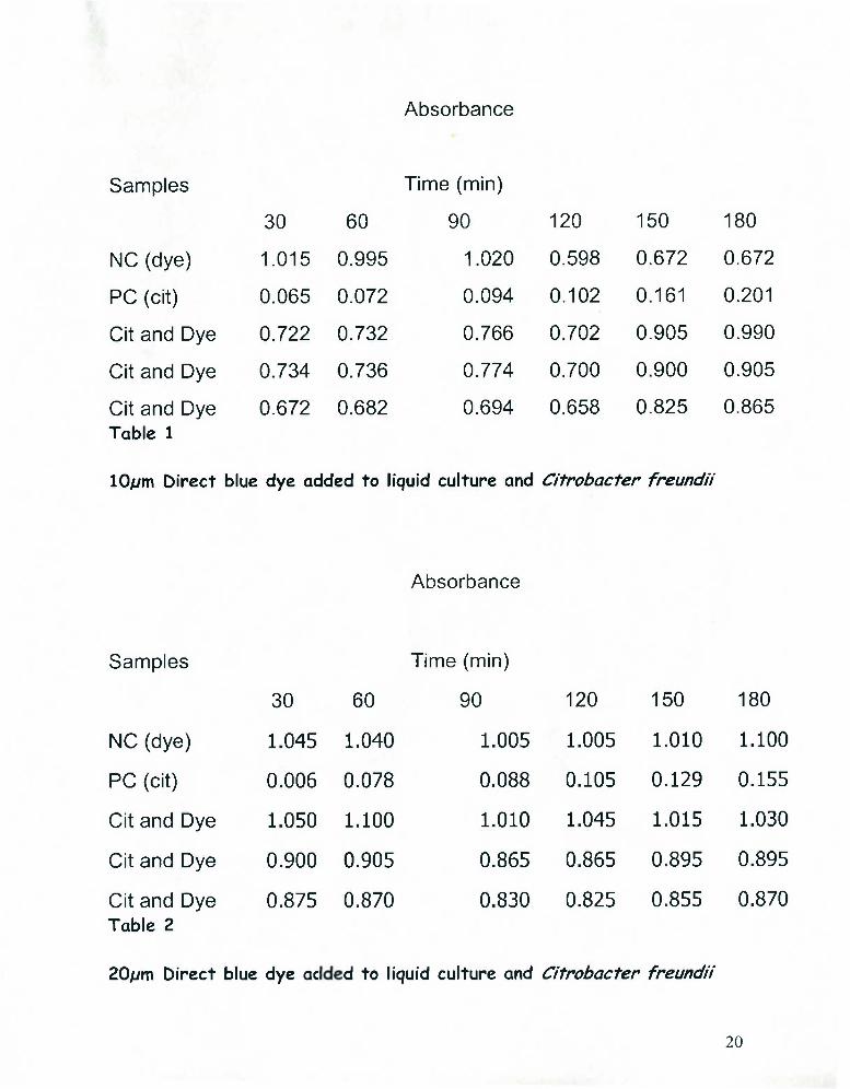
Absorbance
Samples Time (min)
30 60 90 120 150 180
NC (dye) 1.015 0.995 1.020 0.598 0.672 0.672
PC (cit) 0.065 0.072 0.094 0.102 0.161 0.201
Cit and Dye 0.722 0.732 0.766 0.702 0.905 0.990
Cit and Dye 0.734 0.736 0.774 0.700 0.900 0.905
Cit and Dye Table 1
0.672 0.682 0.694 0.658 0.825 0.865
10/vm Direct blue dye added to liquid culture and Citrobacter freundii
Absorbance
Samples
30
NC (dye) 1.045
PC (cit) 0.006
Cit and Dye 1.050
Cit and Dye 0.900
Cit and Dye 0.875 Table 2
20/vm Direct blue dye ac
Time (min)
60 90
1.040 1.005
0.078 0.088
1.100 1.010
0.905 0.865
0.870 0.830
d to liquid culture and
120 150 180
1.005 1.010 1.100
0.105 0.129 0.155
1.045 1.015 1.030
0.865 0.895 0.895
0.825 0.855 0.870
Citrobacter freundii
20

Absorbance
Samples Time (min)
30 60 90 120 150 180
NC (dye) 0.446 0.452 0.448 0.452 0.456 0.682
PC (cit) 0.015 0.018 0.021 0.026 0.046 0.137
Cit and Dye 0.488 0.486 0.480 0.484 0.488 0.744
Cit and Dye 0.468 0.466 0.466 0.468 0.474 0.718
Cit and Dye Table 3
0.506 0.504 0.500 0.504 0.510 0.784
30fjm Direct blue dye added to liquid culture and Citrobacter freundii
Decolonization of Blue 15 dye (lOmM) by C. freundii indicated the
presence of azoreducatse enzyme. The larger the amount of dye the
longer it took for the Citrobacter freundii to decolorize the Blue 15
dye
DNA/ Protein measurement
The concentration of genomic DNA was measured with a UV
21

Spectrophotometer
No Items Results Unit
1 A1 .257 Abs
A2 . 155 Abs
A3 . 051 Abs
C-DNA 9.193 ug/ml
C-Pro 6.647 ug/ml
Ratio 1.966 ug/ml
The ratio for protein measurement should range from 1.5 to 2. Our
protein ratio was 1.966
Chapter 5: Summary and Conclusion
Brief Summary
Genomic DNA was extracted from small bacterial cultures by treating
it with lysozyme or Mutanolysin. The resulting DNA was screened by
22

agarose gel electrophoreses. PCR analysis and restriction endonuclease
digestion was followed for isolating the azoreducatse gene.
Conclusion
• Decolonization of Azo Dye (Direct blue 15)10 - 20/jM
concentrated by Citrobacter freundii.
• Biotransformation of azo dye by Citrobacter freundii indicated
presence of azoreducatse gene.
Isolation of the azoreducatse gene with PCR, restriction
endonucleases, electrophoresis, cloning and transformation are in
progress.
23

Work Cited
1. Blumel, S., Knackmuss Hans-Joachim, Stolz Andreas. 2002.
American Society for Microbiology, vol. 68, 3948-3955
2. Brown, J.P. 1981. Reduction of polymeric azo and nitro dyes
intestinal bacteria. Appl. Environ. Microbiol.41:1283-128
3. Cerniglia, C.E., and R. Fatemah. 1995. Environmental Health Issues,
Vol. 103.
4. Cerniglia, C.E., R. Fatemah, Franklin, Wirt. 1990. Azoreductase
Activity of Anaerobic Bacterial Isolated from Human Intestinal
Microflora,_vol. 56, 2146 -2151
5. Chung, K.T, 5.E. Stevens, and Cerniglia. 1992. The reduction of azo
dyes by the intestinal microflora. Crit. Rev. Microbiol.18:175-190
6. Dubin, P., and K.L. Wright. 1975. Reduction of azo food dyes in
cultures of Proteus Vulgaris. Xeniobiotica 5:563-571
7. Hartman, C. P., G. E. Fulk, and A. W. Andrews. 1978. Azo reduction
of intestinal anaerobe. Mutat. Res. 58:125-132
8. Manning, B. W., C. E. Cerniglia, and T. W. Federle. 1985. Metabolismof