Molecular Pathways: Beta-Adrenergic Signaling in...
7
Molecular Pathways Molecular Pathways: Beta-Adrenergic Signaling in Cancer Steven W. Cole 1,2,3,4 and Anil K. Sood 5,6,7 Abstract Beta-adrenergic signaling has been found to regulate multiple cellular processes that contribute to the initiation and progression of cancer, including inflammation, angiogenesis, apoptosis/anoikis, cell motility and trafficking, activation of tumor-associated viruses, DNA damage repair, cellular immune response, and epithelial–mesenchymal transition. In several experimental cancer models, activation of the sympathetic nervous system promotes the metastasis of solid epithelial tumors and the dissemination of hematopoietic malignancies via b-adrenoreceptor–mediated activation of protein kinase A and exchange protein activated by adenylyl cyclase signaling pathways. Within the tumor microenvironment, b-adrenergic receptors on tumor and stromal cells are activated by catecholamines from local sympathetic nerve fibers (norepineph- rine) and circulating blood (epinephrine). Tumor-associated macrophages are emerging as key targets of b-adrenergic regulation in several cancer contexts. Sympathetic nervous system regulation of cancer cell biology and the tumor microenvironment has clarified the molecular basis for long-suspected relationships between stress and cancer progression, and now suggests a highly leveraged target for therapeutic intervention. Epidemiologic studies have linked the use of b-blockers to reduced rates of progression for several solid tumors, and preclinical pharmacologic and biomarker studies are now laying the groundwork for translation of b-blockade as a novel adjuvant to existing therapeutic strategies in clinical oncology. Clin Cancer Res; 18(5); 1201–6. Ó2011 AACR. Background The b-adrenergic signaling pathway (Fig. 1) mediates sympathetic nervous system (SNS)–induced fight-or-flight stress responses (1, 2). SNS neural fibers innervate most major organ systems and can release micromolar concen- trations of the catecholamine neurotransmitter norepi- nephrine into target tissues in response to physiologic, psychologic, and environmental threats to homeostasis (1–3). Acute SNS activation also elevates catecholamine levels in circulating blood via the release of epinephrine from chromaffin cells of the adrenal medulla and norepi- nephrine spill-over from vascular neuro-muscular junc- tions (1–3). Acute stress responses can elevate norepineph- rine and epinephrine levels by >10-fold within seconds, but basal levels also fluctuate tonically over time in response to organismic and environmental conditions (1–3). In addi- tion to central nervous system (CNS) control of general SNS neural outflow, local regulatory processes also influence SNS nerve fiber activity and catecholamine release and degradation. As a result, norepinephrine and epinephrine concentrations can differ substantially in solid tissues versus blood, as well as across different tissue environments at the same point in time (1–3). The biologic effects of norepinephrine and epinephrine are mediated by a 1 -, a 2 -, and b-adrenergic receptor families, which show distinct patterns of tissue distribution and signal through distinct biochemical pathways (3, 4). The 3 subtypes of b-adrenergic receptor, b 1 , b 2 , and b 3 , are present at many sites of tumor growth and metastasis, such as the brain, lung, liver, kidney, adrenal gland, breast, ovary, prostate, lymphoid tissues, bone marrow, and vasculature. b-adrenergic signaling regulates the function of several cancer-relevant cell types, including epithelial cells, vascular myocytes and pericytes, adipocytes, fibroblasts, neural and glial cells, and most lymphoid and myeloid immune cells (3, 4). Ligation of b-receptors by norepinephrine and epi- nephrine activates the G as guanine nucleotide-binding pro- tein to stimulate adenylyl cyclase synthesis of cyclic AMP (cAMP). The resulting transient cAMP flux can regulate a diverse array of cellular processes via 2 major downstream effector systems (Fig. 1). One cAMP effector involves activation of protein kinase A (PKA), which subsequently phosphorylates serine or threonine residues on target proteins that bear PKA- receptive amino acid motifs [e.g., R-R-X-(S/T)-Y, in which R ¼ arginine, S ¼ serine, T ¼ threonine, X ¼ any amino acid, and Y ¼ hydrophobic amino acid]. PKA regulates a wide variety of cellular processes ranging from general Authors' Affiliations: 1 Division of Hematology-Oncology, Department of Medicine, UCLA School of Medicine, 2 Jonsson Comprehensive Cancer Center, 3 Norman Cousins Center and 4 UCLA Molecular Biology Institute, Los Angeles, California; Departments of 5 Gynecologic Oncology and 6 Cancer Biology, and 7 Center for RNA Interference and Non-Coding RNA, The University of Texas MD Anderson Cancer Center, Houston, Texas Corresponding Author: Steven W. Cole, University of California at Los Angeles, 11-934 Factor Building, Los Angeles, CA 90095-1678. Phone: 310-267-4243; E-mail: [email protected] doi: 10.1158/1078-0432.CCR-11-0641 Ó2011 American Association for Cancer Research. Clinical Cancer Research www.aacrjournals.org 1201 on June 7, 2018. © 2012 American Association for Cancer Research. clincancerres.aacrjournals.org Downloaded from Published OnlineFirst December 20, 2011; DOI: 10.1158/1078-0432.CCR-11-0641
Transcript of Molecular Pathways: Beta-Adrenergic Signaling in...
Molecular Pathways
Molecular Pathways: Beta-Adrenergic Signaling in Cancer
Steven W. Cole1,2,3,4 and Anil K. Sood5,6,7
AbstractBeta-adrenergic signaling has been found to regulate multiple cellular processes that contribute to the
initiation and progression of cancer, including inflammation, angiogenesis, apoptosis/anoikis, cell motility
and trafficking, activation of tumor-associated viruses, DNA damage repair, cellular immune response, and
epithelial–mesenchymal transition. In several experimental cancer models, activation of the sympathetic
nervous system promotes the metastasis of solid epithelial tumors and the dissemination of hematopoietic
malignancies via b-adrenoreceptor–mediated activation of protein kinase A and exchange protein activated
by adenylyl cyclase signaling pathways. Within the tumor microenvironment, b-adrenergic receptors on
tumor and stromal cells are activated by catecholamines from local sympathetic nerve fibers (norepineph-
rine) and circulating blood (epinephrine). Tumor-associated macrophages are emerging as key targets of
b-adrenergic regulation in several cancer contexts. Sympathetic nervous system regulation of cancer cell
biology and the tumormicroenvironment has clarified themolecular basis for long-suspected relationships
between stress and cancer progression, and now suggests a highly leveraged target for therapeutic
intervention. Epidemiologic studies have linked the use of b-blockers to reduced rates of progression for
several solid tumors, and preclinical pharmacologic and biomarker studies are now laying the groundwork
for translation of b-blockade as a novel adjuvant to existing therapeutic strategies in clinical oncology.
Clin Cancer Res; 18(5); 1201–6. �2011 AACR.
BackgroundThe b-adrenergic signaling pathway (Fig. 1) mediates
sympathetic nervous system (SNS)–induced fight-or-flightstress responses (1, 2). SNS neural fibers innervate mostmajor organ systems and can release micromolar concen-trations of the catecholamine neurotransmitter norepi-nephrine into target tissues in response to physiologic,psychologic, and environmental threats to homeostasis(1–3). Acute SNS activation also elevates catecholaminelevels in circulating blood via the release of epinephrinefrom chromaffin cells of the adrenal medulla and norepi-nephrine spill-over from vascular neuro-muscular junc-tions (1–3). Acute stress responses can elevate norepineph-rine and epinephrine levels by >10-fold within seconds, butbasal levels also fluctuate tonically over time in response toorganismic and environmental conditions (1–3). In addi-tion to central nervous system (CNS) control of general SNSneural outflow, local regulatory processes also influence
SNS nerve fiber activity and catecholamine release anddegradation. As a result, norepinephrine and epinephrineconcentrations candiffer substantially in solid tissues versusblood, as well as across different tissue environments at thesame point in time (1–3).
The biologic effects of norepinephrine and epinephrinearemediated bya1-,a2-, and b-adrenergic receptor families,which show distinct patterns of tissue distribution andsignal through distinct biochemical pathways (3, 4). The3 subtypes of b-adrenergic receptor, b1, b2, and b3, arepresent at many sites of tumor growth and metastasis, suchas the brain, lung, liver, kidney, adrenal gland, breast, ovary,prostate, lymphoid tissues, bone marrow, and vasculature.b-adrenergic signaling regulates the function of severalcancer-relevant cell types, including epithelial cells, vascularmyocytes and pericytes, adipocytes, fibroblasts, neural andglial cells, and most lymphoid and myeloid immune cells(3, 4). Ligation of b-receptors by norepinephrine and epi-nephrine activates the Gas guanine nucleotide-binding pro-tein to stimulate adenylyl cyclase synthesis of cyclic AMP(cAMP). The resulting transient cAMP flux can regulate adiverse array of cellular processes via 2 major downstreameffector systems (Fig. 1).
One cAMP effector involves activation of protein kinaseA (PKA), which subsequently phosphorylates serine orthreonine residues on target proteins that bear PKA-receptive amino acid motifs [e.g., R-R-X-(S/T)-Y, in whichR ¼ arginine, S ¼ serine, T ¼ threonine, X ¼ any aminoacid, and Y ¼ hydrophobic amino acid]. PKA regulates awide variety of cellular processes ranging from general
Authors' Affiliations: 1Division of Hematology-Oncology, Department ofMedicine, UCLA School of Medicine, 2Jonsson Comprehensive CancerCenter, 3Norman Cousins Center and 4UCLA Molecular Biology Institute,Los Angeles, California; Departments of 5Gynecologic Oncology and6Cancer Biology, and 7Center for RNA Interference and Non-Coding RNA,The University of Texas MD Anderson Cancer Center, Houston, Texas
Corresponding Author: Steven W. Cole, University of California at LosAngeles, 11-934 Factor Building, Los Angeles, CA 90095-1678. Phone:310-267-4243; E-mail: [email protected]
doi: 10.1158/1078-0432.CCR-11-0641
�2011 American Association for Cancer Research.
ClinicalCancer
Research
www.aacrjournals.org 1201
on June 7, 2018. © 2012 American Association for Cancer Research. clincancerres.aacrjournals.org Downloaded from
Published OnlineFirst December 20, 2011; DOI: 10.1158/1078-0432.CCR-11-0641
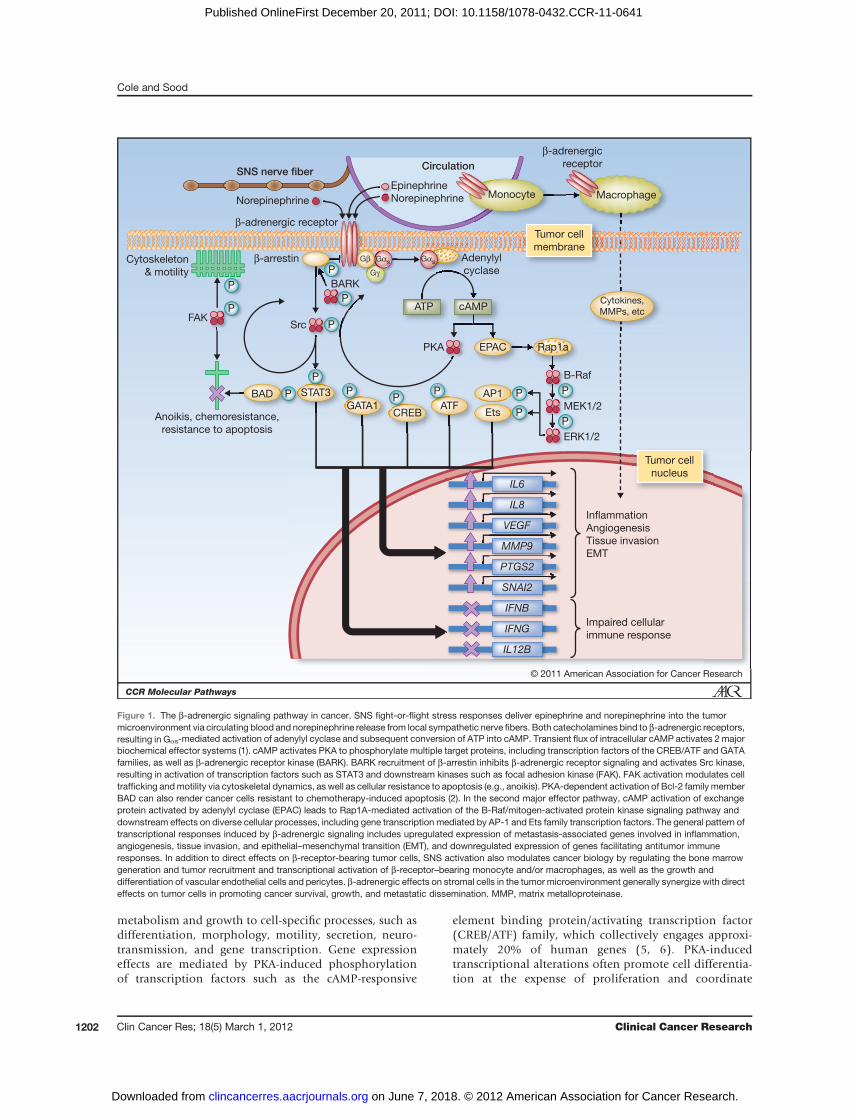
metabolism and growth to cell-specific processes, such asdifferentiation, morphology, motility, secretion, neuro-transmission, and gene transcription. Gene expressioneffects are mediated by PKA-induced phosphorylationof transcription factors such as the cAMP-responsive
element binding protein/activating transcription factor(CREB/ATF) family, which collectively engages approxi-mately 20% of human genes (5, 6). PKA-inducedtranscriptional alterations often promote cell differentia-tion at the expense of proliferation and coordinate
© 2011 American Association for Cancer Research
SNS nerve fiber
Norepinephrine
β-adrenergic receptor
β-arrestin GαsGαsGβ
Gγ
β-adrenergic
receptorCirculation
Tumor cell
membraneAdenylyl
cyclase
ATP cAMP
Rap1aEPAC
Cytokines,
MMPs, etc
PKA
BARK
Src
P
P
P
P
P
P
P
P
P
P
PP P
PAnoikis, chemoresistance,
resistance to apoptosis
STAT3
CREB
AP1
B-Raf
ERK1/2
Tumor cell
nucleus
Inflammation
Angiogenesis
Tissue invasion
EMT
Impaired cellular
immune response
IL12B
IFNG
IFNB
SNAI2
PTGS2
MMP9
VEGF
IL8
IL6
MEK1/2Ets
ATFGATA1BAD
FAK
Cytoskeleton
& motility
MacrophageEpinephrine
Norepinephrine Monocyte
Figure 1. The b-adrenergic signaling pathway in cancer. SNS fight-or-flight stress responses deliver epinephrine and norepinephrine into the tumormicroenvironment via circulating blood and norepinephrine release from local sympathetic nerve fibers. Both catecholamines bind to b-adrenergic receptors,resulting in Gas-mediated activation of adenylyl cyclase and subsequent conversion of ATP into cAMP. Transient flux of intracellular cAMP activates 2 majorbiochemical effector systems (1). cAMP activates PKA to phosphorylate multiple target proteins, including transcription factors of the CREB/ATF and GATAfamilies, as well as b-adrenergic receptor kinase (BARK). BARK recruitment of b-arrestin inhibits b-adrenergic receptor signaling and activates Src kinase,resulting in activation of transcription factors such as STAT3 and downstream kinases such as focal adhesion kinase (FAK). FAK activation modulates celltrafficking andmotility via cytoskeletal dynamics, as well as cellular resistance to apoptosis (e.g., anoikis). PKA-dependent activation of Bcl-2 family memberBAD can also render cancer cells resistant to chemotherapy-induced apoptosis (2). In the second major effector pathway, cAMP activation of exchangeprotein activated by adenylyl cyclase (EPAC) leads to Rap1A-mediated activation of the B-Raf/mitogen-activated protein kinase signaling pathway anddownstream effects on diverse cellular processes, including gene transcription mediated by AP-1 and Ets family transcription factors. The general pattern oftranscriptional responses induced by b-adrenergic signaling includes upregulated expression of metastasis-associated genes involved in inflammation,angiogenesis, tissue invasion, and epithelial–mesenchymal transition (EMT), and downregulated expression of genes facilitating antitumor immuneresponses. In addition to direct effects on b-receptor-bearing tumor cells, SNS activation also modulates cancer biology by regulating the bone marrowgeneration and tumor recruitment and transcriptional activation of b-receptor–bearing monocyte and/or macrophages, as well as the growth anddifferentiation of vascular endothelial cells and pericytes. b-adrenergic effects on stromal cells in the tumor microenvironment generally synergize with directeffects on tumor cells in promoting cancer survival, growth, and metastatic dissemination. MMP, matrix metalloproteinase.
Cole and Sood
Clin Cancer Res; 18(5) March 1, 2012 Clinical Cancer Research1202
on June 7, 2018. © 2012 American Association for Cancer Research. clincancerres.aacrjournals.org Downloaded from
Published OnlineFirst December 20, 2011; DOI: 10.1158/1078-0432.CCR-11-0641
transcriptome-wide responses to stress or homeostatic per-turbation (5). PKA also activates the b-adrenergic receptorkinase (BARK), which subsequently induces b-arrestin totransiently desensitize further b-receptor signaling andactivate the Src/Ras/mitogen-activated protein kinase(MAPK) pathway (7). Recent work also shows that PKAcan directly activate Src (A.K. Sood; unpublished data).A second major cAMP effector involves the guanine
nucleotide exchange protein activated by adenylyl cyclase(EPAC; ref. 8). EPAC activates the Ras-like guanine triphos-phatase Rap1A, which in turn stimulates downstream effec-tors B-Raf, MAP/extracellular signal-regulated kinase (ERK)1/2, and ERK1/2. In addition to the well-known effects ofthe MAPK pathway on cell growth and proliferation, EPACsignaling accounts for many cAMP-induced effects on cellmorphology, motility, and secretion dynamics. The effectsof EPAC can be distinguished from those of PKA throughpharmacologic agonists andmolecularmanipulations (e.g.,target-specific siRNA). Although some overlaps occur,b-adrenergic influences on inflammation, angiogenesis,and invasion seem to be mediated predominantly by PKAinduction of genes encoding cytokines and growth factors,whereas EPAC induces complementary but distinct effectson cell morphology and motility.
Beta-adrenergic regulation of tumor biologyStudies of b-adrenergic influence on tumor biology
were motivated by epidemiologic observations associatingstressful life circumstances with accelerated progression ofincident cancers (9, 10) and studies linking the use ofb-adrenergic antagonists ("b-blockers") with reduced dis-ease progression (11–15). Epidemiologic studies reveal themost consistent relationships between stressful conditionsand progression of already-incident tumors, and relativelylittle data suggest that stress affects the initial incidence ofcancer (9, 10). In vivo laboratorymodels also show themostconsistent effects of experimentally imposed stress on xeno-graft and syngeneic tumor models (i.e., already initiatedtumors; refs. 16, 17), whereas effects on spontaneous inci-dence or primary tumor growth are less common (but dooccur occasionally; refs. 18–21). In mousemodels of breast(22) and prostate carcinomas (16, 17), as well as malignantmelanoma (23, 24) and leukemia (25, 26), b-adrenergicantagonists have been found to block stress-inducedenhancement of tumor progression and/ormetastasis with-out affecting primary tumor growth in vivo or tumor cellproliferation in vitro. b-adrenergic agonists have also beenfound to accelerate in vivo tumor progression andmetastasisin the absence of stress (16, 17, 22, 27).Several cellular andmolecular processes have been found
to mediate b-adrenergic influences on tumor progression(Fig. 1), including recruitment of macrophages into theprimary tumor (22), increased expression of proinflam-matory cytokines such as interleukin-6 (IL-6) and IL-8by tumor cells (28–30) and immune cells (29), VEGF-mediated increases in angiogenesis (27, 31, 32), matrixmetalloproteinase (MMP)–related increases in tissue inva-sion (31, 33, 34), tumor cell mobilization and motility
(17, 35, 36), focal adhesion kinase (FAK)–mediated resis-tance to anoikis apoptosis (37), and BAD-mediated resis-tance to chemotherapy-induced apoptosis (16, 38).b-antagonists alone or in conjunction with nonsteroidalantiinflammatory agents (NSAID) have also been found toinhibit surgery-induced metastasis in animal models(24, 39, 40). Some evidence suggests that b-adrenergicsignaling can also inhibit p53-mediated DNA repair (41),suppress cytotoxic T-lymphocyte and natural killer cellresponses (26), inhibit expression of type I IFNs (22, 42),upregulate the Her2-signaling pathway (43, 44), stimulatearachadonic acid signaling (45), activate gene expression bytumor-promoting viruses (9, 46), and upregulate the SNAI2transcription factor regulating epithelial–mesenchymaltransition (S.W. Cole, S.K. Lutgendorf, and A.K. Sood;unpublished data). Each of the latter dynamics may con-tribute to SNS-induced tumor progression in vivo, but hasnot yet been confirmed to do so in direct inhibitor studies ofmediation. However, it is clear that SNS activation canregulate a wide range of cancer-related molecular pathwaysvia both direct regulation of b-receptor–bearing tumor cellsand regulation of other b-receptor–bearing cells present inthe tumor microenvironment, such as macrophages andvascular cells.
The SNS can potentially regulate tumor b-adrenergicsignaling both via circulating norepinephrine/epinephrineand via local norepinephrine release from SNS nerve fibers.However, growing evidence suggests the latter dynamicplays a dominant role. Analyses of catecholamine levels inhuman ovarian carcinomas document substantially highernorepinephrine levels in tumor tissue than in blood, andthey find no detectable epinephrine in tumor tissue (aswould be expected if blood were the primary source oftumor catecholamines; refs. 47, 48). Intratumor norepi-nephrine levels also correlate with patient psychosocial riskfactors and with tumor gene expression profiles, but bloodlevels of norepinephrine/epinephrine do not (47, 48). Bothobservations suggest a primary role for local nerve fiber-derived norepinephrine in driving b-adrenergic effects ontumor biology. Histologic analyses of catecholaminergicfibers within human breast and ovarian carcinomas showextensive perivascular innervation and occasional radiationof nerve fibers into the tumor parenchyma (E.K. Sloan;unpublished data; ref. 29). This pattern of SNS innervationis similar to that observed in other solid tissues (e.g., lymphnodes; ref. 49) and provides a source of norepinephrine todirectly regulate b-adrenergic receptors on both tumor cellsand stromal cells (particularly tumor-associated macro-phages; ref. 22). Interestingly, data from the lymph nodesetting have shown that chronic stress can increase thedensity of SNS nerve fibers within parenchymal tissue(49). Activated macrophages may also synthesize catecho-lamines (50), but no evidence has yet shown this to occurwithin tumors. An additional pathway by which SNS activ-ity may regulate cancer biology, both within the primarytumor microenvironment and systemically at metastatictarget sites, involves b-adrenergic regulation of myelopoi-esis (51–55) and its effects on monocyte and/or
Beta-Adrenergic Signaling in Cancer
www.aacrjournals.org Clin Cancer Res; 18(5) March 1, 2012 1203
on June 7, 2018. © 2012 American Association for Cancer Research. clincancerres.aacrjournals.org Downloaded from
Published OnlineFirst December 20, 2011; DOI: 10.1158/1078-0432.CCR-11-0641
macrophage trafficking and gene expression (22, 52, 55).This pathway implies that some b-adrenergic influences ontumor biology may originate outside the tumor, via SNSinnervation of the bone marrow hematopoietic environ-ment or catecholamine "conditioning" of traffickingmono-cytes that are ultimately recruited into the tumor microen-vironment (22). Such dynamics would complicate thetargeting of therapeutic interventions based on tumor tissueb-receptor expression, but they also imply that adjuvanttherapy with b-antagonists may suppress systemic supportfor tumor progression.
Clinical–Translational AdvancesBecauseb-adrenergic signalingmodulates tumor progres-
sion via multiple downstream molecular pathways,b-antagonists may provide a highly leveraged adjuvanttherapy strategy with pleiotropic impacts on the primarytumor, its surrounding microenvironment, and metastatictarget sites. The biologic appeal of this concept is enhancedby thewidespread availability of safe, inexpensive, andwell-understood b-antagonists (4). However, several issues needto be resolved to establish the translational potential ofb-blockers as adjuvant therapy for cancer.
The most pressing need involves direct assessment ofb-antagonists’ clinical efficacy in randomized phase II trials.Conflicting results fromcurrently available nonrandomizedobservational studies (11–15, 56) suggest that furtherobservational studies are not likely to definitively establishthe clinical utility of b-blockers in cancer because of meth-odologic difficulties such as (i) confounding by indication(e.g., the primary historic indication for b-blockade, car-diovascular disease, shares common pathophysiologic dri-vers with cancer progression such as smoking, adiposity,and systemic inflammation); (ii) confounding with otherpharmacologic exposures that may affect cancer progres-sion (e.g., angiotensin-converting-enzyme inhibitors); (iii)absence of information on influential risk factors andtreatment parameters (e.g., cardiovascular data sets providelimited informationon cancer progression and/ormortalityrisk factors, and cancer-related data sets provide limitedmeasures of b-blocker agents and/or utilization); and, (iv)time- and practice pattern-related confounding of cancersurvival trends with b-blocker utilization trends (particu-larly for nonselective b-antagonists that aremost likely to beefficacious, as outlined below). Randomized controlledtrials provide the only certain way to overcome such biasesanddefinitively assess the protective effects ofb-antagonists’on clinical cancer progression. The availability of preclinicaldata andapproved, safe, and inexpensiveb-antagonistswithwell-understood pharmacology and minimal side effectsprovides a favorable risk–benefit profile for initial phase IIproof-of-concept trials in clinical oncology.
Clinical trial initiation will require selection of optimaldisease settings and treatment regimens for assessing clin-ical impact. Preclinical laboratorymodels and human phar-maco-epidemiologic studies both suggest thatb-antagonistsare likely to be most effective in inhibiting the micrometa-
static spread of early-stage tumors, as opposed to chemo-prevention of new tumors or reduction of advanced tumorburdens. As such, it makes sense to target tumor types suchas breast or prostate cancer that are routinely detected atearly stage, metastasize via inflammatory and circulatorymechanisms already linked to b-adrenergic signaling, andare sufficiently prevalent to provide high-power detectionofgroup differences amid the low progression and/or recur-rence rates characteristic of early-stagedisease. In the contextof breast cancer, some epidemiologic data suggest thatb-antagonists may be particularly valuable in the contextof estrogen receptor/progesterone receptor/Her2 triple-neg-ative breast cancer (13). Initial trials should also targetdisease settings such as ovarian carcinoma and malignantmelanoma for which extensive preclinical or pharmaco-epidemiologic data already exist and suggest a significanttherapeutic potential.
Optimal b-antagonist regimens also need to be defined,including the specific agent, the timing of its initiation, andthe duration of treatment. Pharmacologic dissection ofpreclinical models of ovarian, breast, and prostate cancerreveal SNS effects to be mediated predominantly by b2- orb3-adrenergic receptors (16, 19, 27, 55). Nonselectiveb-antagonists such as propranolol and nadolol have beenhighly active in these model systems, but the more com-monly prescribed b1-selective agents such as atenolol gen-erally failed to inhibit SNS effects on tumor progression.Similar effects have been observed in pharmaco-epidemi-ologic analyses of breast cancer, with nonselective b-antago-nists showing comparable (13) or greater protective effectsthan b1-selective agents (12). Given these observations, theuse of nonselective antagonists such as propranolol wouldprovide the broadest biologic leverage and minimize therisk of missing an active b-receptor target. CNS adrenergicreceptors seem to play a role in some protective effects ofb-antagonists, suggesting that CNS-penetrant agents such aspropranolol may be preferred over agents that do not crossthe blood–brain barrier such as nadolol. Experimental datashowing that b-antagonists can inhibit surgery-inducedmetastasis (24, 39, 40) suggest initiation prior to surgery(i.e., neoadjuvant) and perhaps in combination with anNSAID. The duration of b-blockade required to reducetumor progression and/or recurrence rates has not beendetermined, but long-term b-blockade has routinely beenused in cardiology and would seem to provide an appro-priate starting point in oncology.
Beta-blocker treatment could potentially be targeted onthe basis of tumor characteristics such as the expressionof b-receptors (57) or their downstream target genes (48),or on the basis of patient characteristics such as stress oranxiety levels (9, 58, 59). However, there is currently noevidence that any patient- or tumor-level characteristicsaffect b-blocker efficacy in clinical oncology. As such,initial randomized clinical trials should target the generaldisease settings in which b-blockade is likely to be mosteffective (as outlined above) and collect additionalpatient- and tumor-specific data to support responderanalyses identifying predictive biomarkers of treatment
Cole and Sood
Clin Cancer Res; 18(5) March 1, 2012 Clinical Cancer Research1204
on June 7, 2018. © 2012 American Association for Cancer Research. clincancerres.aacrjournals.org Downloaded from
Published OnlineFirst December 20, 2011; DOI: 10.1158/1078-0432.CCR-11-0641
efficacy. Several reasons why tumor b-receptor expressionmight not provide an accurate predictive biomarkerinclude the fact that receptor expression does not assessthe amount of SNS norepinephrine/epinephrine ligandimpinging upon the receptor and potential adrenergiceffects at extratumoral sites, such as metastatic targettissues or the bone marrow hematopoietic generation ofsubsequently infiltrating macrophages and lymphocytes(22, 60).Although a variety of translational parameters remain to
be optimized, a growing body of preclinical and pharmaco-epidemiologic data suggests that b-adrenergic antagonistshold considerable promise for inhibiting the pleiotropiceffects of SNS activation on tumor progression and metas-tasis. Over the next few years, we can expect further dataexpanding the range of tumor types examined, identifying
additional mechanisms of b-adrenergic effects on tumorprogression, and initial randomized clinical trials assessingthe efficacy of b-blockade as an adjuvant therapy in clinicaloncology.
Disclosure of Potential Conflicts of InterestNo potential conflicts of interest were disclosed.
AcknowledgmentsSpace constraints have necessitated the omission of many relevant
references.
Grant SupportPreparation of this review was supported by NIH grants CA116778 and
CA109298.
ReceivedOctober 23, 2011; revisedDecember 2, 2011; acceptedDecember2, 2011; published OnlineFirst December 20, 2011.
References1. Weiner H. Perturbing the organism: the biology of stressful experience.
Chicago: University of Chicago Press; 1992.2. Sapolsky RM. Why zebras don't get ulcers: A guide to stress, stress-
related diseases, and coping. New York: Freeman; 1994.3. DalyCJ,McGrath JC.Previously unsuspectedwidespreadcellular and
tissue distribution of b-adrenoceptors and its relevance to drug action.Trends Pharmacol Sci 2011;32:219–26.
4. Baker JG, Hill SJ, Summers RJ. Evolution of b-blockers: from anti-anginal drugs to ligand-directed signalling. Trends Pharmacol Sci2011;32:227–34.
5. Montminy M. Transcriptional regulation by cyclic AMP. Annu RevBiochem 1997;66:807–22.
6. Zhang X, Odom DT, Koo SH, Conkright MD, Canettieri G, Best J, et al.Genome-wide analysis of cAMP-response element binding proteinoccupancy, phosphorylation, and target gene activation in humantissues. Proc Natl Acad Sci U S A 102:4459–64.
7. Luttrell LM, FergusonSS,DaakaY,MillerWE,MaudsleyS,DellaRoccaGJ, et al. Beta-arrestin-dependent formation of beta2 adrenergicreceptor-Src protein kinase complexes. Science 1999;283:655–61.
8. de Rooij J, Zwartkruis FJ, Verheijen MH, Cool RH, Nijman SM, Wittin-ghofer A, et al. Epac is a Rap1 guanine-nucleotide-exchange factordirectly activated by cyclic AMP. Nature 1998;396:474–7.
9. Antoni MH, Lutgendorf SK, Cole SW, Dhabhar FS, Sephton SE,McDonald PG, et al. The influence of bio-behavioural factors ontumour biology: pathways and mechanisms. Nat Rev Cancer2006;6:240–8.
10. Chida Y, Hamer M, Wardle J, Steptoe A. Do stress-related psycho-social factors contribute to cancer incidence and survival? Nat ClinPract Oncol 5:466–75.
11. Powe DG, Voss MJ, Z€anker KS, Habashy HO, Green AR, Ellis IO, et al.Beta-blocker drug therapy reduces secondary cancer formation inbreast cancer and improves cancer specific survival. Oncotarget2010;1:628–38.
12. Barron TI, Connolly RM, Sharp L, Bennett K, Visvanathan K. Betablockers and breast cancermortality: a population- based study. JClinOncol 2011;29:2635–44.
13. Melhem-Bertrandt A, Chavez-Macgregor M, Lei X, Brown EN, Lee RT,Meric-Bernstam F, et al. Beta-blocker use is associatedwith improvedrelapse-free survival in patients with triple-negative breast cancer. JClin Oncol 2011;29:2645–52.
14. De Giorgi V, Grazzini M, Gandini S, Benemei S, Lotti T, Marchionni N,et al. Treatment with b-blockers and reduced disease progression inpatients with thick melanoma. Arch Intern Med 2011;171:779–81.
15. Lemeshow S, Sørensen HT, Phillips G, Yang EV, Antonsen S, Riis AH,et al. b-Blockers and survival among Danish patients with malignantmelanoma: a population-based cohort study. Cancer Epidemiol Bio-markers Prev 2011;20:2273–9.
16. Kulik GA, Hassan S, Karpova Y, Baurin V. Behavioral stressprotects prostate cancer cells from apoptosis. In: Proceedings ofthe 102nd Annual Meeting of the American Association for CancerResearch; 2011 Apr 2–6; Orlando, Florida; Philadelphia (PA): AACR;2011.
17. PalmD, LangK, NiggemannB, Drell TL 4th,Masur K, Zaenker KS, et al.The norepinephrine-driven metastasis development of PC-3 humanprostate cancer cells in BALB/c nude mice is inhibited by beta-block-ers. Int J Cancer 2006;118:2744–9.
18. Hermes GL, Delgado B, Tretiakova M, Cavigelli SA, Krausz T, ConzenSD, et al. Social isolation dysregulates endocrine andbehavioral stresswhile increasing malignant burden of spontaneous mammary tumors.Proc Natl Acad Sci U S A 2009;106:22393–8.
19. CaoL, LiuX, LinEJ,WangC,Choi EY,RibanV, et al. Environmental andgenetic activation of a brain-adipocyte BDNF/leptin axis causes can-cer remission and inhibition. Cell 2010;142:52–64.
20. Boyd AL, Salleh A, Humber B, Yee J, Tomes L, Kerr LR. Neonatalexperiences differentially influence mammary gland morphology,estrogen receptor a protein levels, and carcinogenesis in BALB/cmice. Cancer Prev Res (Phila) 2010;3:1398–408.
21. Schuler LA,AugerAP.Psychosocially influencedcancer: diverse early-life stress experiences and links to breast cancer. Cancer Prev Res(Phila) 2010;3:1365–70.
22. Sloan EK, Priceman SJ,CoxBF, YuS, PimentelMA, TangkanangnukulV, et al. The sympathetic nervous system induces a metastatic switchin primary breast cancer. Cancer Res 2010;70:7042–52.
23. Hasegawa H, Saiki I. Psychosocial stress augments tumor develop-ment through beta-adrenergic activation in mice. Jpn J Cancer Res2002;93:729–35.
24. Goldfarb Y, Sorski L, Benish M, Levi B, Melamed R, Ben-Eliyahu S.Improving postoperative immune status and resistance to cancermetastasis: a combined perioperative approach of immunostimulationand prevention of excessive surgical stress responses. Ann Surg2011;253:798–810.
25. Pimentel MA, Chai MG, Le CP, Cole SW, Sloan EK. Sympatheticnervous system regulation of metastasis. In: Jandial R, Hunter K,editors. Metastatic cancer: integrated organ system and biologicalapproach. Austin, TX: Landes Bioscience; 2012.
26. Inbar S, Neeman E, Avraham R, Benish M, Rosenne E, Ben-Eliyahu S.Do stress responses promote leukemia progression? An animal studysuggesting a role for epinephrine and prostaglandin-E2 throughreduced NK activity. PLoS ONE 2011;6:e19246.
27. Thaker PH, Han LY, Kamat AA, Arevalo JM, Takahashi R, Lu C, et al.Chronic stress promotes tumor growth and angiogenesis in a mousemodel of ovarian carcinoma. Nat Med 2006;12:939–44.
28. NilssonMB, Armaiz-Pena G, Takahashi R, Lin YG, Trevino J, Li Y, et al.Stress hormones regulate interleukin-6 expression by human ovarian
Beta-Adrenergic Signaling in Cancer
www.aacrjournals.org Clin Cancer Res; 18(5) March 1, 2012 1205
on June 7, 2018. © 2012 American Association for Cancer Research. clincancerres.aacrjournals.org Downloaded from
Published OnlineFirst December 20, 2011; DOI: 10.1158/1078-0432.CCR-11-0641
carcinoma cells through a Src-dependent mechanism. J Biol Chem2007;282:29919–26.
29. Cole SW, Arevalo JM, Takahashi R, Sloan EK, Lutgendorf SK, SoodAK, et al. Computational identification of gene-social environmentinteraction at the human IL6 locus. Proc Natl Acad Sci U S A2010;107:5681–6.
30. Shahzad MM, Arevalo JM, Armaiz-Pena GN, Lu C, Stone RL, Moreno-Smith M, et al. Stress effects on FosB- and interleukin-8 (IL8)-drivenovariancancergrowthandmetastasis. JBiolChem2010;285:35462–70.
31. Yang EV, Sood AK, Chen M, Li Y, Eubank TD, Marsh CB, et al.Norepinephrine up-regulates the expression of vascular endothelialgrowth factor, matrix metalloproteinase (MMP)-2, andMMP-9 in naso-pharyngeal carcinoma tumor cells. Cancer Res 2006;66:10357–64.
32. Chakroborty D, Sarkar C, Basu B, Dasgupta PS, Basu S. Catechola-mines regulate tumor angiogenesis. Cancer Res 2009;69:3727–30.
33. LandenCNJr, LinYG, ArmaizPenaGN,DasPD, Arevalo JM, KamatAA,et al. Neuroendocrine modulation of signal transducer and activator oftranscription-3 in ovarian cancer. Cancer Res 2007;67:10389–96.
34. Sood AK, Bhatty R, Kamat AA, Landen CN, Han L, Thaker PH, et al.Stress hormone-mediated invasion of ovarian cancer cells. Clin Can-cer Res 2006;12:369–75.
35. Lang K, Drell TL 4th, Lindecke A, Niggemann B, Kaltschmidt C,Zaenker KS, et al. Induction of a metastatogenic tumor cell type byneurotransmitters and its pharmacological inhibition by establisheddrugs. Int J Cancer 2004;112:231–8.
36. Drell TL4th, JosephJ, LangK,NiggemannB,ZaenkerKS,EntschladenF. Effects of neurotransmitters on the chemokinesis and chemotaxis ofMDA-MB-468 humanbreast carcinoma cells. Breast Cancer Res Treat2003;80:63–70.
37. Sood AK, Armaiz-Pena GN, Halder J, Nick AM, Stone RL, Hu W, et al.Adrenergic modulation of focal adhesion kinase protects humanovarian cancer cells from anoikis. J Clin Invest 2010;120:1515–23.
38. Sastry KS, Karpova Y, ProkopovichS, Smith AJ, Essau B,Gersappe A,et al. Epinephrine protects cancer cells from apoptosis via activation ofcAMP-dependent protein kinase and BAD phosphorylation. J BiolChem 2007;282:14094–100.
39. Glasner A, AvrahamR,RosenneE,BenishM, ZmoraO, ShemerS, et al.Improving survival rates in two models of spontaneous postoperativemetastasis in mice by combined administration of a beta-adrenergicantagonist and a cyclooxygenase-2 inhibitor. J Immunol 2010;184:2449–57.
40. Lee JW, Shahzad MM, Lin YG, Armaiz-Pena G, Mangala LS, Han HD,et al. Surgical stress promotes tumor growth in ovarian carcinoma.ClinCancer Res 2009;15:2695–702.
41. Hara MR, Kovacs JJ, Whalen EJ, Rajagopal S, Strachan RT, Grant W,et al. A stress response pathway regulates DNA damage through b2-adrenoreceptors and b-arrestin-1. Nature 2011;477:349–53.
42. Collado-HidalgoA,SungC,ColeS.Adrenergic inhibition of innate anti-viral response: PKA blockade of Type I interferon gene transcriptionmediates catecholamine support for HIV-1 replication. Brain BehavImmun 2006;20:552–63.
43. Shi M, Liu D, Duan H, Qian L, Wang L, Niu L, et al. The b2-adrenergicreceptor and Her2 comprise a positive feedback loop in human breastcancer cells. Breast Cancer Res Treat 2011;125:351–62.
44. Gu L, Lau SK, Loera S, Somlo G, Kane SE. Protein kinase A activationconfers resistance to trastuzumab in human breast cancer cell lines.Clin Cancer Res 2009;15:7196–206.
45. Cakir Y, PlummerHK3rd, Tithof PK, Schuller HM. Beta-adrenergic andarachidonic acid-mediated growth regulation of human breast cancercell lines. Int J Oncol 2002;21:153–7.
46. ChangM, Brown H, Collado-Hidalgo A, Arevalo J, Galic Z, SymensmaT, et al. Beta-adrenoreceptors reactivate Kaposi's sarcoma-associ-ated herpesvirus lytic replication via PKA-dependent control of viralRTA. J Virol 2005;79:13538–47.
47. Lutgendorf SK, Degeest K, Dahmoush L, Farley D, Penedo F,Bender D, et al. Social isolation is associated with elevated tumornorepinephrine in ovarian carcinoma patients. Brain Behav Immun2011;25:250–5.
48. Lutgendorf SK, DeGeest K, Sung CY, Arevalo JM, Penedo F, Lucci J3rd, et al. Depression, social support, and beta-adrenergic transcrip-tion control in human ovarian cancer. Brain Behav Immun 2009;23:176–83.
49. SloanEK,Capitanio JP, Tarara RP,MendozaSP,MasonWA,ColeSW.Social stress enhances sympathetic innervation of primate lymphnodes: mechanisms and implications for viral pathogenesis. J Neu-rosci 2007;27:8857–65.
50. Flierl MA, Rittirsch D, Nadeau BA, Chen AJ, Sarma JV, Zetoune FS,et al. Phagocyte-derived catecholamines enhance acute inflammatoryinjury. Nature 2007;449:721–5.
51. Cohen MJ, Shankar R, Stevenson J, Fernandez R, Gamelli RL, JonesSB. Bone marrow norepinephrine mediates development of function-ally different macrophages after thermal injury and sepsis. Ann Surg2004;240:132–41.
52. Engler H, Bailey MT, Engler A, Sheridan JF. Effects of repeated socialstress on leukocyte distribution in bone marrow, peripheral blood andspleen. J Neuroimmunol 2004;148:106–15.
53. Laakko T, Fraker P. Rapid changes in the lymphopoietic and granu-lopoietic compartments of the marrow caused by stress levels ofcorticosterone. Immunology 2002;105:111–9.
54. Tang Y, Shankar R, Gamboa M, Desai S, Gamelli RL, Jones SB.Norepinephrine modulates myelopoiesis after experimental thermalinjury with sepsis. Ann Surg 2001;233:266–75.
55. Cole SW. Beta-adrenergic regulation of gene expression in cancer. In:Proceedingsof the 102ndAnnualMeeting of theAmerican Associationfor Cancer Research; 2011 Apr 2–6; Orlando, Florida; Philadelphia(PA): AACR; 2011.
56. Shah SM, Carey IM, Owen CG, Harris T, Dewilde S, Cook DG. Doesb-adrenoceptor blocker therapy improve cancer survival? Findingsfrom a population-based retrospective cohort study. Br J Clin Phar-macol 2011;72:157–61.
57. Powe DG, Voss MJ, Habashy HO, Zanker KS, Green AR, Ellis IO, et al.Alpha- and beta-adrenergic receptor (AR) protein expression is asso-ciated with poor clinical outcome in breast cancer: an immunohisto-chemical study. Breast Cancer Res Treat 2011;130:457–63.
58. AntoniMH, Lutgendorf SK, BlombergB, Stagl J, CarverCS, Lechner S,et al. Transcriptional modulation of human leukocytes by cognitive-behavioral stress management in women undergoing treatment forbreast cancer. Biol Psychiatry 2011 Nov 14. [Epub ahead of print].
59. Lutgendorf SK, Sood AK, Antoni MH. Host factors and cancer pro-gression: biobehavioral signaling pathways and interventions. J ClinOncol 2010;28:4094–9.
60. Lutgendorf SK, Lamkin DM, Jennings NB, Arevalo JM, Penedo F,DeGeest K, et al. Biobehavioral influences onmatrixmetalloproteinaseexpression in ovarian carcinoma. Clin Cancer Res 2008;14:6839–46.
Cole and Sood
Clin Cancer Res; 18(5) March 1, 2012 Clinical Cancer Research1206
on June 7, 2018. © 2012 American Association for Cancer Research. clincancerres.aacrjournals.org Downloaded from
Published OnlineFirst December 20, 2011; DOI: 10.1158/1078-0432.CCR-11-0641
2012;18:1201-1206. Published OnlineFirst December 20, 2011.Clin Cancer Res Steven W. Cole and Anil K. Sood Molecular Pathways: Beta-Adrenergic Signaling in Cancer
Updated version
10.1158/1078-0432.CCR-11-0641doi:
Access the most recent version of this article at:
Cited articles
http://clincancerres.aacrjournals.org/content/18/5/1201.full#ref-list-1
This article cites 52 articles, 23 of which you can access for free at:
Citing articles
http://clincancerres.aacrjournals.org/content/18/5/1201.full#related-urls
This article has been cited by 20 HighWire-hosted articles. Access the articles at:
E-mail alerts related to this article or journal.Sign up to receive free email-alerts
Subscriptions
Reprints and
To order reprints of this article or to subscribe to the journal, contact the AACR Publications Department at
Permissions
Rightslink site. Click on "Request Permissions" which will take you to the Copyright Clearance Center's (CCC)
.http://clincancerres.aacrjournals.org/content/18/5/1201To request permission to re-use all or part of this article, use this link
on June 7, 2018. © 2012 American Association for Cancer Research. clincancerres.aacrjournals.org Downloaded from
Published OnlineFirst December 20, 2011; DOI: 10.1158/1078-0432.CCR-11-0641