Molecular Mechanisms of Pyrimidine Dimer Excision in ...
13
JOURNAL OF BACTEOLOGY, May 1981, p. 692-704 0021-9193/81/050692-13$02.00/0 Vol. 146, No. 2 Molecular Mechanisms of Pyrimidine Dimer Excision in Saccharomyces cerevisiae: Incision of Ultraviolet-Irradiated Deoxyribonucleic Acid In Vivo RICHARD J. REYNOLDSt AND ERROL C. FRIEDBERG Laboratory of Experimental Oncology, Department of Pathology, Stanford University, Stanford, California 94305 Received 4 December 1980/Accepted 23 February 1981 A group of genetically related ultraviolet (UV)-sensitive mutants of Saccha- romyces cerevisiae has been examined in terms of their survival after exposure to UV radiation, their ability to carry out excision repair of pyrimidine dimers as measured by the loss of sites (pyrimidine dimers) sensitive to a dimer-specific enzyme probe, and in terms of their ability to effect incision of their deoxyribo- nucleic acid (DNA) during post-UV incubation in vivo (as measured by the detection of single-strand breaks in nuclear DNA). In addition to a haploid RAD+ strain (S288C), 11 different mutants representing six RAD loci (RADI, RAD2, RAD3, RAD4, RAD14, and RAD18) were examined. Quantitative analysis of excision repair capacity, as determined by the loss of sites in DNA sensitive to an enzyme preparation from M. luteus which is specific for pyrimidine dimers, revealed a profound defect in this parameter in all but three of the strains examined. The radl4-1 mutant showed reduced but significant residual capacity to remove enzyme-sensitive sites as did the rad2-4 mutant. The latter was the only one of three different rad2 alleles examined which was leaky in this respect. The UV-sensitive strain carrying the mutant allele radl8-1 exhibited normal loss of enzyme-sensitive sites consistent with its ament to the RAD6 rather than the RAD3 epistatic group. All strains having mutant alleles of the RADI, RAD2, RAD3, RAD4, and RAD14 loci showed no detectable incubation-dependent strand breaks in nuclear DNA after exposure to UV radiation. These experiments suggest that the RADI, RAD2, RAD3, RAD4 (and probably RAD14) genes are aJl required for the incision of UV-irradiated DNA during pyrimidine dimer excision in vivo. Current models of the molecular mechanism of pyrimidine dimer excision in ultraviolet (UV)- irradiated cells suggest that enzyme-catalyzed incision of deoxyribonucleic acid (DNA) is a very early biochemical event (see references 9 and 10 for recent review). The incision of DNA is thought to be followed by exonucleolytic re- moval of the damaged regions and by synthesis of new DNA using the opposite complementary strand as a template. The excision repair process is completed by the rejoining of newly synthe- sized to extant DNA by DNA ligase. It is obvious that such models predict the presence of single- strand breaks in the DNA of UV-irradiated cells. These single-strand breaks are expected to per- sist until strand rejoining by DNA igase is com- pleted; hence, their detection and subsequent disappearance constitutes a probe for studying the excision of pyrimidine dimers in vivo. t Present address: Department of Physiology, School of Public Health, Harvard University, Boston, MA 02215. The appearance of single-strand breaks in DNA after UV irradiation has been observed in a variety of biological systems including Esche- richia coli (1, 15, 30, 36), bacteriophage X lyso- gens (29, 32), bovine fibroblasts (5), and human fibroblasts (6, 7, 31). The relationship of these DNA strand breaks to the excision of pyrimidine dimers is perhaps best established in E. coli (1, 15, 30, 36). Significantly fewer single-strand breaks are evident in strains of E. coli known to be abnormally UV-sensitive and defective in the excision of pyrimidine dimers, suggesting a re- lationship between the DNA strand breaks and the excision repair of these photoproducts. Fur- ther evidence in support of such a relationship has been provided by Youngs and Smith (36), who demonstrated that the single-strand breaks detected in the DNA of excision-proficient strains of E. coli are photoreactivable, whereas the residual level detected in the DNA of exci- sion deficient strains is not. 692 on January 28, 2018 by guest http://jb.asm.org/ Downloaded from
Transcript of Molecular Mechanisms of Pyrimidine Dimer Excision in ...
JOURNAL OF BACTEOLOGY, May 1981, p. 692-7040021-9193/81/050692-13$02.00/0
Vol. 146, No. 2
Molecular Mechanisms of Pyrimidine Dimer Excision inSaccharomyces cerevisiae: Incision of Ultraviolet-Irradiated
Deoxyribonucleic Acid In VivoRICHARD J. REYNOLDSt AND ERROL C. FRIEDBERG
Laboratory ofExperimental Oncology, Department ofPathology, Stanford University, Stanford, California94305
Received 4 December 1980/Accepted 23 February 1981
A group of genetically related ultraviolet (UV)-sensitive mutants of Saccha-romyces cerevisiae has been examined in terms of their survival after exposure toUV radiation, their ability to carry out excision repair of pyrimidine dimers asmeasured by the loss of sites (pyrimidine dimers) sensitive to a dimer-specificenzyme probe, and in terms of their ability to effect incision of their deoxyribo-nucleic acid (DNA) during post-UV incubation in vivo (as measured by thedetection of single-strand breaks in nuclear DNA). In addition to a haploidRAD+strain (S288C), 11 different mutants representing six RAD loci (RADI, RAD2,RAD3, RAD4, RAD14, and RAD18) were examined. Quantitative analysis ofexcision repair capacity, as determined by the loss of sites in DNA sensitive to anenzyme preparation from M. luteus which is specific for pyrimidine dimers,revealed a profound defect in this parameter in all but three of the strainsexamined. The radl4-1 mutant showed reduced but significant residual capacityto remove enzyme-sensitive sites as did the rad2-4 mutant. The latter was theonly one of three different rad2 alleles examined which was leaky in this respect.The UV-sensitive strain carrying the mutant allele radl8-1 exhibited normal lossof enzyme-sensitive sites consistent with itsament to the RAD6 rather thanthe RAD3 epistatic group. All strains having mutant alleles of the RADI, RAD2,RAD3, RAD4, andRAD14 loci showed no detectable incubation-dependent strandbreaks in nuclearDNA after exposure toUV radiation. These experiments suggestthat the RADI, RAD2, RAD3, RAD4 (and probably RAD14) genes are aJlrequired for the incision of UV-irradiated DNA during pyrimidine dimer excisionin vivo.
Current models of the molecular mechanismof pyrimidine dimer excision in ultraviolet (UV)-irradiated cells suggest that enzyme-catalyzedincision of deoxyribonucleic acid (DNA) is avery early biochemical event (see references 9and 10 for recent review). The incision of DNAis thought to be followed by exonucleolytic re-moval of the damaged regions and by synthesisof new DNA using the opposite complementarystrand as a template. The excision repair processis completed by the rejoining of newly synthe-sized to extant DNA byDNA ligase. It is obviousthat such models predict the presence of single-strand breaks in the DNA of UV-irradiated cells.These single-strand breaks are expected to per-sist until strand rejoining by DNA igase is com-pleted; hence, their detection and subsequentdisappearance constitutes a probe for studyingthe excision of pyrimidine dimers in vivo.
t Present address: Department of Physiology, School ofPublic Health, Harvard University, Boston, MA 02215.
The appearance of single-strand breaks inDNA after UV irradiation has been observed ina variety of biological systems including Esche-richia coli (1, 15, 30, 36), bacteriophage X lyso-gens (29, 32), bovine fibroblasts (5), and humanfibroblasts (6, 7, 31). The relationship of theseDNA strand breaks to the excision ofpyrimidinedimers is perhaps best established in E. coli (1,15, 30, 36). Significantly fewer single-strandbreaks are evident in strains of E. coli known tobe abnormally UV-sensitive and defective in theexcision of pyrimidine dimers, suggesting a re-lationship between the DNA strand breaks andthe excision repair of these photoproducts. Fur-ther evidence in support of such a relationshiphas been provided by Youngs and Smith (36),who demonstrated that the single-strand breaksdetected in the DNA of excision-proficientstrains of E. coli are photoreactivable, whereasthe residual level detected in the DNA of exci-sion deficient strains is not.
692
on January 28, 2018 by guesthttp://jb.asm
.org/D
ownloaded from
PYRIMIDINE DIMER EXCISION IN S. CEREVISIAE 693
The excision repair of UV-irradiated DNA inthe yeast Saccharomyces cerevisiae appears tobe genetically more complex than in procar-yotes. Relative to normal strains, defective ex-cision repair after exposure to UV radiation hasbeen reported in strains mutant at 10 differentloci, including RADI, RAD2, RAD3, RAD4,RAD7, RADIO, RAD14, RAD16, MMSI9, andUVS12 (3, 19-21, 23, 25, 26, 34, 35). With theexception of UVS12 (which has not been exten-sively studied as yet), all of these loci belong toa single epistatic group (generally referred to asthe RAD3 group) and are thus believed to codefor functions involved in the same biochemicalpathway. Two additional loci (CDC8 [22] andCDC9 [14]) may also be involved in the excisionrepair of UV-irradiated DNA; however, the ef-fect of mutations at these loci on the excision ofpyrimidine dimers from nuclear DNA (n-DNA)has not been reported. A mutant of the RAD18locus, a member of the RAD6 epistatic group, isalso abnormally sensitive to killing byUV radia-tion but also has not been directly studied interms of its excision repair capacity. Mutationsthat have been studied at other loci in the RAD6epistatic group are without effect on excision ofpyrimidine dimers (20).The exhaustive genetic analysis of UV radia-
tion sensitivity in S. cerevisiae has not beenaccompanied by biochemical information on theproducts of the various RAD loci referred toabove. Neither has it been precisely establishedat which step in the process of excision repairthe many mutants in the RAD3 epistatic groupare defective. As a prelude to undertaking ex-periments aimed at the isolation and character-ization of DNA repair-specific enzymes fromyeast, we have examined the capacity of a nor-mal strain (RAD+) and a number of mutantstrains to carry out specific steps in the excisionrepair of pyrimidine dimers in vivo. In this paperwe report the results of studies in which we haveused a technique designed to directly measuresmall numbers of DNA single-strand breaks invivo. We present evidence that during the post-UV incubation of normal S. cerevisiae (strainS288C), or of a UV-sensitive strain belonging tothe RAD6 epistatic group (radl8-1), single-strand breaks in n-DNA are readily detectable.All or most of these breaks are not observed inthe DNA of 10 UV-sensitive mutants belongingto the RAD3 epistatic group, suggesting thatthese strains are defective in the incision of theirDNA after exposure to UV radiation. In addi-tion, all except two of these mutants fail to showany detectable loss of sites from their DNAsensitive to a pyrimidine dimer-specific enzymeprobe. To our knowledge this represents the firststudy in which such a large number of UV-
sensitive mutants of S. cerevisiae have beenexamined under identical experimental condi-tions.
MATERIALS AND METHODSYeast strains. The UV-sensitive strains used in
this study were originally isolated by Richard Snow(Department of Genetics, University of California atDavis) and are genetically closely related to the hap-loid RAD+ strain 8288C (33; R. Snow, personal com-munication) that was used as a normal control. Thestrain radl-19 was previously (27) incorrectly desig-nated as Radl-1l. These strains were kindly providedby Dorma Gottlieb and Richard Snow.Media and culturing conditions. A solidified me-
dium (YPD agar) containing 1% (wt/vol) yeast extract(Difco), 2% (wt/vol) peptone (Difco), 2% (wt/vol) glu-cose, and 2% (wt/vol) agar (Difco) was used for main-tenance of cultures and for cell viability determina-tions. Before UV irradiation, cells were routinely cul-tured in a synthetic liquid medium (YNB-D/0.5%)containing 0.67% (wt/vol) yeast nitrogen base withoutamino acids (Difco) and 0.5% (wt/vol) glucose.
For all experiments cultures were inoculated intoYNB-D/0.5% from single colonies grown on YPD agarplates. After 12 to 15 h of growth at 29°C, cultureswere diluted into fresh YNB-D/0.5% at a density of 5x 10⁶ cells/ml, and growth was continued at 29°C withvigorous aeration. Normal exponential growth wasrestored after a single population doubling, and cellsto be examined for the loss of enzyme-sensitive sites(ESS) or for the appearance of single-strand breaks inn-DNA were isotopically labeled. To label DNA, [6-3H]uracil (23 or 27.5 Ci/mmol) or [2-'4C]uracil (54.0mCi/mmol) was added to the culture medium to finalconcentrations of 10 and 2.5 uCi/ml, respectively. Cellswere grown in the labeled medium for approximatelytwo generations (3 to 3.5 h) as suggested by Hatzfeld(12). At the end of the labeling period, cells wereharvested by centrifugation (ca. 400 x g, for 5 min atroom temperature) and suspended in ice-cold 67 mMpotassium phosphate buffer (pH 7.0) at a concentra-tion of 3 x 10' cells/ml.UV irradiation and postirradiation treat-
ments. Cells in 67 mM phosphate buffer (pH 7.0)were irradiated in suspension with stirring at 0°C. Thesource ofUV radiation (predominantly at 254 nm) wasa General Electric germicidal lamp, and the incidentdose rate was determined with an IL 254 germici-dal photometer (International Light, Newburyport,Mass.). The average UV dose rate to cells in suspen-sion was determined from the incident dose rate bythe method of Morowitz (16) but may have beenslightly overcorrected due to light scattering (13). Ir-radiated cells (still in phosphate buffer) were returnedto 29°C and incubated with vigorous aeration forvarious periods from 0 to 3 h. Upon completion ofirradiation and any postirradiation incubations, cellsin phosphate buffer were cooled on ice to 0°C and heldat that temperature until conversion to spheroplasts.
Spheroplast formation. Cells were converted tospheroplasts as described previously (26) except thatreduction with 2-mercaptoethanol was carried out at0°C and cells were incubated with glusalase (EndoLaboratories, Garden City, N.Y.) for 30 min at 30°C.
VOL. 146, 1981
on January 28, 2018 by guesthttp://jb.asm
.org/D
ownloaded from
694 REYNOLDS AND FRIEDBERG
Measurement of cell viability. Viable cell num-bers were determined for unirradiated and for UV-irradiated cultures from the ability of cells to formcolonies on YPD agar plates. Cultures at densities ofapproximately 5 x 10⁷ cells/ml were diluted with 67mM potassium phosphate buffer to the desired celldensities, and samples were spread in duplicate ontoYPD agar plates. Cells on the YPD agar plates wereirradiated at 254 nm and then incubated at roomtemperature for 5 days before direct determination ofcolony numbers.Measurement of single-strand breaks in n-
DNA. The determination of single-strand breaks inyeast n-DNA has been described in detail elsewhere(27). Briefly, log-phase cells grown in YNB-D/0.5%containing either [6-3H]uracil or [2-14C]uracil werewashed free of radioactive medium and suspended in67 mM potassium phosphate buffer (pH 7.0). Thesuspended cells were UV irradiated at 0°C with a doseof 100 J/m2 unless otherwise indicated and then aer-ated at 29°C for varying periods of time. Samples werecollected and held on ice until all incubations werecompleted, at which time they were converted tospheroplasts as described above. The spheroplastswere gently lysed as previously described, and thelysates were layered onto 5 to 20% (wt/vol) alkalinesucrose gradients. After velocity sedimentation, thegradients were fractionated, the amount of radioactiv-ity in each fractionwas measured by liquid scintillationspectrometry, and weight-average molecular weightsof the DNA were calculated from the distribution ofradioactivity through the gradient. The number ofsingle-strand breaks in UV-irradiated DNA was cal-culated relative to the sedimentation profiles of n-DNA from unirradiated, unincubated samples thatwere included in each experiment.Measurement of ESS in n-DNA. Sites sensitive
to attack by a pyrimidine dimer-specific DNA-incisingactivity in extracts of Micrococcus luteus were de-tected and quantitated by a modification of the pro-cedure ofReynolds (26). CelLs were grown as describedabove and irradiated at 3 J/m2 of UV radiation. Pel-leted spheroplasts (2 x 10&) were suspended in 100 plof a solution containing 100 mM NaCL 10 mM ethyl-enedia,inetetraacetic acid, and 50 mM tris(hy-droxymethyl)aminomethane-hydrochloride (pH 7.6).The suspended spheroplasts were lysed immediatelyby the addition of 100 ;1 of 5% (wt/vol) sodium-N-lauroyl sarcosinate (ICN Pharmaceuticals, Inc., Cleve-land, Ohio), 1.0 M NaCL 10 mM ethylenediaminetet-raacetic acid, and 50 mM tris(hydroxymethyl)-aminomethane-hydrochloride (pH 7.6). Lysates (200PI) were deproteinized by extraction with 0.5 ml offresh phenol [neutralized by shaking equal volumes ofwater-saturated phenol and 1.0 M tsis(hydroxymeth-yl)aminomethane-hydrochloride (pH 8.0)]. Beforeseparation of the organic and aqueous phases by cen-trifugation, an additional 100 Al of deionized water wasadded to each sample to reduce the density of theaqueous phase and thereby facilitate separation of thetwo phases. After low-speed centrifugation, the or-ganic phase was removed by aspiration and theaqueous phase plus residual interface material werere-extracted with an additional 0.5 ml of neutrlizedphenol. Approximately 250 pl of the aqueous phase
was recovered after the second extraction and dialyzedin nitrocellulose bags against 20 ml of 50 mM NaCl-1mM ethylenediaminetetraacetic acid-10 mM tris-(hydroxymethyl)aminomethane-hydrochloride (pH7.6), to eliminate residual phenol and to adjust saltand pH to conditions optimal for treatment with theM. luteus UV DNA-incising activity. Samples weredialyzed against three changes of buffer with dialysistimes of approximately 1.5, 6, and 12 h. Preparation ofthe nitrocellulose dialysis bags has been describedpreviously (26).
After dialysis, 100-pl samples were incubated withor without excess amounts of enzyme prepared fromM. luteus as described previously (4, 26). It was re-cently demonstrated that the relevant enzymatic re-action is not the result of a simple endonuclease butrather the combined actions of a pyrimidine-dimerDNA glycosylase and apurinic/apyrimidine endonu-clease(s) found in M. luteus preparations (11). We usethe term "UV DNA-incising activity" to refer to thesecombined activities. Reactions were terminated byplacing samples on ice until just before sedimentation,at which time an additional 100 pl of 20mM ethylene-diaminetetraacetic acid and 1.0 M NaOH were addedto each. Velocity sedimentation of DNA samplesthrough precalibrated alkaline sucrose gradients andcalculation of numbers of ESS from the resultingdistributions of radioactivity through the gradientshave been described elsewhere (26, 27).
RESULTSUV sensitivi of rad mutants. Survival
curves for the strains used in this study (Fig. 1and 2) demonstrate significant differences intheir relative sensitivity to UV radiation. In gen-eral the radl, rad2, rad3, and rad4 mutantsdisplayed similar extreme sensitivities to UVradiation; however, the rad2-4 mutant was sig-nificantly more UV resistant than the rad2-2and rad2-3 mutants. The radl4-1 mutant wasthe least UV sensitive of any of the mutantstrains examined.Presence of single-strand breaks in n-
DNA. The capacity of UV-irradiated S. cerevi-siae to catalyze the formation of single-strandbreaks in DNA was determined primarily byvelocity sedimentation through alkaline sucrosedensity gradients of gently lysed spheroplasts.With this experimental approach the potentialfor shearing of DNA is minimized, and smallnumbers of single-strand breaks can be repro-ducibly demonstrated with careful technique.Typical sedimentation profiles for n-DNA fromthe excision-proficient (RAWD) haploid strainS288C are depicted in Fig. 3A. Nuclear DNAreleased by lysis of spheroplasts of unirradiatedcells sedimented with a weight-average molecu-lar weight of 173 x 106. DNA released from cellsirradiated at 00C with 100 J of UV light per m2before spheroplast formation sedimented moreslowly, with a weight-average molecular weight
J. BACTERIOL.
on January 28, 2018 by guesthttp://jb.asm
.org/D
ownloaded from

PYRIMIDINE DIMER EXCISION IN S. CEREVISIAE 695
c0
U.
CP
a
0 40 80 120Dose (Jm-2)
FIG. 1. Colony-forming ability after UV irradia-tion. Symbols: radl-19 (O), rad2-4 (A), radl4-1 (0),and S288C (RAD) (0).
of 83 x 106. Incubation of cells at 290C afterirradiation but before conversion of the cells tospheroplasts resulted in a further time-depend-ent decrease in the sedimentation rate of n-
DNA. As shown in Fig. 3A, n-DNA from normalcells incubated for 3 h after irradiation sedi-mented with an apparent weight-average molec-ular weight of 61 x 106. This time-dependentreduction in molecular weight in the excision-proficient strain was strictly dependent on in-cubation at temperatures above 00C. No reduc-tion in molecular weight beyond that observedin the DNA from cells converted to spheroplastsimmediately after irradiation was evident whenirradiated celLs were held for up to 24 h at 00Cbefore being converted to spheroplasts (data notshown).
Results obtained with the UV-sensitive radl-2 mutant (Fig. 3B) are representative of manyof the excision-defective strains examined. Thesedimentation profile of the n-DNA releasedfrom spheroplasts of unirradiated radl-2 cells
was indistinguishable from that obtained withunirradiated normal cells. Irradiation of radl-2cells with 100 J of UV light per m2 reduced thesedimentation rate of the n-DNA to a weight-average molecular weight of84 x 106. In contrast
to the results obtained with the normal RAD+strain S288C, however, no further reduction inthe size of the n-DNA was evident when irradi-ated radl-2 cells were incubated at 290C beforeconversion to spheroplasts.Comparison of weight-average molecular
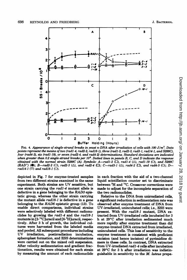
weights calculated from sedimentation profilessimilar to those depicted in Fig. 3 allowed us todetermine the average number of single-strandbreaks in n-DNA induced by a particular exper-imental protocol. The results of such compari-sons for all of the strains examined are shown inFig. 4 and suggest the presence of two classes ofDNA strand breaks after exposure of S. cerevi-siae to UV radiation. Relative to unirradiatedcontrols, small numbers of single-strand breakswere consistently detected in the n-DNA of cellsirradiated and maintained at 00C until theirconversion to spheroplasts. We refer to this classas "incubation-independent" DNA single-strandbreaks. In repeated experiments, the averagenumber of incubation-independent DNA single-strand breaks in the normal strain and in mostUV-sensitive mutant strains was - 1.2 single-strand breaks per 108 molecular weight afterirradiation at 100 J/m2. This value representsabout 1 break for every 400 pyrimidine dimersin n-DNA.A second class of single-strand breaks ap-
peared in the n-DNA of some strains duringincubation at 290C after exposure to UV radia-tion. We designate these as "incubation-depend-ent" single-strand breaks. In the normal strain,between 1 and 2 such single-strand breaks per108 molecular weight were detected during thefirst 3 h of incubation after irradiation with 100J ofUV light per M2. In previous studies (27) wedemonstrated that the number of this class ofstrand breaks was significantly reduced if cellswere incubated under conditions permissive forenzymatic photoreactivation. The photoreactiv-ability of these lesions indicates that they arerelated to the presence of pyrimidine dimers inn-DNA. With the exception of radl8-1, none ofthe mutant strains presented in Fig. 4 exhibitedany detectable incubation-dependent single-strand breaks in n-DNA in repeated experi-ments. The radl8-1 mutant (Fig. 4D) differedfrom the RAD+ strain S288C in that the incu-bation-dependent class of DNA single-strandbreaks appeared with faster kinetics and wasquantitatively greater over the 3-h incubationperiod examined. Preliminary experiments withthe UV-sensitive mutant rad7-1 yielded resultsindistinguishable from those observed withstrain S288C (data not shown). However, theexperimental conditions used with the rad7-1strain were slightly different from those gener-ally employed in these experiments, and further
VOL. 146, 1981
on January 28, 2018 by guesthttp://jb.asm
.org/D
ownloaded from
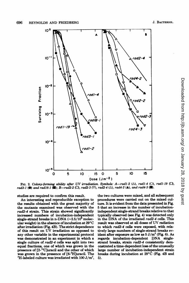
696 REYNOLDS AND FRIEDBERG J. BACTERIOL.
100
10'I \ trod2-4
\ r 2rod4-2O S 10 A
\A0 ~~~~~rodl-4
G .2 rUoi\d rad2-42
\B rad3-/ A rad4-3
rodl-2
lo-511 1 1 ~~~~~~~~II I
0 5 10 IS 0 5 10 15
Dose (Jm-2)FIG. 2. Colony-forming ability after UV irradiation. Symbols: A-radl-2 (A), radl-4 (0), radl-19 (0),
rad3-1 (0) and rad18&1 (U); B-rad2-2 (01), rad2-3 (V), rad2-4 (A), rad4-2 (A), and rad4-3 (U).
studies are required to confirn this result.An interesting and reproducible exception to
the results obtained with the great majority ofthe mutants examined was observed with therad2-4 strain. This strain showed significantlyincreased numbers of incubation-independentsingle-strand breaks in n-DNA (-2.5/108 molec-ular weight) in the absence ofincubation at 290Cafter irradiation (Fig. 4B). The strict dependenceof this result on UV irradiation as opposed toany other variable in the experimental protocolwas demonstrated in an experiment in which asingle culture of rad2-4 cells was split into twoequal fractions, one of which was grown in thepresence of [2-14C]uracil and the other of whichwas grown in the presence of [6-3H]uracil. The3H-labeled culture was irradiated with 100 J/m2,
the two cultures were mixed, and all subsequentprocedures were carried out on the mixed cul-ture. It is evident from the data presented in Fig.5 that an increase in the number of incubation-independent single-strand breaks relative to thattypically observed (see Fig. 4) was detected onlyin the DNA of the irradiated rad2-4 cells. Thisresult was observed at all doses of UV radiationto which rad2-4 cells were exposed, with rela-tively large numbers of single-strand breaks ev-ident after exposure as low as 5 J/m2 (Fig. 6). Asregards incubation-dependent DNA single-strand breaks, strain rad2-4 consistently dem-onstrated a time-dependent loss of the unusuallylarge number of incubation-independent strainbreaks during incubation at 290C (Fig. 4B and5).
on January 28, 2018 by guesthttp://jb.asm
.org/D
ownloaded from
PYRIMIDINE DIMER EXCISION IN S. CEREVISIAE 697
10 Iu0
8
6 _
ffi00 .
0~~~~0
0
aera0ion ofocellsat 29 10s B
41
8
a.
6 A0~\9
0\~%
00
2
0
1.0 .8 .6 4 .2 0
Distance Sedimented
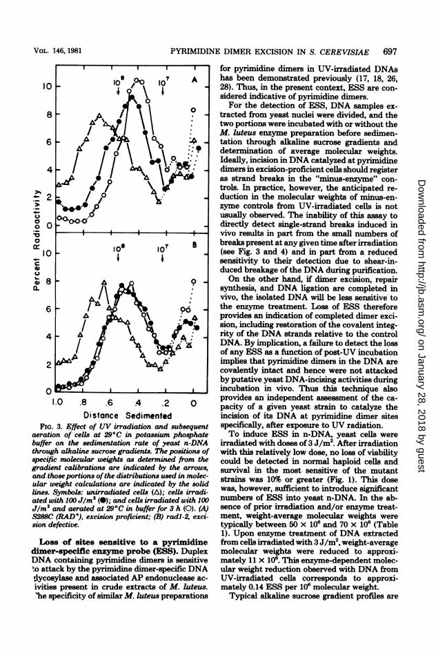
FIG. 3. Effect of UV irradiation and subsequentaeration of cells at 290C in potassium phosphate
buffer on the sedimentation rate of yeast n-DNAthrough alkaline sucrose gradients. The positions ofspecific molecular weights as determined from thegradient calibrations are indicated by the arrows,and those portions ofthe distributions used in molec-ular weight calculations are indicated by the solidlines. Symbols: unirradiated cells (A); cells irradi-ated with 100 J/m2 (-); and cells irradiated with 100J/m2 and aerated at 29°C in buffer for 3 h (0). (A)S288C (RAD+), excision proficient; (B) radl-2, exci-sion defective.
Loss of sites sensitive to a pyrimidinedimer-specific enzyme probe (ESS). DuplexDNA containing pyrimidine dimers is sensitiveto attack by the pyrimidine dimer-specific DNAdlycosylase and associated AP endonuclease ac-ivities present in crude extracts of M. luteus.'he specificity of similar M. luteus preparations
for pyrimidine dimers in UV-irradiated DNAshas been demonstrated previously (17, 18, 26,28). Thus, in the present context, ESS are con-sidered indicative of pyrimidine dimers.For the detection of ESS, DNA samples ex-
tracted from yeast nuclei were divided, and thetwo portions were incubated with or without theM. luteus enzyme preparation before sedimen-tation through alkaline sucrose gradients anddetermination of average molecular weights.Ideally, incision in DNA catalyzed at pyrimidinedimers in excision-proficient cells should registeras strand breaks in the "minus-enzyme" con-trols. In practice, however, the anticipated re-duction in the molecular weights of minus-en-zyme controls from UV-irradiated cells is notusually observed. The inability of this assay todirectly detect single-strand breaks induced invivo results in part from the small numbers ofbreaks present at any given time after irradiation(see Fig. 3 and 4) and in part from a reducedsensitivity to their detection due to shear-in-duced breakage of the DNA during purification.On the other hand, if dimer excision, repair
synthesis, and DNA ligation are completed invivo, the isolated DNA will be less sensitive tothe enzyme treatment. Loss of ESS thereforeprovides an indication of completed dimer exci-sion, including restoration of the covalent integ-rity of the DNA strands relative to the controlDNA. By implication, a failure to detect the lossof any ESS as a function of post-UV incubationimplies that pyrimidine dimers in the DNA arecovalently intact and hence were not attackedby putative yeast DNA-incising activities duringincubation in vivo. Thus this technique alsoprovides an independent assessment of the ca-pacity of a given yeast strain to catalyze theincision of its DNA at pyrimidine dimer sitesspecifically, after exposure to UV radiation.To induce ESS in n-DNA, yeast cells were
irradiated with doses of 3 J/m2. After irradiationwith this relatively low dose, no loss of viabilitycould be detected in normal haploid cells andsurvival in the most sensitive of the mutantstrains was 10% or greater (Fig. 1). This dosewas, however, sufficient to introduce significantnumbers of ESS into yeast n-DNA. In the ab-sence of prior irradiation and/or enzyme treat-ment, weight-average molecular weights weretypically between 50 x 106 and 70 x 106 (Table1). Upon enzyme treatment of DNA extractedfrom cells irradiated with 3 J/m2, weight-averagemolecular weights were reduced to approxi-mately 11 x 106. This enzyme-dependent molec-ular weight reduction observed with DNA fromUV-irradiated cells corresponds to approxi-mately 0.14 ESS per 106 molecular weight.
Typical alkaline sucrose gradient profiles are
VOL. 146, 1981
on January 28, 2018 by guesthttp://jb.asm
.org/D
ownloaded from
698 REYNOLDS AND FRIEDBERG
a0
l
**1
UU)
(I
0 I 2 3 0 1 2 3Buffer Holding (Hours)
FIG. 4. Appearance of single-strand breaks in yeast n-DNA after irradiation of cells with 100 Jlm2. Datapoints represent the means oftwo (radl -4, rad2-2, radl8-1), three (radl -2, rad2-3, rad3-1, radl4-1, and S288C),four (rad4-3), six (radl-19), or seven (rad2-4, and rad4-2) determinations. Standard deviations are indicatedwhen greater than 0.2 single-strand breaks per 108. Dotted lines in panels B, C, and D indicate the responseobtained with the normal strain S288C (A). Symbols: A-radl-2 (0), radl-4 (A), radl-19 (0), and S288C(RAD+) (-); B-rad2-2 (0), rad2-3 (A), and rad2-4 (]); C-rad3-1 (A), rad4-2 (0), and rad4-3 (0); D-radl4-1 (V) and radl8-1 (0).
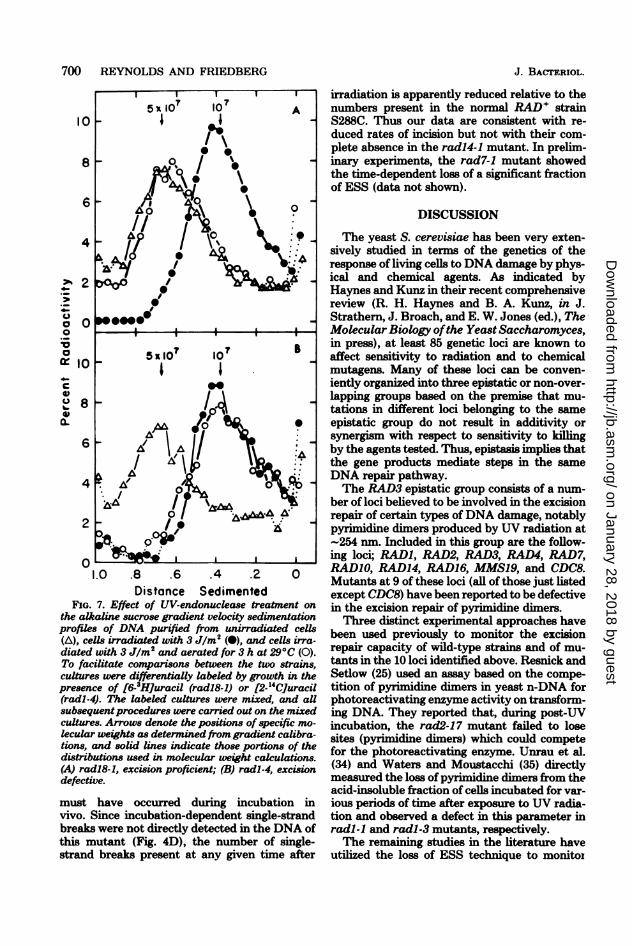
depicted in Fig. 7 for enzyme-treated samplesfrom two different strains examined in the sameexperiment. Both strains are UV sensitive, butone strain carrying the radl-4 mutant allele isdefective in a gene belonging to the RAD3 epis-tatic group, whereas the other strain carryingthe mutant allele radl8-1 is defective in a genebelonging to the RAD6 epistatic group (13). Toenable direct comparisons, individual strainswere selectively labeled with different radionu-clides by growing the radl-4 and the radl8-1mutants in [2-14C]uracil and [6-3H]uracil, respec-tively. After 3 h of growth, the individual cul-tures were harvested from the labeled mediaand pooled. All subsequent procedures includingUV irradiation, postirradiation incubation,spheroplast formation, and ESS determinationswere carried out on the mixed cell suspension.After velocity sedimentation and gradient frac-tionation, results were obtained for each strainby measuring the amount of each radionuclide
in each fraction with the aid of a two-channelliquid scintillation counter set to discriminatebetween 3H and 14C. Crossover corrections weremade to adjust for the incomplete separation ofthe two radionuclides.
Relative to the DNA from unirradiated cells,a significant reduction in sedimentation rate wasobserved after enzyme treatment of DNA fromUV-irradiated, unincubated cells; i.e., ESS werepresent. With the radl8-1 mutant, DNA ex-tracted from UV-irradiated cells incubated for 3h at 29°C after irradiation sedimented muchmore rapidly after enzyme treatment than didenzyme-treated DNA extracted from irradiated,unincubated cells. This loss of sensitivity to theenzyme treatment is consistent with proficientexcision (and hence incision) of pyrimidine di-mers in these cells. In contrast, DNA extractedfrom UV-irradiated radl-4 cells after incubationat 290C for 3 h after irradiation was indistin-guishable in sensitivity to the M. luteus prepa-
J. BACTERIOL.
on January 28, 2018 by guesthttp://jb.asm
.org/D
ownloaded from

PYRIMIDINE DIMER EXCISION IN S. CEREVISIAE 699
1 2Buffer Holding (Hours)
FIG. 5. Dependence of single-strand breaks inrad2-4 upon prior UV irradiation. A single culture ofexponentially growing cells was divided, and growthwas continued in media containing either [6-3Hlur-acil or [2-14Cluracil for two generations. The 3H-labeled cells (0) were either left unirradiated orirradiated with 100 J/m2 and then mixed with theunirradiated "4C-labeled cells (0). All subsequentprocedures were carried out on the mixed culture.
4
3~~~~~~~~~~~~~~0
0~~ ~ ~ ~ ~~rd-
0 10 25 50 100 200Dose (J m-2)
FIG. 6. Dose response for the appearance of singlestrand breaks in the n-DNA of rad2-4 and radl-19.Cultures of rad2-4 (0) and radl-19 (0) were grownin the presence of [2-14C]uracil and [6-3Hluracil,respectively, and then pooled. Irradiation of the cellsand all subsequent procedures were carried out on
the pooled culture.
TABLE 1. ESS in the n-DNA of mutants radl8-1and radl-4a
Post- 106 Mol wt % Total
Stan Dose UV in- ESS/10c6 ESSW(Jm2) cuba-;-Enz. +Enz. (100%-tion (h) _____0.140)
radl8-1 0 58.2 45.3 0.0103 0 53.2 13.1 0.115 823 3 52.5 38.7 0.014 10
radl-4 0 62.4 52.4 0.0073 0 58.5 11.5 0.140 1003 3 57.8 12.0 0.132 94
.ESS were determined from a comparison of DNAaverage molecular weights after incubation of DNAsamples with (+ Enz.) or without (- Enz.) M. luteusextracts as described in the text.
ration from DNA extracted from irradiated, un-incubated cells. The absence of a time-depend-ent change in sensitivity to the enzyme treat-ment indicates a defect in the excision repair ofpyrimidine dimers in the radl-4 mutant.A quantitative analysis of the experiment de-
picted in Fig. 7 is presented in Table 1. Weight-average molecular weights for the minus-enzymecontrols ranged from 52.5 x 10⁶ to 64.3 x 10⁶ inthis experiment without significant deviationupon irradiation or postirradiation treatments ofcelLs. Treatment of DNA from unirradiated cellswith the M. luteus enzyme preparation revealeda small but reproducible amount of activityagainst both 14C- and 3H-labeled DNAs in theabsence of prior cellular irradiation. This deg-radation was very limited with freshly preparedDNA and probably represents activity againstDNA damage resulting from radionuclide decay(W. L. Carrier, personal communication). Simi-lar activity was not detected in a previous studyin which a more rapid DNA purification proce-dure was used (26). The numbers of sites attrib-utable to this activity were never greater than0.01 sites per 10i molecular weight, and all datapresented are uncorrected for this activity.The kinetics of the loss of ESS in a number of
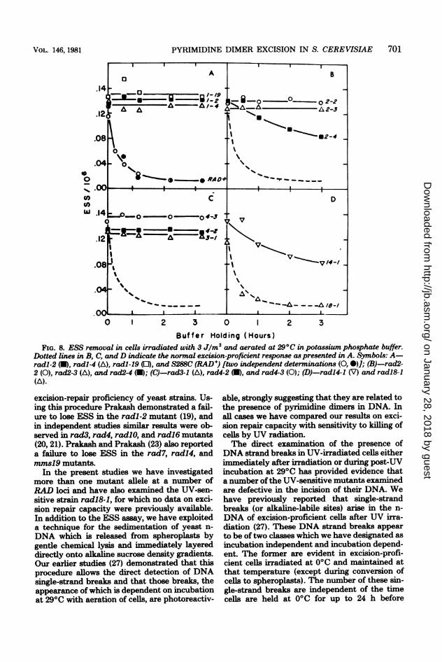
strains used in this study are presented in Fig. 8.Strains radl-2, radl-4, radl-19, rad2-2, rad2-3,rad3-1, rad4-2, and rad4-3 exhibited no signifi-cant loss of ESS during 3 h of post-UV incuba-tion. The rad2-4 (Fig. 8B), radl4-1 (Fig. 8D),and radl8-1 (Fig. 8D) strains did show signifi-cant loss of ESS as a function of the time ofincubation. Both of the former two strains lostabout 40% of total ESS during 3 h of incubationat 290C, whereas the kinetics of loss of sites fromthe DNA of strain radl8-1 were indistinguish-able from those of the normal RAD+ strainS288C. The loss of ESS from the DNA of theradl4-1 mutant suggests that incision events
4
3
0
I I I I
0
o~ -0+ UV
-
- UV0-@ 0@, 0*
-UV 0
VOL. 146, 1981
on January 28, 2018 by guesthttp://jb.asm
.org/D
ownloaded from
700 REYNOLDS AND FRIEDBERG
10
8
6
4
z.,2
oO
:0._.
0
0
a-
8
6
4
2
0
1.0 .8 .6 .4 .2 0Distance Sedimented
FIG. 7. Effect of UV-endonuclease treatment onthe alkaline sucrose gradient velocity sedimentationprofiles of DNA purified from unirradiated cells(A), cells irradiated with 3 Jlm2 (0), and cells irra-diated with 3 JIm2 and aerated for 3 h at 29°C (0).To facilitate comparisons between the two strains,cultures were differentially labeled by growth in thepresence of [6-3HJuracil (radi8-1) or [2-14CJuracil(radl-4). The labeled cultures were mixed, and aUsubsequentprocedures were carried out on the mixedcultures. Arrows denote the positions of specific mo-lecular weights as determined from gradient calibra-tions, and solid lines indicate those portions of thedistributions used in molecular weight calculations.(A) radl8-1, excision proficient; (B) radl-4, excisiondefective.
must have occurred during incubation invivo. Since incubation-dependent single-strandbreaks were not directly detected in the DNA ofthis mutant (Fig. 4D), the number of single-strand breaks present at any given time after
J. BACrERIOL.
irradiation is apparently reduced relative to thenumbers present in the normal RAD' strainS288C. Thus our data are consistent with re-duced rates of incision but not with their com-plete absence in the radl4-1 mutant. In prelim-inary experiments, the rad7-1 mutant showedthe time-dependent loss of a significant fractionof ESS (data not shown).
DISCUSSION
The yeast S. cerevisiae has been very exten-sively studied in terms of the genetics of theresponse of living cells to DNA damage by phys-ical and chemical agents. As indicated byHaynes and Kunz in their recent comprehensivereview (R. H. Haynes and B. A. Kunz, in J.Strathem, J. Broach, and E. W. Jones (ed.), TheMolecular Biology ofthe Yeast Saccharomyces,in press), at least 85 genetic loci are known toaffect sensitivity to radiation and to chemicalmutagens. Many of these loci can be conven-iently organized into three epistatic or non-over-lapping groups based on the premise that mu-tations in different loci belonging to the sameepistatic group do not result in additivity orsynergism with respect to sensitivity to killingby the agents tested. Thus, epistasis implies thatthe gene products mediate steps in the sameDNA repair pathway.The RAD3 epistatic group consists of a num-
ber of loci believed to be involved in the excisionrepair of certain types ofDNA damage, notablypyrimidine dimers produced by UV radiation at-254 nm. Included in this group are the follow-ing loci; RADI, RAD2, RAD3, RAD4, RAD7,RADIO, RAD14, RAD16, MMSI9, and CDC8.Mutants at 9 of these loci (all of those just listedexcept CDC8) have been reported to be defectivein the excision repair of pyrimidine dimers.Three distinct experimental approaches have
been used previously to monitor the excisionrepair capacity of wild-type strains and of mu-tants in the 10 loci identified above. Resnick andSetlow (25) used an assay based on the compe-tition of pyrimidine dimers in yeast n-DNA forphotoreactivating enzyme activity on transform-ing DNA. They reported that, during post-UVincubation, the rad2-17 mutant failed to losesites (pyrimidine dimers) which could competefor the photoreactivating enzyme. Unrau et al.(34) and Waters and Moustacchi (35) directlymeasured the loss of pyrimidine dimers from theacid-insoluble fraction of cells incubated for var-ious periods of time after exposure to UV radia-tion and observed a defect in this parameter inradl-1 and radl-3 mutants, respectively.The remaining studies in the literature have
utilized the loss of ESS technique to monitoI
on January 28, 2018 by guesthttp://jb.asm
.org/D
ownloaded from
PYRIMIDINE DIMER EXCISION IN S. CEREVISIAE 701
.14
.12(
.08
.04~
I I I
A
A A
%%8'-- 0 --,gRAD
B
a-o ° O2-21-%.. AL2-3
i --I a 2-U162
'4
i.14 0-0o- ~o#-4;V5
.08-
o~~~~~~~~ E.04o , .
0 1 2 3 0 1 2 3
Buffer Holding (Hours)FIG. 8. ESS removal in cells irradiated with 3 J/m2 and aerated at 290C in potassium phosphate buffer.
Dotted lines in B, C, and D indicate the normal excision-proficient response as presented in A. Symbols: A-radl-2 (U), radl-4 (A), radl-19 (0), and S288C (RAD +) [two independent determinations (0, )]; (B)-rad2-2 (0), rad2-3 (A), and rad2-4 (U); (C)-rad3-1 (A), rad4-2 (U), and rad4-3 (0); (D)-radl4-1 (V) and radl8-1(A).
excision-repair proficiency of yeast strains. Us-ing this procedure Prakash demonstrated a fail-ure to lose ESS in the radl-2 mutant (19), andin independent studies similar results were ob-served in rad3, rad4, radlO, and radl6 mutants(20, 21). Prakash and Prakash (23) also reporteda failure to lose ESS in the rad7, radl4, andmmsI9 mutants.
In the present studies we have investigatedmore than one mutant allele at a number ofRAD loci and have also examined the UV-sen-sitive strain radl8-1, for which no data on exci-sion repair capacity were previously available.In addition to the ESS assay, we have exploiteda technique for the sedimentation of yeast n-
DNA which is released from spheroplasts bygentle chemical lysis and immediately layereddirectly onto alkaline sucrose density gradients.Our earlier studies (27) demonstrated that thisprocedure allows the direct detection of DNAsingle-strand breaks and that those breaks, theappearance ofwhich is dependent on incubationat 290C with aeration of cells, are photoreactiv-
able, strongly suggesting that they are related tothe presence of pyrimidine dimers in DNA. Inall cases we have compared our results on exci-sion repair capacity with sensitivity to killing ofcells by UV radiation.The direct examination of the presence of
DNA strand breaks in UV-irradiated cells eitherimmediately after irradiation or during post-UVincubation at 290C has provided evidence thata number of the UV-sensitive mutants examinedare defective in the incision of their DNA. Wehave previously reported that single-strandbreaks (or alkaline-labile sites) arise in the n-
DNA of excision-proficient cells after UV irra-diation (27). These DNA strand breaks appearto be of two classes which we have designated as
incubation independent and incubation depend-ent. The former are evident in excision-profi-cient cells irradiated at 00C and maintained atthat temperature (except during conversion ofcells to spheroplasts). The number of these sin-gle-strand breaks are independent of the timecells are held at 00C for up to 24 h before
00
U)'U
VOL. 146, 1981
no%
I111.,.
on January 28, 2018 by guesthttp://jb.asm
.org/D
ownloaded from
702 REYNOLDS AND FRIEDBERG
spheroplast formation and are independent ofthe duration of the steps involved in spheroplastformation. With the exception of the rad2-4mutant, similar numbers of incubation-inde-pendent single-strand breaks were observed inall strains examined. We believe it likely thatthese strand breaks are the result of direct pho-tochemical nondiimer damage to DNA (24).However, further studies are in progress to ex-amine their origin.The second class of DNA strand breaks ob-
served in excision-proficient yeast is dependenton aeration of irradiated cells at 29°C. The num-ber of these breaks is UV dose dependent; how-ever, after longer times of incubation they de-crease, consistent with the notion that they rep-resent transient intermediates in the excisionrepair of pyrimidine dimers. This conclusion isfurther supported by the demonstration of theirphotoreactivability. The absence of this class ofDNA strand breaks in mutants of five of thegenetic loci examined suggests that all five loci(RADI, RAD2, RAD3, RAD4, and RAD14) areinvolved in the incision step of pyrimidine dimerexcision. This conclusion is substantiated by theobservation of incubation-dependent single-strand breaks in the DNA of the excision-profi-cient, UV-sensitive radl8-1 mutant. In prelimi-nary studies we have also observed incubation-dependent strand breaks in the DNA of theexcision-defective rad7-1 mutant. Whether ornot this result represents leakiness of this partic-ular strain, or is characteristic of all mutants atthe RAD7 locus, needs to be established byfurther study.
Strains that failed to demonstrate the pres-ence ofDNA strand breaks by this direct exper-imental approach would also be expected toretain all ESS in their DNA. Quantitative ex-amination of this parameter revealed that mostof the mutants defective in loci belonging to theRAD3 epistatic group had extreme effects onexcision repair capacity after irradiation of cellswith low doses of UV. The severely affectedmutants carried the mutant alleles radl-2, radl-4, radl-19, rad2-2, rad2-3, rad3-1, rad4-2, andrad4-3. No significant loss of ESS could be de-tected in any of these strains during incubationfor 3 h after irradiation at 3 J/m2. The effects ofthese mutant alleles on excision repair of UVdamage is consistent with their extreme UVsensitivities. Two strains carrying the mutantalleles rad2-4 and radl4-1 were able to removea significant fraction of ESS from their DNA,although clearly less than the normal strain,despite the fact that neither strain showedclearly demonstrable incubation-dependentDNA strand breaks. Interestingly, these twostrains were also found to have intermediate UV
sensitivities relative to the extremely UV-sensi-tive strains and to the normal RAD+ strainS288C. The results obtained with the rad2-4strain relative to the other rad2 mutants exam-ined suggest that incomplete molecular blocksin the pathway ofpyrimidine dimer excision maybe reflected in biological endpoints such as sur-vival. The same may be true of the radl4-1mutant, although other alleles at the RAD14locus are not yet available for study. The possi-bility of such "leakiness" should be an importantconsideration in the selection of strains for bio-logical and genetic studies.A UV-sensitive strain carrying a mutant allele
of the RAD18 gene was also examined for ESSremoval. Although the radl8-1 mutant was ex-tremely UV sensitive in our hands, the demon-stration of normal kinetics for the loss of ESS isnot unexpected since the RAD18 locus is a mem-ber of the RAD6 epistatic group, all members ofwhich are thought to be excision repair profi-cient. Normal pyrimidine dimer excision has alsobeen demonstrated in rad6 and rad9 mutants(20).
In conclusion, our studies indicate that a sig-nificant component of the genetic complexityassociated with excision repair of pyrimidinedimers in S. cerevisiae is concentrated aroundthe molecular events required for the incision ofUV-irradiated DNA in vivo. This study providesclear evidence that at least four genetic loci(RADI, RAD2, RAD3, and RAD4) are involvedin this process. This conclusion is supported bythe results of our earlier studies (27) in whichwe showed that mutants defective in the RADI,RAD2, RAD3, and RAD4 loci all contain normallevels of enzyme activity that in a cell-free sys-tem catalyzes the selective excision of thymine-containing pyrimidine dimers from UV-irradi-ated E. coli DNA previously incised by thedimer-specific UV DNA-incising activity of M.luteus. Further studies on the dimer-excisingactivity in cell-free systems will be reported else-where (R. J. Reynolds and E. C. Friedberg,manuscript in preparation). These conclusionsare not in agreement with the recent report byBekker et al. (2) who claim to have demon-strated normal levels of "UV endonuclease" ac-tivity in extracts of a number of rad mutants ofS. cerevisiae. Our results with mutants at theRAD7 and RAD14 loci are not definitive, norhave we yet examined mutants at the RADIO,RAD16, or MMS19 loci. Thus, potentially anadditional five genetic loci may be involved inevents associated with or preceding DNA inci-sion.The genetic complexity associated with the
very early events in the excision repair of pyrim-idine dimers in S. cerevisiae bears a striking
J. BACTERIOL.
on January 28, 2018 by guesthttp://jb.asm
.org/D
ownloaded from
PYRIMIDINE DIMER EXCISION IN S. CEREVISIAE 703
analogy to that demonstrated in human cells bythe study of cell lines obtained from patientswith xeroderma pigmentosum (see reference 8for a recent review). At least seven complemen-tation groups (exclusive of the xeroderna pig-mentosum variant form) have been documentedin this disease. Investigation of the loss of ESSfrom DNA extracted from one member of eachcomplementation group (37) has demonstratedno significant loss of ESS in five of the sevengroups. These results suggest that study of themolecular mechanisms ofexcision repair ofDNAin S. cerevisiae may provide a very relevantmodel system for the understanding of this sub-ject in higher eucaryotes, including humans.
ACKNOVVLEDGMENTSThese studies were supported by Public Health Service
research grant CA-12428 from the National Cancer Instituteand by contract 79EV70032 with the U.S. Department ofEnergy. R.J.R. was supported by a fellowship from The BankofAmerica-Gianinni Foundation and by Public Health Servicetumor biology training grant CA-09151 from the NationalInstitutes of Health.
LITERATURE CITED1. Achey, P., and D. Billen. 1969. Saturation of dark repair
synthesis: accumulation of strand breaks. Biophys. J. 9:647-653.
2. Bekker, M. L, 0. K. Kaboev, A. T. Akhmedov, andL A. Luchkina. 1980. Ultraviolet-endonuclease activ-ity in cell extracts ofSaccharomyces cerevisiae mutantsdefective in excision of pyrimidine dimers. J. Bacteriol.142:322-324.
3. Bekker, M. L, 0. K. Kaboev, and S. V. Koval'tsova.1980. A new mutant of the yeast Saccharomyces cere-visiae defective in excision of UV-damaged sites inDNA. Mol. Gen. Genet. 177:541-544.
4. Carrier, W. L., and R. B. Setlow. 1970. Endonucleasefrom Micrococcus luteus which has activity towardultraviolet-irradiated deoxyribonucleic acid: purifica-tion and properties. J. Bacteriol. 102:178-186.
5. Cleaver, J. E., G. HI Thomas, J. E. Trosko, and J. T.Lett. 1972. Excision repair (dimer excision, strandbreakage and repair replication) in primary cultures ofeukaryotic (bovine) cells. Exp. Cell Res. 74:67-80.
6. Dingman, C. W., and T. Kakunaga. 1976. DNA strandbreaking and rejoining in response to ultraviolet lightin normal human and xeroderma pigmentosum cells.Int. J. Radiat. Biol. 30:55-66.
7. Fornace, A. J., Jr., K. W. Kohn, and IL E. Kann, Jr.1976. DNA single-strand breaks during repair of UVdamage in human fibroblasts and abnormalities of re-pair in xeroderma pigmentosum. Proc. Natl. Acad. Sci.U.S.A. 73:39-43.
8. Friedberg, E. C., U. K. Ehmann, and J. L. Williams.1979. Human diseases associated with DNA repair de-fects. Adv. Rad. Biol. 8:85-174.
9. Hanawalt, P. C., P. K. Cooper, A. K. Ganesan, and C.A. Smith. 1979. DNA repair in bacteria and mamma-lian cells. Annu. Rev. Biochem. 48:783-836.
10. Hanawalt, P. C., E. C. Friedberg, and C. F. Fox. 1978.DNA repair mechanisms. Academic Press, Inc., NewYork.
11. Haseltine, W. A., L. K. Gordan, C. P. Lindan, R. ILGrafstrom, N. L. Shaper, and L. Grossman. 1980.Cleavage of pyrimidine dimers in specific DNA se-quences by a pyrimidine dimer DNA-glycosylase of M.
luteus. Nature (London) 295:634-641.12. Hatzfeld, J. 1973. DNA labelling and its assay in yeast.
Biochim. Biophys. Acta 299:34-42.13. Jagger, J., T. Foum, and S. McCaul. 1975. Ultraviolet
irradiation of suspensions of micro-organisms: possibleerrors involved in the estimation of average fluence percell. Photochem. Photobiol. 21:379-382.
14. Johnston, L H., and K. A. Nasmyth. 1978. Saccharo-myces cerevisiae cell cycle mutant cdc9 is defective inDNA ligase. Nature (London) 274:891-893.
15. Kato, T. 1972. Excision repair characteristics ofrecB res-and uvrC- strains ofEscherichia coli. J. Bacteriol. 112:1237-1246.
16. Morowitz, H. J. 1950. Absorption effects in volume irra-diation of microorganisms. Science 111:229-230.
17. Paterson, M. C., P. IL M. Lohman, and M. L. Sluyter.1973. Use ofUV endonuclease from Micrococcus luteusto monitor the progress of DNA repair in UV-irradiatedhuman cells. Mutat. Res. 19:245-256.
18. Patrick, Mo H., and IL Harm. 1973. Substrate specificityof a bacterial UV endonuclease and the overlap with invitro photoenzymatic repair. Photochem. Photobiol. 18:371-386.
19. Prakash, L. 1975. Repair of pyrimidine dimers in nuclearand mitochondrial DNA of yeast irradiated with lowdoses of ultraviolet light. J. Mol. Biol. 98:781-795.
20. Prakash, L. 1977. Repair of pyrimidine dimers in radia-tion-sensitive mutants rad3, rad4, rad6 and rad9 ofSaccharomyces cerevisiae. Mutat. Res. 45:13-23.
21. Prakash, L 1977. Defective thymine dimer excision inradiation-sensitive mutants radlOand radl6 ofSaccha-romyces cerevisiae. Mol. Gen. Genet. 152:125-128.
22. Prakash, L, D. Hinkle, and S. Prakash. 1979. De-creased UV mutagenesis in cdc8, a DNA replicationmutant of Saccharomyces cerevisiae. Mol. Gen. Genet.172:249-258.
23. Prakash, L, and S. Prakash. 1979. Three additionalgenes involved in pyrimidine dimer removal in Saccha-romyces cerevisiae RAD7, RAD14 and MMS19. Mol.Gen. Genet. 176:351-359.
24. Rahni, R. O., and M. IL Patrick. 1976. Photochemistryof DNA; secondary structure, photosensitization, basesubstitution, and exogenous molecules, p. 97-146. In S.Y. Wang (ed.), Photochemistry and photobiology ofnucleic acids, vol. 2. Academic Press, Inc., New York.
25. Resnick, M. A., and J. K. Setlow. 1972. Repair ofpyrimidine dimer damage induced in yeast by ultravi-olet light. J. Bacteriol. 109:979-986.
26. Reynolds, R. J. 1978. Removal ofpyrimidine dimers fromSaccharomyces cerevisiae nuclear DNA under non-growth conditions as detected by a sensitive, enzymaticassay. Mutat. Res. 50:43-56.
27. Reynolds, R. J., and E. C. Friedberg. 1980. The molec-ular mechanism of pyrimidine dimer excision in Sac-charomyces cerevisiae. I. Studies with intact cells andcell-free systems, p. 121-139. In W. M. Generoso, M. D.Shelby, and F. J. deSerres (ed.), DNA repair and mu-tagenesis in eukaryotes. Plenum Press, New York.
28. Reynolds, R. J., and P. IL IL Lohman. 1978. Micro-coccus luteus UV-endonuclease-sensitive sites in far-and near-UV-irradiated Chinese hamster ovary cells, p.27-30. In P. C. Hanawalt, E. C. Friedberg, and C. F.Fox (ed.), DNA repair mechanisms. Academic Press,Inc., New York.
29. Seeberg, E., and I. Johansen. 1973. Incision in ultravi-olet irradiated circular bacteriophage DNA moleculesin excision proficient and deficient lysogens of E. coli.Mol. Gen. Genet. 123:173-184.
30. Setlow, R. B. 1972. Analysis of irradiated material, p. 71-99. In U. Galo and L. Santamaria (ed.), Researchprogress in organic, biological and medicinal chemistry,vol. 3, part I. North Holland Publishing Co., Amster-dam.
VOL. 146, 1981
on January 28, 2018 by guesthttp://jb.asm
.org/D
ownloaded from
704 REYNOLDS AND FRIEDBERG
31. Setlow, R. B., J. D. Regan, J. German, and W. LCarrier. 1969. Evidence that Xerodermapigmentosumcells do not perform the first step in the repair ofultraviolet damage to their DNA. Proc. Natl. Acad. Sci.U.SA. 64:1035-1041.
32. Shimada, K., H. Ogawa, and J. Tomizawa. 1968. Stud-ies on radiation-sensitive mutants of E. coli. II. Break-age and repair of ultraviolet inradiated intracellularDNA of phage lambda. Mol. Gen. Genet. 101:245-256.
33. Snow, R. 1967. Mutants of yeast sensitive to ultravioletlight. J. Bacteriol. 94:571-575.
34. Unrau, P., R. Wheateroft, and B. S. Cox. 1971. Theexcision of pyrimidine dimers from DNA of ultraviolet-irradiated yeast. Mol. Gen. Genet. 113:359-362.
35. Waters, R., and E. Moustacchi, 1974. The disappear-ance of ultraviolet-induced pyrimidine dimers from thenuclear DNA of exponential and stationary phase cells
of Saccharomyces cerevisiae following various post-ir-radiation treatments. Biochiin Biophys. Acta 363:407-419.
36. Youngs, D. A., and K. C. Smith. 1976. Single-strandbreaks in the DNA of the uvrA and uvrB strains ofEscherichia coli K-12 after ultraviolet irradiation. J.Bacteriol. 24:533-MI.
37. Zelle, B., and P. H. ML Lohman. 1979. Repair of UV-endonuclease-suaceptible sites in the 7 complementa-tion groups of xeroderma pigmentosum A through G.Mutat. Res. 62:363-368.
J. BACTERIOL.
on January 28, 2018 by guesthttp://jb.asm
.org/D
ownloaded from